64 Slice CT
64 Slice CT
64 Slice CT

You also want an ePaper? Increase the reach of your titles
YUMPU automatically turns print PDFs into web optimized ePapers that Google loves.
major importance and the need to obtain direct coronal<br />
or sagittal scans no longer exists. For trauma patients<br />
this has been an amazing revolution. With a single<br />
data acquisition multiple different exams can be rapidly<br />
generated and interpreted.<br />
With a volumetric data set, parameters such as image<br />
slice thickness, viewing plane<br />
and image-rendering algorithm<br />
(MPR, MIP, volume etc.) can<br />
be determined retrospectively,<br />
often on the fly, and are<br />
essentially independent of data<br />
acquisition. The radiologist<br />
now possesses almost<br />
unlimited choices for image<br />
review. This fl exibility has<br />
resulted in dramatic benefits in<br />
image quality and diagnostic<br />
information and therefore<br />
patient care, but has come with<br />
an often overlooked down side:<br />
an explosion of data can clog a<br />
hospital network, send storage<br />
costs through the roof, and<br />
bring workflow to a crawl if<br />
not managed appropriately.<br />
Fortunately, there are<br />
many strategies that can<br />
be employed that preserve<br />
the clinical and diagnostic<br />
benefits of multi-detector <strong>CT</strong><br />
without overburdening the<br />
PACS system or destroying<br />
physician productivity. To<br />
accomplish this, the processes<br />
of data acquisition, image postprocessing<br />
and image review<br />
should all be addressed separately with a different solution<br />
needed for each process.<br />
Data acquisition<br />
Multi-detector <strong>CT</strong> scanners with 16 or more slices are<br />
capable of routine acquisition of volumetric data sets in<br />
almost all patients, for all exams. Volumetric data sets are<br />
defi ned as data sets that have been acquired over a large<br />
amount of anatomy in a short time with isotropic (or near<br />
isotropic) voxels.<br />
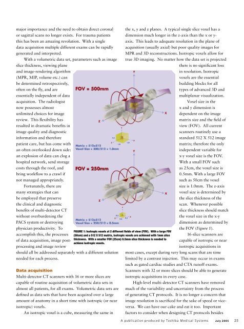
An isotropic voxel is a cube, measuring the same in<br />
the x, y and z planes. A typical single slice voxel has a<br />
dimension much longer in the z-axis than the x or y-<br />
FIGURE 1: Isotropic voxels at 2 different fi elds of view (FOV). With a large FOV<br />
(50cm) and a 512 X 512 matrix, isotropic voxels are achieved with 1mm slice<br />
thickness. With a smaller FOV (25cm) 0.5mm slice thickness is needed to<br />
achieve isotropic voxels.<br />
axis. This leads to adequate resolution in the plane of<br />
acquisition (usually axial) but poor quality images for<br />
MPR and 3D reconstructions. Isotropic voxels allow for<br />
true 3D imaging. No matter how the data set is projected<br />
there is no signifi cant loss<br />
in resolution. Isotropic<br />
voxels are the essential<br />
building blocks for all<br />
types of advanced 3D and<br />
multiplanar visualization.<br />
Voxel size in the<br />
x and y dimension is<br />
dependent on the image<br />
matrix size and the fi eld of<br />
view (FOV). All current<br />
scanners routinely use a<br />
standard 512 X 512 image<br />
matrix; therefore the only<br />
independent variable for<br />
x-y voxel size is the FOV.<br />
With a small FOV such<br />
as 25cm, the voxel size is<br />
0.5mm. With a large FOV<br />
such as 50cm the voxel<br />
size is 1.0mm. The z-axis<br />
voxel size is determined by<br />
the slice thickness of the<br />
scan. Whenever possible<br />
slice thickness should match<br />
the voxel size in the x-y<br />
dimension as determined by<br />
the FOV (Figure 1).<br />
16-slice scanners are<br />
capable of isotropic or near<br />
isotropic acquisitions in<br />
most cases, except during very long scans that are time<br />
limited by a contrast injection. This may occur in exams<br />
such as gated cardiac studies and <strong>CT</strong>A runoff exams.<br />
Scanners with 32 or more slices should be able to generate<br />
isotropic acquisitions in every case.<br />
High-level multi-detector <strong>CT</strong> scanners have removed<br />
much of the variability and uncertainty from the process<br />
of generating <strong>CT</strong> protocols. It is no longer a concern that<br />
image resolution is sacrifi ced for the sake of speed or viceversa.<br />
We can have our cake and eat it too. Important<br />
factors to consider when designing <strong>CT</strong> protocols besides<br />
A publication produced by Toshiba Medical Systems<br />
July 2005 25