Non-invasive Ventilation – A Century of Experience
The use of NIV is expected to increase considerably in the coming years, and clinicians, researchers and manufacturers will have to work closely to gain further knowledge and experience in dealing with its many challenges. It is of course beyond our reach to make NIV 100% successful, but we should strive to continue along the path of the considerable and impressive progress which has been made over just two decades to provide our patients with the highest possible level of care when using this technique.
The use of NIV is expected to increase considerably in the coming years, and clinicians, researchers and manufacturers will have to work closely to gain further knowledge and experience in dealing with its many challenges. It is of course beyond our reach to make NIV 100% successful, but we should strive to continue along the path of the considerable and impressive progress which has been made over just two decades to provide our patients with the highest possible level of care when using this technique.

Create successful ePaper yourself
Turn your PDF publications into a flip-book with our unique Google optimized e-Paper software.
D-9197-2009<br />
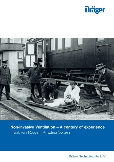
<strong>Non</strong>-<strong>invasive</strong> <strong>Ventilation</strong> <strong>–</strong> A century <strong>of</strong> experience<br />
Frank van Rooyen, Krisztina Soltész
Important note<br />
Medical knowledge is subject to constant change due to research and<br />
clinical experience. The author <strong>of</strong> this booklet has taken great care<br />
to make certain that the views, opinions and assertions included,<br />
particularly those concerning applications and effects, correspond<br />
with the current state <strong>of</strong> knowledge. However, this does not absolve<br />
readers from their obligation to take clinical measures on their own<br />
responsibility.<br />
All rights to this booklet are reserved by Dräger Medical GmbH,<br />
in particular the right <strong>of</strong> reproduction and distribution. No part <strong>of</strong><br />
this booklet may be reproduced or stored in any form either by<br />
mechanical, electronic or photographic means without the express<br />
permit <strong>of</strong> Dräger Medical GmbH, Germany.
<strong>Non</strong>-<strong>invasive</strong> <strong>Ventilation</strong> <strong>–</strong><br />
A century <strong>of</strong> experience<br />
Frank van Rooyen, Krisztina Soltész
CONTENT |<br />
CONTENT<br />
Foreword 06<br />
1.0 Introduction 09<br />
2.0 NIV (<strong>Non</strong>-<strong>invasive</strong> ventilation) basics 11<br />
3.0 History, trends and challenges <strong>of</strong> NIV 13<br />
4.0 Patient classification and categories <strong>of</strong> acute respiratory failure 16<br />
4.1 Patient selection 16<br />
4.2 Differentiating acute respiratory failure 17<br />
4.3 Hypoxemic respiratory failure 19<br />
4.3.1 Cardiogenic pulmonary oedema 19<br />
4.3.2 Pneumonia 19<br />
4.3.3 Acute lung injury (ALI) and acute respiratory distress syndrome (ARDS) 20<br />
4.3.4 Respiratory failure in patients with compromised immunity 20<br />
4.3.5 Decompensated OSA (Obstructive sleep apnea) 21<br />
4.4 Hypercapnic respiratory failure 22<br />
4.4.1 COPD (Chronic obstructive pulmonary disease) 22<br />
4.4.2 Asthma 23<br />
4.5 Postoperative respiratory failure 23<br />
4.6 Facilitation <strong>of</strong> weaning 25<br />
4.7 Special procedures 25<br />
5.0 Prerequisites for NIV therapy <strong>–</strong> the first steps 26
04|05<br />
6.0 The interface 30<br />
6.1 Choice <strong>of</strong> interface: vented versus non-vented systems 32<br />
6.2 Major challenges for patient interfaces 35<br />
6.2.1 Minimizing leaks 36<br />
6.2.2 Maximize comfort 36<br />
6.2.3 Minimize dead space 37<br />
6.2.4 Patient Interface: ClassicStar® and NovaStar® NIV full-face masks 38<br />
6.3 Full-face mask versus nasal mask and nasal pillow 39<br />
6.4 Total-face mask 41<br />
6.5 Helmet 43<br />
6.6 Interface use in chronic settings (home care environment) 45<br />
6.7 <strong>Non</strong>-<strong>invasive</strong> ventilation in pediatrics <strong>–</strong> the interface 46<br />
6.8 Summary on patient interface 47<br />
7.0 Humidification 48<br />
7.1 Active heated humidifiers 48<br />
7.2 Heated moisture exchangers HME filters 48<br />
8.0 The medical team 50<br />
9.0 Monitoring NIV therapy 51<br />
10.0 How to apply and set NIV 52<br />
11.0 Practical aspects <strong>of</strong> NIV 60<br />
12.0 Leak compensation and trigger adaptation 63<br />
13.0 Summary: The benefits <strong>of</strong> NIV 69<br />
14.0 Appendix 70<br />
15.0 References 75<br />
16.0 Abbreviations 82
FOREWORD |<br />
Foreword<br />
Since the first use <strong>of</strong> positive end-expiratory pressure ventilation in the late<br />
1930's and early 1940's to treat cardiogenic pulmonary edema and other<br />
forms <strong>of</strong> non-hypercapnic respiratory failure, non-<strong>invasive</strong> ventilation (NIV)<br />
has come a very long way. Negative pressure NIV was used extensively during<br />
the major poliomyelitis epidemics <strong>of</strong> the 1950's, as well as later on in the<br />
chronic home care setting, but positive pressure NIV has become the<br />
accepted technique over the past twenty years [1].<br />
The major evolution <strong>of</strong> NIV took place in the period ranging from the late<br />
1980's and early 1990's to the present day in the fields <strong>of</strong> both chronic and<br />
acute respiratory failure.<br />
Besides the accumulated evidence allowing an optimal strategy regarding<br />
clinical indications, long-term domiciliary NIV has been made much<br />
more efficient and user-friendly thanks to two important concomitant<br />
developments: the availability <strong>of</strong> dependable, powerful, compact and<br />
portable bilevel turbine-type ventilators, and major progress in NIV masks,<br />
in terms <strong>of</strong> materials used, adaptation to the patient's facial features, and<br />
overall comfort.<br />
During that time, similar advances were made in the use <strong>of</strong> NIV to treat<br />
acute respiratory failure (ARF) [1]. The first studies were performed in<br />
patients with decompensated chronic obstructive pulmonary disease<br />
(COPD) [2]. Subsequently, evidence <strong>of</strong> the usefulness <strong>of</strong> the technique in<br />
avoiding endotracheal intubation in these patients accumulated, resulting in<br />
its acceptance as a standard <strong>of</strong> care in severely decompensated COPD [3, 4].<br />
Studies performed in patients without COPD but with hypercapnic ARF<br />
yielded comparable results. In the setting <strong>of</strong> non-hypercapnic ARF, results <strong>of</strong><br />
various studies have been somewhat contradictory and less convincing than<br />
in hypercapnic ARF [3, 4]. This is not surprising considering the large
06|07<br />
heterogeneity <strong>of</strong> conditions manifested by non-hypercapnic ARF. However,<br />
both pathophysiological evidence and documented favorable outcomes point<br />
towards an increased recognition <strong>of</strong> the potential benefit derived from NIV<br />
in selected groups <strong>of</strong> patients [4, 5].<br />
NIV is most <strong>of</strong>ten applied using a spontaneous-assisted mode such as<br />
pressure support (PS), proven to be equally efficient and more comfortable<br />
than controlled modes. However, to achieve an optimal interaction between<br />
the patient's respiratory activity and the intensity and timing <strong>of</strong> the<br />
ventilator's response can prove difficult and may result in patient-ventilator<br />
asynchrony, a situation which is worsened by the presence <strong>of</strong> leaks at the<br />
patient-mask interface [6]. Furthermore, leaks interfere with certain key<br />
ventilator functions such as triggering and cycling [7], which can worsen<br />
asynchrony. In turn, this leads to an increase in the work <strong>of</strong> breathing, discomfort<br />
and intolerance, the latter being associated with failure <strong>of</strong> NIV [8].<br />
Therefore, successful and safe application <strong>of</strong> NIV in the setting <strong>of</strong> ARF<br />
requires minimization <strong>of</strong> leaks and their consequences on ventilator<br />
function, and optimization <strong>of</strong> patient-ventilator interaction.<br />
The challenge involved is considerable. Indeed, having established that NIV<br />
is an indispensable tool in the treatment <strong>of</strong> several causes <strong>of</strong> ARF, we must<br />
now push the limits <strong>of</strong> our technology and know-how to provide NIV at its<br />
best to our patients. Several encouraging recent studies suggest some paths<br />
to follow in order to meet this challenge. Clinicians and researchers have<br />
gained better understanding <strong>of</strong> the pathophysiology <strong>of</strong> the control <strong>of</strong><br />
ventilation and the mechanisms underlying patient-ventilator asynchrony,<br />
as well as the means to improve this problem [6, 9] . Some ventilator<br />
manufacturers have developed "NIV modes" which can, in many cases,<br />
attenuate the deleterious consequences <strong>of</strong> leaks on ventilator performance<br />
and asynchrony [7]. Finally, the automation <strong>of</strong> certain tasks such as titration<br />
<strong>of</strong> the level <strong>of</strong> ventilator support, detection <strong>of</strong> asynchrony and trigger and
FOREWORD |<br />
1.0 INTRODUCTION<br />
cycling correction is being developed and the concept has already proved<br />
promising in clinical trials [10, 11].<br />
The use <strong>of</strong> NIV is expected to increase considerably in the coming years, and<br />
clinicians, researchers and manufacturers will have to work closely to gain<br />
further knowledge and experience in dealing with its many challenges. It<br />
is <strong>of</strong> course beyond our reach to make NIV 100% successful, but we should<br />
strive to continue along the path <strong>of</strong> the considerable and impressive progress<br />
which has been made over just two decades to provide our patients with the<br />
highest possible level <strong>of</strong> care when using this technique.<br />
PD. Dr. Philippe Jolliet<br />
Senior Member <strong>of</strong> the ICU Staff<br />
University Hospital Geneva, Switzerland<br />
D-9193-2009
08|09<br />
1.0 Introduction<br />
In the past, patients suffering from acute respiratory failure were traditionally<br />
<strong>invasive</strong>ly ventilated via endotracheal intubation. In order to suppress<br />
spontaneous breathing and facilitate mechanical ventilation, sedation and<br />
sometimes muscle relaxation were used as well. Now, thanks to advances<br />
in our understanding <strong>of</strong> pulmonary mechanics and pathophysiology, the<br />
benefits <strong>of</strong> maintaining spontaneous breathing for as long as possible have<br />
become increasingly apparent.<br />
It was in 1989 when Dräger introduced the Biphasic Positive Airway Pressure<br />
(BIPAP*) ventilation mode in the Evita ventilator. It was the first step toward<br />
a “Room to Breathe” concept which was later augmented with Dräger’s<br />
AutoFlow ® and automatic tube compensation (ATC) features. In 1995, as a<br />
response to new findings on lung protection during mechanical ventilation,<br />
Dräger introduced the innovative Evita 4. It was the first and only ventilator<br />
on the market that supported and stimulated spontaneous breathing during<br />
the entire respiration cycle in both volume and pressure controlled<br />
ventilation modes. Small tidal volumes and airway pressures below 35 cm<br />
H 2 O were now broadly accepted goals in an effort to develop effective new<br />
protective ventilation strategies. Studies followed which demonstrated the<br />
benefits on gas exchange and lung recruitment with spontaneous breathing<br />
during BIPAP ventilation.<br />
All the while, a different but similar form <strong>of</strong> BIPAP, known as BiPAP, was<br />
being used with great success in the home care ventilation setting for the<br />
non-<strong>invasive</strong> treatment <strong>of</strong> OSA (Obstructive Sleep Apnea). Slowly, intensivists<br />
began to apply this non-<strong>invasive</strong> treatment to critically ill patients in order to<br />
avoid intubation or to <strong>of</strong>fer non-<strong>invasive</strong> support after extubation.<br />
* Trademark used under license
1.0 INTRODUCTION | 2.0 NIV (NON-INVASIVE VENTILATION) BASICS<br />
Several subsequent trials showed that NIV could reduce the length <strong>of</strong> stay on<br />
the ICU (Intensive Care Unit) as well as with intubation associated<br />
nosocomial pneumonia [12, 13, 14, 15].<br />
This booklet will explain how the concept <strong>of</strong> NIV can be applied in hospitals<br />
in intensive care, medium care or emergency department in daily practice.<br />
It gives a clear overview <strong>of</strong> the different forms <strong>of</strong> respiratory failure which<br />
can be effectively treated with NIV. It will provide precise selection criteria<br />
for potential patients and describe in detail how NIV should be applied on<br />
the basis <strong>of</strong> those criteria. Because the psychological effects <strong>of</strong> mask<br />
ventilation and patient motivation are vital factors which, in many cases,<br />
can make the difference between success and failure, it will also take an<br />
in-depth look at the different types <strong>of</strong> masks, how to use them and what to<br />
watch for during therapy. Finally, the key characteristics <strong>of</strong> a modern<br />
ventilator and the potential benefits <strong>of</strong> NIV are described.<br />
Initially, NIV may be somewhat more labor-intensive than conventional<br />
<strong>invasive</strong> ventilation, but benefits such as reduced length <strong>of</strong> stay on the ICU<br />
[16], shorter ventilation times [16] and the lower incidence <strong>of</strong> nosocomial<br />
pneumonia [17] and their subsequent positive effects in terms <strong>of</strong> both cost<br />
and outcome can make NIV well worth the effort.<br />
The objective <strong>of</strong> this booklet is to provide the reader with a practical<br />
guideline for the use <strong>of</strong> non-<strong>invasive</strong> ventilation and is intended for personal<br />
study only. All medical statements and values in this handbook have been<br />
taken from the literature. It is beyond the scope <strong>of</strong> this booklet to validate<br />
the content and quality <strong>of</strong> these studies and no responsibility will be taken<br />
for errors or inaccuracies.<br />
* BIPAP Trademark used under license
10|11<br />
Admission<br />
Discharge<br />
MT-1826-2008<br />
The therapy phases for a ventilated patient throughout the continuum from admission to discharge<br />
2.0 NIV basics<br />
WHAT IS NIV?<br />
NIV <strong>–</strong> non-<strong>invasive</strong> ventilation is a broad term for any ventilation therapy<br />
applied in a non-<strong>invasive</strong> way, e.g. via a mask, nasal prongs or a helmet.<br />
Therefore, NIV, or NPPV (<strong>Non</strong>-<strong>invasive</strong> Positive Pressure <strong>Ventilation</strong>), is also<br />
very <strong>of</strong>ten referred to as “mask ventilation”. This is in contrast to “<strong>invasive</strong><br />
ventilation”, where an endotracheal tube or a tracheal canula serves as an<br />
<strong>invasive</strong> interface between the patient and the ventilator.<br />
WHEN TO USE NIV?<br />
NIV can be used either before intubation or following extubation. The<br />
classical use <strong>of</strong> NIV therapy in a hospital setting is following extubation,<br />
in particular in cases where spontaneous breathing is not sufficient for<br />
adequate gas exchange. Here, the goal <strong>of</strong> NIV is to promote the weaning<br />
process and avoid reintubation [18]. Increasingly, the early use <strong>of</strong> NIV on the<br />
ICU now serves as a first line interventional tool to prevent intubation [12].<br />
Ideally, the entire spectrum <strong>of</strong> ventilation therapy (prevent <strong>–</strong> stabilize <strong>–</strong> wean <strong>–</strong><br />
recover) can be adequately covered using NIV.<br />
Another classical application <strong>of</strong> NIV therapy is in the home care<br />
environment, where it is primarily used to provide respiratory support in<br />
patients with OSA.
2.0 NIV (NON-INVASIVE VENTILATION) BASICS | 3.0 HISTORY, TRENDS AND CHALLENGES OF NIV<br />
WHAT IS NEEDED FOR NIV?<br />
There are three prerequisites for NIV, the first and most important being a<br />
conscious, cooperative patient. The second is a NIV-capable ventilator<br />
equipped with leak compensation and smart measures to avoid autotriggering.<br />
Thirdly, a well-fitting mask is vital for the success <strong>of</strong> NIV therapy.<br />
These prerequisites will be described in more detail later on in this booklet.
12|13<br />
D-9191-2009<br />
Dräger iron lung model E 52 with electric drive (1952)<br />
3.0 History, trends and challenges <strong>of</strong> NIV<br />
The first apparatus used for non-<strong>invasive</strong> ventilation was a whole body<br />
ventilator in which the patient was enclosed up to the neck in an airtight<br />
box. Invented by the Scottish physician John Dalziel (1838), this body<br />
ventilator relied on the cyclic application <strong>of</strong> positive and negative pressure<br />
to the body to support spontaneous breathing. Both huge and expensive, this<br />
manually operated ventilator did not come into widespread use. Based on<br />
this early work, Philip Drinker developed the first electrically-driven iron<br />
lung in 1929. This ventilator was later improved on by J.H. Emerson in<br />
Cambridge, Massachusetts in 1931, who made it lighter, less expensive and
3.0 HISTORY, TRENDS AND CHALLENGES OF NIV |<br />
D-9194-2009<br />
MT-0562-2008<br />
The Dräger pulmotor in use.<br />
NIV ventilation with Carina®.<br />
added a manual backup drive system in case <strong>of</strong> a power failure. It was this<br />
type <strong>of</strong> ventilator which was predominantly used during the poliomyelitis<br />
epidemic <strong>of</strong> the 1950’s, but a few were still in use all the way up to the<br />
beginning <strong>of</strong> this century.<br />
As the need for a more portable model <strong>of</strong> the iron lung grew, R. Eisen menger<br />
was inspired to develop the cuirass ventilator, which was patented in 1928.<br />
These chest shell ventilators were later improved on by Fairchild-Huxley<br />
and Monoghan and introduced in 1949. This type <strong>of</strong> non-<strong>invasive</strong> ventilation<br />
is still in use today in the home care setting.<br />
In 1907, a pioneer in the field <strong>of</strong> developing ventilators, Heinrich Dräger,<br />
received a patent on the Pulmotor, a non-<strong>invasive</strong> ventilator. It was the birth<br />
<strong>of</strong> the mobile rescue ventilator. The first models had a fixed pressure cycling<br />
period and were powered by pressurized oxygen. Negative pressure was used<br />
to support exhalation. When measured against today’s insights on protective<br />
ventilation, this ventilator was hopelessly inadequate, but it may have<br />
facilitated lung drainage and helped re-establish spontaneous breathing in<br />
its early applications in near-drowning victims. These prototypes were<br />
developed as single hose systems, which in turn caused CO 2 rebreathing
14|15<br />
issues. When production started up in 1908, Heinrich Dräger’s son Bernard<br />
changed the single hose to a dual hose system. Thirty-eight years later,<br />
Pulmotor number 12.000 left the production line in Lübeck.<br />
Today, a wide variety <strong>of</strong> non-<strong>invasive</strong> ventilators are available which can be<br />
differentiated into two groups: sophisticated intensive care ventilators with a<br />
NIV option, and the less complicated NIV devices for use in the sub-acute and<br />
home care environment. The first group uses a high pressure gas inlet for<br />
both oxygen and air provided by a central gas supply, while the second group<br />
uses low and/or high pressure oxygen inlets only. For compressed air, these<br />
ventilators are equipped with either a blower or turbine technology. Turbine<br />
driven ventilators achieve higher inspiratory pressure and flow than blower<br />
operated devices, which are predominantly used for patients with OSA in a<br />
home care setting.<br />
Current trends in hospitals show that NIV is being used more frequently and<br />
for a wider range <strong>of</strong> conditions than ever before. Patients with chronic<br />
obstructive pulmonary disease make up a large portion <strong>of</strong> the total<br />
population treated with NIV therapy on the ICU.<br />
Following the introduction <strong>of</strong> better equipment and improved masks,<br />
interest in NIV has increased sharply. Enhanced leak management and a<br />
reduction in the incidence <strong>of</strong> facial lesions caused by poorly-fitting masks<br />
have served to further push the popularity <strong>of</strong> NIV.
4.0 PATIENT CLASSIFICATION AND CATEGORIES OF ACUTE RESPIRATORY FAILURE |<br />
4.0 Patient classification and categories <strong>of</strong> acute<br />
respiratory failure<br />
In the past, the standard management for patients in the ICU suffering<br />
from severe acute respiratory failure included endotracheal intubation<br />
and mechanical ventilation. Today, it is becoming increasingly common<br />
to consider NIV as a first option. In order to decide whether therapy with<br />
NIV is indicated, a careful analysis <strong>of</strong> both the type and the underlying<br />
cause <strong>of</strong> respiratory failure is necessary. The success <strong>of</strong> NIV depends on<br />
several factors:<br />
<strong>–</strong> Proper patient selection<br />
<strong>–</strong> Correct classification <strong>of</strong> the respiratory failure type<br />
<strong>–</strong> Knowledge <strong>of</strong> the underlying pathology<br />
<strong>–</strong> Appropriate interface selection<br />
<strong>–</strong> Skills and expertise <strong>of</strong> the care team<br />
<strong>–</strong> Proper timing, i.e. when to start or stop NIV<br />
<strong>–</strong> Ventilator capabilities with regard to leak management and auto-triggering<br />
4.1 PATIENT SELECTION<br />
Selecting the right patient, applying adequate ventilation support with an<br />
appropriate setting as well as the use <strong>of</strong> an optimal patient interface are all<br />
essential factors that can make the difference between failure and success.<br />
A detailed understanding <strong>of</strong> the various causes <strong>of</strong> ARF and the underlying<br />
pathology are essential for the decision making process. A first set <strong>of</strong> selection<br />
criteria might be based primarily on clinical indicators, such as the severity<br />
<strong>of</strong> dyspnea, tachypnea, the recruitment <strong>of</strong> accessory muscles and the<br />
presence <strong>of</strong> paradoxical abdominal breathing.
16|17<br />
COMPLY WITH THE FOLLOWING ENTRY CRITERIA BEFORE STARTING NIV<br />
Indications for NIV*<br />
moderate or severe dyspnea<br />
respiratory rate ≥ 25 breath/min<br />
use <strong>of</strong> accessory muscles<br />
presence <strong>of</strong> paradoxical breathing<br />
PaCO 2 > 45 mm Hg / 6 kPa<br />
PaO 2 /FiO 2 < 300<br />
7.25 ≤ pH < 7.35<br />
*NIV is indicated if two criteria are applicable<br />
Contra<strong>–</strong>indications NIV<br />
respiratory arrest<br />
impaired consciousness<br />
hemodynamic unstable<br />
cardiac ischemia<br />
arrythmias<br />
hypotensive shock<br />
excessive secretions<br />
inability to clear airway<br />
agitated and not cooperative<br />
facial abnormalities that are affecting the<br />
acceptance and fit <strong>of</strong> the mask, e.g. burns,<br />
surgery, anatomic<br />
The type <strong>of</strong> ventilator accessible for therapy, its ventilation mode options and<br />
the available interfaces must also be taken into consideration, the later<br />
being <strong>of</strong> particular importance, as it has been shown that the patient<br />
interface <strong>of</strong>ten determines the success rate <strong>of</strong> NIV therapy.<br />
4.2 DIFFERENTIATING ARF<br />
Acute respiratory failure (ARF) is defined as the inability to maintain<br />
adequate pulmonary gas exchange and is characterized by abnormal levels<br />
<strong>of</strong> arterial PaO 2 and PaCO 2 blood gas tensions. The determination <strong>of</strong> arterial<br />
blood gases is important because it helps to differentiate between Type 1 and<br />
Type 2 failure.<br />
A patient with Type 1 failure is hypoxemic and has a normal or low PaCO 2 .<br />
Oxygenation is affected by intrapulmonary venous admixture caused by<br />
pneumonia, emphysema or acquired respiratory distress syndrome (ARDS),<br />
which results in ventilation-perfusion mismatch. In most situations, oxygen<br />
therapy with or without CPAP (Continuous Positive Airway Pressure) therapy<br />
is sufficient to improve the situation.
4.0 PATIENT CLASSIFICATION AND CATEGORIES OF ACUTE RESPIRATORY FAILURE |<br />
Acute<br />
respiratory<br />
failure<br />
(see chapter 4.2)<br />
Hypoxemic<br />
respiratory<br />
failure<br />
(see chapter 4.3)<br />
Hypercapnic<br />
respiratory<br />
failure<br />
(see chapter 4.4)<br />
Postoperative<br />
respiratory<br />
failure<br />
(see chapter 4.5)<br />
Facilitate<br />
weaning<br />
(see chapter 4.6)<br />
Special<br />
procedures<br />
(see chapter 4.7)<br />
Cardiogenic<br />
Pulmonary<br />
Edema<br />
(see chapter 4.3.1)<br />
Pneumonia<br />
(see chapter 4.3.2)<br />
Acute lung injury<br />
(ALI) and acute<br />
respiratory distress<br />
syndrome<br />
(ARDS)<br />
(see chapter 4.3.3)<br />
Respiratory<br />
failure in<br />
immunocom<br />
promised<br />
patients<br />
(see chapter 4.3.4)<br />
Decompensated<br />
obstructive<br />
sleep apnea<br />
(OSA)<br />
(see chapter 4.3.5)<br />
Chronic<br />
obstructive<br />
pulmonary<br />
disease<br />
(COPD)<br />
(see chapter 4.3.5)<br />
Asthma<br />
(see chapter 4.4.2)<br />
D-9201-2009<br />
+<br />
Type 1 failure<br />
Type 2 failure<br />
Type 1 failure<br />
Hypoxaemia without hypercapnia<br />
PaO2 < 8 kPa<br />
3.5 kPa < PaCO2 < 6.0 kPa<br />
PA-aO2 = increased<br />
Type 2 failure<br />
Hypoxaemia with hypercapnia<br />
PaO2 < 8kPa<br />
PaCO2 > 6kPa<br />
PA-aO2 = normal<br />
Differenciating ARF<br />
Patients suffering from Type 2 failure show a combination <strong>of</strong> hypoxemia and<br />
hypercapnia caused by airway obstruction, as in asthma and COPD, as well<br />
as a loss <strong>of</strong> diffusion area, as is the case with emphysema. These patients<br />
require ventilatory support, given as PS, BIPAP or even volume controlled<br />
ventilation (VCV) in order to restore acceptable arterial blood gas values.<br />
Acute respiratory failure can be differentiated into 5 sub-classifications, each<br />
having one or more subgroups.
18|19<br />
4.3 HYPOXEMIC RESPIRATORY FAILURE<br />
Hypoxic respiratory failure is present in patients with resting dyspnea,<br />
a PaO 2 / FiO 2 < 300 mmHg and a respiratory rate > 30/min. A large,<br />
randomized study conducted by Antonelli et al. concluded that in 70% <strong>of</strong><br />
the selected patients, intubation was avoided by using NIV therapy [19].<br />
There are many conditions which are unrelated to COPD which can lead to<br />
hypoxic respiratory failure. Many subgroups can be defined, the most<br />
important <strong>of</strong> which are mentioned hereafter:<br />
4.3.1 CARDIOGENIC PULMONARY EDEMA<br />
Patients with cardiogenic pulmonary edema have a reduced functional residual<br />
capacity which can be effectively treated with non-<strong>invasive</strong> ventilation [20].<br />
The use <strong>of</strong> continuous positive airway pressure (CPAP) with pressures<br />
between 10-13 cm H 2 O will recruit collapsed alveoli and help reduce the<br />
work <strong>of</strong> breathing. Additional pressure support eases spontaneous breathing<br />
and helps reduce respiratory rate and alleviate dyspnea rapidly [21, 22].<br />
4.3.2 PNEUMONIA<br />
Pneumonia is an infection <strong>of</strong> the lung caused by bacteria, viruses, fungi or<br />
a combination <strong>of</strong> these pathogens. In adults, the most common form is<br />
bacterial streptococcal pneumonia, whereas in children, pneumonia caused<br />
by infections with Mycoplasma sp. is more frequent. In the elderly, patients<br />
suffering from dysphagia, <strong>of</strong>ten seen following a stroke or in Parkinson’s<br />
disease, are affected most <strong>of</strong>ten.<br />
Ventilator associated pneumonia (VAP) is <strong>of</strong>ten considered to be a greater<br />
potential threat because <strong>of</strong> the enhanced virulence observed in bacteria<br />
found in hospitals.<br />
In addition to antibiotic therapy, NIV can be used in patients with pneumonia<br />
who are suffering from ARF. The <strong>of</strong>ten unpredictable course <strong>of</strong> pneumonia<br />
requires a carefully monitoring <strong>of</strong> the patient.
4.0 PATIENT CLASSIFICATION AND CATEGORIES OF ACUTE RESPIRATORY FAILURE |<br />
In the literature, there is some dispute as to the effects <strong>of</strong> NIV in patients<br />
with pneumonia. In some studies, NIV improves outcome, especially in<br />
patients with COPD [23, 24, 25]. On the other hand, several randomized,<br />
controlled trials demonstrated a higher failure rate for NIV, with more<br />
patients requiring intubation [19, 26]. In most situations, non-<strong>invasive</strong> CPAP<br />
therapy is sufficient and improves oxygenation, reduces the respiratory rate<br />
and alleviates dyspnea.<br />
4.3.3 ACUTE LUNG INJURY (ALI) AND ACUTE RESPIRATORY DISTRESS<br />
SYNDROME (ARDS)<br />
ALI and ARDS are inflammatory disorders <strong>of</strong> the lung. ALI is caused by<br />
direct injury <strong>of</strong> the lung caused by pneumonia, aspiration or trauma and is<br />
characterized by:<br />
<strong>–</strong> bilateral pulmonary congestion (x-ray)<br />
<strong>–</strong> pulmonary capillary wedge pressure ≤ 18 mmHg<br />
<strong>–</strong>PaO 2 /FiO 2 < 300 (ALI)<br />
<strong>–</strong>PaO 2 /FiO 2 < 200 (ARDS)<br />
ARDS is a more severe form <strong>of</strong> ALI.<br />
The success rate using NIV in this group <strong>of</strong> patients is about 50% [28]. In a<br />
recent prospective, multicenter study, it was found that NIV, used as a first<br />
line option, prevented intubation in 54% <strong>of</strong> the patients treated [27].<br />
4.3.4 RESPIRATORY FAILURE IN PATIENTS WITH COMPROMISED IMMUNITY<br />
In this group, patients develop hypoxemic respiratory failure as a response to<br />
organ or bone-marrow transplantation. The benefits <strong>of</strong> NIV versus intubation<br />
on patient outcome are mainly related to the reduced risk <strong>of</strong> septic shock,<br />
ventilator associated pneumonia (VAP) or nosocomial infections [28].
20|21<br />
HCO 3<br />
(mM)<br />
35<br />
30<br />
P CO2 = 60 mmHg<br />
P CO2 = 40 mmHg<br />
P CO2 = 20 mmHg<br />
25<br />
20<br />
Respiratory<br />
Acidosis<br />
Normal<br />
Respiratory<br />
Alkalosis<br />
15<br />
D-9203-2009<br />
7 7.08 7.16 7.24 7.32 7.4 7.48 7.56 7.64 7.72 7.8<br />
pH<br />
Davenport diagram<br />
4.3.5 DECOMPENSATED OBSTRUCTIVE SLEEP APNEA (OSA)<br />
Patients with obstructive sleep apnea (OSA) can develop ARF if the periods<br />
<strong>of</strong> hypoxemia and hypercapnia are getting longer and finally resulting into<br />
cardiopulmonary failure. Particularly obese patients with OSA can develop<br />
an ARF if breathing cost too much energy and if hypercapnia is associated<br />
with pulmonary hypertension. These patients are responding very well to a<br />
non-<strong>invasive</strong> CPAP therapy [26].<br />
4.4 HYPERCAPNIC RESPIRATORY FAILURE<br />
Hypercapnic respiratory failure is a Type 2 failure and is usually caused by<br />
hypoventilation. For reasons <strong>of</strong> clarity, this booklet will deal only with those<br />
forms <strong>of</strong> hypercapnic respiratory failure caused by tangible underlying<br />
pulmonary disease. Prolonged hypoventilation results in an acute respiratory<br />
acidosis with an increase <strong>of</strong> bicarbonate [HCO 3 ] ion concentration and a<br />
decrease in blood pH. It is important to differentiate between acute and<br />
chronic respiratory acidosis because treatment is different. In chronic<br />
respiratory acidosis, one can observe increased PaCO 2 but nearly normal pH<br />
(see page 17, Differentiating ARF).
4.0 PATIENT CLASSIFICATION AND CATEGORIES OF ACUTE RESPIRATORY FAILURE |<br />
This is possible because the kidneys can compensate the pH by stepping up<br />
acid excretion (renal compensation). The response time for this mechanism<br />
is slow (> 48h), and stands in contrast to the much faster respiratory<br />
mechanism which can react within minutes. Hypercapnic respiratory failure<br />
can be differentiated to the following subgroups:<br />
4.4.1 CRONIC OBSTRUCTIVE PUMONARY DISEASE (COPD)<br />
Chronic Pulmonary Obstructive Disease (COPD) is a preventable condition<br />
seen mainly in patients with a history <strong>of</strong> smoking. This subgroup represents<br />
the largest patient population to benefit from NPPV. The incidence <strong>of</strong> COPD<br />
increases with both age and severity <strong>of</strong> the smoking habit. Other, less<br />
frequent causes <strong>of</strong> COPD include viral or bacterial infections and allergic<br />
reactions to inhaled particles. The disease is characterized by inflammation<br />
<strong>of</strong> the bronchi, resulting in increased secretion production and airway<br />
narrowing. Symptoms include continuous coughing and dyspnea.<br />
An allergic reaction can induce an exacerbation <strong>of</strong> COPD. Based on the<br />
results <strong>of</strong> several randomized trials it is recommended to consider NIV as<br />
a first option in patients with COPD suffering from acute hypercapnic<br />
respiratory failure with a PaCO 2 > 6.0 kPa and SpO 2 ≤ 88% [29, 30]. For this<br />
group <strong>of</strong> patients, a large amount <strong>of</strong> evidence supports the idea that NIV can<br />
reduce intubation rates, length <strong>of</strong> hospital stay and the incidence <strong>of</strong> airway<br />
infections [13, 31].
22|23<br />
4.4.2 ASTHMA<br />
Asthma is a chronic disease characterized by episodes <strong>of</strong> airway constriction<br />
and inflammation, sometimes accompanied by excessive mucus production.<br />
An asthma attack can be triggered by a wide variety <strong>of</strong> external stimuli such<br />
as cold air, sports, stress, smoke, and a large number <strong>of</strong> allergens. Inhalation<br />
therapy with bronchodilators is effective and alleviates airway constriction.<br />
Typical signs <strong>of</strong> an acute asthma attack include wheezing, prolonged<br />
expiration, increased heart rate and the extensive use <strong>of</strong> accessory muscles.<br />
The latter can be clearly recognized during inspiration by observing the<br />
sternocleidomastoid muscles, which are literally drawn in by the patient’s<br />
greatly increased respiratory effort.<br />
For patients with severe asthma suffering from acute respiratory failure,<br />
there is less evidence that NIV is always the best approach to begin with.<br />
However, several trials in patients with a status asthmaticus have<br />
demonstrated gas exchange improvement and a reduction <strong>of</strong> intubation<br />
rates [32, 33]. It is recommended to monitor these patients carefully and to<br />
intubate promptly if the situation deteriorates, or if there is no improvement<br />
after two hours <strong>of</strong> NIV therapy.<br />
4.5 POSTOPERATIVE RESPIRATORY FAILURES<br />
NIV is <strong>of</strong>ten used as a preventive therapy to restore a breathing pattern<br />
following surgery. Temporary upper airway obstruction due to swelling <strong>of</strong><br />
the glottis is a frequent phenomenon and can cause ARF. For such patients,<br />
NIV with PSV (Pressure Support <strong>Ventilation</strong>) is a good alternative to<br />
reintubation, as it can rapidly improve lung mechanics and gas exchange<br />
[34, 35]. The literature shows predominantly positive results when NIV is<br />
used in postoperative patients. However, NIV should be discontinued in<br />
patients with ARF and concomitant surgical complications requiring major<br />
re-intervention. This group <strong>of</strong> patients will require intubation and controlled<br />
ventilation in any case.
4.0 PATIENT CLASSIFICATION AND CATEGORIES OF ACUTE RESPIRATORY FAILURE |<br />
Post-operative<br />
non-<strong>invasive</strong> ventilation<br />
Curative<br />
Preventive<br />
ARF present<br />
Objective: avoid intubation<br />
ARF not present, but risk to develop<br />
Objective: avoid ARF<br />
D-9209-2009<br />
CPAP<br />
5 - 8 cm H 2O<br />
PSV<br />
10 - 12 cm H 2O<br />
CPAP<br />
3 - 5 cm H 2O<br />
PSV<br />
10 cm H 2O<br />
Yearbook <strong>of</strong> ISICEM 2008.<br />
Cardiac surgery<br />
Following cardiac surgery, patients frequently show altered breathing<br />
patterns. This is mainly due to pain and diaphragm dysfunction and leads to<br />
smaller tidal volumes and a higher respiratory rate in order to maintain<br />
adequate gas exchange. This in turn causes alveolar hypoventilation and the<br />
development <strong>of</strong> atelectasis. Preventive treatment with NIV is possible using<br />
CPAP therapy with 3-5 cm H 2 O and, if necessary, additional pressure support<br />
ventilation PSV with a pressure <strong>of</strong> 10 cm H 2 O.<br />
Thoracic surgery<br />
For patients who develop respiratory failure following lung transplantation,<br />
some studies show positive results with a significant reduction in the need<br />
for <strong>invasive</strong> ventilation from 50% to 21%. Prophylactic use <strong>of</strong> NIV following
24|25<br />
lung resection have shown to result in a much lower incidence <strong>of</strong> atelectasis<br />
than in a control group [36].<br />
Abdominal surgery<br />
Patients who have undergone gastroplasty, cholecystomy or thoracoabdominal<br />
surgery show significant improvement when treated with NIV during the<br />
first 24 hours after surgery with moderate PSV therapy (10-12 cm H 2 O) [60].<br />
Caution must be exercised when applying NIV at higher levels <strong>of</strong> PSV in the<br />
early postoperative phase in patients with new gastrointestinal anastomosis,<br />
since the accidental insufflation <strong>of</strong> air into the GI tract can leak through the<br />
sutures into the abdomen. In these patients, non-<strong>invasive</strong> CPAP therapy alone<br />
is preferred, and if PSV is deemed necessary, the pressure should be kept<br />
below 8 cm H 2 O. In general, NIV used as a curative therapy shows very<br />
positive results, with a reduction <strong>of</strong> intubation rates <strong>of</strong> up to 70% [51, 52].<br />
4.6 FACILITATION OF WEANING<br />
Following extubation, non-<strong>invasive</strong> ventilation is being used increasingly as<br />
the next step in the weaning process. Outcomes <strong>of</strong> several randomized,<br />
controlled studies have shown that NIV reduces the time needed to<br />
successfully wean patients from the ventilator, thereby reducing the length <strong>of</strong><br />
stay on the ICU, the duration <strong>of</strong> hospitalization as a whole as well as the risks<br />
and costs <strong>of</strong> infections associated with endotracheal intubation [2, 27, 30].<br />
4.7 SPECIAL PROCEDURES<br />
NIV is commonly used in critical ill patients before planned intubation.<br />
Here, NIV serves to pre-oxygenate the patient, helping to avoid hypoxic<br />
respiratory failure during the course <strong>of</strong> intubation.<br />
Along these lines, NIV is <strong>of</strong>ten used in preparation for other short procedures<br />
such as endotracheal suctioning or bronchoscopy, where a drop in oxygen<br />
saturation is to be expected.
5.0 PREREQUISITES NIV THERAPY <strong>–</strong> THE FIRST STEPS |<br />
5.0 Prerequisites for NIV therapy <strong>–</strong> the first steps<br />
THE PATIENT<br />
After selecting the right patient for NIV, it is important to inform the patient<br />
as to what will happen next. Proper instruction, encouragement and<br />
motivation are essential factors for success. Before commencing NIV therapy,<br />
the patient should first be allowed to experience the feeling <strong>of</strong> the face mask.<br />
With the caregiver holding the mask, the patient should take 5-10 breaths,<br />
letting the patient become accustomed to the support <strong>of</strong> a ventilator.<br />
Start NIV with very low PEEP and PS levels to allow the patient to accept NIV.<br />
After a number <strong>of</strong> breathes slowly increase PEEP and PS to the desired<br />
settings, carefully monitoring patients response.<br />
The patient should be very closely monitored for at least the next 30 minutes,<br />
and frequent checks should be performed to ensure that the patient is com -<br />
fortable. If this is the case, observation intervals can be gradually increased.<br />
However, some patients may claim to be comfortable when the opposite is<br />
true. It is therefore important to correlate such statements with clinical<br />
observations. Highly motivated patients, doing their very best to breathe<br />
spontaneously, can sometimes exert themselves to the point <strong>of</strong> complete<br />
exhaustion. This is a potentially serious situation which may require<br />
conversion to a more <strong>invasive</strong> ventilation mode or even rapid intubation.<br />
INTERFACE AND COMFORT<br />
It is recommended to select a mask with a s<strong>of</strong>t cushion (e.g. gel cushions are<br />
highly comfortable) <strong>of</strong> appropriate size. Preferably, the mask can be further<br />
molded to achieve a better fit, reducing leakage. With a well-fitting mask, the<br />
cushion provides equal pressure distribution over the face. This is less irritating<br />
for the patient and there is a reduced risk <strong>of</strong> skin lesions. Gentle tightening<br />
<strong>of</strong> the straps is usually all that is necessary; the need for excessive tightening<br />
is an indication that the mask is not <strong>of</strong> the appropriate size or shape to the<br />
face (for more information on masks, see the chapter 6.0 The interface).
26|27<br />
Evita Infinity ® V500<br />
Evita XL<br />
Evita 4 edition<br />
Evita 2 dura<br />
Savina<br />
Carina ® and Carina ® home<br />
MT-154-2001<br />
MT-4300-2007<br />
MT-0472-2008<br />
MT-766-99<br />
MT-0023-2008<br />
MT-0514-2008 | 0513-2008<br />
MT-1188-2008<br />
MT-0495-2008<br />
MT-1029-2008<br />
Oxylog ® 3000<br />
Oxylog ® 2000 plus<br />
Babylog 8000 plus<br />
THE VENTILATOR<br />
Today, NIV on the ICU is being used increasingly as a first line intervention<br />
option to prevent respiratory function from deteriorating to the point<br />
where intubation becomes necessary [37, 38, 39, 40]. Nearly all current<br />
Dräger ventilators, such as the Evita Infinity ® V500, Evita XL, Evita 4 edition,<br />
Evita 2 dura, Savina, Carina ® as well as the transport ventilators Oxylog ® 3000<br />
and Oxylog 2000 plus are suitable for non-<strong>invasive</strong> ventilation. For the<br />
Savina, as well as the Evita and Oxylog ® 2000, NIV requires an optional<br />
s<strong>of</strong>tware upgrade. This upgrade provides leak compensation for leaks <strong>of</strong> up<br />
to 180 l/min (100 l/min for Oxylogs) and includes a smart algorithm for<br />
automatic adjustment <strong>of</strong> trigger sensitivity.
5.0 PREREQUISITES NIV THERAPY <strong>–</strong> THE FIRST STEPS |<br />
MT-0970-2007<br />
MT-0971-2007<br />
NovaStar® NIV full-face mask with standard<br />
elbow.<br />
NovaStar® NIV full-face mask with anti-asphyxia<br />
valve and exhalation ports.<br />
Furthermore, it allows enhanced alarm management, data and waveform<br />
monitoring. Automatic leak compensation and trigger adjustment ensure<br />
that patients can breathe spontaneously at the highest level <strong>of</strong> comfort while<br />
maintaining a degree <strong>of</strong> trigger sensitivity that would normally only be<br />
possible in a leak-free circuit.<br />
The Evita Infinity ® V500, Evita XL, Evita 4 edition, Evita 2 dura and Savina<br />
are used with a double limb system, i.e. an inspiratory hose and an<br />
expiratory hose connected with a y-piece to the mask. All mentioned<br />
ventilators using dual limb systems and with a built-in safety valve for<br />
ventilator failure require a NIV mask with so called standard elbow<br />
(SE).These masks are also known as non-vented NIV masks (see chapter 6.1<br />
Choice <strong>of</strong> interface: vented versus non-vented systems).
28|29<br />
The other category <strong>of</strong> NIV masks includes interfaces with integrated antiasphyxia-valve<br />
(AAV) and exhalation ports (EP). These types <strong>of</strong> masks are<br />
usually used in combination with single limb systems and are named vented<br />
NIV masks.<br />
If the single limb system has an integrated leakage valve and the ventilator<br />
has a built-in safety valve for ventilator failure, as it is the case for the<br />
Carina ® system, a non-vented mask can be used in the hospital or<br />
institutional environment.
6.0 THE INTERFACE |<br />
6.0 The interface<br />
NIV represents a true advance in the management <strong>of</strong> both chronic and acute<br />
respiratory failure [41]. Apart from the choice <strong>of</strong> ventilator type, mode and<br />
settings, another crucial issue is the optimal interface. A broad range <strong>of</strong><br />
patient interfaces is available, but until now, little attention has been focused<br />
on how to choose the right interface. Furthermore, there is no generally<br />
accepted consensus on interface management [42]. The following<br />
comments will focus on these five types <strong>of</strong> interfaces in a hospital or<br />
institutional setting.
30|31<br />
In general, there are five different types <strong>of</strong> factory-made patient interfaces:<br />
The full-face mask<br />
The full-face mask covers<br />
nose and mouth. For this<br />
reason, this mask type is<br />
<strong>of</strong>ten called oronasal mask.<br />
The nasal mask<br />
The nasal mask covers the<br />
nose only, as its name<br />
indicates.<br />
The nasal pillow<br />
The nasal pillow <strong>–</strong> also called<br />
nasal prong <strong>–</strong> usually consist <strong>of</strong><br />
silicone rubber and is introduced<br />
directly into the nostril.<br />
MT-5074-2008<br />
MT-5076-2008<br />
MT-0970-2007<br />
MT-5072-2008<br />
D-7066-2009<br />
The total-face mask<br />
The total-face mask covers<br />
the entire face.<br />
The helmet<br />
The helmet is a transparent<br />
cylinder made <strong>of</strong> s<strong>of</strong>t PVC which<br />
covers the entire head <strong>of</strong> the<br />
patient. Cushion-like elements<br />
ensure a nearly airtight seal at<br />
the neck and shoulder areas.
6.0 THE INTERFACE |<br />
6.1 CHOICE OF INTERFACE: VENTED VERSUS NON-VENTED SYSTEMS<br />
The choice <strong>of</strong> interface is influenced by numerous factors including the type<br />
<strong>of</strong> respiratory failure, patient condition, tolerance and type <strong>of</strong> ventilator.<br />
Once the interface (nasal, full-face, total-face etc.) is chosen, the type <strong>of</strong><br />
interface system (non-vented or vented) must be selected. Technically, this is<br />
stipulated by the breathing system (incl. ventilator and breathing circuit)<br />
used. There are two major prerequisites for interfaces used in NIV:<br />
<strong>–</strong> Sufficient CO 2 management (elimination)<br />
<strong>–</strong> Adequate safety precautions in case <strong>of</strong> ventilator failure in order to avoid<br />
asphyxia / CO 2 narcosis.<br />
Table 3 summarizes the most common combinations used with NIV in the<br />
hospital environment. However, always consult the instructions for use <strong>of</strong><br />
the ventilator and its accessories before commencing therapy.
32|33<br />
TABLE 3 <strong>–</strong> TYPES OF BREATHING SYSTEMS<br />
System Built-in Type <strong>of</strong> Inter- Interface Sufficiant Adequate safety<br />
type Safety circuit face type CO 2 precautions in<br />
valve in form management case <strong>of</strong> ventilator<br />
the failure (Asphyxia /<br />
ventilator<br />
CO 2 narcosis)<br />
Active Available 2 limb non- Interface Less relevant as Built-in safety valve<br />
circuit vented (nasal, full- inspiration / in the ventilator<br />
system face, total- expiration take provides the patient<br />
face masks) place separately access to room air<br />
with standard<br />
in case <strong>of</strong> ventilator<br />
elbow<br />
failure<br />
Not 1 limb non- Interface Maintained by Expiration Valve<br />
available circuit vented (nasal, full- expiration valve allows the patient<br />
system face, total- to access room air<br />
with “true” face masks) in case <strong>of</strong> ventilator<br />
expiration with standard failure (pressure<br />
valve elbow drop)<br />
Passive Not 1 limb non- Nasal mask Relevant to a Patient can breathe<br />
available circuit vented with lesser extent; through his/her<br />
system with standard CO 2 elimination mouth in case <strong>of</strong><br />
leakage elbow is maintained by ventilator failure<br />
valve<br />
leakage valve<br />
Not 1 limb non- Full-face Maintained by Only in hospital<br />
available circuit vented mask with leakage valve environment!<br />
system standard Adequate alarm<br />
with elbow management must<br />
leakage<br />
be available for<br />
valve<br />
ventilator failure!<br />
Medical stuff must<br />
immediately enable<br />
the patient to have<br />
access to room air<br />
in case <strong>of</strong> ventilator<br />
failure.<br />
Not 1 limb vented Full-face Maintainted by Anti-asphyxia valve<br />
available circuit mask with leakage valve allows the patient<br />
system anti-asphyxia for access to room<br />
with valve only (no air in case <strong>of</strong> ventilaleakage<br />
exhalation tor failure (pressure<br />
valve port) < 3cm H 2 O)<br />
Not 1 limb vented Full-face / Maintained by Anti-asphyxia valve<br />
available “simple” total-face exhalation ports allows the patient<br />
circuit masks with incorporated in for access to room<br />
system anti-asphyxia the mask air in case <strong>of</strong> ventilavalve<br />
AND<br />
tor failure (pressure<br />
built-in < 3cm H 2 O)<br />
exhalation<br />
ports in the<br />
masks
6.0 THE INTERFACE |<br />
One must differentiate between non-vented and vented NIV masks. Masks<br />
are usually called non-vented if they have a so called standard elbow. The<br />
mask elbow is usually colored blue to “warn” the user that it does not have<br />
an anti-asphyxia valve or exhalation ports.<br />
Masks with anti-asphyxia valve and incorporated exhalation ports are<br />
usually called vented. The anti-asphyxia valve is a safety valve allowing the<br />
patient to breathe room air if the ventilator fails (pressure < 3 cm H 2 O). The<br />
exhalation ports maintain CO 2 flush during NIV. The mask elbow is usually<br />
white / transparent so that the user can observe the position <strong>of</strong> the antiasphyxia<br />
valve. Exhalation ports are usually incorporated into the mask body<br />
or mask elbow.<br />
The breathing system (ventilator plus breathing circuit) is usually referred<br />
to as “active” if there is an (active) safety valve integrated in the ventilator<br />
or breathing circuit which allows the patient to breathe room air in case <strong>of</strong><br />
ventilator failure (e. g. breathing circuits with true expiration valve,<br />
ventilators with built-in safety valve). If there is no active safety valve built in<br />
to the ventilator or the breathing circuit, the breathing system is usually<br />
referred to as “passive” (e. g. breathing circuit with a leakage valve only).<br />
Although, there usually are sufficient alarm systems for ventilator failure<br />
and CO 2 monitoring during NIV ventilation in the hospital and institutional<br />
environment, the incorrect set up <strong>of</strong> the entire breathing system (incl.<br />
ventilator, breathing circuits, and NIV masks) can result in the clinical risk<br />
that the patient does not receive sufficient ventilation. Insufficient<br />
ventilation may lead to severe clinical consequences. For this reason, all<br />
product instructions for use must be consulted prior to use as well as the<br />
entire breathing system should only be installed by clinicians who have<br />
extensive experience in NIV ventilation.
34|35<br />
The masks described above (non-vented or vented) are in widespread use in<br />
hospital settings. However, full-face masks equipped with an anti-asphyxia<br />
valve only (no exhalation ports) are also available. These masks are generally<br />
used with an additional leakage valve system placed within the breathing<br />
circuit or between the circuit and the interface. The CO 2 maintenance is<br />
carried out by the leakage valve, whilst the anti-asphyxia valve in the mask<br />
ensures the access to room air in case <strong>of</strong> ventilator failure. However, a more<br />
modern solution is a full-face mask with an anti-asphyxia valve and<br />
incorprated expiration ports and a simple single gas delivery circuit. This<br />
system is also associated with less dead space and more effective CO 2<br />
reduction. Furthermore, a ventilator with a single limb circuit and a<br />
leakage valve is very <strong>of</strong>ten used in combination with nasal masks (e. g.<br />
chronic settings or weaning).<br />
6.2 MAJOR CHALLENGES FOR PATIENT INTERFACES<br />
The following sections discuss the major challenges faced in conjunction<br />
with NIV interfaces.
6.0 THE INTERFACE |<br />
6.2.1 MINIMIZING LEAKS<br />
Minimizing leakage is the most important challenge in NIV. Leaks result<br />
either from a poor seal between the mask and the skin or through the open<br />
mouth, reducing alveolar ventilation and synchrony between the patient and<br />
the ventilator [41]. The amount <strong>of</strong> leakage should be monitored and will<br />
influence the choice <strong>of</strong> mask type.<br />
While using a nasal mask or nasal pillow, an open mouth can cause a loss<br />
<strong>of</strong> inspiratory volume. If mouth leakage is continued over long periods, it<br />
can lead to critical swelling <strong>of</strong> the nasal mucous membranes if high nasal<br />
airflow is present. In this case, a more suitable solution could be the use <strong>of</strong><br />
a full-face mask.<br />
If oxygen is being applied, leaks will <strong>of</strong> course cause a loss <strong>of</strong> oxygen in the<br />
airflow. Therefore, when oxygen is applied from an external source, given<br />
at a defined flow rate via a T-piece, care must be taken to ensure that the<br />
resultant oxygen concentration is sufficient for the patient. Correcting the<br />
problem may involve improving the seal <strong>of</strong> the mask, increasing the oxygen<br />
flow, or both.<br />
In addition, it should be ensured that high-velocity airflow does not escape<br />
from the mask upwards into the patient’s eyes, which can cause eye<br />
irritation and conjunctivitis [45].<br />
6.2.2 MAXIMIZE COMFORT<br />
The next major challenge is finding an interface which is comfortable, and<br />
free <strong>of</strong> unwanted side-effects. Although NIV is generally perceived as more<br />
comfortable for patients than <strong>invasive</strong> ventilation, mask intolerance remains<br />
a major cause <strong>of</strong> NIV failure. Failure rates range from below 10% to over<br />
40%, despite the best efforts <strong>of</strong> skilled caregivers [46].
36|37<br />
Mask-induced pressure sores, which typically occur over the nasal bridge,<br />
are usually caused by over-tightening the headgear straps. Pressure sores are<br />
an important disadvantage and may lead to reduced tolerance [41]. There<br />
are different types <strong>of</strong> skin lesions, ranging from slight redness over the nasal<br />
bridge to open ulcers. Although the nasal bridge is the most sensitive area,<br />
skin breakdown can also appear on the patient’s face, in particular over the<br />
patient’s zygomatic bone.<br />
While the efficacy <strong>of</strong> short-term NIV therapy takes clear precedence over<br />
comfort, it is agreed that a well-fitting and comfortable mask is vital, both<br />
for therapeutic success as well as prevention <strong>of</strong> skin lesions.<br />
Different dressings have been evaluated for their ability to prevent nasal<br />
bridge abrasion, especially in ARF. The fit and comfort <strong>of</strong> the mask can be<br />
improved by including mask cushions and seal/support rings. Variations<br />
include inflatable cushion masks, gel masks and customizable masks which<br />
can be molded to fit the patient’s individual facial contours [42]. Newer,<br />
more compact masks and masks with gel seals are better tolerated by<br />
patients and are gaining popularity. Clincal studies have yet to evaluate<br />
their efficacy in NIV.<br />
Regardless <strong>of</strong> the mask selected, proper fit is <strong>of</strong> paramount importance to<br />
optimize patients comfort. Sizing gauges should be used to facilitate proper<br />
sizing and minimize strap tension. Caregivers must be prepared to try<br />
different types and sizes <strong>of</strong> masks in an effort to enhance patient comfort [43].<br />
6.2.3 MINIMIZE DEAD SPACE<br />
The third challenge is the issue <strong>of</strong> dead space, which is an inherent problem<br />
<strong>of</strong> larger, non-<strong>invasive</strong> interfaces when directly compared with an endotracheal<br />
tube. Carbon dioxide rebreathing is seen in ventilators with a single<br />
gas delivery circuit and no true exhalation valve. For nasal masks or nasal<br />
pillow, CO 2 rebreathing is relevant, albeit to a lesser extent; the greatest<br />
effect is seen in full-face masks, total-face masks and the helmet.
6.0 THE INTERFACE |<br />
CO 2 rebreathing can be reduced by taking a few key factors into consideration.<br />
It has been shown that continuous flow throughout the expiration phase<br />
reduces total dynamic dead space to almost physiological levels in most face<br />
masks. When positive pressure is not maintained throughout expiration,<br />
dynamic dead space was reduced during NIV, but to a lesser degree [47].<br />
Furthermore, the position <strong>of</strong> the exhalation port has an influence on dynamic<br />
dead space: the exhalation port located within the full-face mask was more<br />
effective in eliminating CO 2 from the full-face mask and the circuit than the<br />
leakage valve exhalation port located within the circuit. Although, there have<br />
not been general data available addressing the effects <strong>of</strong> exhalation port<br />
position and mask inner volume on CO 2 rebreathing so far [44].<br />
The clinical impact <strong>of</strong> mask-associated dead space in continuous flow<br />
circuits using an intentional leak to eliminate CO 2 versus systems with<br />
valves, where dead space may have a higher impact, has not yet been fully<br />
explored. Different types <strong>of</strong> full-face masks are now available e. g. masks<br />
with AAV and exhalation ports. Some <strong>of</strong> these masks have addressed the issue<br />
<strong>of</strong> dead space and have increased leak rates; which may improve the quality<br />
<strong>of</strong> NIV [42]. However, additional laboratory and clinical studies are necessary<br />
to confirm the above described observation.<br />
6.2.4 PATIENT INTERFACES: CLASSICSTAR® AND NOVASTAR® NIV<br />
FULL-FACE MASKS<br />
Fitting the mask to the patient rather than making the patient fit the mask<br />
was the goal <strong>of</strong> a new line <strong>of</strong> Dräger patient interfaces: The ClassicStar ® and<br />
NovaStar ® non-<strong>invasive</strong> ventilation full-face masks.<br />
The ClassicStar ® mask cushion can be inflated or deflated to match the<br />
patient’s facial contours, resulting in an improved, anatomical fit. The<br />
NovaStar ® mask has a fine silicone gel filled cushion which maximizes<br />
patient comfort built into the flexible, transparent mask shell. This pliable<br />
ring allows the NovaStar ® mask to be shaped to match the individual<br />
patient’s face, providing a truly customized fit.
38|39<br />
MT-0575-2007<br />
MT-0970-2007<br />
ClassicStar® NIV full-face mask<br />
NovaStar® NIV full-face mask<br />
Additional features, e.g. access for nasogastric tube, adjustable forehead<br />
support etc. further improve the performance <strong>of</strong> the ClassicStar ® and<br />
NovaStar ® NIV full-face masks.<br />
6.3 FULL-FACE MASK VERSUS NASAL MASK AND NASAL PILLOWS<br />
Reviews <strong>of</strong> published studies show the breakdown <strong>of</strong> interfaces mostly used<br />
in acute settings: [41]<br />
<strong>–</strong> Full-face masks 63%<br />
<strong>–</strong> Nasal masks 31%<br />
<strong>–</strong> Nasal pillows 6%<br />
These results support the commonly held belief that in acute settings, the<br />
full-face mask is preferred over nasal masks. This may be due to the fact that<br />
dyspnoeic patients are predominantly mouth breathers and therefore<br />
predisposed to greater air leakage and reduced effectiveness during nasal<br />
mask ventilation [43]. Using a full-face mask usually <strong>of</strong>fers improved alveolar<br />
ventilation. However, the nasal mask may be better tolerated by the patient [48].
6.0 THE INTERFACE |<br />
MT-0572-2008<br />
D-7066-2009<br />
Nasal mask<br />
Nasal pillow<br />
Nasal masks add less dead space and help minimize the feeling <strong>of</strong><br />
claustrophobia. They also allow eating / drinking and expectoration.<br />
Sometimes, chin straps are used with nasal masks in order to reduce mouth<br />
leakage. However, this combination is rarely effective. The improvement in<br />
arterial blood gas tensions appears to be slower in some studies using nasal<br />
masks in comparison with full-face mask.<br />
Nasal pillows or plugs are alternative types <strong>of</strong> nasal masks which use s<strong>of</strong>t<br />
rubber or silicone pledgets that are inserted directly into the nostrils [43].<br />
Though nasal pillows may cause less claustrophobia than nasal masks, they<br />
show similar advantages and disadvantages to nasal masks when compared<br />
with full-face masks.<br />
The full-face mask allows delivering <strong>of</strong> higher ventilation pressures with less<br />
leakage. They also require less patient cooperation and permit mouth<br />
breathing. Compared to nasal masks, the more common application <strong>of</strong> fullface<br />
masks in ARF is a reflection <strong>of</strong> the higher quality <strong>of</strong> ventilation (at least<br />
during the initial phase <strong>of</strong> intervention) in terms <strong>of</strong> minute ventilation and<br />
improved blood gases [42].
40|41<br />
MT-0970-2007<br />
MT-5074-2008<br />
Full-face mask NovaStar®<br />
Total-face mask<br />
6.4 TOTAL-FACE MASK<br />
In an attempt to improve performance <strong>of</strong> the interface, the total-face mask<br />
was compared with the full-face and nasal masks [49]. Results showed<br />
that the total-face mask may improve ventilation <strong>–</strong> the reduction in PaCO 2<br />
was greatest when NIV was delivered with total-face mask. In addition,<br />
patient discomfort, the level <strong>of</strong> mask / mouth leaks and dyspnea were most<br />
effectively reduced during NIV employing a total-face mask. Possible<br />
explanations could include:<br />
<strong>–</strong> An unobstructed patient field <strong>of</strong> vision<br />
<strong>–</strong> The ability to communicate verbally<br />
<strong>–</strong> Less discomfort due to air flow directed into the eyes when leaks occur<br />
with a face mask<br />
<strong>–</strong> The sensation <strong>of</strong> air flowing over the entire face (which relieves the<br />
sensation <strong>of</strong> dyspnea) [49].<br />
The dead space volume <strong>of</strong> the total-face mask used in the study was approx.<br />
1.500 ml, which is relatively large when compared to the nasal mask (105 ml)
6.0 THE INTERFACE |<br />
and full-face mask (250 ml) (all masks in question were different from the<br />
above showed pictures) [51].*<br />
Complications related to the increased dead space (e.g. increased sensation<br />
<strong>of</strong> dyspnea or adverse effects on blood gas tensions) were not observed. One<br />
would expect complications such as eye irritation and gastric distention to<br />
be more common during non-<strong>invasive</strong> ventilation with the total-face mask.<br />
This, however, was not observed [49].<br />
Similar results were presented in another study [50]. Despite having a larger<br />
dead space volume than other masks (full-face and nasal), no patient<br />
demonstrated further increases in PaCO 2 or more labored breathing while<br />
using the total-face mask.<br />
On the other hand side, the following should be taken into consideration: in<br />
order to minimize the potential adverse effects <strong>of</strong> increased dead space, all<br />
patients were treated with a continuous base flow <strong>of</strong> oxygen and air mixture<br />
(according to the patient’s FiO 2 requirement), which served to wash out the<br />
dead space. Also, an ancillary flow <strong>of</strong> oxygen may have been delivered<br />
directly into the mask ports itself. In order to avoid further CO 2 rebreathing,<br />
all patients were placed on ventilators fitted with a dual-limb circuit<br />
(separate inspiratory and expiratory limbs) [50].<br />
The results suggest that in patients with acute hypercapnic respiratory<br />
failure who did not tolerate NIV using nasal or full-face masks, the total-face<br />
mask might improve patient acceptance <strong>of</strong> NIV therapy, possibly enhancing<br />
gas exchange and respiratory mechanics, thereby avoiding the need for<br />
endotracheal intubation and mechanical ventilation. The total-face mask<br />
could be complementary to full-face masks which are used in the majority <strong>of</strong><br />
cases in the hospital environment.<br />
* Dead spaces <strong>of</strong> nasal, full-face, and total-face masks can vary depending on manufacturer,<br />
design, and sizes <strong>of</strong> the masks. Reduction in dead space by facial structures when wearing the<br />
mask was not taken into consideration.
42|43<br />
MT-5076-2008<br />
Helmet<br />
6.5 HELMET<br />
The helmet is a new interface with a potential for increasing the success<br />
rate <strong>of</strong> non-<strong>invasive</strong> ventilation by improving tolerance.<br />
Theoretically, the helmet has important advantages:<br />
<strong>–</strong> Improved tolerance (satisfactory interaction <strong>of</strong> the patient with the<br />
environment)<br />
<strong>–</strong> Fixation system (may reduce the risk <strong>of</strong> skin lesions)<br />
<strong>–</strong> Universal fit (can be applied to any patient regardless <strong>of</strong> facial anatomy) [48].<br />
The helmet was the subject <strong>of</strong> considerable enthusiasm after several<br />
publications reported the successful NPPV treatment <strong>of</strong> patients suffering<br />
from hypoxemic acute respiratory failure. In these studies, use <strong>of</strong> the helmet<br />
was associated with better tolerance and fewer complications than a full-face<br />
mask. In a pilot trial, it was reported that the helmet reduced the rate <strong>of</strong><br />
skin lesions. It allows complete freedom <strong>of</strong> head movement, verbal<br />
communication, reading and normal alimentation. The helmet was better<br />
tolerated than the full-face mask and may even improve collaboration between<br />
patient and caregivers, and with that, the overall quality <strong>of</strong> care [51].
6.0 THE INTERFACE |<br />
Results <strong>of</strong> a later study showed that both the helmet and the full-face mask<br />
improve PaO 2 /FiO 2 ratios and can reduce PaCO 2 levels significantly. The<br />
reduction in PaCO 2 was less pronounced in the group <strong>of</strong> patients treated<br />
with the helmet than those treated with full-face mask. In the study, NPPV<br />
via helmet was less effective for hypercapnia than full-face NPPV. Two<br />
possible factors may have influenced this result: Firstly, carbon dioxide<br />
rebreathing and secondly, inspiratory effort [52].<br />
A recent study reported similar results. Regardless <strong>of</strong> the interface used, non<strong>invasive</strong><br />
ventilation improved gas exchange and reduced inspiratory effort.<br />
The helmet, however, was less efficient than the full-face mask in reducing<br />
inspiratory effort and had a somewhat adverse effect on patient-ventilator<br />
synchrony, as indicated by the longer delays to trigger on and cycle <strong>of</strong>f the<br />
mechanical assistance as well as the number in ineffective efforts [53].<br />
No differences were seen between the two interfaces with regard to patient<br />
comfort. Helmet and full-face mask were tolerated equally well and both<br />
were effective in ameliorating gas exchange [53].
44|45<br />
Despite all enthusiasm for the helmet, there are still reasons for caution.<br />
<strong>–</strong> When NPPV is applied with the helmet, both the external and the middle<br />
ear are directly exposed to inspiratory positive pressure. This could<br />
theoretically expose the middle and inner ear to the risk <strong>of</strong> mechanical<br />
damage. Also, the noise level may be disturbing for some patients.<br />
<strong>–</strong> Another major concern is the risk <strong>of</strong> rebreathing caused by the large dead<br />
space within the helmet.<br />
<strong>–</strong> There are also potential ventilator triggering and cycling issues caused by<br />
the large compressible volume within the circuit.<br />
Until these issues are resolved, the helmet should not be recommended for<br />
the treatment <strong>of</strong> hypercapnic respiratory failure with NPPV [39].<br />
Although the use <strong>of</strong> the helmet has remained controversial and several<br />
studies have not come out with a clear recommendation for patients with<br />
acute hypercapnic respiratory failure, the positive achievements <strong>of</strong> this new<br />
interface in patients with acute hypoxic respiratory failure should be taken<br />
into consideration.<br />
6.6 INTERFACE USE IN CHRONIC SETTINGS (HOME CARE ENVIRONMENT)<br />
In contrast the breakdown <strong>of</strong> interfaces majorly used in chronic settings is:<br />
<strong>–</strong> Full-face masks 6%<br />
<strong>–</strong> Nasal masks 73%<br />
<strong>–</strong> Nasal pillows 11%<br />
<strong>–</strong> Mouthpiece 5% [41]<br />
The characteristics and advantages <strong>of</strong> each <strong>of</strong> these interfaces have been<br />
discussed previously. In general, patients rate nasal masks as more<br />
comfortable for long-term (chronic) applications [54].
6.0 THE INTERFACE |<br />
6.7 NON-INVASIVE VENTILATION IN PEDIATRICS <strong>–</strong> THE INTERFACE<br />
NIV in the pediatric environment has become an option in the last few years<br />
and its use is on the rise.<br />
In contrast to studies in adults, nasal masks seem to be preferred, both in<br />
acute and chronic settings, this in spite <strong>of</strong> the fact that the children in these<br />
studies were <strong>of</strong>ten past the age <strong>of</strong> obligatory nose breathing. The use <strong>of</strong> a<br />
full-face masks carries the risk that the lower jaw will be pushed backwards,<br />
possibly resulting in airway obstruction. Furthermore, the risk <strong>of</strong> aspiration,<br />
issues with regard to proper fitting <strong>of</strong> full-face masks, problems with<br />
cooperation and increased dead space could serve to explain the more<br />
frequent choice <strong>of</strong> nasal masks [55]. In a newer study, continuous positive<br />
airway pressure was used with a modified helmet in 15 children with ARF.<br />
Results showed that the modified helmet was well tolerated and improved<br />
oxygenation in children (1 month to 5 years). This study was innovative and<br />
introduced a new NIV interface in the pediatric setting. However, as the<br />
study was a case series, several important points were not addressed [56].<br />
Mask design should aim for the lowest skin contact pressure compatible<br />
with effective ventilation. Pressure marks should be looked for and adverse<br />
effects on the maxillary bone monitored. Changing mask position and/or<br />
shape can be options to minimize facial lesions. Tightening the mask straps<br />
to minimize leak to the extent that skin injury and cranial deformation<br />
occurs should be avoided. High priority should be given to choosing the<br />
smallest mask possible in order to minimize dead space and facilitate trigger<br />
function [55].<br />
In spite <strong>of</strong> the crucial role they play, comparative data on non-<strong>invasive</strong><br />
interfaces in the pediatric population is practically nonexistent. In addition,<br />
only a small number <strong>of</strong> interfaces are available for pediatric use (compared<br />
to the number available for adults). Further research and development are<br />
urgently needed in this area [55].
46|47<br />
6.8 SUMMARY ON PATIENT INTERFACE<br />
The choice <strong>of</strong> interface for NPPV has changed radical since the first custommade<br />
masks appeared in the 1980s. Nowadays a wide range <strong>of</strong> sizes and styles<br />
are available. New and innovative interface are released frequently [57].<br />
From the available evidence, it can not be said that any interface is clearly<br />
superior to any other in terms <strong>of</strong> important outcomes such as intubation<br />
rate or mortality.<br />
The full-face interface may be more effective than the nasal interface for<br />
adult ARF patients [39]. In general, they seem to be the first choice<br />
approach. Results seem to give indication that on patients with acute<br />
hypercapnic respiratory failure, who could not tolerate NPPV using full-face<br />
or nasal masks, the total-face mask, could be used to improve patient<br />
acceptance. Though, the helmet as a relatively new patient interface it can<br />
not be recommended for NPPV treatment <strong>of</strong> patients with hypercapnic<br />
respiratory failure; its positive results should be taken into consideration in<br />
patients with acute hypoxic respiratory failure.<br />
The mask design may affect the risk <strong>of</strong> facial skin breakdown. Based on<br />
anecdotal experience, using the correct size, not securing the headgear too<br />
tightly, and using wound-care tape on the bridge <strong>of</strong> the nose (full-face, nasal<br />
masks) are all important considerations to avoid facial skin breakdown [39].<br />
Newer moldable masks with gel seals are well tolerated by patients and are<br />
gaining popularity. However, no clinical studies evaluated their efficacy in<br />
providing NPPV.<br />
Numerous studies have compared the different aspects <strong>of</strong> interfaces. The<br />
research findings can be used as an indicator <strong>of</strong> which type <strong>of</strong> interface may<br />
be applicable, under what circumstances the interface would be changed<br />
and whether interfaces treat the breathing disorder in the same way [57].<br />
All consulted studies have their strengths and limitations which are not subject to discussion. These<br />
studies should merely help to provide an insight into the use <strong>of</strong> patient interfaces during NPPV.
7.0 HUMIDIFICATION |<br />
7.0 Humidification<br />
Although NIV does not bypass the upper airway and humidification <strong>of</strong><br />
inhaled air will take place naturally, it is still recommended to use a<br />
humidification system. Particularly in patients breathing through the<br />
mouth, artificial humidification is required. Depending on the amount and<br />
viscosity <strong>of</strong> secretions, an active humidifier or a heated moisture exchanger<br />
can be chosen.<br />
7.1 ACTIVE HEATED HUMIDIFIERS<br />
For patients with large leaks around the mask, high inspiratory flow and O 2<br />
concentrations above 40%, an active humidifier is preferred because <strong>of</strong><br />
their large capacity to produce heated and humidified air [58]. Many<br />
patients suffering from an ARF are dehydrated and have difficulty clearing<br />
secretions. Well humidified air at a temperature between 25-30°C is an<br />
attributing factor for success in NIV therapy, as it improves both secretion<br />
clearance and the tolerance <strong>of</strong> the NIV therapy [20].<br />
7.2 HEATED MOISTURE EXCHANGERS <strong>–</strong> (HME) FILTERS<br />
An HME is a convenient solution for humidifying inhaled air. They are<br />
efficient and produce little resistance, but the necessary expansion <strong>of</strong> the<br />
breathing circuit increases dead space volume by 50-100 ml. This comes in<br />
addition to the dead space <strong>of</strong> the mask, which is usually about 200-300 ml<br />
for a full-face mask (see footnote page 40). When a dual-limb circuit is used,<br />
an additional 20-30 ml must be added to account for the short connection<br />
tube between the y-piece and mask. In a worst case scenario, the circuit<br />
dead space can double or even triple the physiologic dead space, which is<br />
equal to about 150 ml.
48|49<br />
The patient will attempt to compensate for this additional dead space by<br />
increasing minute volume, subsequently resulting in an increase <strong>of</strong> the work<br />
<strong>of</strong> breathing (WOB) compared breathing with a heated humidifier at<br />
comparable PaCO 2 values [59, 60].<br />
The detrimental effect <strong>of</strong> the increased dead space is ameliorated when a<br />
ventilator is used that requires vented masks (AAV) (see chapter 6.1 choice<br />
<strong>of</strong> interface).<br />
The single-limb circuit makes the small connection tube to the mask<br />
redundant in most situations.
8.0 THE MEDICAL TEAM | 9.0 MONITORING NIV THERAPY<br />
8.0 The medical team<br />
Success with NIV therapy depends very much on the skills and experience <strong>of</strong><br />
the medical team. A specialist physician generally decides which patients<br />
should be treated with NIV and is responsible for patient supervision. The<br />
specialist chooses which mode <strong>of</strong> support is needed and gives orders for<br />
ventilator settings. The physician should be assisted by trained nurses or<br />
respiratory therapists (RT) familiar with NIV therapy.<br />
Training is required for all staff who will be working with NIV. ICU’s are<br />
generally staffed by a multi-disciplinary team. The goal for the team is to<br />
reach the same advanced skill level.<br />
In addition to a solid background in critical care ventilation, additional<br />
training is necessary in:<br />
<strong>–</strong> selection criteria for NIV<br />
<strong>–</strong> selection criteria for interfaces<br />
<strong>–</strong> function, operation and NIV equipment settings<br />
<strong>–</strong> assessing patient response to NIV and how to react if treatment fails<br />
<strong>–</strong> understanding ventilator function with respect to leak compensation,<br />
triggering and free breathing
50|51<br />
9.0 Monitoring NIV therapy<br />
The level <strong>of</strong> monitoring necessary depends on several factors, including<br />
status <strong>of</strong> the patient, the severity <strong>of</strong> ARF and the treatment setting, e.g. ICU,<br />
emergency or intermediate care ward.<br />
Standard monitoring<br />
Extended monitoring<br />
<strong>–</strong> ABG <strong>–</strong> ABG<br />
<strong>–</strong> SpO 2 <strong>–</strong> SpO 2<br />
<strong>–</strong> Airway pressure <strong>–</strong> Airway pressure<br />
<strong>–</strong> Minute volume <strong>–</strong> Minute volume<br />
<strong>–</strong> Apnoe time with back-up ventilation <strong>–</strong> Apnoe time with back-up ventilation<br />
<strong>–</strong> Frequency<br />
<strong>–</strong>FiO 2<br />
<strong>–</strong> Flow and pressure waveforms<br />
<strong>–</strong>ECG<br />
<strong>–</strong>NIBP<br />
<strong>–</strong> Temperature<br />
Target values for most adult patients are:<br />
<strong>–</strong> SpO 2 ≥ 92%, for COPD SpO 2 ≥ 88%.<br />
<strong>–</strong> 7.35 ≤ pH ≤ 7.45<br />
<strong>–</strong>14 ≤ RR ≤ 25 breaths/min<br />
<strong>–</strong> 4.6 kPa ≤ PaCO 2 ≤ 6.0 kPa<br />
<strong>–</strong> 6 ml/kg ≤ Vt ≤ 8ml/kg<br />
In cases where NIV failure is a possibility extended monitoring should be<br />
used, since the condition <strong>of</strong> patients with ARF can deteriorate very rapidly.<br />
A critical care ventilator should be on standby to facilitate a rapid switch to<br />
<strong>invasive</strong> ventilation.
10.0 How to apply and set NIV |<br />
D-9190-2009<br />
Evita Infinity® V500: select and confirm the NIV mode from the Standby menu<br />
10.0 How to apply and set NIV<br />
Once the patient has been selected (chapter 4) and informed as to what will<br />
take place, has practiced using the mask and is as comfortable as possible, it<br />
is time to switch on the ventilator.<br />
SETTING UP THE VENTILATOR<br />
For all Dräger ventilators, NIV is an option, with the exception <strong>of</strong> the<br />
Carina ® , where it comes as a standard feature.
52|53<br />
D-9192-2009<br />
Evita XL: select and confirm the NIV mode from the Standby menu<br />
Set up and check the ventilator according to the instructions for use. When<br />
using Dräger Evita ventilators for NIV therapy select and confirm the NIV<br />
mode from the Standby menu. A switch from <strong>invasive</strong> to non-<strong>invasive</strong><br />
ventilation and vice versa is possible only from here. Depending on the type<br />
<strong>of</strong> Evita ventilator an indicator representing a patient with a mask is a sign<br />
that NIV is switched on.<br />
For the Dräger Savina ventilator, NIV is switched on via the Standby menu as<br />
well. From the menu Patient connection, then select Mask/NIV.
10.0 How to apply and set NIV |<br />
D-9189-2009 | D-9195-2009<br />
Carina®: select NIV from<br />
the Standby menu<br />
Savina: select and confirm the NIV mode from the Standby menu<br />
With the Dräger Carina ® ventilator, it is important to first verify that the<br />
selector for leakage valve or exhalation valve tubing systems at the bottom <strong>of</strong><br />
the ventilator is in the position <strong>of</strong> leakage valve. Then NIV can be selected<br />
via the Standby menu. Using the Configuration and Method button, the NIV<br />
mode can be switched on.<br />
Once on NIV Standby, the mode settings can be prepared and tested with a<br />
demonstration lung. After a check <strong>of</strong> the entire setup, the ventilator needs to<br />
be placed in Standby again; the device is now ready for use.<br />
VOLUME CONTROLLED SETTINGS<br />
Depending on the classification and the severity <strong>of</strong> acute respiratory failure,<br />
a volume or pressure controlled mode can be chosen. There is no clear<br />
evidence which mode is associated with better outcome. For both, it is<br />
important not to exceed a pressure above 20-25 cm H 2 O; otherwise, there is<br />
a risk <strong>of</strong> gastric insufflation. Therefore, for a volume controlled strategy,<br />
total airway pressure including PEEP, should not exceed 25 cm H 2 O. As in<br />
<strong>invasive</strong> ventilation, protective ventilation strategies should be applied with<br />
NIV therapy as well, with a target volume setting <strong>of</strong> 6-8 ml/kg for adult<br />
patients and 4-6 ml/kg for pediatric patients.
54|55<br />
D-9212-2009<br />
Alarm adjustment at the Evita XL<br />
With the Evita, the body weight setting from the Standby menu can be used<br />
as a starting point for ventilation settings, which also includes an automatic<br />
adjustment <strong>of</strong> alarm limits.<br />
In pressure controlled ventilation modes such as SIMVAutoFlow, PCV+, BIPAP<br />
or pressure support ventilation PS, the inspiratory pressure should not exceed<br />
20-25 cm H 2 O including PEEP. In pressure controlled ventilation, check that<br />
the resultant tidal volume is in accordance with the expectations. Begin with<br />
a PEEP or CPAP <strong>of</strong> 4-5 cm H 2 O and a Pinsp or Pasb/PS <strong>of</strong> 8-12 cm H 2 O [61].<br />
ALARM ADJUSTMENT<br />
Set alarms for pressure, minute volume, tidal volume and frequency<br />
according to the ventilation settings and patient range. For Evita ventilators<br />
equipped with NIV plus, it is possible to turn <strong>of</strong>f the alarms for MV low, Vt<br />
high and T apnea in order to avoid nuisance alarms. Additionally, a delay time<br />
can be set for Paw low (airway pressure low), allowing short disconnections<br />
for skin care, alimentation or speaking without triggering a disconnect<br />
alarm.
10.0 How to apply and set NIV |<br />
BACKUP OR APNEA VENTILATION<br />
Set the apnea time and the minimum minute volume setting Vt and<br />
frequency as a back up in case the patient stops breathing spontaneously.<br />
POSITION OF THE PATIENT<br />
The patient should be in a semi-recumbent position <strong>of</strong> about 45°. This<br />
position promotes spontaneous breathing as abdominal pressure is reduced<br />
and FRC is therefore increased. There is less risk <strong>of</strong> upper airway<br />
obstruction and coughing is facilitated, improving airway clearance.<br />
MASK SELECTION<br />
See chapter on interfaces. There are 5 types <strong>of</strong> factory-made interfaces<br />
available for NIV, most <strong>of</strong> them in different sizes:<br />
<strong>–</strong> nasal pillow<br />
<strong>–</strong> nasal mask<br />
<strong>–</strong> full-face mask<br />
<strong>–</strong> total-face<br />
<strong>–</strong> helmet<br />
For each type, there are advantages and disadvantages. The following<br />
criteria are important for the use <strong>of</strong> any interface:<br />
<strong>–</strong> mask adapts well to the patient facial contours and thus, comfortable and<br />
well-tolerated<br />
<strong>–</strong> optimal balance between leakage and headgear strap tension<br />
<strong>–</strong> precautionary measures are taken against the development <strong>of</strong> pressure<br />
sores and skin breakdown<br />
<strong>–</strong> adequate safety precautions in case <strong>of</strong> ventilator failure (e. g. built-in safety<br />
valve in the ventilator, anti-asphyxia valve in the mask) allowing the patient<br />
to breathe freely in case <strong>of</strong> ventilator failure<br />
<strong>–</strong> Sufficient CO 2 management during NIV
56|57<br />
If the Dräger NovaStar ® NIV full-face mask is used, mask fit can be uniquely<br />
enhanced by modeling the mask to the shape <strong>of</strong> the patient’s face. If the<br />
Dräger ClassicStar ® NIV full-face mask is used, the mask cushion can be<br />
adjusted to the patient’s face resulting in an improved, anatomical fit.<br />
When using Dräger ventilators, NIV masks (full-face) with standard elbow<br />
(non-vented) are to be used.<br />
INSTALLATION - PREPARATION<br />
Before each use:<br />
Verify that the ventilator, including the alarms and safety systems, has been<br />
validated prior to use. Have the ventilator set up ready for use and in the<br />
Standby or Standby plus mode (Evita XL with NIV plus option). Inspect the<br />
mask prior to each use.<br />
Appropriate mask fitting is essential for a successful non-<strong>invasive</strong> ventilation<br />
therapy. The Dräger Sizing Gauge (mask fitting template) helps you to<br />
choose the most suitable mask size for your patient.<br />
Place the mask over the face <strong>of</strong> the patient and hold it in position. Evita<br />
Infinity ® V500, Evita XL and Carina ® with NIV plus will immediately support<br />
ventilation with the first inhalation (Auto wake up).<br />
For all other ventilators activate the ventilator immediately after placing the<br />
mask over the face <strong>of</strong> the patient. A second person may be required to<br />
synchronize ventilator activation with the patient’s first breath.
10.0 How to apply and set NIV |<br />
MT-0569-2008<br />
MT-2802-2008<br />
Mask ventilation with Carina® and<br />
NovaStar® NIV full-face mask<br />
NovaStar® NIV full-face mask with<br />
standard elbow<br />
Adjust the length <strong>of</strong> the strap to approximately the width <strong>of</strong> the face. Pull the<br />
upper strap over the head <strong>of</strong> the patient with your hand or swivel it around<br />
and snap it into its place. Then pull the second strap around the head and<br />
attach it to the mask while keeping the mask in position with the other<br />
hand.<br />
Customizing the Dräger NovaStar ® NIV full-face mask:<br />
The pliable ring and the mask material allow for bending <strong>of</strong> the mask and<br />
re-shaping it according to the patient’s face characteristics to provide the<br />
most comfortable fit.<br />
Customizing the Dräger ClassicStar ® NIV full-face mask:<br />
In order to modify the cushion pressure, insert the Dräger pump ball (or a<br />
syringe) in the cushion valve, allowing the air volume in the cushion to be<br />
increased or decreased and thus, shape the mask to the patient’s face contours.<br />
If necessary, change the position <strong>of</strong> the forehead support and forehead pad<br />
to eliminate leaks at the bridge <strong>of</strong> the nose. Nose-bridge leaks must not be<br />
decreased by over-tightening the headgear straps.
58|59<br />
Check the mask carefully for leaks and readjust the shape if necessary.<br />
Check the headgear straps for equal tension, making sure they are not too<br />
tight.<br />
The same procedure can be used with nasal masks. Here, synchronization<br />
with the first breath is not critical as the patient can inhale through the<br />
mouth. Prerequisites for successful nasal mask therapy include breathing<br />
through its nose while keeping the mouth closed.
11.0 PRACTICAL ASPECTS OF NIV |<br />
11.0 Practical aspects <strong>of</strong> NIV<br />
MONITORING THE COURSE OF THERAPY<br />
<strong>–</strong> Begin with a low pressure support level <strong>of</strong> about 4-6 cm H 2 O and increase<br />
in steps <strong>of</strong> 2-3 cm H 2 O until the desired support level is reached. This<br />
allows the patient to become accustomed to the ventilatory support.<br />
<strong>–</strong> Start with a low PEEP <strong>of</strong> 2-3 cm H 2 O and increase with steps <strong>of</strong> 1-2 cm H 2 O<br />
at an interval time <strong>of</strong> about 10 minutes to get the patient accustomed.<br />
Increase PEEP till each breath is triggered and/or a appropriate level is<br />
achieved.<br />
<strong>–</strong> Check the mask for leaks and reduce them if necessary by repositioning<br />
the mask, using supporting cushions or molding it to better fit the<br />
patient’s facial contours (NovaStar ® only).<br />
<strong>–</strong> Motivate the patient to breathe quietly through the mask and provide<br />
feedback. Inform the patient that he/she is doing well and provide<br />
encouragement and praise. Due to the learning curve involved with<br />
assisted breathing, it is very important to communicate with the patient<br />
on his/her performance particularly during the first hour.<br />
<strong>–</strong> In the initial phase, check the pr<strong>of</strong>ile <strong>of</strong> breaths delivered by varying the<br />
Ti, ramp/rise time, termination criteria and trigger sensitivity. Ask the<br />
patient what is more comfortable following each change.<br />
<strong>–</strong> If there are major tolerance problems which cannot be solved in the first<br />
15-30 minutes, discontinue NIV and switch over to <strong>invasive</strong> ventilation.<br />
Setting up NIV is somewhat more labor intensive, but maintaining NIV<br />
therapy requires the same amount <strong>of</strong> nursing time as <strong>invasive</strong> ventilation.
60|61<br />
<strong>–</strong> Before beginning NIV, perform an arterial blood gas analysis and note the<br />
initial values for pH, PaO 2 and PaCO 2 .<br />
<strong>–</strong> Check oxygenation with SpO 2 monitoring and adjust % O 2 and/or PEEP to<br />
improve oxygenation.<br />
<strong>–</strong> Observe how the patient is breathing and watch for the use <strong>of</strong> accessory<br />
muscles. If evident, apply faster insufflations or higher support levels, both<br />
within the normal safety boundaries for ventilation.<br />
<strong>–</strong> Perform an arterial blood gas (ABG) analysis after 1 hour and monitor<br />
SpO 2 , PaO 2 , PaCO 2 , and pH values. Repeat the ABG as needed or following<br />
major setting changes.<br />
<strong>–</strong> Check to ensure that tidal volume and respiration rate are in accordance<br />
with your clinical expectations.<br />
<strong>–</strong> Observe the flow waveform and check to ensure that expiratory flow<br />
returns smoothly back to zero before the next breath commences. If this is<br />
not the case, reduce rate, Ti, P ASB or Pinsp.<br />
If expiratory flow does not return smoothly to zero, volume is trapped with<br />
each breath, resulting in the build-up <strong>of</strong> intrinsic PEEP. This can later cause<br />
trigger and synchronization problems. Without proper adjustment, it can<br />
result in NIV failure due to patient exhaustion.<br />
It should be noted that in patients with severe COPD, a PEEPi <strong>of</strong> sometimes<br />
up to10-15 cm H 2 O can be observed. In such patients, work <strong>of</strong> breathing<br />
(WOB) can be reduced by increasing PEEP levels.<br />
To increase patient tolerance<br />
<strong>–</strong> Check to ensure that the mask is not too tight and is not leaking excessively<br />
(preferably Mvleak < 20 l/min for adults, < 10 l/min for pediatrics, < 30%<br />
<strong>of</strong> minute volume for neonates). Find a compromise between leakage and<br />
headgear strap tension.<br />
<strong>–</strong> Check the patients face for changes in skin color and pressure sores.
12.0 LEAKAGE COMPENSATION AND TRIGGER ADAPTATION |<br />
If an improvement <strong>of</strong> fit is not possible and the risk <strong>of</strong> developing a skin<br />
lesion appears unacceptably high, use support cushions, another type <strong>of</strong><br />
mask and/or cushion, or discontinue non-<strong>invasive</strong> ventilation and change to<br />
<strong>invasive</strong> ventilation.<br />
After 1 hour, if the patient has not responded to therapy and PaO 2 is still too<br />
low, consider the following:<br />
1. Increase FiO 2<br />
2. Increase PEEP<br />
3. Increase Ti, Tpause or Flow for a longer Pplateau (BIPAP)<br />
Options 2 and 3 will result in an increased mean airway pressure that<br />
should improve oxygenation but can effect haemodynamics.<br />
If PaCO 2 is too high due to respiratory hypercapnia, increase minute<br />
ventilation by:<br />
<strong>–</strong> Increasing the respiratory rate<br />
and/or<br />
<strong>–</strong> increasing Vt<br />
Depending on the patient’s condition, if there is no improvement at all after<br />
1 hour <strong>of</strong> non-<strong>invasive</strong> ventilation, consider discontinuing NIV therapy and<br />
switch over to <strong>invasive</strong> ventilation.<br />
Unfortunately, many patients do not have a standalone oxygenation or a<br />
ventilation problem. Usually, a combination <strong>of</strong> both can be observed. This<br />
is why it is important to have a good understanding <strong>of</strong> the cause <strong>of</strong> each <strong>of</strong><br />
them, how they are related to each other and how to treat the condition<br />
most effectively.
62|63<br />
12.0 Leakage compensation and trigger adaptation<br />
One <strong>of</strong> the technical challenges for any NIV ventilator is to involve strategies<br />
for dealing with leaks. Aside from the fact that leaks require some form <strong>of</strong><br />
compensation in order to maintain PEEP, they are also responsible for<br />
monitoring and trigger problems. In particular, the ventilator’s leak<br />
compensation mechanisms are tested during any form <strong>of</strong> volume controlled<br />
ventilation (CMVassist or A/C) [38].<br />
COMPENSATING THE VOLUME<br />
To solve these inherent problems, Dräger critical care ventilators utilize a<br />
smart algorithm which eliminates leak and triggering related problems.<br />
Like all other ventilators, Dräger ventilators measure volumes outside the<br />
lung. As a consequence, one may not assume that the difference between a<br />
set inspiratory volume (Vt) and a measured exhaled volume (Vte)<br />
corresponds to the leakage volume. Likewise, the measured exhaled volume<br />
cannot automatically be considered equal to the volume that has ventilated<br />
the lung (alveolar ventilation). These assumptions fail to take pressure and<br />
time effects into account. During inspiration, the pressure is usually higher<br />
than during expiration, resulting in increased leakage around the mask than<br />
during expiration. Time has a similar effect because the leak volume is the<br />
time (s) x the leakage flow (l/s).<br />
The following example can serve to simplify the problem. A patient is<br />
ventilated with an inspiratory volume <strong>of</strong> (Vt) 700 ml with a certain amount<br />
<strong>of</strong> leakage around the mask. From this volume delivered by the ventilator, a<br />
certain amount has leaked away and is not reaching the lung. If the patient<br />
exhales, we measure a volume, e.g. Vte = 500 ml and one could erroneously<br />
assume that this is always the volume that was entered the lung. This is in<br />
fact the case when the leak is only present during inspiration. The volume<br />
entering the lung was actually higher, perhaps 550 ml, but a part has leaked<br />
away during expiration and did not pass the flow sensor. This explains why
12.0 LEAKAGE COMPENSATION AND TRIGGER ADAPTATION |<br />
MV<br />
(measured, displayed)<br />
MV del <strong>–</strong> MV = MV leak<br />
(displayed in % MV)<br />
exp.<br />
insp.<br />
MV delivered<br />
(controlled, not displayed)<br />
V te x f<br />
(measured, displayed)<br />
V ti x f<br />
(measured, displayed)<br />
D-9202-2009<br />
MV Patient<br />
(not displayed)<br />
Automatic trigger adaptation to leakages<br />
the leakage flow is not simply the difference between f x (Vt set <strong>–</strong> Vte).<br />
Thus, when applying volume controlled ventilation with 6-8 ml/kg strategy,<br />
the ventilator must make a number <strong>of</strong> measurements and corrections to<br />
ensure correct alveolar ventilation. The ventilator applies a higher volume<br />
(not visible) than was set in order to correct for inspiratory leaks. This is true<br />
for <strong>invasive</strong> as well as all non-<strong>invasive</strong> volume controlled ventilation modes.<br />
A detailed explanation <strong>of</strong> the corrections made, compensating for humidity<br />
and temperature between Vti, delivered by the ventilator and measured<br />
exhaled volume (Vte) from the patient, would be beyond the scope <strong>of</strong> this<br />
booklet.<br />
COMPENSATING CPAP / PEEP<br />
Leaks are not only a problem in volume controlled ventilation but also occur<br />
during pressure controlled ventilation, since in both types <strong>of</strong> ventilation, the<br />
leak has an effect on the set PEEP/CPAP levels, which should remain
64|65<br />
l/min<br />
50<br />
25<br />
0<br />
<strong>–</strong>25<br />
<strong>–</strong>50<br />
0<br />
2<br />
4<br />
6<br />
8<br />
sec<br />
D-9204-2009<br />
SyncPlus®: Flow gradient triggering<br />
constant. That means the ventilator needs to be equipped with an algorithm<br />
to compensate immediately a drop in CPAP/PEEP due to a leakage. When<br />
such a mechanism is not in place derecruitment can happen after each<br />
exhalation [38].<br />
COMPENSATING AUTO-TRIGGERING<br />
The challenge for all ventilators is to correctly differentiate between a drop<br />
in PEEP/CPAP caused by a leak or caused by an inspiratory effort. Because<br />
both generate a flow, this is immediately picked up by the flow sensor,<br />
triggering support from the ventilator.<br />
In this situation, it is generally recommended to set the trigger sensitivity to<br />
a level lower than the leak flow in order to avoid auto-triggering. As long as<br />
the leak is constant, this would theoretically be sufficient. However, in daily<br />
practice, this is never the case. Instead, it can differ almost from breath to<br />
breath and requires adjustment each time without setting it at a value that
12.0 LEAKAGE COMPENSATION AND TRIGGER ADAPTATION |<br />
is below the expected maximum leakage; this would in turn increase the<br />
amount <strong>of</strong> effort required to trigger support to unacceptably high levels.<br />
Currently, all Dräger ventilators feature a smart solution which makes<br />
manual re-adjustment <strong>of</strong> the flow trigger superfluous. Automatic leak and<br />
trigger adaptation makes use <strong>of</strong> the leak flow measurement and<br />
continuously adjusts the trigger sensitivity to a level which precisely cancels<br />
out the leak flow.<br />
An example will explain what this means:<br />
Let us assume to have a pressure support ventilation with a flow trigger set<br />
to 1 l/min. If there is no leakage, the patient must draw at least a flow <strong>of</strong><br />
1 l/min to trigger support.<br />
Now a leak is detected, in this case it is measured to be 15 l/min, and the<br />
flow trigger sensitivity is automatically moved to a level <strong>of</strong> 16 l/min. The<br />
result eliminates auto-triggering and the patient feels the same trigger<br />
sensitivity as before.<br />
In other words, if the leak increases or decreases, the trigger sensitivity will<br />
follow it at the same set distance.<br />
The Carina ® ventilator features a unique additional trigger algorithm<br />
SyncPlus ® that works in parallel with the flow trigger system. This additional<br />
trigger algorithm was deemed necessary because the Carina ® can be used<br />
with a leakage valve in a single hose system as well as with an exhalation<br />
valve. SyncPlus ® combines a multitude <strong>of</strong> detection criteria and incorporates<br />
flow, pressure and flow gradient into a balanced trigger detection algorithm.<br />
This sophisticated algorithm adapts quickly to the needs <strong>of</strong> the patient and<br />
can differentiate between artefacts and even the smallest spontaneous<br />
breathing efforts. SyncPlus ® helps to enhance therapy responsiveness, while<br />
the need for caregiver intervention is minimized.
66|67<br />
Trigger sensitivity<br />
Leakage flow<br />
D-9205-2009<br />
Automatic leakage and trigger sensitivity adaptation<br />
INSPIRATION TERMINATION<br />
Just as leaks cause problems during inspiration, they also interfere with the<br />
termination <strong>of</strong> PS breaths. It could happen, due to the leakage, that the flow<br />
termination criteria are never met and the ventilator remains in an<br />
insufflation phase.<br />
For example, most ventilators terminate a PS breath when the inspiratory<br />
flow is reduced to 25% <strong>of</strong> initial peak flow. If the leak is larger than this, the<br />
ventilator continues to insufflate. Thus, an inspiratory termination trigger,<br />
adjustable from 5-70% <strong>of</strong> the maximal inspiratory flow, must be set.<br />
In addition to its function in leak compensation, this inspiratory termination<br />
trigger is also required for pulmonary diseases such as asthma. These patients<br />
prefer fast and high flow support which is terminated at their natural expiration<br />
time, usually shorter than the standard termination criterion <strong>of</strong> 25% <strong>of</strong> peak<br />
flow. A prolonged inspiration shortens the natural expiration time, resulting<br />
in hyperinflation and increased patient work <strong>of</strong> breathing (WOB) [62].
13.0 SUMMARY: THE BENEFITS OF NIV |<br />
Flow (l/min)<br />
Peak Inspiration Flow (PIF)<br />
Inspiration Termination<br />
(lowest value = 5 %PIF)<br />
D-9196-2009<br />
D-9207-2009<br />
Flow (l/min)<br />
Peak Inspiration Flow (PIF)<br />
Inspiration Termination<br />
(Standard = 25 %PIF)<br />
D-9208-2009 D-9206-2009<br />
Flow (l/min)<br />
Peak Inspiration Flow (PIF)<br />
Inspiration Termination<br />
(highest value = 70 %PIF)<br />
Adjustable inspiration termination during pressure support<br />
SMOOTH TRANSITION FROM INSPIRATION TO EXPIRATION<br />
For more comfortable spontaneous breathing during non-<strong>invasive</strong><br />
ventilation, flow acceleration during inspiration has to be adapted by the<br />
ramp setting to the patient’s spontaneous breathing pattern. The transition<br />
from inhalation to exhalation is in the NIV plus option smoothed to increase<br />
the comfort <strong>of</strong> spontaneous breathing.
68|69<br />
13.0 Summary: The benefits <strong>of</strong> NIV<br />
NIV is used more and more at the ICU’s as a first line treatment in case <strong>of</strong><br />
an acute respiratory failure. From the literature there is much evidence<br />
that NIV can be used successfully on many patients with different types <strong>of</strong><br />
respiratory failures as long as a proper selection has taken place and the<br />
underlying disease is known.<br />
Important prerequisites for the success <strong>of</strong> a NIV therapy are a cooperative<br />
and well informed patient, a ventilator with a good leakage management in<br />
combination with an automatic adaptation <strong>of</strong> the trigger sensitivity and a<br />
trained medical team.<br />
There is clear evidence for NIV that it result in to a reduction <strong>of</strong> infectious<br />
complications, reduced weaning time, reduced length <strong>of</strong> stay at the ICU and<br />
reduced intubation rates [16]. These benefits are going attendant with costs<br />
reductions.<br />
Selection <strong>of</strong> the interface is a critical point in NIV. There is no general<br />
recommendation about which type <strong>of</strong> interface has to be used for which type<br />
<strong>of</strong> respiratory failure or patient. Each type <strong>of</strong> interface has its place in the NIV<br />
therapy, with specific advantages and disadvantages. For all types <strong>of</strong> masks it<br />
is important to try different sizes in case you have problems with leakages or<br />
with tolerance by the patient. Use s<strong>of</strong>t cushions (gel is preferred) and don’t<br />
pull the headgear straps too tight. Be always alerted on rebreathing and<br />
additional death space. Make sure that you have chosen the right combination<br />
<strong>of</strong> mask (SE, AAV, vented or non-vented), breathing circuit and ventilator.<br />
Last prerequisite for success is a ventilator that is able to ensure optimal<br />
synchronization under different leakage situations. The correct selection<br />
and setting <strong>of</strong> the ventilation mode and an adequate humidification are<br />
small things that can make a huge difference in the success rate <strong>of</strong> NIV.
14.0 APPENDIX |<br />
1. Appendix I <strong>–</strong> Hypercapnic respiratory failure<br />
COPD<br />
Light<br />
Mild<br />
Severe<br />
ABG<br />
pH > 7.35<br />
Dyspnea<br />
ABG<br />
7.30 < pH < 7.35<br />
PaO2 < 8.0 kPa<br />
PaCO2 > 6kPa<br />
RR < 30 breaths/min<br />
ABG<br />
pH < 7.30<br />
PaO2 < 8.0 kPa<br />
PaCO2 > 6kPa<br />
RR > 30 breaths/min<br />
No NIV<br />
Conventional treatment<br />
O2 therapy<br />
Nebulising bronchdilatators<br />
Conventional treatment<br />
Improvement after 60 min<br />
Yes<br />
No<br />
Continue<br />
conventional<br />
treatment<br />
NPPV<br />
NPPV<br />
Setting<br />
PEEP/CPAP 4-6 cm H2O<br />
ASB/PSV 8-15 cm H2O<br />
FiO2 % resulting to a SaO2 ≥ 92%<br />
Back-up ventilation: Vt 6 ml/kg<br />
Freq 12-16 breaths/min<br />
I:E 1:2<br />
Setting<br />
PEEP/CPAP 6-8 cm H2O<br />
ASB/PSV 15-20 cm H2O<br />
FiO2 % resulting to a SaO2 ≥ 92%<br />
Back-up ventilation: Vt 6 ml/kg<br />
Freq 12-16 breaths/min<br />
I:E 1:2<br />
D-9198-2009<br />
Equipment<br />
Evita’s or<br />
Savina with<br />
NIV option,<br />
Carina®<br />
Interface<br />
Full-face mask<br />
Monitoring<br />
ECG<br />
NIBP<br />
Temp<br />
SaO2<br />
MV<br />
Paw<br />
Freq<br />
EtCO2<br />
No improvement after 1- 4 hours<br />
Intubation
70|71<br />
2. Appendix II <strong>–</strong> Hypoxemic respiratory failure<br />
ALI/ARDS<br />
ALI<br />
<strong>–</strong> Tachypnea<br />
<strong>–</strong> pH < 7,35<br />
<strong>–</strong> PaCO2 > 6kPa<br />
<strong>–</strong> PaO2/FiO2 < 300<br />
ARDS<br />
<strong>–</strong> Tachypnoe<br />
<strong>–</strong> pH < 7,35<br />
<strong>–</strong> PaCO2 > 6kPa<br />
<strong>–</strong> PaO2/FiO2 < 200<br />
NPPV<br />
Equipment<br />
Evita or Savina<br />
with NIV option<br />
Interface<br />
Full-face mask<br />
Setting<br />
PEEP/CPAP 4-8 cm H2O<br />
ASB/PSV 10-15 cm H2O<br />
FiO2 % resulting to a SaO2 ≥ 92%<br />
Back-up ventilation: Vt 6 ml/kg<br />
Freq 12-16 breaths/min<br />
I:E 1:2<br />
Monitoring<br />
ECG<br />
NIBP/IBP<br />
Temp<br />
SaO2<br />
MV<br />
Paw<br />
Freq<br />
EtCO2<br />
Improvement<br />
After 1-2 hours<br />
Yes<br />
No<br />
D-9199-2009<br />
Continue NIV<br />
Intubate patient
14.0 APPENDIX |<br />
3. Appendix III <strong>–</strong> Hypercapnic respiratory failure<br />
Asthma<br />
Acute severe<br />
Life threatening<br />
<strong>–</strong> RR ≥ 25/min<br />
<strong>–</strong> HR ≥ 110/min<br />
<strong>–</strong> Tachypnea<br />
<strong>–</strong> Can’t speak full sentence in one breath<br />
<strong>–</strong> pH < 7.35<br />
<strong>–</strong> RR > 25/min<br />
<strong>–</strong> HR > 110/min<br />
<strong>–</strong> SpO2 < 92%<br />
<strong>–</strong> PaO2 < 8 kPa<br />
<strong>–</strong> PaCO2 4.6 <strong>–</strong> 8.0 kPa<br />
Conventional medical therapy<br />
Maximal medical therapy + NIV<br />
Equipment<br />
<strong>–</strong> Carina®<br />
<strong>–</strong> O2-flowmeter 0-15 l/min<br />
<strong>–</strong> Nasal or full-face mask<br />
Settings<br />
<strong>–</strong> Flowmeter: High flow <strong>of</strong> 100% O2<br />
resulting in a SpO2 > 92%<br />
<strong>–</strong> Carina/Evita/<br />
Savina: CPAP 2-5 cm H2O<br />
PSV 5-15 cm H2O<br />
FiO2 resulting into SpO2 ≥ 92%<br />
Equipment<br />
Evita with O2-flow option<br />
Savina<br />
O2-flow meter<br />
Nasal mask or full-face mask<br />
Yes<br />
Improvement<br />
No<br />
Continue therapy<br />
D-9200-2009<br />
Monitoring<br />
ECG<br />
NIBP<br />
SpO2<br />
RR<br />
ABG<br />
Ppeak<br />
Pplat<br />
PEEPi<br />
MV<br />
Vt<br />
Invasive ventilation<br />
Evita, Savina<br />
CMV<br />
Vt 6 <strong>–</strong> 8 ml/kg<br />
Freq 10 <strong>–</strong> 14 breaths/min<br />
Insp flow ≥ 60 l/min<br />
PEEP: 3-5 cm H20<br />
I .E ≥ 1 : 2
72|73<br />
4. Appendix IV <strong>–</strong> Cardiogenic Pulmonary Edema<br />
Cardiogenic<br />
Pulmonary Edema<br />
Dyspnea<br />
pH ≤ 7.35<br />
PaO2/FiO2 < 200<br />
PaCO2 ≤ 6.0 kPa<br />
RR ≥ 35 breaths/min<br />
HR ≥ 100<br />
Dyspnoe<br />
pH ≤ 7.35<br />
PaO2/FiO2 < 200<br />
PaCO2 ≥ 6.0 kPa<br />
RR ≥ 35 breaths/min<br />
HR ≥ 100<br />
CPAP<br />
NPPV<br />
D-9218-2009<br />
Carina<br />
NovaStar®/ClassicStar®<br />
Nasal/full-face mask<br />
CPAP 6-10 cm H20<br />
FiO2 <strong>–</strong>> PaO2 ≥ 10.6 kPa<br />
SaO2 ≥ 92%<br />
Monitoring<br />
ECG<br />
HR<br />
NIBP/IBP<br />
RR<br />
PaCO2<br />
PaO2<br />
SaO2<br />
T<br />
pH<br />
MV<br />
VT<br />
RR<br />
Ppeak<br />
PEEP<br />
Carina<br />
NovaStar®/ClassicStar®<br />
Full-face mask<br />
CPAP 6-10 cm H2O<br />
PSV 5-10 cm H2O<br />
FiO2 <strong>–</strong>> PaO2 ≥ 10.6 kPa<br />
SaO2 ≥ 90%
14.0 APPENDIX |<br />
5. Appendix V <strong>–</strong> Workflow NIV<br />
Yes<br />
Patient with respiratory distress<br />
Post extubation, post operative, acute respiratory failure,<br />
exacerbation chronic respiratory failure<br />
Hypercapnic respiratory failure<br />
COPD, Asthma<br />
No<br />
Hypoxemic respiratory failure<br />
ALI, ARDS, CPE, Pneumonia<br />
Yes<br />
Patient needs<br />
ventilatory<br />
support<br />
No<br />
Standard<br />
conventional<br />
medical therapy<br />
Yes<br />
Contra<strong>–</strong>indications NIV<br />
<strong>–</strong> respiratory arrest<br />
<strong>–</strong> impaired consciousness<br />
<strong>–</strong> hemodynamic unstable<br />
<strong>–</strong> cardiac ischemia<br />
<strong>–</strong> arrythmias<br />
<strong>–</strong> hypotensive shock<br />
<strong>–</strong> excessive secretions<br />
<strong>–</strong> inability to clear airway<br />
<strong>–</strong> agitated and not cooperative<br />
<strong>–</strong> facial abnormalities that are affecting<br />
the acceptance and fit <strong>of</strong> the mask,<br />
e.g. burns, surgery, anatomic<br />
No<br />
Yes<br />
Start NIV therapy<br />
<strong>–</strong> Connct patient to hemodynamic monitor<br />
<strong>–</strong> Prepare ventilator or CPAP device<br />
<strong>–</strong> Measure face contours<br />
<strong>–</strong> Decide on kind and size <strong>of</strong> mask<br />
<strong>–</strong> Set %O2<br />
<strong>–</strong> Set PSV + CPAP<br />
<strong>–</strong> Set alarms on ventilator<br />
<strong>–</strong> Make patient comfortable with NIV/mask<br />
<strong>–</strong> Connect patient on the ventilator<br />
Do not<br />
intubate status<br />
Yes<br />
Palliative NIV ?<br />
No<br />
Invasive<br />
ventilation<br />
Check:<br />
<strong>–</strong> Clinical status on going until patient<br />
is stable<br />
<strong>–</strong> Leakages<br />
<strong>–</strong> Tolerance NIV<br />
<strong>–</strong> ABG after 1 hour and then after change<br />
<strong>of</strong> NIV setting or each 4-8 hours<br />
Consider<br />
home care NIV<br />
Yes<br />
Consider to<br />
extubate and use<br />
NIV for weaning<br />
Yes<br />
Yes<br />
Yes<br />
Respiratory failure improving<br />
No<br />
Chronic<br />
respiratory<br />
failure ?<br />
No<br />
Stable <strong>of</strong>f NIV<br />
Yes No Yes<br />
D-9210-2009<br />
Continue NIV Start weaning Stable <strong>of</strong>f NIV<br />
Yes Yes Yes<br />
End NIV<br />
Therapy
74|75<br />
15.0 Reference list<br />
1. Mehta S, Hill N (2001) non-<strong>invasive</strong> ventilation. Am J Respir Crit Care<br />
Med 163:540-577<br />
2. Brochard L, Mancebo J, Wysocki M, L<strong>of</strong>aso F, Conti G, Rauss A,<br />
Simonneau G, Benito S, Gasparetto A, Lemaire F, Isabey D, Harf A<br />
(1995) non-<strong>invasive</strong> ventilation for acute exacerbations <strong>of</strong> chronic<br />
obstructive pulmonary disease. N Engl J Med 333:817-822<br />
3. Liesching T, Kwok H, Hill N (2003) Acute applications <strong>of</strong> non-<strong>invasive</strong><br />
positive pressure ventilation. Chest 124:699-713<br />
4. Ambrosino N, Vagheggini G (2008) non-<strong>invasive</strong> positive pressure<br />
ventilation in the acute care setting: where are we? Eur Respir J 31:874-<br />
886<br />
5. Keenan S, Sinuff T, Cook D, Hill N (2004) Does non-<strong>invasive</strong> positive<br />
pressure ventilation improve outcome in acute hypoxemic respiratory<br />
failure? A systematic review. Crit Care Med 32(12):2516-23<br />
6. Tobin M, Jubran A, Laghi F (2001) Patient-ventilator interaction. Am J<br />
Respir Crit Care Med 163:1059-1063<br />
7. Vignaux L, Tassaux D, Jolliet P (2007) Performance <strong>of</strong> non-<strong>invasive</strong><br />
ventilation modes on ICU ventilators during pressure support: a bench<br />
model study. Intensive Care Med 33:1444-1451<br />
8. Carlucci A, Richard J, Wysocki M, Lepage E, Brochard L (2001) non<strong>invasive</strong><br />
versus conventional mechanical ventilation. An epidemiologic<br />
survey. Am J Respir Crit Care Med 163:874-80<br />
9. Kondili E, Prinianakis G, Georgopoulos D (2003) Patient-ventilator<br />
interaction. Br J Anaesth 91:106-119<br />
10. Battisti A, Roeseler J, Tassaux D, Jolliet P (2006) Automatic adjustment<br />
<strong>of</strong> pressure support by a computer-driven knowledge based system<br />
during non-<strong>invasive</strong> ventilation: a feasibility study. Intensive Care Med<br />
33:632-638
15.0 REFERENCE LIST |<br />
11. Mulqueeny Q, Ceriana P, Carlucci A, Fanfulla F, Delmastro M, Nava S<br />
(2007) Automatic detection <strong>of</strong> ineffective triggering and double<br />
triggering during mechanical ventilation. Intensive Care Med<br />
33(11):2014-8<br />
12. Ram FSF et al; <strong>Non</strong>-<strong>invasive</strong> positive pressure ventilation for treatment<br />
<strong>of</strong> respiratory failure due to severe acute exacerbations <strong>of</strong> asthma.<br />
The Cochrane Library 2005, Issue 4<br />
13. Lightowler J, Wedzicha JA et al; <strong>Non</strong>-<strong>invasive</strong> positive pressure<br />
ventilation to treat respiratory failure resulting from exacerbations <strong>of</strong><br />
chronic obstructive pulmonary disease: Cochrane systematic review and<br />
meta-analysis. BMJ 2003; 326;18<br />
14. Guidelines for the Management <strong>of</strong> Adults with Hospital-acquired,<br />
Ventilator-associated, and Healthcare-associated Pneumonia. Am J<br />
Respir Crit Care Med Vol. 171.pp 388-416, 2005<br />
15. <strong>Non</strong><strong>invasive</strong> <strong>Ventilation</strong> in Severe Hypoxemic Respiratory Failure. Ferrer<br />
M, Esquinas A, Leon M, Gonzalez G, Alarcon A, and Torres A; Am J<br />
Respir Crit Care Med Vol. 171.pp 388-416, 2005<br />
16. Lellouche F et al; A multicenter randomized trial <strong>of</strong> computer-driven<br />
protocolized weaning from mechanical ventilation.<br />
Am J Respir Crit Care Med Vol 174. pp 894 -900, 2006<br />
17. Hunter J D; Ventilator associated pneumonia. Postgrad. Med. J. 2006;<br />
82, 172-178<br />
18. Ferrer M, et al; <strong>Non</strong><strong>invasive</strong> ventilation after intubation and mechanical<br />
ventilation. Eur Respir J 2002; 19: 959<strong>–</strong>965<br />
19. Antonelli M, Conti G, Moro ML et al; Predictors <strong>of</strong> failures <strong>of</strong><br />
non<strong>invasive</strong> positive pressure ventilation in patients with acute<br />
hypoxemic respiratory failure. A multi center study. ICM 2001; 27: 1718-<br />
1728<br />
20. Crane SD, Elliot MW, Gilligan P, et al; <strong>Non</strong><strong>invasive</strong> ventilation in<br />
cardiogenic pulmonary edema: A multicenter randomized trial; Am<br />
Journal Respir Crit Care Med 2003; 168: 1432-1437
76|77<br />
21. Crane SD, Elliot MW, Gilligan P, et al; Randomised controlled<br />
comparison <strong>of</strong> CPAP, BiPAP and standard treatment in emergency<br />
department patients with acute cardiogenic pulmonary edema;<br />
Emergency Medicine Journal 2004 ; 21 : 155-161<br />
22. Mehta S, Jay GD, Woolard RH, et al; Randomizid, prospective trial<br />
bilevel versus CPAP in acute pulmonary edema; CCM 1997; 25: 620-628<br />
23. Jolliet P, Blanca A, Pasquina P, Chevrolet JC; <strong>Non</strong>-<strong>invasive</strong> pressure<br />
support ventilation in severe community-acquired pneumonia;<br />
Intensive Care Med 2001; 27: 812-821<br />
24. Confalonieri M, Potena A, Carbone G; Acute respiratory failure in<br />
patients with severe community-acquired; Am J Respir Crit Care Med<br />
1999; 160: 1585-<br />
25. Ferrer M, Esquinas A, Leon M et al; <strong>Non</strong><strong>invasive</strong> ventilation in severe<br />
hypoxemic respiratory failure. A randomized clinical trial; Am J Resp<br />
Crit Care Med 2003; 168: 1438-1444<br />
26. T. Honrubia, FJ Garcia Lopez, N. Franco, et al; <strong>Non</strong><strong>invasive</strong> vs.<br />
conventional mechanical ventilation for acute respiratory failure; Chest<br />
2005;128: 3916-3924<br />
27. Massimo Antonelli; A multiple center survey on the use in clinical<br />
practice <strong>of</strong> non<strong>invasive</strong> ventilation as a first-line intervention for acute<br />
respiratory distress syndrom; Critical Care Medicine 2007; 35: 18-25<br />
28. Nourdine K, Combes P, Carton MJ, et al; Does non<strong>invasive</strong> ventilation<br />
reduce the ICU nosocomial infection risk; Intensive Care Med 1999 ;<br />
25 :567-573<br />
29. Plant PK, Owen JL, Elliot MW; Early use <strong>of</strong> non-<strong>invasive</strong> ventilation for<br />
acute exacerbations <strong>of</strong> chronic obstructive pulmonary disease on genral<br />
respiratory wards: a multicentre randomised controlled trial.; Lancet,<br />
2000; 355: 1931-1935<br />
30. Brochard L, Mancebo J, Wysocki M, et al; <strong>Non</strong><strong>invasive</strong> ventilation for<br />
acute exacerbations <strong>of</strong> chronic obstructive pulmonary disease.; New<br />
Eng J Med 1995 ; 333 : 817-822
15.0 REFERENCE LIST |<br />
31. Keenan SP, Sinuff T, Cook DJ, et al; Which patients with acute<br />
exacerbations <strong>of</strong> chronic obstructive pulmonary disease benefit from<br />
non<strong>invasive</strong> positive pressure ventilation? A systematic review <strong>of</strong> the<br />
literature.; Annals <strong>of</strong> Internal Medicine, 2003; 138: 861-870<br />
32. Laurence Vigneaux; Performance <strong>of</strong> non-<strong>invasive</strong> modes on ICU<br />
ventilators during pressure support: a bench model study.; ICM 23007,<br />
33<br />
33. Juliana C. Ferreira Robert M. Kacmarek; Trigger performance <strong>of</strong> midlevel<br />
ICU mechanical ventilators during assisted ventilation: a bench<br />
study.; ICM 2008<br />
34. Pennock BE, Crawshaw l, Kaplan PD; <strong>Non</strong><strong>invasive</strong> nasal mask<br />
ventilation for acute respiratory failure. Chest 1994; 105; 441-444<br />
35. Joris JL, Sottiaux TM, Chiche JD, Desaive CJ, Lamy ML; Effect <strong>of</strong> bilevel<br />
positive airway pressure (BiPAP) nasal ventilation on the<br />
postoperative pulmonary restrictive syndrome in obese patients<br />
undergoing gastroplasty. Chest 1997; 111: 665-670<br />
36. Robert M. Kacmarek; <strong>Non</strong>-<strong>invasive</strong> Positive Pressure <strong>Ventilation</strong>: The<br />
little things do make the difference! Respiratory Care 2003;48: 919-921<br />
37. Demoule A, Girou E, Richard JC, Taillé, Brochard L; Increased use <strong>of</strong><br />
non-<strong>invasive</strong> ventilation in French intensive care units. ICM 2006; 32:<br />
1747-1755<br />
38. Etienne Javouhy; <strong>Non</strong>-<strong>invasive</strong> ventilation as primary ventilation support<br />
for infants with severe bronchiolitis. ICM 2008<br />
39. Hess D; The evidence for non<strong>invasive</strong> positive pressure ventilation in<br />
the care <strong>of</strong> patients in acute respiratory failure; A systematic review <strong>of</strong><br />
the literature. Respiratory Care, 2004; 49: 810-828<br />
40. Antonelli M, Conti G, Esquinas MD, Montini L, Maggiore SM, Gonzalez-<br />
Diaz G, et al, A multi-center survey on the use in clinical practice <strong>of</strong><br />
non<strong>invasive</strong> ventilation as a first-line intervention for acute respiratory<br />
distress syndrome. Crit Care Med, 2007;35: 18-25<br />
41. Elliott, M. W.: The interface: crucial for successful non-<strong>invasive</strong><br />
ventilation in: Eur Respir J 2004; 23: 7-8
78|79<br />
42. Schönh<strong>of</strong>er, B., Sortor-Leger, S.: Equipment needs for non-<strong>invasive</strong><br />
mechanical ventilation in: Eur Respir J, 2002, 20: 1029-36<br />
43. Hill, N. S., Mehta, S.: State <strong>of</strong> the Art non-<strong>invasive</strong> <strong>Ventilation</strong> in: A J<br />
Respir Crit Care Med 2001; 163: 540-577<br />
44. Guilherme P., Schettino P., Chatmongkolchart, S., Hess D. R., Kacmarek<br />
R. M.: Position <strong>of</strong> exhalation port and mask design affect CO2<br />
rebreathing during non-<strong>invasive</strong> positive pressure ventilation in: Crit<br />
Care Med, 2003, 31:8: 2178-2182<br />
45. Schönh<strong>of</strong>er, B.: Nicht-<strong>invasive</strong> Beatmung Grundlagen und moderne<br />
Praxis, 2006, UNI-MED, Bremen, P. 59-60<br />
46. Hill, N. S.: Saving face: better interface for non-<strong>invasive</strong> ventilation in:<br />
Intensive Care Med, 2002, 28: 227-229<br />
47. Staatci, E., Miller, D. M., Stell, I. M., Lee, K. C., Moxham J.: Dynamic<br />
dead space in face masks used with non-<strong>invasive</strong> ventilators a lung<br />
model study in: Eur Respir J, 2004, 23: 129-135<br />
48. Antonelli M., Pennisi, M., A., Conti, G.: New advances in the use <strong>of</strong> non<strong>invasive</strong><br />
ventilation for acute hypoxemic respiratory failure in…. Eur<br />
Respir J, 2003, 22 : Suppl. 42, 65s-71s<br />
49. Criner, G. J., Travaline, J. M., Brennan K. J., Kreimer, D. T. : Efficacy <strong>of</strong><br />
a New Full-face Mask for non-<strong>invasive</strong> Positive Pressure <strong>Ventilation</strong> in:<br />
Chest, 1994, 106: 1109-1115<br />
50. Roy, B., Cordova, F. C., Travaline, J. M., D’Alonzo G. E., Criner G. J.:<br />
Full-face Mask for non-<strong>invasive</strong> Positive Pressure <strong>Ventilation</strong> in Patients<br />
with Acute Respiratory Failure in: JAOA, 2007, 4: 148-156<br />
51. Antonelli, M., Conti, G., Pelosi, P., Gregoretti, C., Pennisi, M., A., Costa,<br />
R., Severgnini, P., Chiaranda, M., Proietti, R. : New treatment <strong>of</strong> acute<br />
hypoxemic respiratory failure : non-<strong>invasive</strong> pressure support ventilation<br />
delivered by helmet <strong>–</strong> A pilot controlled trial in : Crit Care Med, 2002,<br />
30:3: 602-608
15.0 REFERENCE LIST |<br />
52. Antonelli, M., Pennisi, M. A., † Pelosi, P., ‡ Gregoretti, C., Squadrone, V.,<br />
Rocco, M., Cecchini, L., Chiumello, D., †† Severgnini, P., ‡‡ Proietti, R.,<br />
Navalesi, P., Conti, G.: non-<strong>invasive</strong> Positive Pressure <strong>Ventilation</strong> Using a<br />
Helmet in Patients with Acute Exacerbation <strong>of</strong> Chronic Obstructive<br />
Pulmonary Disease in Anesthesiology, 2004, 100: 16-24<br />
53. Navalesi, P., Costa, R., Ceriana, P., Carlucci, A., Prinianakis, G.,<br />
Antonelly, M., Conti, G., Nava, S.,: non-<strong>invasive</strong> ventilation in chronic<br />
obstructive pulmonary disease patients: helmet versus facial mask in:<br />
Intensive Care Medicine, 2007, 33:1: 74-81(8)<br />
54. Hill, N., S.: non-<strong>invasive</strong> <strong>Ventilation</strong> for Chronic Obstructive Pulmonary<br />
Disease in: Respiratory Care, 2004, 49:1: 72-89<br />
55. Norregaard, O.: non-<strong>invasive</strong> ventilation in children in: Eur Respir J,<br />
2002, 20: 1332-1342<br />
56. Brunow, W., Johnston, C.: The fundamental role <strong>of</strong> interface in non<strong>invasive</strong><br />
positive pressure ventilation in: Pediatr Crit Care Med, 2006,<br />
7:5: 495<br />
57. Buchanan, F., Gibson, R.: You only need one type <strong>of</strong> mask <strong>–</strong> if only it<br />
were so easy in: The buyers’ Guide to Respiratory Care Products, 2007,<br />
P. 110-139<br />
58. François Lellouche Laurent Brochard¸ Effect <strong>of</strong> the humidification<br />
device on the work <strong>of</strong> breathing during non-<strong>invasive</strong> ventilation. ICM<br />
2002, 28<br />
59. Le Bourdelles G, Le Mière E, Fiquet B, Djedaїni K, Suamon G, Coste F,<br />
Dreyfuss D; Comparison <strong>of</strong> the effects <strong>of</strong> heat and moisture exchangers<br />
and heated humidifiers on ventilation and gas exchange during<br />
weaning trials from mechanical ventilation. Chest 1996; vol 110: 1294-<br />
1298<br />
60. Iotti GA, Olivei MC, Palo A, Galbusera C, Veronesi R, Comelli A, Brunner<br />
JX, Braschi A; Unfavorable mechanical effects <strong>of</strong> heat and moisture<br />
exchangers in ventilated patients. Intensive Care Medicine 1997; 23:<br />
399-405
80|81<br />
61. B.R: Celli, W. MacNee; Standards for the diagnosis and treatment <strong>of</strong><br />
patients with COPD: a summary <strong>of</strong> the ATS/ERS position paper. ERS<br />
2004; 23<br />
62. Davide Chiumello et al.; Effect <strong>of</strong> different cycling-<strong>of</strong>f criteria and PEEP<br />
during pressure support ventilation in patient with chronic obstructive<br />
disease. Critical care Medicine, 2007,Vol 35, No 11<br />
63. Jaber S., Chanques G., Jung B.: Postoperative non-<strong>invasive</strong> <strong>Ventilation</strong>.<br />
Yearbook <strong>of</strong> Intensive Care and Emergency Medicine, 2008; 310-319<br />
64. Varon J., Walsh G., Fromm R. J.: Feasability 0f non<strong>invasive</strong> mechanical<br />
ventilation in the treatment <strong>of</strong> acute respiratory failure in postoperative<br />
cancer patients. J Crit Care, 1998; 13: 55-57<br />
65. Squadrone V., Coha M., Cerutti E.: Contineous positive airway pressure<br />
for treatment <strong>of</strong> postoperative hypoxemia: a randomized controlled.<br />
JAMA, 2005; 293:589-595<br />
66. The use <strong>of</strong> non-<strong>invasive</strong> ventilation in the management <strong>of</strong> patients with<br />
chronic obstructive pulmonary disease admitted to hospital with acute<br />
type II respiratory failure. British Thorax Socienty, October 2008
16.0 ABBREVIATIONS |<br />
16.0 Abbreviations<br />
AAV<br />
ALI<br />
ARDS<br />
ARF<br />
ATCTM<br />
BIPAP<br />
COPD<br />
CPAP<br />
EP<br />
GI<br />
HME<br />
NIV<br />
NPPV<br />
OSA<br />
PSV<br />
Anti-Asphyxia Valve<br />
Acute Lung Injury<br />
Acute Respiratory Distress Syndrome<br />
Acute Respiratory Failure<br />
Automatic Tube Compensation<br />
Biphasic Positive Airway Pressure <strong>Ventilation</strong><br />
Chronic Obstructive Pulmonary Disease<br />
Continuous Positive Pressure <strong>Ventilation</strong><br />
Exhalation Ports<br />
Gastrointestinal anastomosis<br />
Heated Moisture Exchanger (Filter)<br />
<strong>Non</strong>-<strong>invasive</strong> <strong>Ventilation</strong><br />
<strong>Non</strong>-<strong>invasive</strong> Positive Pressure <strong>Ventilation</strong><br />
Obstructive Sleep Apnea<br />
Pressure Support <strong>Ventilation</strong>
82|2<br />
SE<br />
VAP<br />
WOB<br />
Standard Elbow<br />
Ventilator Associated Pneumonia<br />
Work <strong>of</strong> breathing
HEADQUARTERS<br />
Drägerwerk AG & Co. KGaA<br />
Moislinger Allee 53<strong>–</strong>55<br />
23558 Lübeck, Germany<br />
www.draeger.com<br />
REGION EUROPE CENTRAL<br />
AND EUROPE NORTH<br />
Dräger Medical GmbH<br />
Moislinger Allee 53<strong>–</strong>55<br />
23558 Lübeck, Germany<br />
Tel +49 451 882 0<br />
Fax +49 451 882 2080<br />
info@draeger.com<br />
REGION EUROPE SOUTH<br />
Dräger Médical S.A.S.<br />
Parc de Haute Technologie d’Antony 2<br />
25, rue Georges Besse<br />
92182 Antony Cedex, France<br />
Tel +33 1 46 11 56 00<br />
Fax +33 1 40 96 97 20<br />
dlmfr-contact@draeger.com<br />
REGION MIDDLE EAST, AFRICA,<br />
CENTRAL AND SOUTH AMERICA<br />
Dräger Medical GmbH<br />
Branch Office Dubai<br />
Dubai Healthcare City<br />
P.O. Box 505108<br />
Dubai, United Arab Emirates<br />
Tel + 971 436 24 762<br />
Fax + 971 436 24 761<br />
contactuae@draeger.com<br />
REGION ASIA / PACIFIC<br />
Draeger Medical South East Asia Pte Ltd<br />
25 International Business Park<br />
#04-27/29 German Centre<br />
Singapore 609916, Singapore<br />
Tel +65 6572 4388<br />
Fax +65 6572 4399<br />
asia.pacific@draeger.com<br />
REGION NORTH AMERICA<br />
Draeger Medical, Inc.<br />
3135 Quarry Road<br />
Telford, PA 18969-1042, USA<br />
Tel +1 215 721 5400<br />
Toll-free +1 800 437 2437<br />
Fax +1 215 723 5935<br />
info.usa@draeger.com<br />
90 51 941 | 05.10-3 | Marketing Communications | GM | LSL | LE | © 2010 Drägerwerk AG & Co. KGaA