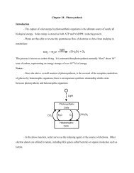
Chapter 9 - Lipids and Biological Membranes
Chapter 9 - Lipids and Biological Membranes
Chapter 9 - Lipids and Biological Membranes

Create successful ePaper yourself
Turn your PDF publications into a flip-book with our unique Google optimized e-Paper software.
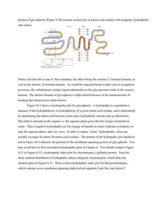
portion of glycophorin (Figure 9-20) consists exclusively of amino acid residues with nonpolar, hydrophobicside chains:Notice also that this is one of three domains, the others being the exterior, C-terminal domain, aswell as the interior, N-terminal domain. As would be expected based on their role in recognitionprocesses, the carbohydrate residues (green diamonds) on this glycoprotein reside in the exteriordomain. The interior domain of glycophorin is alpha helical because of the intramolecular H-bonding that characterizes alpha helices.Figure 10-3 shows a hydropathy plot for glycophorin. A hydropathy is a quantitativemeasure of the hydrophobicity or hydrophilicity of a given amino acid residue, <strong>and</strong> is determinedby partitioning the amino acid between water <strong>and</strong> a hydrophobic solvent such as chloroform.The relative amounts in the organic vs. the aqueous phase gives the free energy of transfer towater. Thus a negative hydrophathy (or free energy of transfer to water) indicates a tendency toseek the aqueous phase, <strong>and</strong> vice versa. In order to reduce “noise,” hydrophathy values areactually averages for about 20 amino acid residues. The portion of the hydropathy plot shaded inred in Figure 10-3 indicates the position of the membrane-spanning portion of glycophorin. Youmay recall that we first encountered hydropathy plots in <strong>Chapter</strong> 6. You should compare Figure9-21 to Figure 6-35, a hydropathic index plot for chymotrypsin, a globular protein. Note thefairly r<strong>and</strong>om distribution of hydropathic indices along the chymotrypsin, which lacks theshaded region in Figure 9-21. What would a hydropathic index plot for Bacteriorhodopsin,which contains seven membrane-spanning alpha helical segments, look like (see below)?