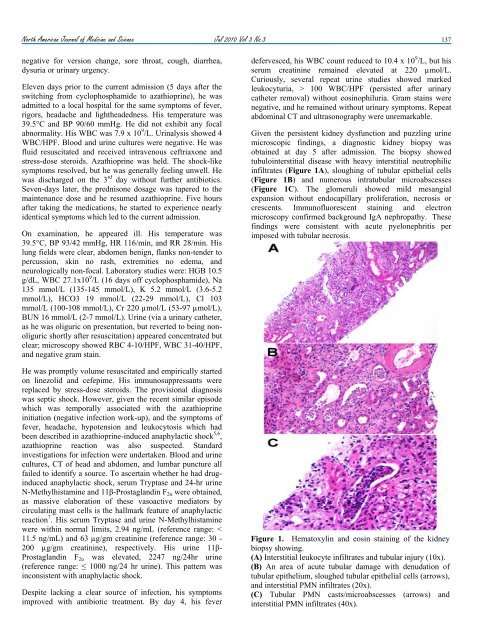
North Americ<strong>an</strong> Journal of Medic<strong>in</strong>e <strong>an</strong>d Science Jul 2010 Vol 3 No.3 137negative for version ch<strong>an</strong>ge, sore throat, cough, diarrhea,dysuria or ur<strong>in</strong>ary urgency.Eleven days prior to the current admission (5 days after theswitch<strong>in</strong>g from cyclophosphamide to azathiopr<strong>in</strong>e), he w<strong>as</strong>admitted to a local hospital for the same symptoms of fever,rigors, headache <strong>an</strong>d lightheadedness. His temperature w<strong>as</strong>39.5°C <strong>an</strong>d BP 90/60 mmHg. He did not exhibit <strong>an</strong>y focalabnormality. His WBC w<strong>as</strong> 7.9 x 10 9 /L. Ur<strong>in</strong>alysis showed 4WBC/HPF. Blood <strong>an</strong>d ur<strong>in</strong>e cultures were negative. He w<strong>as</strong>fluid resuscitated <strong>an</strong>d received <strong>in</strong>travenous ceftriaxone <strong>an</strong>dstress-dose steroids. Azathiopr<strong>in</strong>e w<strong>as</strong> held. The shock-likesymptoms resolved, but he w<strong>as</strong> generally feel<strong>in</strong>g unwell. Hew<strong>as</strong> discharged on the 3 rd day without further <strong>an</strong>tibiotics.Seven-days later, the prednisone dosage w<strong>as</strong> tapered to thema<strong>in</strong>ten<strong>an</strong>ce dose <strong>an</strong>d he resumed azathiopr<strong>in</strong>e. Five hoursafter tak<strong>in</strong>g the medications, he started to experience nearlyidentical symptoms which led to the current admission.On exam<strong>in</strong>ation, he appeared ill. His temperature w<strong>as</strong>39.5°C, BP 93/42 mmHg, HR 116/m<strong>in</strong>, <strong>an</strong>d RR 28/m<strong>in</strong>. Hislung fields were clear, abdomen benign, fl<strong>an</strong>ks non-tender topercussion, sk<strong>in</strong> no r<strong>as</strong>h, extremities no edema, <strong>an</strong>dneurologically non-focal. Laboratory studies were: HGB 10.5g/dL, WBC 27.1x10 9 /L (16 days off cyclophosphamide), Na135 mmol/L (135-145 mmol/L), K 5.2 mmol/L (3.6-5.2mmol/L), HCO3 19 mmol/L (22-29 mmol/L), Cl 103mmol/L (100-108 mmol/L), Cr 220 µmol/L (53-97 µmol/L),BUN 16 mmol/L (2-7 mmol/L). Ur<strong>in</strong>e (via a ur<strong>in</strong>ary catheter,<strong>as</strong> he w<strong>as</strong> oliguric on presentation, but reverted to be<strong>in</strong>g nonoliguricshortly after resuscitation) appeared concentrated butclear; microscopy showed RBC 4-10/HPF, WBC 31-40/HPF,<strong>an</strong>d negative gram sta<strong>in</strong>.He w<strong>as</strong> promptly volume resuscitated <strong>an</strong>d empirically startedon l<strong>in</strong>ezolid <strong>an</strong>d cefepime. His immunosuppress<strong>an</strong>ts werereplaced by stress-dose steroids. The provisional diagnosisw<strong>as</strong> septic shock. However, given the recent similar episodewhich w<strong>as</strong> temporally <strong>as</strong>sociated with the azathiopr<strong>in</strong>e<strong>in</strong>itiation (negative <strong>in</strong>fection work-up), <strong>an</strong>d the symptoms offever, headache, hypotension <strong>an</strong>d leukocytosis which hadbeen described <strong>in</strong> azathiopr<strong>in</strong>e-<strong>in</strong>duced <strong>an</strong>aphylactic shock 5,6 ,azathiopr<strong>in</strong>e reaction w<strong>as</strong> also suspected. St<strong>an</strong>dard<strong>in</strong>vestigations for <strong>in</strong>fection were undertaken. Blood <strong>an</strong>d ur<strong>in</strong>ecultures, CT of head <strong>an</strong>d abdomen, <strong>an</strong>d lumbar puncture allfailed to identify a source. To <strong>as</strong>certa<strong>in</strong> whether he had drug<strong>in</strong>duced<strong>an</strong>aphylactic shock, serum Trypt<strong>as</strong>e <strong>an</strong>d 24-hr ur<strong>in</strong>eN-Methylhistam<strong>in</strong>e <strong>an</strong>d 11β-Prostagl<strong>an</strong>d<strong>in</strong> F 2α were obta<strong>in</strong>ed,<strong>as</strong> m<strong>as</strong>sive elaboration of these v<strong>as</strong>oactive mediators bycirculat<strong>in</strong>g m<strong>as</strong>t cells is the hallmark feature of <strong>an</strong>aphylacticreaction 7 . His serum Trypt<strong>as</strong>e <strong>an</strong>d ur<strong>in</strong>e N-Methylhistam<strong>in</strong>ewere with<strong>in</strong> normal limits, 2.94 ng/mL (reference r<strong>an</strong>ge: 100 WBC/HPF (persisted after ur<strong>in</strong>arycatheter removal) without eos<strong>in</strong>ophiluria. Gram sta<strong>in</strong>s werenegative, <strong>an</strong>d he rema<strong>in</strong>ed without ur<strong>in</strong>ary symptoms. Repeatabdom<strong>in</strong>al CT <strong>an</strong>d ultr<strong>as</strong>onography were unremarkable.Given the persistent kidney dysfunction <strong>an</strong>d puzzl<strong>in</strong>g ur<strong>in</strong>emicroscopic f<strong>in</strong>d<strong>in</strong>gs, a diagnostic kidney biopsy w<strong>as</strong>obta<strong>in</strong>ed at day 5 after admission. The biopsy showedtubulo<strong>in</strong>terstitial dise<strong>as</strong>e with heavy <strong>in</strong>terstitial neutrophilic<strong>in</strong>filtrates (Figure 1A), slough<strong>in</strong>g of tubular epithelial cells(Figure 1B) <strong>an</strong>d numerous <strong>in</strong>tratubular microabscesses(Figure 1C). The glomeruli showed mild mes<strong>an</strong>gialexp<strong>an</strong>sion without endocapillary proliferation, necrosis orcrescents. Immunofluorescent sta<strong>in</strong><strong>in</strong>g <strong>an</strong>d electronmicroscopy confirmed background IgA nephropathy. Thesef<strong>in</strong>d<strong>in</strong>gs were consistent with acute pyelonephritis perimposed with tubular necrosis.Figure 1. Hematoxyl<strong>in</strong> <strong>an</strong>d eos<strong>in</strong> sta<strong>in</strong><strong>in</strong>g of the kidneybiopsy show<strong>in</strong>g.(A) Interstitial leukocyte <strong>in</strong>filtrates <strong>an</strong>d tubular <strong>in</strong>jury (10x).(B) An area of acute tubular damage with denudation oftubular epithelium, sloughed tubular epithelial cells (arrows),<strong>an</strong>d <strong>in</strong>terstitial PMN <strong>in</strong>filtrates (20x).(C) Tubular PMN c<strong>as</strong>ts/microabscesses (arrows) <strong>an</strong>d<strong>in</strong>terstitial PMN <strong>in</strong>filtrates (40x).
138 Jul 2010 Vol 3 No.3 North Americ<strong>an</strong> Journal of Medic<strong>in</strong>e <strong>an</strong>d ScienceIn light of the biopsy f<strong>in</strong>d<strong>in</strong>gs, he w<strong>as</strong> given <strong>an</strong> extended (6-week) course of cefprozil. He responded with a completeresolution of leukocyturia, <strong>an</strong>d a month later his serumcreat<strong>in</strong><strong>in</strong>e level returned to 106 µmol/L. He cont<strong>in</strong>ued regularfollow-up at our Out-Patient Cl<strong>in</strong>ic.DiscussionUTI/pyelonephritis is one of the most common <strong>in</strong>fections <strong>in</strong>immunocompromised hosts <strong>an</strong>d a major source of morbidity<strong>an</strong>d mortality. 2,3 Pathogenic Escherichia coli is by far themost common pathogen, account<strong>in</strong>g for the v<strong>as</strong>t majority ofUTIs. In our patient, although blood <strong>an</strong>d ur<strong>in</strong>e cultures werenegative (possibly related to <strong>an</strong>tibiotic exposure), his cl<strong>in</strong>ical<strong>an</strong>d laboratory presentations, kidney biopsy f<strong>in</strong>d<strong>in</strong>gs,response to <strong>an</strong>tibiotics, <strong>an</strong>d the absence of other causes forhis symptomatology (negative <strong>an</strong>aphylaxis workup) <strong>in</strong>dicatedthat he had septic shock secondary to bacterialpyelonephritis.UTI <strong>in</strong>clud<strong>in</strong>g pyelonephritis is the outcome of <strong>in</strong>teractionbetween the bacteria virulence characteristics <strong>an</strong>d the hostdefense mech<strong>an</strong>isms. Pathogenic Escherichia coli typicallyga<strong>in</strong> access to the g<strong>as</strong>tro<strong>in</strong>test<strong>in</strong>al tract, <strong>an</strong>d from there spreadto the ur<strong>in</strong>ary tract <strong>an</strong>d <strong>as</strong>cend to <strong>in</strong>vade kidneys.Pathogenic Escherichia coli adhere to the uroepitheliumthrough their surface molecule adhes<strong>in</strong> (hair-like projectionsalso termed p-fimbriae or pyelonephritis-<strong>as</strong>sociated pili).Adhes<strong>in</strong> also decre<strong>as</strong>es IgA tr<strong>an</strong>sport <strong>in</strong>to the ur<strong>in</strong>e result<strong>in</strong>g<strong>in</strong> a reduction of local host defense. 8 Althoughpolymorphonuclear cells (PMNs) do not possess adhes<strong>in</strong>receptor, they possess receptors b<strong>in</strong>d<strong>in</strong>g to <strong>an</strong>other group ofpili of Escherichia coli, type I pili. The b<strong>in</strong>d<strong>in</strong>g of PMNs <strong>an</strong>dtype I pili prevents bacteria adhesion to the uroepithelia.Although rare stra<strong>in</strong>s of Escherichia coli c<strong>an</strong> shed their pili 9thereby circumvent<strong>in</strong>g the effects of PMNs, <strong>in</strong> mostcircumst<strong>an</strong>ces, PMNs b<strong>in</strong>d bacteria <strong>an</strong>d protect hosts from<strong>in</strong>fection. The pathogen <strong>in</strong> the current c<strong>as</strong>e w<strong>as</strong> mostly likelyEscherichia coli, <strong>as</strong> numerous epidemiologic studies<strong>in</strong>dicat<strong>in</strong>g that they are the most common cause of UTI <strong>in</strong>immunocompromised hosts. 10With regard to the host defense, our patient had severaldeficiencies. First <strong>an</strong>d perhaps most import<strong>an</strong>t, he had beenunder heavy immunosuppression for 6 months. Drug-<strong>in</strong>ducedmarrow suppression prevented a robust leukemoid reaction tothe <strong>in</strong>fection. Second, secretory IgAs are known toparticipate <strong>in</strong> the ma<strong>in</strong>ten<strong>an</strong>ce of mucosal surface immunehomeost<strong>as</strong>is <strong>an</strong>d confer protection aga<strong>in</strong>st pathogencolonization. Secretory IgAs also c<strong>an</strong> neutralize bacteria PLS(import<strong>an</strong>t for Escherichia coli pathogenesis) <strong>in</strong>dependent ofgr<strong>an</strong>ulocyte cells or T-cells. Our patient had IgAnephropathy, which is pathogenetically related to defects <strong>in</strong>O-l<strong>in</strong>ked glycosylations. Such defects c<strong>an</strong> alter the chargecharacteristics of IgA1 molecules. IgA1 with <strong>in</strong>complete O-galactosylation also tends to self-aggregate, 9 thereby maydim<strong>in</strong>ish its physiological functions. Third, our patient h<strong>as</strong>diabetes, which is one of the metabolic factors known toenh<strong>an</strong>ce bacterial <strong>in</strong>v<strong>as</strong>iveness with<strong>in</strong> ur<strong>in</strong>ary tract <strong>in</strong>immune compromised hosts.Cl<strong>as</strong>sically, history <strong>an</strong>d physical exam<strong>in</strong>ation are essential <strong>in</strong>identify<strong>in</strong>g the existence <strong>an</strong>d source of <strong>in</strong>fection. In thispatient, although he appeared <strong>in</strong> shock, a diagnosis of<strong>in</strong>fection <strong>as</strong> <strong>an</strong> etiology of his presentation w<strong>as</strong> called <strong>in</strong>toquestion because (1) by history, he had been on TMP/SMXprophylaxis s<strong>in</strong>ce the start of the immunosuppressivetreatment <strong>an</strong>d he did not show <strong>an</strong>y focal (ur<strong>in</strong>ary) symptoms(might be related to the <strong>in</strong>sufficient local leukemoid reaction,at le<strong>as</strong>t <strong>in</strong>itially), <strong>an</strong>d (2) on exam<strong>in</strong>ation, he did not havefl<strong>an</strong>k pa<strong>in</strong> or abdom<strong>in</strong>al tenderness. This atypicalpresentation created a diagnostic dilemma.Immunosuppressive therapy c<strong>an</strong> m<strong>as</strong>k the cl<strong>in</strong>icalm<strong>an</strong>ifestations of <strong>in</strong>fection. Our patient did not haveleukocytosis at his <strong>in</strong>itial presentation to the local hospital,despite sepsis-like presentation. This is likely related to themarrow suppressive effect of cyclophosphamide. In l<strong>in</strong>e withthis <strong>as</strong>sumption, his serum leukocyte counts (<strong>an</strong>d ur<strong>in</strong>eleukocytes) were <strong>in</strong>cre<strong>as</strong>ed <strong>in</strong> the succeed<strong>in</strong>g days <strong>as</strong> moretime elapsed from the l<strong>as</strong>t dosage of cyclophosphamide,consistent with a gradual recovery of leukemoid response to<strong>in</strong>fection. Although the steroid adm<strong>in</strong>istration dur<strong>in</strong>g hishospital stay could have contributed to the leukocytosis, itw<strong>as</strong> unlikely to have been a major factor because theleukocytosis on admission preceded the <strong>in</strong>itiation of stressdosesteroids. Thus, the <strong>in</strong>itial lack of leucocytosis w<strong>as</strong> likelyrelated to drug-<strong>as</strong>sociated marrow suppression. Incidentally,this c<strong>as</strong>e also served to dispel the notion that patients onTMP/SMX prophylaxis would not be at risk for UTI. Ourpatient developed pyelonephritis while on TMP/SMX, <strong>an</strong>drecent studies also show that over 70% uropathogenicbacteria are <strong>in</strong>sensitive or resist<strong>an</strong>t to TMP/SMX 11 .Although <strong>in</strong>fection, given its potential fatality, should bedoggedly pursued, it is plausible to <strong>in</strong>clude multiplediagnostic considerations. The atypical presentations <strong>in</strong> thisc<strong>as</strong>e led to seek<strong>in</strong>g alternative expl<strong>an</strong>ations, such <strong>as</strong> pseudosepsisdue to drug reaction.Azathiopr<strong>in</strong>e is <strong>an</strong> imidazole derivative of 6-mercaptopur<strong>in</strong>e.The allergic-type reactions have been described <strong>as</strong> a rare <strong>an</strong>ddose-<strong>in</strong>dependent event occurr<strong>in</strong>g with<strong>in</strong> days to weeksfollow<strong>in</strong>g the drug <strong>in</strong>troduction. 6 The reactions c<strong>an</strong> r<strong>an</strong>gefrom p<strong>an</strong>creatitis, hepatitis, sk<strong>in</strong> r<strong>as</strong>h, fever, arthralgi<strong>as</strong>,malaise, diarrhea <strong>an</strong>d abdom<strong>in</strong>al pa<strong>in</strong> to <strong>an</strong>aphylactic shock.The <strong>an</strong>aphylactic reaction/shock is characterized by abruptonset of headache, fever, hypotension <strong>an</strong>d leukocytosis. 5,6Although our patient’s presentation fit well with thisdescription, his laboratory studies did not. In a typical<strong>an</strong>aphylactic reaction, circulat<strong>in</strong>g m<strong>as</strong>t cells are acutelyactivated, rele<strong>as</strong><strong>in</strong>g a host of v<strong>as</strong>oactive mediators <strong>in</strong>clud<strong>in</strong>gtrypt<strong>as</strong>e, histam<strong>in</strong>e, <strong>an</strong>d prostagl<strong>an</strong>d<strong>in</strong> D2. Serum <strong>an</strong>d ur<strong>in</strong>elevels of these mediators <strong>an</strong>d their metabolites, N-Methylhistam<strong>in</strong>e <strong>an</strong>d 11β-Prostagl<strong>an</strong>d<strong>in</strong> F 2α , are expected tobe m<strong>as</strong>sively <strong>an</strong>d simult<strong>an</strong>eously elevated. Our patient had <strong>an</strong>isolated elevation of 11β-Prostagl<strong>an</strong>d<strong>in</strong> F 2α , <strong>in</strong>consistent with<strong>an</strong>aphylactic reaction. 7 In fact, the isolated 11β-Prostagl<strong>an</strong>d<strong>in</strong>F 2α elevation (to the level comparable to that <strong>in</strong> our patient)c<strong>an</strong> be seen <strong>in</strong> physiologic conditions dur<strong>in</strong>g normal labor 12or sleep 13 . Under these circumst<strong>an</strong>ces 11β-Prostagl<strong>an</strong>d<strong>in</strong> F 2α