Electrospun Chitosan–Alginate Nanofibers with In Situ ...
Electrospun Chitosan–Alginate Nanofibers with In Situ ...
Electrospun Chitosan–Alginate Nanofibers with In Situ ...

You also want an ePaper? Increase the reach of your titles
YUMPU automatically turns print PDFs into web optimized ePapers that Google loves.
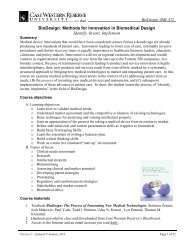
TEA-2010-0086-ver9-Jeong_1P.3d 08/30/10 1:25pm Page 5Table 1. Electrospinning Conditions of <strong>Chitosan–Alginate</strong>–Poly(Ethylene Oxide) Nanofibrous ScaffoldsElectrospinning conditionsSamplecodeChitosan aconcentration(wt%)Alginate bconcentration(wt%)PEO b concentration(wt%) (Mw ¼ 900 kDa)Final solutionvolume ratio(chitosan:alginate:PEO)Final wt%(chitosan:alginate:PEO)Voltage(kV)Needlesize (G)Tip-to-collectordistance (cm)Flow rate(mL/min)CP 2080 5.0 4.0 20:00:80 1.00:3.20CP 3070 30:00:70 1.50:2.80CP 4060 40:00:60 2.00:2.40CP 5050 50:00:50 2.50:2.00AP 2080 2.0 4.0 00:20:80 0.40:3.20 13 20 10 0.02AP 3070 00:30:70 0.60:2.80AP 4060 00:40:60 0.80:2.40AP 5050 00:50:50 1.00:2.00CAP 104050 5.0 2.0 4.0 10:40:50 0.50:0.80:2.00CAP 252550 25:25:50 1.25:0.50:2.00CAP 401050 40:10:50 2.00:0.20:2.00CAP 202060 20:20:60 1.00:0.40:2.40CAP 151570 15:15:70 0.75:0.30:2.80CAP 101080 10:10:80 0.50:0.20:3.20a Chitosan was dissolved in 1 M acetic acid.b Alginate and PEO were dissolved in dH 2O.Mw, molecular weight; PEO, poly(ethylene oxide).5
TEA-2010-0086-ver9-Jeong_1P.3d 08/30/10 1:25pm Page 66 JEONG ET AL.FIG. 2. Scanning electronphotomicrographs of electrospunCP (a–d) and AP (e–h)nanofibers. (a) CP 2080, (b) CP3070, (c) CP 4060, (d) CP 5050,(e) AP 2080, (f) AP 3070, (g) AP4060, and (h) AP 5050. Scalebars represent 3 mm. CP, chitosan–PEO;AP, alginate–PEO.F2 cpolysaccharides ( Fig. 2). For all blending ratios shown, uniformnanofibers could be obtained. If the volume ratio ofPEO was decreased below 50%, it was found that all thenanofibers contained bead structures (data not shown); thus,the PEO volume ratio was maintained at 50% or higher forFIG. 3. (a) Viscosity and (b) conductivity of the CP and APsolutions at varying volume ratios of PEO in the blend. Valuesrepresent mean standard deviation.this study. The viscosity and conductivity of these solutionswere measured to characterize the solution properties thatallowed for optimal nanofiber formation. Uniform nanofiberswere formed when the volume ratio of the PEO wasbetween 50%–80%. These solutions had viscosities approximatelybetween 0.15 and 0.7 Pa s( Fig. 3a), indicating thatthis was an ideal range for electrospinning. The addition ofPEO increased the viscosity of the alginate solutions butdecreased the viscosity of the chitosan solutions. Additionally,it was found that solutions <strong>with</strong> more PEO exhibitedlower conductivity, which resulted in the formationof uniform nanofibers during the electrospinning process(Fig. 3b).Once it was determined that the ideal condition for electrospinningCP and AP was to maintain a PEO volume ratio>50%, alginate–chitosan polyelectrolyte complexed nanofiberswere then fabricated and their morphologies examined.Chitosan and alginate were both blended <strong>with</strong> PEO, andvarious ratios of these solutions were used for electrospinning( Fig. 4). <strong>In</strong> two conditions, completely uniform nanofiberswere obtained (Fig. 4d, e). Two others exhibitedprimarily uniform nanofibers <strong>with</strong> only a few beadedstructures (Fig. 4b, f). <strong>In</strong> the two mixtures where the volumeratio of chitosan and alginate were not the same (CAP 104050and CAP 401050), bead-containing nanofibers were obtained(Fig. 4a, c).Although the PEO was required for the electrospinning,this polymer did not contribute functionality to the formednanofibers, and thus it was desired to remove it from thefibers. To obtain nanofibers containing only alginate andchitosan <strong>with</strong>out PEO, the nanofiber scaffolds were incubatedin diH 2 Oat378C for 5 days to extract the water-solublePEO. To ensure that the nanofibrous structure was still intact,the scaffolds were again examined using SEM ( Fig. 5).For all blending ratios, it was apparent that the nanofibrousstructure was indeed intact. The scaffolds that previouslycontained beaded structures <strong>with</strong>in the nanofibers no longerhad the beads present.To examine the gel properties of the chitosan–alginatepolyelectrolyte complexes at the various ratios used in electrospinning,their storage and loss moduli were measuredover time <strong>with</strong> a rheometer ( Fig. 6). <strong>In</strong> all blends, the storagemodulus was always greater than the loss modulus, indicatingthat the polyelectrolytes complex and form a hydrogelimmediately upon mixing. 11 CAP 104050 and CAP 401050,b F3b F4b F5b F6
TEA-2010-0086-ver9-Jeong_1P.3d 08/30/10 1:26pm Page 7ELECTROSPUN CHITOSAN–ALGINATE NANOFIBERS 7FIG. 4. Scanning electronphotomicrographs of CAP nanofibers.(a) CAP 104050, (b)CAP 252550, (c) CAP 401050,(d) CAP 202060, (e) CAP151570, and (f) CAP 101080.Scale bars represent 6 mm, and1 mm in the inset. CAP, chitosan–alginate–PEO.F7 cthe two mixtures that formed bead-containing nanofibersupon electrospinning, both exhibited the highest storagemoduli of any of the blends (Fig. 6a, c). The two mixturesthat formed the uniform nanofibers <strong>with</strong>out beads, CAP202060 and CAP 151570, had the lowest storage moduli (Fig.6d, e).The average fiber diameter of each of the electrospunscaffold compositions was measured using the SEM images( Fig. 7). <strong>In</strong> all cases, the fiber diameters were larger beforeextraction of the PEO compared to after extraction. Additionally,in the scaffolds that did not contain beadedstructures before PEO extraction (CAP 202060 and CAP151570), the fiber diameter before extraction increased as theamount of PEO in the blend increased. The CAP 101080 onlyslightly increased compared to the CAP 252550. Also, althoughnot shown in Figure 7, all of the fibers after PEOextraction had statistically significant differences in their fiberdiameters, but they are all on the order of 100 nm still.To confirm the chemical composition of the electrospunfibers, ATR FT-IR was used. The spectra of the pure components(chitosan, alginate, and PEO) were compared tothose of the electrospun chitosan–PEO–alginate fibers bothbefore and after PEO extraction. The peaks that indicate theamide and carboxyl groups of the chitosan and alginate arehighlighted in Figure 8. The chitosan showed peaks at 3365and 3302 cm 1 due to the O–H and N–H stretch, and at 1654and 1593 cm 1 due to the amide bonds (Fig. 8a). The alginateshowed a peak at 3327 cm 1 from the O–H stretch, another at1603 cm 1 due to the antisymmetric carboxyl stretch and oneat 1413 cm 1 due to the symmetric carboxyl stretch (Fig. 8b).The spectra of the electrospun fibers before PEO extraction(Fig. 8d) exhibited peaks that were also present in the purePEO sample (Fig. 8c). However, after the PEO extractionthese peaks were no longer present (Fig. 8e), indicating thatthe PEO has indeed been successfully extracted from thesenanofibers leaving only peaks corresponding to the complexedchitosan and alginate. Further, the spectra of the fibersafter PEO extraction show a broad peak at 1596 cm 1and a peak at 1416 cm 1 (Fig. 8e). These result from thecarboxyl groups of the alginate overlapping <strong>with</strong> the signalfrom the chitosan, indicating the presence of both compounds.The chitosan used in this study was not a water-solublesalt. Thus, it is expected that the nanofibers containingb F8FIG. 5. Scanning electronphotomicrographs of CAP nanofibersafter PEO extraction.(a) CAP 104050, (b) CAP252550, (c) CAP 401050, (d)CAP 202060, (e) CAP 151570,and (f) CAP 101080. Scale barsrepresent 6 mm, and 1 mm in theinset.
TEA-2010-0086-ver9-Jeong_1P.3d 08/30/10 1:26pm Page 88 JEONG ET AL.FIG. 6. Elastic storage (G 0 ) and viscous loss (G 00 ) moduli change over time during formation of CAP hydrogel blends. (a)CAP 104050, (b) CAP 252550, (c) CAP 401050, (d) CAP 202060, (e) CAP 151570, and (f) CAP 101080. Values representmean standard deviation.F9 cchitosan and alginate would have less swelling in an aqueousenvironment compared to those composed only of water-solublealginate. The swelling ratio did decrease as theamount of chitosan increased ( Fig. 9). The scaffolds containing10% chitosan, 40% alginate, and 50% PEO by volumeshowed a higher degree of swelling than the other chitosan–alginate scaffolds, likely due to the higher amount of hydrophilicalginate present in these fibers.For use of these nanofibers in tissue engineering applications,it is important to examine how cells interact <strong>with</strong>the nanofibrous scaffolds. Alginate is naturally nonadhesiveto cells. The addition of the chitosan was expected topromote cell adhesion since it is a polycation that is ableto adsorb serum proteins, which can subsequently allowcells to adhere to the material. Mouse preosteoblast cells(MC3T3s) were seeded onto the surface of alginate-onlynanofibers, chitosan–alginate nanofibers, and tissue cultureplastic as a positive control. The cells were stained <strong>with</strong> aLive/Dead stain to assess their viability on these surfaces( Fig. 10a–i). On each of the surfaces, all of the cellsb F10FIG. 7. Average fiber diameter of chitosan–alginate nanofibersbefore and after PEO extraction in deionized water at378C for 5 days. Values represent mean standard deviation.FIG. 8. Attenuated total reflectance Fourier transform infraredspectra of (a) chitosan, (b) alginate, (c) PEO, (d) CAPnanofibers before PEO extraction (CAP 252550), and (e)chitosan–alginate nanofibers after PEO extraction (CAP252550).
TEA-2010-0086-ver9-Jeong_1P.3d 08/30/10 1:26pm Page 1010 JEONG ET AL.FIG. 10. Live/Dead stainingof MC3T3 preosteoblast cellscultured for 24, 72, and 120 h on(a–c) TCP, (d–f) PEO-extractedAP 5050, and (g–i) PEO-extractedCAP 252550 nanofibers(scale bars represent 40 (m). (j)Cell proliferation of MC3T3scultured on TCP, AP 5050, andCAP 252550 for up to 120 h, asdetermined by an MTS assay,which measures the cell metabolicactivity. *p < 0.05. Valuesrepresent mean standarddeviation. TCP, tissue cultureplastic.Color images availableonline at www.liebertonline.com/ten.FIG. 11. Scanning electron micrographsof CAP nanofibers fabricated<strong>with</strong> water-soluble chitosan salt and(a) cell adhesion ligand (GRGDSP)-modified alginate or (b) unmodifiedalginate. Scale bars represent 3 mm.
TEA-2010-0086-ver9-Jeong_1P.3d 08/30/10 1:26pm Page 11ELECTROSPUN CHITOSAN–ALGINATE NANOFIBERS 11studies was only soluble in an acidic solution. Since thischitosan was not a water-soluble salt, it limited the swellingof the scaffolds in aqueous solutions. As an alternative tousing chitosan that is only soluble at lower pH, it wasdemonstrated that a water-soluble chitosan salt could insteadbe used. This may be useful for applications where it isdesirable to encapsulate biologically active factors or cells inthe nanofibers during the electrospinning. Additionally, itwas demonstrated that the alginate could be modified <strong>with</strong> apeptide containing the cell-adhesive RGD sequence. Ourfuture work will include examining whether the addition ofthis cell adhesion ligand and others could further promoteand control the adhesion, spreading, proliferation, migration,and differentiation of cells on these scaffolds. Further, thiswork examined only the ability of two different chitosans toform polyionic complexes <strong>with</strong> alginate during eletrospinning;in future, it will be informative to examine additionalchitosans, especially <strong>with</strong> differing degrees of deacetylation,as using chitosans <strong>with</strong> different properties may affect theelectrospinning process.Even <strong>with</strong>out the use of alginate containing a cell-adhesivepeptide sequence, the nanofibers obtained in this studyshowed great potential as biomaterial scaffolds capable ofsupporting cell adhesion and proliferation. When preosteoblastcells were seeded on the nanofibrous scaffolds,those seeded on the chitosan–alginate scaffolds exhibitedboth greater adhesion after 5 h and greater proliferation overthe course of 120 h compared to cells seeded on pure alginatescaffolds. This was due to the presence of chitosan, whichallowed for protein adsorption and subsequent attachmentand spreading of cells. Alginate is naturally nonadhesive tocells, and so they are unable to adhere or proliferate verywell on this material by itself. Thus, the use of the polyelectrolytecomplex enhances the utility of these scaffolds intissue engineering applications.Overall, this study demonstrates for the first time theability to electrospin chitosan–alginate polyelectrolytecomplexes. The optimal conditions for the electrospinningwere determined, and the resultant nanofibers thoroughlycharacterized. These nanofibers were crosslinked in situdue to the polyionic complexation of the chitosan and alginate,and thus did not require any additional chemicalcrosslinking step. The nanofibrous scaffolds were able topromote the adhesion and proliferation of cells, and theyoffergreatpromiseforuseasscaffoldsintissueregenerationstrategies. Future work will include optimization ofconditions for electrospinning <strong>with</strong> water-soluble chitosansalts, the use of peptide-modified alginate to permit additionalcontrol over cell interactions <strong>with</strong> the scaffolds, andthe incorporation of bioactive factors that can be deliveredto guide cellular behavior for specific tissue engineeringapplications.AcknowledgmentsThe authors acknowledge funding support for this workfrom Biomedical Research and Technology Transfer Grant08–081 from the Ohio Department of Development (E.A.)and a National Science Foundation Graduate Research Fellowship(M.D.K.). We thank Ms. Birgit Andersen (NorthCarolina State University) for assistance <strong>with</strong> the GPCanalysis.Disclosure StatementNo competing financial interests exist.References1. Mano, J.F., Silva, G.A., Azevedo, H.S., Malafaya, P.B., Sousa,R.A., Silva, S.S., et al. Natural origin biodegradable systemsin tissue engineering and regenerative medicine: presentstatus and some moving trends. J R Soc <strong>In</strong>terface 4, 999,2007.2. Augst, A.D., Kong, H.J., and Mooney, D.J. Alginate hydrogelsas biomaterials. Macromol Biosci 6, 623, 2006.3. Hashimoto, T., Suzuki, Y., Tanihara, M., Kakimaru, Y., andSuzuki, K. Development of alginate wound dressings linked<strong>with</strong> hybrid peptides derived from laminin and elastin.Biomaterials 25, 1407, 2004.4. Lee, W.R., Park, J.H., Kim, K.H., Kim, S.J., Park, D.H., Chae,M.H., et al. The biological effects of topical alginate treatmentin an animal model of skin wound healing. Wound RepairRegen 17, 505, 2009.5. Bouhadir, K.H., Lee, K.Y., Alsberg, E., Damm, K.L., Anderson,K.W., and Mooney, D.J. Degradation of partially oxidizedalginate and its potential application for tissueengineering. Biotechnol Prog 17, 945, 2001.6. Alsberg, E., Anderson, K.W., Albeiruti, A., Rowley, J.A., andMooney, D.J. Engineering growing tissues. Proc Natl AcadSci USA 99, 12025, 2002.7. Krebs, M.D., Salter, E., Chen, E., Sutter, K.A., and Alsberg, E.Calcium phosphate-DNA nanoparticle gene delivery fromalginate hydrogels induces in vivo osteogenesis. J BiomedMater Res A 92, 1131, 2010.8. Alsberg, E., Anderson, K.W., Albeiruti, A., Franceschi, R.T.,and Mooney, D.J. Cell-interactive alginate hydrogels forbone tissue engineering. J Dent Res 80, 2025, 2001.9. Prang, P., Muller, R., Eljaouhari, A., Heckmann, K., Kunz,W., Weber, T., et al. The promotion of oriented axonalregrowth in the injured spinal cord by alginate-basedanisotropic capillary hydrogels. Biomaterials 27, 3560,2006.10. Mosahebi, A., Simon, M., Wiberg, M., and Terenghi, G. Anovel use of alginate hydrogel as Schwann cell matrix. TissueEng 7, 525, 2001.11. Moura, M.J., Figueiredo, M.M., and Gil, M.H. Rheologicalstudy of genipin cross-linked chitosan hydrogels. Biomacromolecules8, 3823, 2007.12. Lee, K.Y., Jeong, L., Kang, Y.O., Lee, S.J., and Park, W.H.Electrospinning of polysaccharides for regenerative medicine.Adv Drug Deliv Rev 61, 1020, 2009.13. Lim, S.H., Liao, I.C., and Leong, K.W. Nonviral gene deliveryfrom nonwoven fibrous scaffolds fabricated by interfacialcomplexation of polyelectrolytes. Mol Ther 13,1163, 2006.14. Saether, H.V., Holme, H.K., Maurstad, G., Smidsrod, O., andStokke, B.T. Polyelectrolyte complex formation using alginateand chitosan. Carbohydr Polym 74, 813, 2008.15. Douglas, K.L., and Tabrizian, M. Effect of experimental parameterson the formation of alginate-chitosan nanoparticlesand evaluation of their potential application as DNA carrier.J Biomater Sci 16, 43, 2005.16. Lee, K.L., Park, W.H., and Ha, W.S. Polyelectrolyte complexesof sodium alginate <strong>with</strong> chitosan or its derivatives formicrocapsules. J Appl Polym Sci 63, 425, 1997.17. Gaserod, O., Smidsrod, O., and Skjak-Braek, G. Microcapsulesof alginate-chitosan—I. A quantitative study of the
TEA-2010-0086-ver9-Jeong_1P.3d 08/30/10 1:26pm Page 1212 JEONG ET AL.interaction between alginate and chitosan. Biomaterials 19,1815, 1998.18. Simsek-Ege, F.A., Bond, G.M., and Stringer, J. Polyelectrolytecomplex formation between alginate and chitosan asa function of pH. J Appl Polym Sci 88, 346, 2003.19. Rowley, J.A., Madlambayan, G., and Mooney, D.J. Alginatehydrogels as synthetic extracellular matrix materials. Biomaterials20, 45, 1999.20. Majima, T., Funakosi, T., Iwasaki, N., Yamane, S.T., Harada,K., Nonaka, S., et al. Alginate and chitosan polyion complexhybrid fibers for scaffolds in ligament and tendon tissueengineering. J Orthop Sci 10, 302, 2005.21. Fukuda, J., Khademhosseini, A., Yeo, Y., Yang, X., Yeh, J.,Eng, G., et al. Micromolding of photocrosslinkable chitosanhydrogel for spheroid microarray and co-cultures. Biomaterials27, 5259, 2006.22. Ashammakhi, N., Ndreu, A., Nikkola, L., Wimpenny, I., andYang, Y. Advancing tissue engineering by using electrospunnanofibers. Regen Med 3, 547, 2008.23. Chunder, A., Sarkar, S., Yu, Y., and Zhai, L. Fabrication ofultrathin polyelectrolyte fibers and their controlled releaseproperties. Colloids Surf B: Biointerfaces 58, 172, 2007.24. Penchev, H., Paneva, D., Manolova, N., and Rashkov, I.Novel electrospun nanofibers composed of polyelectrolytecomplexes. Macromol Rapid Commun 29, 677, 2008.25. Kim, E.K.F., Wan, A.C.A., Le Visage, C., Liao, I.C., and Leong,K.W. Proliferation and differentiation of human mesenchymalstem cell encapsulated in polyelectrolyte complexationfibrous scaffold. Biomaterials 27, 6111, 2006.26. Shao, X., and Hunter, C.J. Developing an alginate/chitosanhybrid fiber scaffold for annulus fibrosus cells. J BiomedMater Res 82, 701, 2007.27. Wang, J.Z., Huang, X.B., Xiao, J., Li, N., Yu, W.T., Wang, W.,et al. Spray-spinning: a novel method for making alginate/chitosan fibrous scaffold. J Mater Sci Mater Med 21, 497, 2010.28. Zhang, Y.Z., Su, B., Ramakrishna, S., and Lim, C.T. Chitosannanofibers from an easily electrospinnable UHMWPEO-dopedchitosan solution system. Biomacromolecules 9, 136, 2008.29. Bhattarai, N., Li, Z., Edmondson, D., and Zhang, M. Alginatebasednanofibrous scaffolds: structural, mechanical, andbiological properties. Adv Mater 18, 1463, 2006.30. Lu, J., Zhu, Y., Guo, Z., Hu, P., and Yu, J. Electrospinning ofsodium alginate <strong>with</strong> poly(ethylene oxide). Polymer 47,8026, 2006.31. Safi, S., Morshed, M., Hosseini Ravandi, S.A., and Ghiaci, M.Study of electrospinning of sodium alginate, blended solutionsof sodium alginate/poly(vinyl alcohol) and sodiumalginate/poly(ethylene oxide). J Appl Polym Sci 104, 3245,2007.32. Jeong, S.I., Krebs, M.D., Bonino, C.A., Khan, S.A., and Alsberg,E. <strong>Electrospun</strong> alginate nanofibers <strong>with</strong> controlled celladhesion for tissue engineering. Macromol Biosci 2010[Epub ahead of print].33. Ottoy, M.H., Varum, K.M., Christensen, B.E., Anthonsen,M.W., and Smidsrod, O. Preparative and analytical sizeexclusionchromatography of chitosans. Carbohydr Polym31, 253, 1996.34. Rubenstein, M., and Colby, R. Polymer Physics. New York:Oxford University Press; 2003.35. Caykara, T., Demirci, S., Eroglu, M.S., and Guven, O.Poly(ethylene oxide) and its blends <strong>with</strong> sodium alginate.Polymer 46, 10750, 2005.Address correspondence to:Eben Alsberg, Ph.D.Departments of Biomedical Engineering and Orthopaedic SurgeryCase Western Reserve UniversityWickenden Building, Room 20410900 Euclid Ave.Cleveland, OH 44106E-mail: eben.alsberg@case.eduReceived: February 09, 2010Accepted: July 29, 2010Online Publication Date:b AU2