CR-1 Mark II retinal camera brochure (PDF, 2MB) - Canon
CR-1 Mark II retinal camera brochure (PDF, 2MB) - Canon
CR-1 Mark II retinal camera brochure (PDF, 2MB) - Canon
Create successful ePaper yourself
Turn your PDF publications into a flip-book with our unique Google optimized e-Paper software.
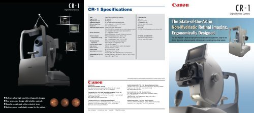
<strong>CR</strong>-1 SpecificationsTypeAngle of viewMagnification viewOperation distanceMinimum pupil sizeImage sizeAttachable digital <strong>camera</strong>Sensor resolutionPatient’s dioptercompensation rangeWorking distanceadjustmentInternal fixation targetLight sourceBuilt-in monitorPower supplyOperating environmentDimensions (W x D x H)WeightDigital <strong>retinal</strong> <strong>camera</strong>, Non mydriatic45 degreesX2 (Digital)35 mm from the front of objective lensø 4 mm (Approx. ø 3.7 mm in SP mode)ø 14 mm on the sensor<strong>Canon</strong> EOS digital SLR <strong>camera</strong>(For information on available <strong>camera</strong> models, please consultyour local authorized <strong>Canon</strong> sales representative.)10.1 megapixels or higher(Resolution depends on model of attached <strong>camera</strong>.)Without compensation lens: -10D to +15DWith “-” compensation lens: -7D to -31DWith “+” compensation lens: +11D to +33DAnterior eye display: split lines adjustmentRetinal display: working distance dotsLED dot matrix, greenIRED for observation, Xenon tube for photography5.7-inch color LCD monitorAC100-240 50/60Hz 1-0.4ATemperature: 10˚C to 35˚CHumidity: 30% RH to 80% RH (with no condensation)320 mm x 530 mm x 550 mm (12.7 in. x 20.7 in. x 21.9 in.)Approx. 21.5 kg (47.4 lb.)COMPONENTSMain unitObjective lens capCamera mount capChin rest paper (100 sheets)Power cableDust coverCD-ROM (Retinal imaging control software NM)OPTIONAL ACCESSORIESExternal eye fixation lamp EL-1Chin rest paper (500 sheets)The State-of-the-Art inNon-Mydriatic Retinal Imaging,Ergonomically DesignedThe Non-Myd <strong>CR</strong>-1 features high-performance specs in an ergonomic, easy-to-usedesign to provide enhanced quality, efficiency and comfort during <strong>retinal</strong> exams.Simulated images and specifications are subject to change without notice.■ Delivers ultra-high resolution diagnostic images■ New ergonomic design with intuitive controls■ Easy to operate and achieve desired views■ Quicker, more comfortable exams for the patientCANON INC.MEDICAL EQUIPMENT GROUP30-2, Shimomaruko 3-chome, Ohta-ku, Tokyo 146-8501, JapanTelephone: +81-3-3757-8497 Fax: +81-3-5482-3960CANON MEDICAL SYSTEMS A division of CANON U.S.A., Inc.15955 Alton Parkway, Irvine, CA 92618-3731, U.S.A.Telephone: (1) 949-753-4160, toll-free within U.S.A.: 1-800-970-7227Fax: (1) 949-753-4184http://www.cusa.canon.com/eye-careCANON EUROPA N.V. Medical Systems DivisionBovenkerkerweg 59-61, 1185 XB Amstelveen, The NetherlandsTelephone: +31-(0) 20-545-8926 Fax: +31-(0) 20-545-8220http://www.canon-europa.com/MedicalCode: 0120W800 © CANON INC. 2008 0308SZ0 PRINTED IN JAPANCANON SINGAPORE PTE. LTD. Medical Equipment Dept.1 Harbour Front Avenue, #04-01 Keppel Bay Tower, Singapore 098632Telephone: +65-6796-3549 Fax: +65-6271-4226http://www.canon-asia.comCANON (CHINA) CO, LTD. Medical Division15F Jinbao Beijing No.89 Jinbao Street, Dongcheng District,Beijing 100005, ChinaTelephone: (86) 10-8513-9999 Fax: (86) 10-8513-9914http://www.canon.com.cnCANON AUSTRALIA PTY. LTD. Optical Division1 Thomas Holt Drive, North Ryde, Sydney, NSW 2113, AustraliaTelephone: +61-2-9805-2000 Fax: +61-2-9805-2444
Designed and engineered foroutstanding image quality and easeof-use,plus greater patient comfortEver since developing the world’s first non-mydriatic <strong>retinal</strong> <strong>camera</strong> in 1976, <strong>Canon</strong> has been apioneer in the field of <strong>retinal</strong> imaging. <strong>Canon</strong>’s latest advancement, the non-mydriatic digitalKey features■ True 45-degree imageThe <strong>CR</strong>-1 features aredesigned opticalsystem that achieveshigh-resolutiondiagnostic images at a45° angle of view.■ Bright, easy-view 5.7-inch swivel LCD monitorAnterior eye and stereo photography are availableEasy alignment and focusingImage capture is fast and easy thanks to a simple two-step procedure.First, you align the two halves of a split pupil image. Then you switch tothe <strong>retinal</strong> display, and adjust the split lines and working distance dots toachieve the correct focus and distance to the retina for clear, sharpimages every time.Working distance dotsSplit lines<strong>CR</strong>-1, combines state-of-the-art optics and <strong>retinal</strong> imaging technology with the renowned EOS▲▲High-quality, high-resolution imagesdigital SLR system to provide industry-leading image quality and efficiency. All this, in an all-newergonomic design that’s more comfortable for the patient and easier to use than ever before.The <strong>CR</strong>-1 features a redesigned optical system that achievesextremely detailed, high-resolution diagnostic images of the retina foraccurate detection and monitoring of ocular conditions includingdiabetic retinopathy, glaucoma, and macular degeneration. The highpixel count of the attached EOS digital SLR <strong>camera</strong> delivers detailedimage quality even when magnified. Once captured, images aretransferred to a connected PC for review.Comfortable, ergonomicdesignThe all-new design of the <strong>CR</strong>-1 integratesadvanced specifications into an ergonomic unit thatfacilitates operation by motorizing proceduresusually performed by hand. The intuitive controls,viewing monitor, and the streamlined form have allbeen designed for improved ease of use andcomfort. It’s also patient-friendly. Because <strong>retinal</strong>images are easy to obtain, exams can becompleted in less time.■ 2x digital magnification“2x” mode provides a closer, more magnified view of theretina. It works by automatically cropping out the peripheraledges of the image so that the region of interest is larger inthe frame. Closeups are extremely clear and detailed thanksto the high pixel count of the attached digital <strong>camera</strong>.■ Motorized chin restThe motorized chin rest can be movedup and down to accommodate thepatient's height using a pair of buttonslocated on the operation panel.■ Front patient protection coverThe smooth front cover panel protects the patient from theinstrument’s moving parts.■ Small Pupil ModePhotograph through undilated pupils as small as 3.7 mm.■ Diopter compensationAccommodates a wide patient diopter correction range.■ High-resolution <strong>Canon</strong> EOS digitalSLR <strong>camera</strong>■ Internal fixation target adjustmentThe finely calibrated internal eye fixation target ofthe <strong>CR</strong>-1 provides the patient with a fixed,consistent point of focus throughout the imagecapture procedure, making it quick and easy toachieve a clear and stable image. The fixationtarget LED can be freely adjusted via the operationpanel to position the eye exactly as desired. Onepush of a button returns the target to its defaultposition.A swivel type external eyefixation target is available asan option and sold separately.■ One-hand joystick operation■ Illuminated operation panelThe illuminated operation panelenables easy one-handedoperation in darkened rooms.Step 1 Step 2Streamlined system and workflowThe efficiency of the <strong>CR</strong>-1 goes beyond image capture. The networkcapability and control software of the <strong>CR</strong>-1 work to streamline the entirediagnostic workflow, allowing you to conveniently review, analyze, print,store and transmit images to remote viewing locations. The DICOMcompliantnetwork interface enables easy integration with existing imagemanagement systems and allows connection to a variety of networkconfigurations such as LAN, WAN and PACS.The bundled Retinal Imaging Control Software for the <strong>CR</strong>-1 puts tools forcomprehensive study management and image capture control at yourfingertips, in an intuitive graphicalinterface that’s simple andstraightforward to use. The PCbasedsoftware provides quick,easy input and access to allinformation and images requiredto assist in your diagnosis.Workflow of a typical examDICOM worklist serverPatient records, study ordersImage captureprocess using <strong>CR</strong>-1Input/receive patient dataPosition patientRough alignmentFine alignment and focus adjustmentBundled Retinal Imaging Control SoftwareAdjust exam chair/chin rest/<strong>camera</strong>Confirm pupil dia., split pupil alignment(working distance), switch to <strong>retinal</strong> displayRetinal image adjustment(working distance and focus)■ Shorter working distanceA shorter working distance allows closerinteraction with the patient, plus easy accessto the patient’s eyes.■ Flash intensity adjustment, plusLow Flash ModeNine steps of flash adjustment are available, in addition to alow flash mode. Low flash intensity makes it easy to retakephotos or take images of both eyes, when necessary.Viewer systemCapture imageConfirm imageSave image






![Consultancy Brochure [PDF, 254 KB] - Canon Ireland](https://img.yumpu.com/36277858/1/189x260/consultancy-brochure-pdf-254-kb-canon-ireland.jpg?quality=85)
