to download the entire Volume 25 as a single PDF file - Journal of ...
to download the entire Volume 25 as a single PDF file - Journal of ...
to download the entire Volume 25 as a single PDF file - Journal of ...

Create successful ePaper yourself
Turn your PDF publications into a flip-book with our unique Google optimized e-Paper software.
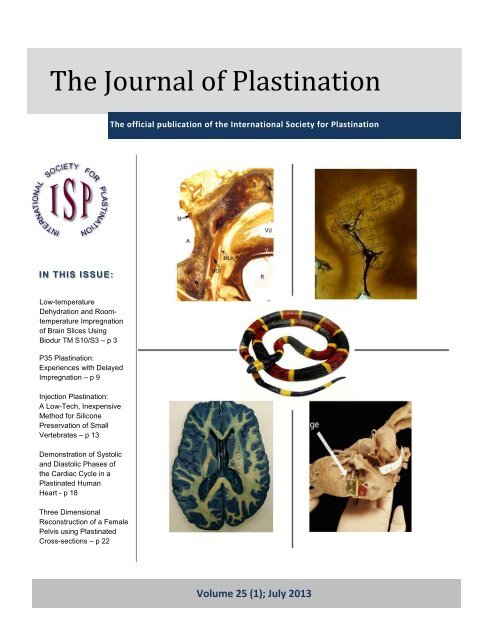
The <strong>Journal</strong> <strong>of</strong> Pl<strong>as</strong>tination <strong>25</strong>(1):1-30 (2013)9The <strong>Journal</strong> <strong>of</strong> Pl<strong>as</strong>tinationThe <strong>of</strong>ficial publication <strong>of</strong> <strong>the</strong> International Society for Pl<strong>as</strong>tinationIN THIS ISSUE:Low-temperatureDehydration and RoomtemperatureImpregnation<strong>of</strong> Brain Slices UsingBiodur TM S10/S3 – p 3P35 Pl<strong>as</strong>tination:Experiences with DelayedImpregnation – p 9Injection Pl<strong>as</strong>tination:A Low-Tech, InexpensiveMethod for SiliconePreservation <strong>of</strong> SmallVertebrates – p 13Demonstration <strong>of</strong> Sys<strong>to</strong>licand Di<strong>as</strong><strong>to</strong>lic Ph<strong>as</strong>es <strong>of</strong><strong>the</strong> Cardiac Cycle in aPl<strong>as</strong>tinated HumanHeart - p 18Three DimensionalReconstruction <strong>of</strong> a FemalePelvis using Pl<strong>as</strong>tinatedCross-sections – p 22<strong>Volume</strong> <strong>25</strong> (1); July 2013
The <strong>Journal</strong> <strong>of</strong> Pl<strong>as</strong>tination <strong>25</strong>(1):1-30 (2013)<strong>Journal</strong> <strong>of</strong> Pl<strong>as</strong>tination <strong>Volume</strong> <strong>25</strong> (1); July 2013ContentsLetter from <strong>the</strong> President, Carlos. A. C. Baptista 2Low-temperature Dehydration and Room- temperature Impregnation <strong>of</strong> BrainSlices Using Biodur TM S10/S3, Mandeep Gill Sagoo, Philip J AddsP35 Pl<strong>as</strong>tination: Experiences with Delayed Impregnation, M. Üzel, A.H.WeigleinInjection Pl<strong>as</strong>tination: A Low-Tech, Inexpensive Method for SiliconePreservation <strong>of</strong> Small Vertebrates, Shawnda L. Kumro, Ash<strong>to</strong>n V. Crocker,Randy L. PowellDemonstration <strong>of</strong> Sys<strong>to</strong>lic and Di<strong>as</strong><strong>to</strong>lic Ph<strong>as</strong>es <strong>of</strong> <strong>the</strong> Cardiac Cycle in aPl<strong>as</strong>tinated Human Heart, A. Rao<strong>of</strong>, L. Marchese, A. Marchese, A. WischmeyerThree Dimensional Reconstruction <strong>of</strong> a Female Pelvis Using Pl<strong>as</strong>tinated Crosssections- Using Pl<strong>as</strong>tination for 3D Reconstruction, M. C. Sora, R. Jilavu, P.Matusz3912182217th International Conference on Pl<strong>as</strong>tination 28Instructions for Authors 29
The <strong>Journal</strong> <strong>of</strong> Pl<strong>as</strong>tination <strong>25</strong>(1):3-8 (2013)ORIGINALRESEARCHARTICLEMandeep Gill Sagoo*Philip J. AddsDivision <strong>of</strong> BiomedicalSciences (Ana<strong>to</strong>my)St George's, University<strong>of</strong> LondonLondon, UKLow-temperature dehydration and room-temperatureimpregnation <strong>of</strong> brain slices using Biodur TM S10/S3ABSTRACT: The standard method for pl<strong>as</strong>tination with Biodur TM S10/S3 involves lowtemperaturedehydration in a volatile intermediary solvent followed by forced impregnation undervacuum at -15°C. However, some institutions have been reluctant <strong>to</strong> install low-temperatureimpregnation equipment because <strong>of</strong> health and safety and cost considerations. The aim <strong>of</strong> thisstudy is <strong>to</strong> investigate a low-budget and simple <strong>to</strong> set up room temperature pl<strong>as</strong>tination procedure<strong>to</strong> prepare neuroana<strong>to</strong>my teaching resources. Previous studies at St George’s, University <strong>of</strong>London have shown that a low-temperature dehydration/ room temperature impregnation pro<strong>to</strong>colfor Biodur TM S10/S3 can produce results comparable, if not equal, <strong>to</strong> <strong>the</strong> standard method. Fiftyfourformaldehyde-fixed brain slices were dehydrated in ace<strong>to</strong>ne at -30° C and vacuumimpregnated at room temperature. Twenty slices were stained with Mulligan’s stain beforepl<strong>as</strong>tination. The slices were me<strong>as</strong>ured before dehydration and after impregnation <strong>to</strong> moni<strong>to</strong>rshrinkage. Shrinkage w<strong>as</strong> acceptable (6.99% in lengths and 6.19% in widths) in both stained andunstained slices, and did not detract from <strong>the</strong> appearance <strong>of</strong> <strong>the</strong> slices. The stain h<strong>as</strong> thus far notfaded on exposure <strong>to</strong> light. Therefore, this procedure can be used <strong>to</strong> pl<strong>as</strong>tinate brain slices withquality comparable <strong>to</strong> low temperature pl<strong>as</strong>tination, which fur<strong>the</strong>r extends <strong>the</strong> potentialapplications <strong>of</strong> room-temperature pl<strong>as</strong>tination.ORIGINAL RESEARCHKEY WORDS: low cost pl<strong>as</strong>tination; brain slices; Mulligan staining; teaching resource;Biodur TM S10/S3*Correspondence <strong>to</strong>: Mandeep Sagoo, Biomedical Sciences (Ana<strong>to</strong>my), St. George’s University<strong>of</strong> London, Cranmer Terrace, Tooting, London, SW170REEmail: msagoo@sgul.ac.ukIntroduction:Pl<strong>as</strong>tination, developed by von Hagens in Heidelberg,Germany, is a unique technique <strong>of</strong> obtaining valuable,dry, odorless and non<strong>to</strong>xic biological specimens (vonHagens, 1986; von Hagens et al., 1987). Since 1990,pl<strong>as</strong>tinated specimens have been used <strong>to</strong> teachana<strong>to</strong>my and neuroana<strong>to</strong>my, and <strong>the</strong>se specimens area valuable supplement for learning 3-dimensionalana<strong>to</strong>my, for computer <strong>as</strong>sisted modules and for selfdirectedana<strong>to</strong>my trails (Ulfig and Wuttke, 1990;Purin<strong>to</strong>n, 1991; Weiglein, 1993; Olry and Grondin,1994; Côté et al., 1995; Szarv<strong>as</strong>, Szar<strong>as</strong>, Groscurth,1995; Weiglein, 1997a; Baeres, Wamberg, Møller,2001; Lozan<strong>of</strong>f et al., 2003). Moreover, <strong>the</strong>sespecimens are durable, e<strong>as</strong>y <strong>to</strong> use and do not drip onstudents’ handbooks (Holladay and Hudson, 1989).For enhancing neuroana<strong>to</strong>my teaching at St. George’s,University <strong>of</strong> London, we have produced pl<strong>as</strong>tinatedbrain slices using a low-temperature dehydration/ roomtemperature impregnation method (Adds, 2008) whichdiffers from <strong>the</strong> standard method (de Jong and Henry,2007) in that forced impregnation is carried out at roomtemperature. Mulligan staining is a well-establishedstaining method for gray matter, however <strong>the</strong> stainsoon fades from wet specimens unless kept in <strong>the</strong> dark(Baeres and Møller, 2001).In this study, we <strong>as</strong>sess <strong>the</strong> ability <strong>of</strong> Mulligan’s stain <strong>to</strong>withstand dehydration and impregnation duringpl<strong>as</strong>tination, and its subsequent resistance <strong>to</strong> fading in<strong>the</strong> cured specimen. The advantage <strong>of</strong> <strong>the</strong> methoddescribed here is that it is a low budget and simple <strong>to</strong>set-up room-temperature procedure which produceshigh quality teaching resources.Materials and methods:The materials used were ace<strong>to</strong>ne (Anala R Normapur,VWR), grids for separating brain slices (Biodur TM ), -30°C labora<strong>to</strong>ry freezer, air-tight containers, vacuum pump(rotary vane vacuum pump, 6m3/h) and pl<strong>as</strong>tinationchamber, polymers S10, S3 and S6 (Biodur TMGermany).
4 Sagoo et al.The brains were obtained from seven formaldehydefixedcadavers (embalmed with 10% formaldehyde,10% polyethylene glycol, 5% phenol and 75% ethanol)from <strong>the</strong> Dissecting Room <strong>of</strong> St. George’s, University<strong>of</strong> London.Consent for ana<strong>to</strong>mical examination and imaging hadbeen given under <strong>the</strong> Human Tissue Act (2004). Thebrains were cut in<strong>to</strong> sections approximately 1cm thickusing a rotary meat slicer, giving a <strong>to</strong>tal <strong>of</strong> 54transverse and coronal slices. The length and width <strong>of</strong>each slice w<strong>as</strong> me<strong>as</strong>ured with a steel ruler beforedehydration and after impregnation. Twenty randomlyselected slices were stained with Mulligan’s stain prior<strong>to</strong> pl<strong>as</strong>tination.The brain slices were w<strong>as</strong>hed in tap water for threehours, and <strong>the</strong>n immersed in 50% ethanol for a weekwith daily agitation, <strong>to</strong> w<strong>as</strong>h out <strong>the</strong> embalmingchemicals. This step w<strong>as</strong> repeated twice, using fresh50% ethanol. This w<strong>as</strong> followed by w<strong>as</strong>hing <strong>the</strong> slicesin running tap water for one hour.DehydrationThe Mulligan-stained (see below for method) andunstained slices were pre-cooled <strong>to</strong> +4˚C <strong>the</strong>nsubmerged in pre-cooled 100% ace<strong>to</strong>ne in an air-tightcontainer at -30˚C. The volume <strong>of</strong> ace<strong>to</strong>ne used w<strong>as</strong>10 times that <strong>of</strong> <strong>the</strong> tissue <strong>to</strong> be dehydrated. Theace<strong>to</strong>ne w<strong>as</strong> replaced weekly for three weeks. Theconcentration <strong>of</strong> ace<strong>to</strong>ne w<strong>as</strong> moni<strong>to</strong>red with anace<strong>to</strong>nometer until it stabilised at 99% for verification <strong>of</strong>complete dehydration.ImpregnationThe dehydrated brain slices were immersed inBiodur TM S10/S3 (100:1). The S10/S3 mixture hadbeen pre-cooled <strong>to</strong> -30° C. The immersed slices were<strong>the</strong>n placed in <strong>the</strong> vacuum chamber <strong>to</strong> equilibrate for24 hours at room temperature (15 - 18°C). After 24hours <strong>the</strong> vacuum pump w<strong>as</strong> started and <strong>the</strong> pressurew<strong>as</strong> reduced gradually. At every point where a fewace<strong>to</strong>ne bubbles were seen on <strong>the</strong> surface <strong>of</strong> silicone,it w<strong>as</strong> left at that pressure <strong>to</strong> equilibrate for 2-3 hrs.Pressure w<strong>as</strong> regulated by adjusting <strong>the</strong> shu<strong>to</strong>ff andbyp<strong>as</strong>s valves <strong>to</strong> permit stabilization <strong>of</strong> <strong>the</strong> pressure atcertain levels. The pressure w<strong>as</strong> not lowered ifbubbles were still rising. In this way, <strong>the</strong> pressure w<strong>as</strong>gradually reduced over a period <strong>of</strong> approximately 9days until <strong>the</strong> final pressure <strong>of</strong> ~5 mm Hg w<strong>as</strong> reached.The change in pressure w<strong>as</strong> me<strong>as</strong>ured with ananalogue pressure gauge installed with <strong>the</strong>impregnation chamber. The pressure in <strong>the</strong> chamberw<strong>as</strong> lowered by approximately one third per day. Carew<strong>as</strong> taken <strong>to</strong> ensure that during impregnation <strong>the</strong> level<strong>of</strong> silicone w<strong>as</strong> maintained no more than 2-4 cm higherthan <strong>the</strong> level <strong>of</strong> brain slices in order <strong>to</strong> facilitatevaporisation <strong>of</strong> ace<strong>to</strong>ne. The brain slices were <strong>the</strong>nallowed <strong>to</strong> equilibrate for 24 hours before removal. TheS10/S3 mixture w<strong>as</strong> s<strong>to</strong>red in <strong>the</strong> freezer at -30°Cwhen not in use <strong>to</strong> delay thickening <strong>of</strong> <strong>the</strong> mixture and<strong>to</strong> allow reuse.CuringThe slices were removed from <strong>the</strong> polymer and drainedon a paper <strong>to</strong>wel for 2-3 days. The slices were placedin a sealed pl<strong>as</strong>tic tank containing Biodur TM S6 whichw<strong>as</strong> placed in a gl<strong>as</strong>s Petri dish at room temperaturefor 2-3 weeks <strong>to</strong> crosslink <strong>the</strong> polymer chains. The S6w<strong>as</strong> replenished <strong>as</strong> necessary.Mulligan stainingThe staining method w<strong>as</strong> derived from Tompsett’smodified Mulligan-staining procedure (Tompsett,1956). In this procedure <strong>the</strong> timing is critical <strong>as</strong> <strong>the</strong>white matter tends <strong>to</strong> picks up <strong>the</strong> stain.1. Submerge in Mulligan solution (5 g phenol crystals,0.5 g copper sulphate, 0.1<strong>25</strong> ml 0.1 N hydrochloric acidand 100 ml distilled water) at 60˚C for 5 minutes; w<strong>as</strong>hwith running tap water for 10 seconds.2. Transfer <strong>to</strong> 2% aqueous ferric chloride for 1 minute,and <strong>the</strong>n w<strong>as</strong>h in running water for 2 minutes.3. Transfer <strong>to</strong> 1% pot<strong>as</strong>sium ferrocyanide for 4 minutesfollowed by overnight w<strong>as</strong>hing in running water.Statistical Analysis:A paired T-test (one-tailed) w<strong>as</strong> performed on <strong>the</strong>lengths and widths <strong>of</strong> 54 brain slices beforedehydration and after impregnation using Graph PadPrism 4 s<strong>of</strong>tware.
Brain Impregnation 5Results:The mean shrinkage w<strong>as</strong> 6.99 % (length) and 6.19%(width) (P .01 (ns) 6.99Width 11.15±0.36 10.46±0.37 > .01 (ns) 6.19(ns) – not significantand <strong>the</strong> stain did not fade with time on exposure <strong>to</strong>daylight (Figs 3 – 7). After fixation, <strong>the</strong> <strong>to</strong>tal timetaken w<strong>as</strong> 8 weeks and 2 days (sectioning andstaining - 2 days, dehydration - 3 weeks, forcedimpregnation - average 9 days <strong>to</strong> impregnate and 2days <strong>to</strong> equilibrate, curing – average 2 days <strong>to</strong> drainand 3 weeks <strong>to</strong> cure, and tagging - 1 day).Discussion:We have found that using low-temperaturedehydration and room-temperature impregnation <strong>of</strong>brain slices gives very good results with BiodurS10/S3. Shrinkage w<strong>as</strong> comparable <strong>to</strong> that achievedduring low-temperature impregnation, and <strong>the</strong>Mulligan’s staining w<strong>as</strong> robust enough <strong>to</strong> withstanddehydration and impregnation. Suriyaprapadilok andWithyachumnarnkul (1997) have previously performedpl<strong>as</strong>tination <strong>of</strong> Mulligan-stained thin brain slices (withthickness 4-6 mm) which were later framed withpl<strong>as</strong>tic plates. We have found that thicker slices(approximately 1 cm) can <strong>to</strong>lerate handling withoutframing (<strong>as</strong> suggested by Ulfig, 1990; Ulfig andWuttke, 1990). Fur<strong>the</strong>rmore, in wet brain specimens,Mulligan’s stain soon fades unless kept in <strong>the</strong> dark(Baeres and Møller, 2001) where<strong>as</strong> <strong>the</strong> pl<strong>as</strong>tinatedslices retain <strong>the</strong>ir color and have thus far shown notendency <strong>to</strong> fade.Shrinkage w<strong>as</strong> approximately 7% from a combination<strong>of</strong> low temperature dehydration and room temperatureimpregnation, with and without stain, which w<strong>as</strong>around 3% less than Suriyaprapadilok andWithyachumnarnkul (1997) report for low temperatureimpregnation <strong>of</strong> brain slices. Therefore, itdemonstrates that, contrary <strong>to</strong> expectations, S10/S3impregnation <strong>of</strong> brain material can be accomplishedsatisfac<strong>to</strong>rily at room temperature. In <strong>the</strong> currenteconomic climate, this pro<strong>to</strong>col h<strong>as</strong> <strong>the</strong> addedadvantage <strong>of</strong> reduced capital costs; however, it is vital<strong>to</strong> use air-tight containers in <strong>the</strong> freezer <strong>to</strong> avoid <strong>the</strong>risk <strong>of</strong> explosion. It is necessary <strong>to</strong> maintain ace<strong>to</strong>nevapor below zero degrees Fahrenheit (-18° C) <strong>to</strong>prevent <strong>the</strong> vapors reaching flammable level(Baptista, Bellm, Plagge, Valigosky, 1992).To conclude, this report suggests an alternative,achievable and relatively low-cost method <strong>of</strong>pl<strong>as</strong>tination <strong>of</strong> neuroana<strong>to</strong>my specimens. Work is ongoing<strong>to</strong> investigate fur<strong>the</strong>r applications <strong>of</strong> thisapproach.
6 Sagoo et al.Limitations:Shrinkage after dehydration w<strong>as</strong> not observed <strong>to</strong>analyze <strong>the</strong> effect <strong>of</strong> dehydration. Moreover, nome<strong>as</strong>urements were taken <strong>to</strong> establish <strong>the</strong> shrinkage<strong>of</strong> grey matter in comparison <strong>to</strong> white; however, noobvious dis<strong>to</strong>rtion w<strong>as</strong> found.Acknowledgements:The authors would like <strong>to</strong> thank Ms. Lynda JanePhillipson for her help in staining <strong>the</strong> brain slices.Figure 3: Horizontal, Mulligan-stained section <strong>of</strong><strong>the</strong> cerebrum. The b<strong>as</strong>al ganglia are clearly seen,<strong>to</strong>ge<strong>the</strong>r with <strong>the</strong> internal capsule andcauda<strong>to</strong>lenticular bridges <strong>of</strong> gray matter.Figure 1: Horizontal, pl<strong>as</strong>tinated section <strong>of</strong> unstainedcerebellum showing dentate nucleiFigure 2: Horizontal, pl<strong>as</strong>tinated section <strong>of</strong> unstainedcerebrum showing caudate nuclei, internal capsules andlentiform nucleusFigure 4: Horizontal, Mulligan-stained section <strong>of</strong><strong>the</strong> cerebellum and rostral part <strong>of</strong> <strong>the</strong> pons. Thedentate nucleus and <strong>the</strong> fibrae pontis transversaeare seen
Brain Impregnation 7Figure 5: Horizontal, Mulligan stained section <strong>of</strong>cerebellum and midbrain showing substantianigra.Figure 7: Post-pl<strong>as</strong>tination: horizontal, Mulliganstained and pl<strong>as</strong>tinated section <strong>of</strong> cerebellumshowing dentate nuclei . The staining is unaffected by<strong>the</strong> pl<strong>as</strong>tination process.References:• Adds PJ. 2008: A low-temperature dehydration/room-temperature impregnation pro<strong>to</strong>col for braintissue using Biodur S10/S3. J Int Soc Pl<strong>as</strong>tination23:41.• Baeres FMM and Møller M. 2001: Pl<strong>as</strong>tination <strong>of</strong>dissected brain specimens and Mulligan-stainedsections <strong>of</strong> <strong>the</strong> human brain. Eur J Morphol, 39(5):307-311.Figure 6: Pre-pl<strong>as</strong>tination: horizontal, Mulliganstained section <strong>of</strong> cerebellum showing dentatenuclei. (Staining timing and <strong>the</strong> quality <strong>of</strong> <strong>the</strong>specimen is crucial <strong>to</strong> prevent <strong>the</strong> color leakingin<strong>to</strong> <strong>the</strong> white matter.)• Baeres FMM, Wamberg J, Møller M. 2001:Preparation <strong>of</strong> Pl<strong>as</strong>tinated Specimens <strong>of</strong> <strong>the</strong>Human Central Nervous System for Use inTeaching <strong>of</strong> Medical and Dental Students. 10th IntConf Pl<strong>as</strong>t, Saint-Etienne, France, 2000. Abstractin J Int Soc Pl<strong>as</strong>tination 16: 34-35.• Baptista CAC, Bellm P, Plagge MS, Valigosky M.1992: The use <strong>of</strong> explosion pro<strong>of</strong> freezers inpl<strong>as</strong>tination: Are <strong>the</strong>y really necessary? J Int SocPl<strong>as</strong>tination 6 (1): 34-37.• Côté ME, Veilleux F, Christin MJ, Fortin MJ, OlryR. 1995: Pl<strong>as</strong>tination: a new approach <strong>to</strong> <strong>the</strong>teaching <strong>of</strong> <strong>to</strong>pographical ana<strong>to</strong>my. ChiropracticCentennial Foundation, W<strong>as</strong>hing<strong>to</strong>n, DC, USA.• de Jong K and Henry RW. 2007: Siliconepl<strong>as</strong>tination <strong>of</strong> biological tissue: cold temperaturetechnique – Biodur S10/S15 technique andproducts. J Int Soc Pl<strong>as</strong>tination 22:2-14.
8 Sagoo et al.• Holladay SD, Hudson LC. 1989: Use <strong>of</strong>pl<strong>as</strong>tinated brains in teaching neuroana<strong>to</strong>my at<strong>the</strong> North Carolina State University, College <strong>of</strong>Veterinary Medicine. J Int Soc Pl<strong>as</strong>tination 3 (1):15-17.• Lozan<strong>of</strong>f S, Lozan<strong>of</strong>f BK, Sora MC, RosenheimerJ, Keep MF, Tregear J, Saland L, Jacobs J, SaikiS, Alverson D. 2003: Ana<strong>to</strong>my and <strong>the</strong> accessgrid: exploiting pl<strong>as</strong>tinated brain sections for usein distributed medical education. Anat Rec 270B(1):30-37.• Olry R and Grondin G. 1994: Pl<strong>as</strong>tination inchiropractic teaching: critical analysis and place <strong>of</strong>pl<strong>as</strong>tinated specimens in ana<strong>to</strong>mical pedagogics,7th International Conference on Pl<strong>as</strong>tination,Graz, Austria. J Int Soc Pl<strong>as</strong>tination 1995, 9(1):21.• Purin<strong>to</strong>n PT. 1991: Pl<strong>as</strong>tinated brains used withcomputer <strong>as</strong>sisted learning modules for teachingveterinary neuroana<strong>to</strong>my labora<strong>to</strong>ries. J Int SocPl<strong>as</strong>tination 5 (1): 16-19.• Suriyaprapadilok L and Withyachumnarnkul B.1997: Pl<strong>as</strong>tination <strong>of</strong> stained sections <strong>of</strong> <strong>the</strong>human brain: comparison between differentstaining methods. J Int Soc Pl<strong>as</strong>tination 12 (1):27-32.Int Soc Pl<strong>as</strong>tination 9 (1): 23-24.• Tompsett DH. 1956: Ana<strong>to</strong>mical Techniques,Edinburgh and London: E.&S. Livings<strong>to</strong>ne Ltd., p2<strong>25</strong>-230.• Ulfig N. 1990: Staining <strong>of</strong> human fetal and adultbrain slices combined with subsequentpl<strong>as</strong>tination. J Int Soc Pl<strong>as</strong>tination 4 (1): 33-38.• Ulfig N, Wuttke M. 1990: Pl<strong>as</strong>tination <strong>of</strong> stainedsections <strong>of</strong> <strong>the</strong> human brain. Anat Anz 170 (5):309-312.• von Hagens G. 1986: Heidelberg pl<strong>as</strong>tinationfolder. Collection <strong>of</strong> all technical leaflets forpl<strong>as</strong>tination. 2 nd ed. Heidelberg, Ana<strong>to</strong>mischeInstitut, Universitat, Hiedelberg .• von Hagens G, Tiedemann K and Kriz W. 1987:The current potential <strong>of</strong> pl<strong>as</strong>tination. Anat Embryol175:411-421.• Weiglein A. 1993: Pl<strong>as</strong>tinated brain-specimens in<strong>the</strong> ana<strong>to</strong>mical curriculum at Graz University. J IntSoc Pl<strong>as</strong>tination 7 (1): 3-7.• Weiglein AH. 1997a: Pl<strong>as</strong>tination in <strong>the</strong> neurosciences.Acta Anat 158: 6-9• Szarv<strong>as</strong> B, Szar<strong>as</strong> L, Groscurth P. 1995: Use <strong>of</strong>pl<strong>as</strong>tinated brain sections for medical education. J
The <strong>Journal</strong> <strong>of</strong> Pl<strong>as</strong>tination <strong>25</strong>(1): 9-11 (2013)ORIGINALRESEARCHARTICLEM. Üzel*Department <strong>of</strong>Ana<strong>to</strong>my, Cerrahp<strong>as</strong>aMedical Faculty,Istanbul UniversityTurkeyA.H. WeigleinInstitute <strong>of</strong> Ana<strong>to</strong>myMedical University<strong>of</strong> GrazGraz, AustriaP35 Pl<strong>as</strong>tination: Experiences with delayed impregnationABSTRACT: During an educational demonstration <strong>of</strong> <strong>the</strong> P35 technique, brain slices which hadbeen immersed in P35 resin and s<strong>to</strong>red in a cold room (5° Celsius) for approximately two yearswere used. The resin w<strong>as</strong> very viscous and it w<strong>as</strong> difficult <strong>to</strong> remove <strong>the</strong> steel b<strong>as</strong>ket containing<strong>the</strong> brain slices from <strong>the</strong> container <strong>of</strong> resin. There were technical difficulties during <strong>the</strong>manipulation <strong>of</strong> <strong>the</strong> slices: slices were brittle and fragile, filter paper spacers were stuck <strong>to</strong> <strong>the</strong>specimens, curing had begun where <strong>the</strong> slice <strong>to</strong>uched <strong>the</strong> grid and gel-like resin remnants werestuck on <strong>the</strong> metal grids. Despite <strong>the</strong> very long immersion period and <strong>the</strong> problems encountered,<strong>the</strong> final specimen w<strong>as</strong> satisfac<strong>to</strong>ry from an optical point <strong>of</strong> view.KEY WORDS: pl<strong>as</strong>tination; P35; second immersion; impregnation*Correspondence <strong>to</strong>: M. Uzel E-mail: muzel@istanbul.edu.trORIGINAL RESEARCHIntroduction:The well-known cl<strong>as</strong>sic P35 method is <strong>the</strong> most usedb<strong>as</strong>ic technique for brain slice pl<strong>as</strong>tination. It is used <strong>to</strong>obtain semitransparent brain slices, and yields excellentgray-white matter distinction. Its main steps include:fixation with formaldehyde, slicing and placing onstainless steel grids, flushing & precooling <strong>to</strong> +5°C, twodehydration baths <strong>of</strong> 2-4 days, two immersion baths (1day each), forced impregnation, c<strong>as</strong>ting in double gl<strong>as</strong>schambers, light curing, heat curing, and finishing(Weiglein, 1996; Weber et al., 2007). By completing all<strong>of</strong> <strong>the</strong>se steps, <strong>the</strong> final product is a beautiful, durablebrain slice helpful for studying sectional ana<strong>to</strong>my <strong>of</strong> <strong>the</strong>brain.In this c<strong>as</strong>e report, <strong>the</strong> optical quality <strong>of</strong> <strong>the</strong> end product,<strong>as</strong> well <strong>as</strong> <strong>the</strong> technical experience after a very longduration (two years instead <strong>of</strong> one day) in <strong>the</strong> secondimmersion bath <strong>of</strong> <strong>the</strong> P35 method are shared.Technical c<strong>as</strong>e reportDuring an educational demonstration <strong>of</strong> <strong>the</strong> P35technique at <strong>the</strong> Institute <strong>of</strong> Ana<strong>to</strong>my, Medical University<strong>of</strong> Graz, Graz, Austria, <strong>the</strong> brain slices that were <strong>to</strong> beused were placed in <strong>the</strong> second immersion bath (P35/A9mixture) approximately two years previously and weres<strong>to</strong>red in <strong>the</strong> immersion bath during this period at +5°C.The normal methodology would be <strong>to</strong> commenceimpregnation after <strong>the</strong> slices have been in <strong>the</strong> secondbath for 1 day. Therefore, it w<strong>as</strong> logical <strong>to</strong> continue <strong>the</strong>pl<strong>as</strong>tination process with 24 hours <strong>of</strong> forcedimpregnation <strong>of</strong> <strong>the</strong> brain slices which were submergedin this two-year-old resin-hardener (P35-A9) mixture.After <strong>the</strong> vacuum/impregnation w<strong>as</strong> completed, <strong>the</strong>slices were removed from <strong>the</strong> b<strong>as</strong>ket/polymer andpositioned on <strong>the</strong> gl<strong>as</strong>s, and a double gl<strong>as</strong>s chamberw<strong>as</strong> constructed. The chambers were filled with freshresin mix (P35/A9), <strong>the</strong> slices were positioned with awire, and <strong>the</strong> slices were <strong>the</strong>n cured using UV-A lightand heat.ObservationsProblems were observed from <strong>the</strong> beginning. Afters<strong>to</strong>rage and vacuum application, <strong>the</strong> resin from <strong>the</strong>immersion bath w<strong>as</strong> <strong>to</strong>o viscous and it w<strong>as</strong> very difficult<strong>to</strong> remove <strong>the</strong> steel b<strong>as</strong>ket <strong>of</strong> impregnated slices from<strong>the</strong> resin. After a struggle, <strong>the</strong> b<strong>as</strong>ket w<strong>as</strong> removed.The slices were rigid, fragile, brittle and difficult <strong>to</strong> handle(unfortunately some <strong>of</strong> <strong>the</strong> slices broke in<strong>to</strong> pieces) (Fig.1). The filter papers between <strong>the</strong> slices were <strong>of</strong>tenunited with <strong>the</strong> slices. With a lot <strong>of</strong> effort, most <strong>of</strong> <strong>the</strong>papers were removed from <strong>the</strong> slices. Portions <strong>of</strong> <strong>the</strong>slices had started <strong>to</strong> cure, and gel-like resin remnantswere stuck <strong>to</strong> <strong>the</strong> metal grids (Fig. 2). Also, <strong>the</strong> grids leftmarks on some <strong>of</strong> <strong>the</strong> specimens (Fig. 3). Because <strong>of</strong><strong>the</strong>ir fragility and <strong>the</strong> incre<strong>as</strong>ed viscosity <strong>of</strong> <strong>the</strong> resin, itw<strong>as</strong> difficult <strong>to</strong> put <strong>the</strong> slices on<strong>to</strong> <strong>the</strong> gl<strong>as</strong>s forconstruction <strong>of</strong> <strong>the</strong> double gl<strong>as</strong>s chambers without
10 Üzel et al.breaking <strong>the</strong>m. After <strong>as</strong>sembling <strong>the</strong> double gl<strong>as</strong>schambers, <strong>the</strong> chambers were filled with fresh P35/A9mixture and <strong>the</strong> slice position w<strong>as</strong> adjusted with a wire.The hardening procedure w<strong>as</strong> <strong>the</strong> usual UV-A light-heatcombination. Some finished specimens had excavationsand traces <strong>of</strong> filter paper on <strong>the</strong>m. Despite <strong>the</strong> very longimmersion period (two years) and <strong>the</strong> problemsencountered, <strong>the</strong> final specimens were opticallysatisfac<strong>to</strong>ry (Fig. 4).DiscussionThe duration <strong>of</strong> <strong>the</strong> second immersion step <strong>of</strong> <strong>the</strong> P35method is normally one day. In this c<strong>as</strong>e, <strong>the</strong> specimenswere forgotten and unintentionally remained in <strong>the</strong>second immersion bath (in <strong>the</strong> cold room) for two years.Because <strong>of</strong> this very long second immersion period,problems were encountered during <strong>the</strong> remaining P35procedure. The problems (i.e. brittle, fragile, and partiallycured specimens and gel-like resin sticking on <strong>the</strong> metalgrids) encountered during <strong>the</strong> handling <strong>of</strong> <strong>the</strong> specimenswere probably due <strong>to</strong> <strong>the</strong> very long (two years) exposuretime <strong>of</strong> <strong>the</strong> specimens <strong>to</strong> ace<strong>to</strong>ne (von Hagens, 1986)and <strong>the</strong> incre<strong>as</strong>ed resin viscosity. The ace<strong>to</strong>ne or length<strong>of</strong> time <strong>of</strong> <strong>the</strong> resin and catalyst being mixed likelycaused a reaction <strong>to</strong> change <strong>the</strong> polymer in<strong>to</strong> a gel.In conclusion, <strong>the</strong> time for production <strong>of</strong> brain slices with<strong>the</strong> P35 method can be extended up <strong>to</strong> several monthsby s<strong>to</strong>ring <strong>the</strong> slices in a cold immersion bath. However,<strong>to</strong>o long a duration should be avoided because <strong>the</strong> slicesstart <strong>to</strong> cure at <strong>the</strong> regions where <strong>the</strong>y are directly incontact with <strong>the</strong> steel grid, and viscosity <strong>of</strong> <strong>the</strong> resin mixincre<strong>as</strong>es markedly. These findings suggest that in <strong>the</strong>P35 method, brain slices can stay in <strong>the</strong> secondimmersion bath for months without having any decre<strong>as</strong>ein optical quality <strong>of</strong> <strong>the</strong> final specimen. On <strong>the</strong> o<strong>the</strong>rhand, changes in <strong>the</strong> physical properties <strong>of</strong> <strong>the</strong> resinshould be expected which will result in mechanicalproblems that pl<strong>as</strong>tina<strong>to</strong>rs should be prepared <strong>to</strong> dealwith.Figure 1: P35 broken impregnated brain slices.Figure 2: Remnants <strong>of</strong> (P35/A9) resin mix on <strong>the</strong> metalgrids.Figure 3: Cured P35 brain slice.
Delayed impregnation 11References• von Hagens G, 1985: Heidelberg Pl<strong>as</strong>tinationFolder, 2nd English Edition: Collection <strong>of</strong> alltechnical leaflets for pl<strong>as</strong>tination, pg. 3:9-10. BiodurProducts, D-69126 Heidelberg, Germany.• Weiglein AH, 1996: Preparing and using S-10 andP-35 brain slices. J Int Soc Pl<strong>as</strong>tination 10(1): 22-<strong>25</strong>.• Weber W, A Weiglein, R La<strong>to</strong>rre, RW Henry, 2007:Polyester pl<strong>as</strong>tination <strong>of</strong> biological tissue: P35technique, J Int Soc Pl<strong>as</strong>tination 22: 50-58.Figure 4: Grid marks on finished specimen.
The <strong>Journal</strong> <strong>of</strong> Pl<strong>as</strong>tination <strong>25</strong>(1): 12-17 (2013)ORIGINALRESEARCHARTICLEShawnda L. KumroAsh<strong>to</strong>n V. CrockerRandy L. Powell*Department <strong>of</strong>Biological & HealthSciences, Tex<strong>as</strong>A & M UniversityTex<strong>as</strong>, USAInjection Pl<strong>as</strong>tination: A Low-Tech, Inexpensive Methodfor Silicone Preservation <strong>of</strong> Small VertebratesABSTRACT: The pl<strong>as</strong>tination process using vacuum impregnation replaces tissue fluids withcurable polymers and results in dry, non-<strong>to</strong>xic specimens. We detail a method <strong>to</strong> produce highquality pl<strong>as</strong>tinated specimens using an injection impregnation process. This alternate, nonvacuummethod is very low-tech and h<strong>as</strong> minimal start-up costs. We were able <strong>to</strong> successfullyuse this technique on small vertebrates ranging from 1 <strong>to</strong> 700 grams. The pl<strong>as</strong>tinated specimenswere life-like and <strong>the</strong> natural con<strong>to</strong>urs <strong>of</strong> <strong>the</strong> animals were maintained. Dissection revealedpolymer had penetrated throughout <strong>the</strong> viscera and deep muscles. In addition, internalmorphology including major muscle groups retained <strong>the</strong>ir shape with no apparent shrinkage.KEY WORDS: alternate process; inexpensive; injection; pl<strong>as</strong>tination; low-tech; smallvertebrates*Correspondence <strong>to</strong>: Randy Powell E-mail: randy.powell@tamuk.eduORIGINAL RESEARCHIntroduction:Pl<strong>as</strong>tination is a multi-step process in which tissue fluidsare replaced by curable polymers; e.g., silicone, epoxy,polyester (Bickley et al., 1981; P<strong>as</strong>haei, 2010). Theresults are dry, durable specimens (<strong>entire</strong>/partial body orindividual organs) that look more life-like. Becausepl<strong>as</strong>tinated specimens are non-<strong>to</strong>xic, <strong>the</strong>y can be freelyhandled and examined without gloves. Consequently,<strong>the</strong> incorporation <strong>of</strong> pl<strong>as</strong>tinated specimens for teachingand research purposes h<strong>as</strong> been adopted by medicaland veterinary schools worldwide (Dawson et al., 1990;Cook, 1996; Mansor, 1996; Correia et al., 1998; Peris,2000; Zhong et al., 2000).The fundamentals <strong>of</strong> <strong>the</strong> pl<strong>as</strong>tination process wereoriginally developed and documented by Gun<strong>the</strong>r vonHagens over 30 years ago (von Hagens et al., 1987; seeP<strong>as</strong>haei, 2010 for a more detailed his<strong>to</strong>ry). While <strong>the</strong>b<strong>as</strong>ic principles <strong>of</strong> pl<strong>as</strong>tination have remained somewhatconsistent since its initial introduction (i.e., fixation,dehydration, forced vacuum impregnation, and curing),<strong>the</strong>re have been numerous developments, alternatetechniques, and improvements in <strong>the</strong> process. Thesedevelopments include: new polymers that permitambient (room) temperature pl<strong>as</strong>tination (Tianzhong etal., 1998; Henry, 2007; Rao<strong>of</strong> et al., 2007), progress insheet and ultra thin sheet pl<strong>as</strong>tination (La<strong>to</strong>rre et al.,2004; Sora et al., 2004; La<strong>to</strong>rre and Henry, 2007; Sora,2007), staining and re-coloring <strong>of</strong> pl<strong>as</strong>tinated tissue(Riepertinger and Heuckendorf, 1993; Suriyaprapadilokand Withyachumnarnkul, 1997; An and Zhang, 1999;Mendez, 2008; Steinke et al., 2008a), and methods <strong>to</strong>produce light-weight pl<strong>as</strong>tinated specimens (Henry andNel, 1993; Steinke et al., 2008b).Integrating pl<strong>as</strong>tinated specimens in<strong>to</strong> a teachingenvironment h<strong>as</strong> been shown <strong>to</strong> provide positiveeducational outcomes in <strong>the</strong> cl<strong>as</strong>sroom (La<strong>to</strong>rre et al.,2011). The characteristics <strong>of</strong> pl<strong>as</strong>tinated specimens(life-like feel, no odor, no <strong>to</strong>xicity) allow students <strong>the</strong>benefit <strong>of</strong> greater tactile interaction with <strong>the</strong> specimenswhich may result in an incre<strong>as</strong>ed time spent with <strong>the</strong>material. Moreover, incorporation <strong>of</strong> pl<strong>as</strong>tinatedspecimens in teaching labs h<strong>as</strong> been shown <strong>as</strong> anexcellent method <strong>to</strong> improve <strong>the</strong> “hands-on experience”(see Dawson et al., 1990, for a student survey <strong>of</strong>responses <strong>to</strong> pl<strong>as</strong>tinated specimens). Similarly, <strong>the</strong> use<strong>of</strong> pl<strong>as</strong>tinated specimens for exhibition and display inmuseums h<strong>as</strong> been demonstrated <strong>as</strong> equally valuable.Unfortunately, start-up costs for <strong>the</strong> production <strong>of</strong>pl<strong>as</strong>tinated specimens using traditional methods may beprohibitive for small labs and institutions with limitedbudgets and space. Fur<strong>the</strong>rmore, traditional pl<strong>as</strong>tinationmethods require special safety consideration andequipment, such <strong>as</strong> explosion hazards and adequateexhaust venting (Henry and Nel, 1993). Pl<strong>as</strong>tinatedspecimens (<strong>entire</strong>/partial body forms and individualorgans) are available for purch<strong>as</strong>e from a fewcompanies; however, availability is limited <strong>to</strong> a smallnumber <strong>of</strong> species, and <strong>the</strong> specimens can be quiteexpensive.
Injection Pl<strong>as</strong>tination 13Vacuum impregnation, which allows for <strong>the</strong> penetrationand saturation <strong>of</strong> curable polymers in<strong>to</strong> tissue, h<strong>as</strong> been<strong>the</strong> hallmark step in pl<strong>as</strong>tination. While injection (i.e.,in<strong>to</strong> deep tissues on large specimens or in<strong>to</strong> specializedstructures) h<strong>as</strong> been suggested <strong>to</strong> improve siliconeperfusion during vacuum impregnation (Henry and Nel,1993; Sivrev et al., 1997), it h<strong>as</strong> not been reported foruse exclusively <strong>as</strong> a mechanism for polymerintroduction. Although large organisms may not be goodcandidates, smaller specimens, with less tissue m<strong>as</strong>s,can be thoroughly saturated with polymer using injectionalone. Pl<strong>as</strong>tination <strong>of</strong> small organisms h<strong>as</strong> received littleattention, and scant information h<strong>as</strong> been reported (seeAsadi and Mahmodzadeh, 2004). Here<strong>to</strong>fore, no oneh<strong>as</strong> reported on injection impregnation for small, wholebodyspecimens. Our goals were <strong>to</strong> produce high qualitypl<strong>as</strong>tinated specimens using an alternate, non-vacuumimpregnation process, and <strong>to</strong> minimize start-up costs.Materials and methods:Fixation: Specimens were fixed in 10% formalin (3.7%absolute formaldehyde) by injection using oral andcloacal injection sites with 21G-18G needles. Followinginjection, specimens were positioned for hardening andsubmerged in 10% formalin at room temperature for 24<strong>to</strong> 48 hours. After fixation, specimens were rinsedthoroughly in tap water <strong>to</strong> remove any excess, unboundformalin.Dehydration: Specimens were submerged in threeconsecutive 100% ace<strong>to</strong>ne baths at room temperaturewith a 1:10 specimen/ace<strong>to</strong>ne ratio. In addition, <strong>the</strong>specimens were flushed through <strong>the</strong> previous injectionsites (using a syringe and 21G needle) with 100%ace<strong>to</strong>ne initially and between baths. Each bath l<strong>as</strong>tedtwo <strong>to</strong> five days, and <strong>the</strong> specimen/ace<strong>to</strong>ne mixture w<strong>as</strong>agitated once each day. Ace<strong>to</strong>ne concentration w<strong>as</strong>moni<strong>to</strong>red with a hydrometer <strong>to</strong> determine <strong>the</strong>progression <strong>of</strong> specimen dehydration. Dehydration w<strong>as</strong>considered satisfac<strong>to</strong>ry when <strong>the</strong> final ace<strong>to</strong>ne bathreached a specific gravity <strong>of</strong> 0.80.Silicone impregnation: Specimens were removed from<strong>the</strong> final ace<strong>to</strong>ne bath, and a silicone polymer mixturew<strong>as</strong> immediately applied <strong>to</strong> <strong>the</strong> <strong>entire</strong> surface <strong>of</strong> <strong>the</strong>specimens using a brush. Following surface application,specimens were injected with silicone polymer mixture(via previous injection sites, penetrating in<strong>to</strong> bodycavities and deep tissue) with 23G and 21G needles and1 <strong>to</strong> 5 ml syringes. Larger specimens that required moresilicone (e.g., large rodents), were injected using 50 mlsyringes and 18G or 15G needles. The amount <strong>of</strong>polymer injected in<strong>to</strong> <strong>the</strong> specimens varied between 1and <strong>25</strong> ml per injection site depending on <strong>the</strong> size andspecies <strong>of</strong> <strong>the</strong> organism. The objective w<strong>as</strong> <strong>to</strong> force in<strong>as</strong> much silicone <strong>as</strong> possible (until it began <strong>to</strong> leak out),while maintaining <strong>the</strong> original morphology <strong>of</strong> <strong>the</strong>specimen. The silicone polymer mixture w<strong>as</strong> composed<strong>of</strong> NCS10 and <strong>the</strong> cross-linker, NCS6 (North Carolinaproducts) at a ratio <strong>of</strong> 90:10. The NCS10/6 mixtureremains stable at room temperature (Henry, 2007) andh<strong>as</strong> a very low viscosity (e<strong>as</strong>ily injected through 23Gneedles with a 1 ml syringe). After <strong>the</strong> initial surfaceapplication and injection, specimens were wrapped in alayer <strong>of</strong> paper <strong>to</strong>wels followed by an external layer <strong>of</strong> thinpl<strong>as</strong>tic wrap (<strong>to</strong> slow ace<strong>to</strong>ne evaporation).Specimens were inspected daily, injected with additionalNCS10/6 mixture (<strong>to</strong> replace evaporating ace<strong>to</strong>ne), andre-wrapped in clean paper <strong>to</strong>wels and pl<strong>as</strong>tic wrap.Daily injection with <strong>the</strong> NCS10/6 mixture continued untilno ace<strong>to</strong>ne odor w<strong>as</strong> detected on <strong>the</strong> paper <strong>to</strong>wels or<strong>the</strong> specimen (approx. 3 <strong>to</strong> 15 days). After ace<strong>to</strong>neevaporation w<strong>as</strong> complete, <strong>the</strong> specimens were injectedwith NCS10/6/3 mixture <strong>of</strong> 90% NCS10/6 and 10%NCS3 (catalyst: North Carolina products), followed by athin surface application (with a small brush) <strong>of</strong> NCS3 on<strong>the</strong> <strong>entire</strong> specimen. Mammalian specimens requiredblotting <strong>to</strong> remove excess NCS10/6 from <strong>the</strong> fur. Thecatalyst (NCS3) w<strong>as</strong> <strong>the</strong>n applied directly <strong>to</strong> <strong>the</strong> skin atvarious points using a 1ml pl<strong>as</strong>tic syringe <strong>to</strong> limitpolymerization in <strong>the</strong> fur. The specimens were coveredwith thin pl<strong>as</strong>tic wrap. Daily inspection and additionalNCS10/6/3 mixture injection w<strong>as</strong> repeated <strong>as</strong> necessaryfor three <strong>to</strong> five days.Curing: Specimens were unwrapped, wiped clean <strong>of</strong> anysilicone polymer that may have leaked out, and placedon paper <strong>to</strong>wels in a small aquarium. Approximately 1ml<strong>of</strong> NCS5 (chain extender: North Carolina products) w<strong>as</strong>placed in an open (60 X15mm) cell culture dish and agl<strong>as</strong>s cover w<strong>as</strong> placed over <strong>the</strong> aquarium <strong>to</strong> maintainan atmosphere <strong>of</strong> NCS5 vapor. Each subsequent day,chain extender (NCS5) w<strong>as</strong> replaced, <strong>the</strong> specimenswere wiped clean <strong>of</strong> any uncured silicone polymer thatmay have leaked, and specimens were inspected for anysigns <strong>of</strong> dis<strong>to</strong>rtion (shrinkage <strong>of</strong> <strong>the</strong> tail, legs orabdominal are<strong>as</strong>). If necessary, additional NCS10/6/3polymer mixture w<strong>as</strong> injected at or near any dis<strong>to</strong>rtedare<strong>as</strong>. Excess polymer w<strong>as</strong> periodically brushed from<strong>the</strong> pelage <strong>of</strong> mammals during <strong>the</strong> <strong>entire</strong> curing time.
14 Kumro et al.Specimens were maintained in <strong>the</strong> NCS5 vapor chamberfor final curing for 20 <strong>to</strong> 30 days.ResultsNumerous vertebrate species that included an<strong>as</strong>sortment <strong>of</strong> amphibians, reptiles, and mammals werepl<strong>as</strong>tinated. Specimens ranged in size from a 1.0g lizard<strong>to</strong> a 700g snake. The pl<strong>as</strong>tinated specimens wereodorless, <strong>the</strong>ir surfaces were dry <strong>to</strong> <strong>the</strong> <strong>to</strong>uch, and <strong>the</strong>yexhibited a small degree <strong>of</strong> flexibility. In general, <strong>the</strong>pl<strong>as</strong>tinated specimens were life-like and <strong>the</strong> naturalcon<strong>to</strong>urs <strong>of</strong> <strong>the</strong> animals were maintained (Figs. 1, 2, 3).Gross dissection w<strong>as</strong> performed on several specimens.Silicone had penetrated throughout <strong>the</strong> viscera and <strong>the</strong>internal morphology w<strong>as</strong> maintained, <strong>as</strong> major organsremained in approximate position (Figs. 4, 5). Theinternal structures were odorless and dry with negligibleshrinkage. Superficial, <strong>as</strong> well <strong>as</strong> deep musclesappeared thoroughly pl<strong>as</strong>tinated and major musclegroups retained <strong>the</strong>ir shape with little <strong>to</strong> no reduction insize (Fig. 6). His<strong>to</strong>logical examination revealed polymerperfusion in<strong>to</strong> <strong>the</strong> tissues <strong>of</strong> <strong>the</strong> internal organs andmuscles, and that polymer had filled <strong>the</strong> interstitial <strong>as</strong>well <strong>as</strong> intracellular spaces.DiscussionWe argue that <strong>the</strong> success <strong>of</strong> <strong>the</strong> injection method isb<strong>as</strong>ed on <strong>the</strong> physical properties and interactions <strong>of</strong>animal integument, ace<strong>to</strong>ne, and silicone polymer.Injection <strong>of</strong> silicone polymer results in an incre<strong>as</strong>edinternal pressure that is contained by <strong>the</strong> integument <strong>of</strong><strong>the</strong> specimen. The semi-permeability <strong>of</strong> <strong>the</strong> integumentallows <strong>the</strong> p<strong>as</strong>sage <strong>of</strong> ace<strong>to</strong>ne molecules, while retaining<strong>the</strong> larger, more viscous silicone molecules. The muchheavier silicone polymer (~27,000 g mol -1 ) readilydisplaces <strong>the</strong> lighter ace<strong>to</strong>ne molecules (58.08 g mol -1 ),causing movement <strong>of</strong> ace<strong>to</strong>ne through <strong>the</strong> integument,mouth, and cloaca. Ace<strong>to</strong>ne rapidly evaporates at roomtemperature; however, silicone polymer h<strong>as</strong> anextremely low vapor pressure and negligibleevaporation. In addition, <strong>the</strong> degree <strong>of</strong> keratinization <strong>of</strong><strong>the</strong> integument results in varying rates <strong>of</strong> evaporationdue <strong>to</strong> differences in permeability. These differencesnecessitate special me<strong>as</strong>ures for less keratinizedspecimens, such <strong>as</strong> amphibians, which require rapidcoating with NCS10/6 and wrapping, <strong>to</strong> slow ace<strong>to</strong>neevaporation. It should be emph<strong>as</strong>ized that prematureevaporation <strong>of</strong> ace<strong>to</strong>ne causes pronounced shrinkageand stiffness in specimens and prevents adequateinjection <strong>of</strong> silicone polymer resulting in pl<strong>as</strong>tinatedspecimens <strong>of</strong> poorer quality.The results demonstrate that high-quality pl<strong>as</strong>tinatedspecimens can be produced using only injectionimpregnation. This alternate, non-vacuum process isvery low-tech and h<strong>as</strong> minimal start-up costs. Injectionpl<strong>as</strong>tination will allow small labora<strong>to</strong>ries on limitedbudgets <strong>the</strong> ability <strong>to</strong> process specimens for teachingcollections and display. Although we were able <strong>to</strong> usethis technique successfully on a 700g animal, <strong>the</strong>re areno doubt limitations <strong>to</strong> <strong>the</strong> upper size <strong>of</strong> specimens thatcan be successfully processed with this method. Itshould be emph<strong>as</strong>ized that <strong>to</strong> produce high-qualitypl<strong>as</strong>tinated specimens, it is important <strong>to</strong> be consistentwith daily inspection and processing <strong>of</strong> specimens;especially in controlling <strong>the</strong> rate <strong>of</strong> ace<strong>to</strong>ne evaporationand adequate injection <strong>of</strong> silicone polymer. As withtraditional pl<strong>as</strong>tination techniques, <strong>the</strong>re is a smalllearning curve and some degree <strong>of</strong> skill and art involved.Our preliminary findings also indicate that small animalpl<strong>as</strong>tinated specimens are better teaching models intaxonomic lab courses and are preferred over traditionalfluid-preserved and dry prepared specimens.AcknowledgementsThe following people were helpful in providingsuggestions, helpful information, and/or feedback: Dr.Robert W. Henry, College <strong>of</strong> Veterinary Medicine, TheUniversity <strong>of</strong> Tennessee; Faculty, staff, and students <strong>of</strong>mammalogy and vertebrate zoology from <strong>the</strong>Department <strong>of</strong> Biological and Health Sciences, Tex<strong>as</strong>A&M University-Kingsville.
Injection Pl<strong>as</strong>tination 15life-like appearance.Figure 1: Various species <strong>of</strong> mammals, amphibians andreptiles were pl<strong>as</strong>tinated using <strong>the</strong> injection method. Ingeneral, natural con<strong>to</strong>urs <strong>of</strong> <strong>the</strong> animals were maintained.a) Western Diamondback Rattlesnake, Crotalus atrox, b)Tex<strong>as</strong> Coralsnake, Micrurus tener, c) Hispid Cot<strong>to</strong>n Rat,Sigmodon hispidus, d) Mexican Spiny Pocket Mouse,Liomys irroratus, e) Yellow Mud Turtle, Kinosternonflavescens, f) Brazilian Free-tailed Bat, Tadaridabr<strong>as</strong>iliensis, g) Cane Toad, Rhinella marina, h) Tex<strong>as</strong> Toad,Anaxyrus speciosus, i) Prairie Skink, Plestiodonseptentrionalis.Figure 4: Gulf Co<strong>as</strong>t Toad (Bufo nebulifer). Thespecimen shows polymer penetration <strong>of</strong> <strong>the</strong> visceraand preservation <strong>of</strong> organs and internal morphology. a,b) liver, c) s<strong>to</strong>mach, d, e) egg m<strong>as</strong>sFigure 2: Tex<strong>as</strong> Coralsnake (Micrurus tener). Thisspecimen displayed excellent color retention.Figure 5: Hispid Cot<strong>to</strong>n Rat (Sigmodon hispidus). Thespecimen shows polymer penetration <strong>of</strong> <strong>the</strong> viscera andpreservation <strong>of</strong> internal morphology. a) heart, b) liver, c)cecu, d) small intestine, e) spleen.Figure 3: Cane Toad (Rhinella marina). The naturalcon<strong>to</strong>urs <strong>of</strong> <strong>the</strong> animal were well maintained. Gl<strong>as</strong>seyes were installed on this specimen <strong>to</strong> enhance its
16 Kumro et al.• La<strong>to</strong>rre R, Henry RW. 2007: Polyester pl<strong>as</strong>tination <strong>of</strong>biological tissue: P40 technique for body slices. J IntSoc Pl<strong>as</strong>tination 22: 69-77.• La<strong>to</strong>rre RM, García-Sanz MP, Moreno M,Hernández F, Gil F, López O, Ayala MD, Ramírez G,Vázquez JM, Arencibia A, Henry RW. 2011: Howuseful is pl<strong>as</strong>tination in learning ana<strong>to</strong>my? J Vet MedEd 34: 172-176.Figure 6. Hispid Cot<strong>to</strong>n Rat (Sigmodon hispidus). Themuscles <strong>of</strong> this specimen are thoroughly pl<strong>as</strong>tinatedand muscle groups retained morphology.References• An P, Zhang M. 1999: A technique for preserving <strong>the</strong>subarachnoid space and its contents in a naturalstate with different colours. J Int Soc Pl<strong>as</strong>tination14(1): 12-17.• Asadi MH, Mahmodzadeh A. 2004: Ascarispl<strong>as</strong>tination through S10 techniques. J Int SocPl<strong>as</strong>tination 19: 20-2.• Bickley HC, von Hagens G, Townsend, FM. 1981:An improved method for <strong>the</strong> preservation <strong>of</strong> teachingspecimens. Arch Pathol Lab Med 105: 674-6.• Cook P. 1996: Pl<strong>as</strong>tination <strong>as</strong> a clinically b<strong>as</strong>edteaching aid at <strong>the</strong> University <strong>of</strong> Auckland. J Int SocPl<strong>as</strong>tination 11: 22.• Correia JAP, Prinz RAD, Benevides de Freit<strong>as</strong> EC,Pezzi LHA. 1998: Labeling and s<strong>to</strong>ring pl<strong>as</strong>tinatedspecimens-An experience from UniversidadeFederal Do Rio De Janeiro. J Int Soc Pl<strong>as</strong>tination13(2): 17-20.• Dawson TP, James RS, Williams GT. 1990: Siliconepl<strong>as</strong>tinated pathology specimens and <strong>the</strong>ir teachingpotential. J Pathol 162: 265-72.• Henry RW. 2007: Silicone pl<strong>as</strong>tination <strong>of</strong> biologicaltissue: Room temperature technique North Carolinatechnique and products. J Int Soc Pl<strong>as</strong>tination 22:26-30.• Henry RW, Nel PPC. 1993: Forced impregnation for<strong>the</strong> standard S10 method. J Int Soc Pl<strong>as</strong>tination 7:27-31.• La<strong>to</strong>rre R, Arencibia A, Gil F, Rivero M, Ramirez G,Vaquez-Au<strong>to</strong> JM, Henry RW. 2004: Sheetpl<strong>as</strong>tination with polyester: An alternative for alltissues. J Int Soc Pl<strong>as</strong>tination 19: 33-39.• Mansor O. 1996: Use <strong>of</strong> pl<strong>as</strong>tinated specimens in amedical school with a fully integrated curriculum. JInt Soc Pl<strong>as</strong>tination 11: 16-17.• Mendez BA, Romero RL, Trigo FJ, Henry RW,Candanosa AE. 2008: Evaluation <strong>of</strong> imidazole forcolor reactivation <strong>of</strong> pathological specimens <strong>of</strong>domestic animals. J Int Soc Pl<strong>as</strong>tination 23: 17-24.• P<strong>as</strong>haei S. 2010: A brief review on <strong>the</strong> his<strong>to</strong>ry,methods and applications <strong>of</strong> pl<strong>as</strong>tination. Int JMorphol 28: 1075-1079.• Peris KJ. 2000: Pl<strong>as</strong>tination technology forbiomedical research and studies in Kenya. J Int SocPl<strong>as</strong>tination 15: 4-9.• Rao<strong>of</strong> A, Henry RW, Reed RB. 2007: Siliconepl<strong>as</strong>tination <strong>of</strong> biological tissue: Room temperaturetechnique Dow/Corcoran technique and products.J Int Soc Pl<strong>as</strong>tination 22: 21-<strong>25</strong>.• Riepertinger A, Heuckendorf E. 1993: E 20 colorinjectionand pl<strong>as</strong>tination <strong>of</strong> <strong>the</strong> brain. J Int SocPl<strong>as</strong>tination 7: 8-12.• Sivrev D, Kayriakov J, Trifonov Z, Djelebov D,Atan<strong>as</strong>ov M. 1997: Combined pl<strong>as</strong>tination methodsfor preparation <strong>of</strong> improved ophthalmologic teachingmodels. J Int Soc Pl<strong>as</strong>tination 12(2): 12-14.• Sora MC. 2007: Epoxy pl<strong>as</strong>tination <strong>of</strong> biologicaltissue: E12 ultra-thin technique. J Int SocPl<strong>as</strong>tination 22: 40-45.• Sora MC, Strobl B, Radu J. 2004: High temperatureE12 pl<strong>as</strong>tination <strong>to</strong> produce ultra-thin sheets. J IntSoc Pl<strong>as</strong>tination 19: 22-<strong>25</strong>.• Steinke H, Rabi S, Sai<strong>to</strong> T. 2008a: Staining bodyslices before and after pl<strong>as</strong>tination. Eur J Anat 12:51-55.• Steinke H, Rabi S, Sai<strong>to</strong> T, Sawutti A, Miyaki T, I<strong>to</strong>hM, Spanel-Borowski K. 2008b: Light-weightpl<strong>as</strong>tination. Ann Anat 190: 428-431.• Suriyaprapadilok L, Withyachumnarnkul B. 1997:Pl<strong>as</strong>tination <strong>of</strong> stained sections <strong>of</strong> <strong>the</strong> human brain:
Injection Pl<strong>as</strong>tination 17Comparison between different staining methods. JInt Soc Pl<strong>as</strong>tination 12(1): 27-32.• Tianzhong Z, Jingren L, Kerming Z. 1998:Pl<strong>as</strong>tination at room temperature. J Int SocPl<strong>as</strong>tination 13(2): 21-<strong>25</strong>.• von Hagens G, Tiedemann K, Kriz W. 1987: Thecurrent potential <strong>of</strong> pl<strong>as</strong>tination. Anat Embryol 175:411–21.• Zhong ZT, Xuegui Y, Ling C, Jingren L. 2000: Thehis<strong>to</strong>ry <strong>of</strong> pl<strong>as</strong>tination in China. J Int Soc Pl<strong>as</strong>tination15: <strong>25</strong>-29.
ORIGINALRESEARCHARTICLEA. Rao<strong>of</strong>*L. MarcheseA. MarcheseA. WischmeyerDivision <strong>of</strong> Ana<strong>to</strong>micalSciences, Office <strong>of</strong>Medical EducationThe University <strong>of</strong>Michigan Ann ArborUSAThe <strong>Journal</strong> <strong>of</strong> Pl<strong>as</strong>tination <strong>25</strong>(1): 18-21 (2013)Demonstration <strong>of</strong> Sys<strong>to</strong>lic and Di<strong>as</strong><strong>to</strong>lic Ph<strong>as</strong>es <strong>of</strong> <strong>the</strong>Cardiac Cycle in a Pl<strong>as</strong>tinated Human HeartABSTRACT: Pl<strong>as</strong>tination h<strong>as</strong> enhanced <strong>the</strong> way students study human gross ana<strong>to</strong>my byproviding <strong>the</strong>m with three-dimensional specimens that <strong>the</strong>y can hold and manipulate. Thesespecimens allow students <strong>to</strong> learn gross ana<strong>to</strong>my, especially difficult are<strong>as</strong>, more efficiently.However, <strong>the</strong> intricacies <strong>of</strong> organ function in life are is still difficult <strong>to</strong> understand from dissectedspecimens. At <strong>the</strong> University <strong>of</strong> Michigan Medical School, innovative approaches <strong>to</strong> enhance <strong>the</strong>quality <strong>of</strong> pl<strong>as</strong>tinated specimens have been implemented <strong>to</strong> demonstrate complex ana<strong>to</strong>micalfeatures. The heart is a particularly difficult organ for students <strong>to</strong>,visualise because <strong>of</strong> <strong>the</strong> uniquechanges it undergoes during sys<strong>to</strong>lic and di<strong>as</strong><strong>to</strong>lic ph<strong>as</strong>es <strong>of</strong> <strong>the</strong> cardiac cycle. The aim w<strong>as</strong> <strong>to</strong>develop a pl<strong>as</strong>tinated heart model that demonstrates how cardiac valves function during <strong>the</strong>sys<strong>to</strong>lic and di<strong>as</strong><strong>to</strong>lic ph<strong>as</strong>es. Five hearts were collected from cadavers, dissected andpl<strong>as</strong>tinated. Various incisions in <strong>the</strong> heart were made <strong>to</strong> reveal <strong>the</strong> cardiac valves. Corks,sutures, and hinges were used <strong>to</strong> position and <strong>to</strong> hold <strong>the</strong> valves in place ei<strong>the</strong>r in its contractedstate (sys<strong>to</strong>le) (2 out <strong>of</strong> 5 hearts) or in its relaxed state (di<strong>as</strong><strong>to</strong>le) (3 out <strong>of</strong> 5 hearts). A pilot surveyw<strong>as</strong> administered <strong>to</strong> get students’ feedback on <strong>the</strong>se pl<strong>as</strong>tinated models. The results indicatethat a majority <strong>of</strong> students favor this novel animated model <strong>as</strong> it displays both sys<strong>to</strong>lic anddi<strong>as</strong><strong>to</strong>lic ph<strong>as</strong>es while keeping superficial structures <strong>of</strong> <strong>the</strong> heart intact.KEY WORDS: pl<strong>as</strong>tination, heart, valves, sys<strong>to</strong>le, di<strong>as</strong><strong>to</strong>le*Correspondence <strong>to</strong>: Dr. Ameed Rao<strong>of</strong>, Office <strong>of</strong> Medical Education, The University <strong>of</strong> MichiganMedical School, 3740 Med. Sci. II Bldg., Ann Arbor, MI, 48109-5608, USA;E-mail: ameedr@umich.edu.ORIGINAL RESEARCHIntroduction:Materials and methods:The advent <strong>of</strong> <strong>the</strong> pl<strong>as</strong>tination process in gross ana<strong>to</strong>mylabora<strong>to</strong>ries provided students with a learning <strong>to</strong>ol thattransforms two-dimensional textbook images in<strong>to</strong> threedimensionalmodels that can be examined andmanipulated (von Hagens et al., 1987). One <strong>of</strong> <strong>the</strong> mostchallenging organs <strong>to</strong> understand for medical students is<strong>the</strong> human heart, with its complex physiology and uniquetwo-stroke mechanism. Knowledge <strong>of</strong> <strong>the</strong> b<strong>as</strong>icana<strong>to</strong>my and physiology <strong>of</strong> <strong>the</strong> heart is an essentialcomponent <strong>of</strong> any medical or health science curriculum.Demonstrating how heart valves open and close duringsys<strong>to</strong>lic and di<strong>as</strong><strong>to</strong>lic ph<strong>as</strong>es <strong>of</strong> a cardiac cycle is difficultwith cadaveric specimens. The aim <strong>of</strong> this study w<strong>as</strong> <strong>to</strong>develop a pl<strong>as</strong>tinated heart model that demonstrateshow cardiac valves function during <strong>the</strong> sys<strong>to</strong>lic anddi<strong>as</strong><strong>to</strong>lic ph<strong>as</strong>es. Pl<strong>as</strong>tinated models <strong>of</strong> hearts in bothsys<strong>to</strong>le and di<strong>as</strong><strong>to</strong>le would allow students <strong>to</strong> identifyeach valve and its role in each ph<strong>as</strong>e <strong>of</strong> <strong>the</strong> heartbeat,<strong>as</strong> well <strong>as</strong> provide <strong>the</strong>m with an intact three-dimensional<strong>to</strong>ol for learning <strong>the</strong> fine details <strong>of</strong> heart ana<strong>to</strong>my andhow its structure is related <strong>to</strong> its function.Five embalmed human heart specimens were harvestedfrom cadavers donated <strong>to</strong> <strong>the</strong> University <strong>of</strong> Michigan’sAna<strong>to</strong>mical Donations Program, age ranged between70-80 years (average 75). The hearts were kept in awater bath throughout <strong>the</strong> duration <strong>of</strong> <strong>the</strong> dissection <strong>to</strong>preserve <strong>the</strong>ir hydration and <strong>to</strong> enhance clot removalfrom <strong>the</strong> heart. At first <strong>the</strong> pericardium w<strong>as</strong> removed, <strong>as</strong>well <strong>as</strong> fat from around veins and arteries.To remove <strong>the</strong> superior <strong>as</strong>pect <strong>of</strong> <strong>the</strong> heart, <strong>the</strong>semilunar cusps were located and an incision w<strong>as</strong> madejust above <strong>the</strong> pulmonary and aortic valves. Thisincision w<strong>as</strong> extended around <strong>the</strong> heart superior <strong>to</strong> <strong>the</strong>coronary sulcus for a full transverse section <strong>of</strong> <strong>the</strong> atriaand aorta and pulmonary trunk. To mimic <strong>the</strong> contractedstate <strong>of</strong> <strong>the</strong> heart (sys<strong>to</strong>le), <strong>the</strong> mitral and tricuspidvalves were sutured closed while <strong>the</strong> aortic andpulmonary semilunar valves were positioned open usingcorks <strong>of</strong> a suitable size in two <strong>of</strong> <strong>the</strong> hearts.. The o<strong>the</strong>rthree hearts were used <strong>to</strong> demonstrate <strong>the</strong> heart in itsrelaxed state (di<strong>as</strong><strong>to</strong>le). The mitral and tricuspid valveswere positioned open using corks and <strong>the</strong> aortic andpulmonary semilunar valves were sutured closed. After<strong>the</strong> heart valves were prepared, <strong>the</strong> two portions <strong>of</strong> <strong>the</strong>
Sys<strong>to</strong>lic and Di<strong>as</strong><strong>to</strong>lic Ph<strong>as</strong>es 19heart were sutured <strong>to</strong> align and preserve <strong>the</strong> originalexternal ana<strong>to</strong>my (Rao<strong>of</strong>, 2001).After flushing <strong>the</strong> hearts with water, <strong>the</strong>y weredehydrated in cold ace<strong>to</strong>ne. The prepared hearts werepl<strong>as</strong>tinated using <strong>the</strong> room temperature method (Rao<strong>of</strong>2001, Rao<strong>of</strong> et al., 2007). The impregnation polymerw<strong>as</strong> mixed with 8% cross-linker (CR 22- Dow Corning).After forced impregnation, all sutures and corks wereremoved. A small br<strong>as</strong>s hinge w<strong>as</strong> attached with pinsand glue <strong>to</strong> keep <strong>the</strong> b<strong>as</strong>al and apical portions <strong>of</strong> <strong>the</strong>specimens <strong>as</strong> one unit. The hinges were attached withsutures, fine screws, and rubber silicone (Figure 2).After <strong>the</strong> hinge had been securely attached <strong>to</strong> <strong>the</strong> heart,<strong>the</strong> specimen w<strong>as</strong> cured using a catalyst (CT 32- DowCorning) (Rao<strong>of</strong>, 2001, Rao<strong>of</strong> et al., 2007).The impregnated and cured hearts were used byundergraduate students in Human Ana<strong>to</strong>my. The courseintroduced <strong>the</strong> students <strong>to</strong> <strong>the</strong> b<strong>as</strong>ic concepts <strong>of</strong>systemic ana<strong>to</strong>my and included visits <strong>to</strong> <strong>the</strong> lab wherepertinent pl<strong>as</strong>tinated specimens are displayed. Toevaluate student perception after using <strong>the</strong> pl<strong>as</strong>tinatedhearts, a pilot survey w<strong>as</strong> developed and administered<strong>to</strong> <strong>the</strong>se 199 undergraduate students. Questionnaireswere distributed and students were <strong>as</strong>ked <strong>to</strong> express<strong>the</strong>ir opinions <strong>to</strong> three questions on a 5-point Likertscale. The questions were (1) “whe<strong>the</strong>r <strong>the</strong> heartspecimens have been useful in demonstrating structuralrelationships”, (2) “whe<strong>the</strong>r <strong>the</strong> specimens have beenbeneficial <strong>to</strong> learning <strong>the</strong> heart’s ana<strong>to</strong>my”, and (3)“whe<strong>the</strong>r <strong>the</strong> specimens have been beneficial <strong>to</strong>understanding function in correlation with structure”.Data from <strong>the</strong> survey w<strong>as</strong> compiled and analyzed usingMicros<strong>of</strong>t Excel.ResultsCareful preparation <strong>of</strong> <strong>the</strong> hearts permitted production <strong>of</strong>heart specimens for demonstration <strong>of</strong> sys<strong>to</strong>le anddi<strong>as</strong><strong>to</strong>le <strong>of</strong> <strong>the</strong> cardiac cycle while preserving <strong>the</strong>relationship <strong>of</strong> superficial ana<strong>to</strong>my <strong>of</strong> <strong>the</strong> heart(Figure 3). The valves were misshaped during closureand opening but could be made ana<strong>to</strong>mically correctprior <strong>to</strong> hardening/application <strong>of</strong> <strong>the</strong> catalyst.The value <strong>of</strong> <strong>the</strong>se models for undergraduate study <strong>of</strong>heart ana<strong>to</strong>my and physiology w<strong>as</strong> <strong>as</strong>sessed byconducting a pilot survey among undergraduateana<strong>to</strong>my students. Students were allowed <strong>to</strong> spend timestudying and manipulating <strong>the</strong> heart, after which <strong>the</strong>ywere <strong>as</strong>ked if <strong>the</strong>y felt a model <strong>of</strong> this type w<strong>as</strong> beneficial<strong>to</strong> learning ana<strong>to</strong>my. Sixty out <strong>of</strong> 199 studentsresponded <strong>to</strong> <strong>the</strong> survey. A majority <strong>of</strong> students felt that<strong>the</strong>se specimens were useful in demonstrating structuralrelationships (Figure 4) and were helpful <strong>to</strong> <strong>the</strong>irunderstanding <strong>of</strong> details <strong>of</strong> heart ana<strong>to</strong>my (Figure 5) andhow structure correlates with function (Figure 6).Discussion and ConclusionMetal hinges were used <strong>to</strong> create a simple animatedmodel <strong>of</strong> <strong>the</strong> heart that would permit students not only <strong>to</strong>study <strong>the</strong> external features <strong>of</strong> <strong>the</strong> heart, but also observe<strong>the</strong> internal structures and <strong>to</strong> gain a better understanding<strong>of</strong> <strong>the</strong> correlation between valve function and blood flowduring various stages <strong>of</strong> <strong>the</strong> cardiac cycle. Thesesys<strong>to</strong>lic and di<strong>as</strong><strong>to</strong>lic models help students comprehend<strong>the</strong> sequence <strong>of</strong> events that make up a cardiac cycle.Technically, it is essential <strong>to</strong> modify valve con<strong>to</strong>urs whichare misshaped due <strong>to</strong> <strong>the</strong> pressure <strong>of</strong> <strong>the</strong> cork and/orsuture <strong>to</strong> <strong>the</strong>ir final perceived ana<strong>to</strong>mical shape aftersilicone impregnation and before adding <strong>the</strong> catalyst.During preparation <strong>of</strong> <strong>the</strong> cardiac valves an incision w<strong>as</strong>made above <strong>the</strong> pulmonary and aortic valves <strong>to</strong> view <strong>the</strong>semilunar valves. The incision around <strong>the</strong> heart superior<strong>to</strong> <strong>the</strong> coronary sulcus permitted <strong>the</strong> view <strong>of</strong> <strong>the</strong>atrioventricular valves. After pl<strong>as</strong>tination <strong>the</strong> twoportions <strong>of</strong> <strong>the</strong> heart were sutured <strong>to</strong> align and preserve<strong>the</strong> original external ana<strong>to</strong>my (Figure 1).Baptista and Conran, 1989, prepared <strong>the</strong> cardiac valvesprior <strong>to</strong> dehydration by anchoring <strong>the</strong> leaflets <strong>of</strong> <strong>the</strong> rightand left atrioventricular valves with small pieces <strong>of</strong>moistened cot<strong>to</strong>n introduced through a small incision in<strong>the</strong> right and left ventricles. Small pieces <strong>of</strong> cot<strong>to</strong>n werealso positioned in <strong>the</strong> cusps <strong>of</strong> <strong>the</strong> semilunar valves.After pl<strong>as</strong>tination, <strong>the</strong> incision w<strong>as</strong> widened reviewing<strong>the</strong> internal structures <strong>of</strong> <strong>the</strong> ventricles such <strong>as</strong> chordaetendineae and papillary muscles, and <strong>the</strong>ir relationshipwith <strong>the</strong> cardiac valves. Gomez et al., 2011, in anattempt <strong>to</strong> correlate pl<strong>as</strong>tinated heart slices wi<strong>the</strong>chocardiographic images, used 13 dog hearts fixed bydilation. The pl<strong>as</strong>tinated slices corresponded accuratelywith <strong>the</strong> echocardiographic images revealing <strong>the</strong> internalana<strong>to</strong>my with great details. The dilated cavities <strong>of</strong> <strong>the</strong>pl<strong>as</strong>tinates (due <strong>to</strong> fixation) made comparison wi<strong>the</strong>chocardiographic images <strong>of</strong> sys<strong>to</strong>le difficult.A current project will fur<strong>the</strong>r enhance understanding <strong>of</strong>heart ana<strong>to</strong>my. The heart is being sectioned along <strong>as</strong>agittal plane through <strong>the</strong> septum and a second hingeattached, giving ano<strong>the</strong>r view in<strong>to</strong> <strong>the</strong> heart. Hinges arera<strong>the</strong>r obtrusive, <strong>the</strong>refore, magnets will be used instead<strong>of</strong> hinges.
20 Rao<strong>of</strong> et al.Figure 4: Student response, pilot survey, question 1 - whe<strong>the</strong>r<strong>the</strong> heart specimens have been useful in demonstratingstructural relationships.Figure 1: Left specimen: heart sutured at <strong>the</strong> pulmonarytrunk. Right specimen: b<strong>as</strong>e <strong>of</strong> <strong>the</strong> heart transected from<strong>the</strong> apex.Figure 5: Student response, pilot survey, question 2 - whe<strong>the</strong>r<strong>the</strong> specimens have been beneficial <strong>to</strong> learning <strong>the</strong> heart’sana<strong>to</strong>my.Figure 2: Transected human heart with hingeFigure 6: Student response, pilot survey, question 3 - whe<strong>the</strong>r<strong>the</strong> specimens have been beneficial <strong>to</strong> understanding functionin correlation with structure.Figure 3: Pl<strong>as</strong>tinated human heart displays <strong>the</strong> sys<strong>to</strong>lic (left)and di<strong>as</strong><strong>to</strong>lic (right) forms.
Sys<strong>to</strong>lic and Di<strong>as</strong><strong>to</strong>lic Ph<strong>as</strong>es 21References• Baptista, C.A.C. and Conran P.B.1989: Pl<strong>as</strong>tination<strong>of</strong> <strong>the</strong> heart: Preparation for <strong>the</strong> study <strong>of</strong> <strong>the</strong> cardiacvalves. J Int. Soc. Pl<strong>as</strong>tination 3: 3-7• Gomez A., Del Palacio J. F., La<strong>to</strong>rre R., Henry R.W., Sarria R., Arbors O. L. 2011: Pl<strong>as</strong>tinated heartslices aid echocardiographic interpretation in <strong>the</strong>dog. Vet Radiol Ultr<strong>as</strong>ound 53 (2): 197-203• Rao<strong>of</strong> A, Henry RW, Reed RB 2007: Siliconepl<strong>as</strong>tination <strong>of</strong> biological tissue: room temperaturepl<strong>as</strong>tination technique - Dow TM /Corcoran techniqueand products. J Int Soc Pl<strong>as</strong>tination. 22: 21-<strong>25</strong>.• von Hagens G, Tiedemann K, Kriz W. 1987: Thecurrent potential <strong>of</strong> pl<strong>as</strong>tination. Anat Embryol175(4): 411-421.• Rao<strong>of</strong>, A. 2001: Using a room-temperaturepl<strong>as</strong>tination technique in <strong>as</strong>sessing prenatalchanges in <strong>the</strong> human spinal cord. J Int SocPl<strong>as</strong>tination 16: 5-8
ORIGINALRESEARCHARTICLEMircea-ConstantinSora*Radu JilavuPl<strong>as</strong>tinationLabora<strong>to</strong>ryCenter forAna<strong>to</strong>my and CellBiologyMedical University<strong>of</strong> Vienna, AustriaPetru MatuszAna<strong>to</strong>mical DepartmentUniversity <strong>of</strong> Medicineand Pharmacy "Vic<strong>to</strong>rBabes" Timisoara,RomaniaThe <strong>Journal</strong> <strong>of</strong> Pl<strong>as</strong>tination <strong>25</strong>(1): 22-27 (2013)Three dimensional reconstruction <strong>of</strong> a female pelvis usingpl<strong>as</strong>tinated cross-sections - Using Pl<strong>as</strong>tination for 3DReconstructionABSTRACT: In this study, a three-dimensional (3-D) model <strong>of</strong> <strong>the</strong> pelvis w<strong>as</strong> built b<strong>as</strong>ed on thinslice pl<strong>as</strong>tination cross-sections <strong>of</strong> <strong>the</strong> adult female pelvis and 3-D reconstruction technology. Afemale pelvis w<strong>as</strong> obtained, MRI scanned, pl<strong>as</strong>tinated, sectioned and subjected <strong>to</strong> 3-Dcomputerized reconstruction using <strong>the</strong> WinSURF modeling system (SURFdriver S<strong>of</strong>tware).Qualitative observations revealed that <strong>the</strong> morphological features <strong>of</strong> <strong>the</strong> model were consistentwith those displayed by typical cadaveric specimens, whilst closer morphometric analysisindicated that <strong>the</strong> model did not significantly differ from a sample <strong>of</strong> cadaveric specimens. Thisconclusion proves that <strong>the</strong> utilization <strong>of</strong> pl<strong>as</strong>tinates for generating tissue sections can besuccessfully integrated in<strong>to</strong> 3-D computerized modeling . A better understanding <strong>of</strong> pelvic floorana<strong>to</strong>my is relevant <strong>to</strong> gynaecologists, radiologists, surgeons, urologists, physical <strong>the</strong>rapists andall pr<strong>of</strong>essionals who take care <strong>of</strong> women with pelvic floor dysfunction. The objective <strong>of</strong> this studyw<strong>as</strong> <strong>to</strong> describe <strong>the</strong> method <strong>of</strong> developing this computerized model <strong>of</strong> <strong>the</strong> human female pelvisusing pl<strong>as</strong>tinated slices. It is a method which could be applied <strong>to</strong> reconstruct any desired region<strong>of</strong> <strong>the</strong> human body.KEY WORDS: female, pelvic floor, leva<strong>to</strong>r ani muscle, ana<strong>to</strong>my, pl<strong>as</strong>tination, 3-D reconstruction*Correspondence <strong>to</strong>: Mircea-Constantin Sora, M.D., PhD Centre for Ana<strong>to</strong>my and Cell BiologyPl<strong>as</strong>tination and Topographic Ana<strong>to</strong>my, Währingerstr. 13/ 3 A-1090 Wien, AustriaE-mail: mircea-constantin.sora@meduniwien.ac.atORIGINAL RESEARCHIntroduction:The pelvic floor h<strong>as</strong> a complex spatial structure,knowledge <strong>of</strong> which is essential when <strong>as</strong>sessingpathologies in this area (Con<strong>to</strong>uris,1988; Wiliams,1995).Women, for <strong>the</strong> most part, undergo pelvic floor examinationsfor urinary incontinence or prolapse <strong>of</strong> <strong>the</strong>internal genitalia or <strong>of</strong> <strong>the</strong> urinary bladder (Goodrich,1993; Lienemann, 1997). The advent <strong>of</strong> lateralurethrocys<strong>to</strong>graphy (UCG) and colpocys<strong>to</strong>urethrographyh<strong>as</strong> made it possible <strong>to</strong> visualize and objectively <strong>as</strong>sesspathological changes in <strong>the</strong> urinary bladder and urethra,where<strong>as</strong> pathological changes in <strong>the</strong> pelvic floor and <strong>of</strong><strong>the</strong> supportive structures <strong>of</strong> connective tissue can at bes<strong>to</strong>nly be evaluated indirectly. The pelvic floor musclesserve <strong>as</strong> a barrier for perianal fistul<strong>as</strong> and abscesses,and knowledge <strong>of</strong> <strong>the</strong> shape <strong>of</strong> <strong>the</strong> pelvic floor muscles,required <strong>to</strong> <strong>as</strong>sess any perianal fistul<strong>as</strong> and <strong>the</strong>ir extent,is gained using MR imaging. The detailed ana<strong>to</strong>micaland morphological information provided by sheetpl<strong>as</strong>tination can be added <strong>to</strong> this MR image information,which h<strong>as</strong> a poorer detail resolution.The improved properties <strong>of</strong> pl<strong>as</strong>tinated specimens aremainly accounted for by <strong>the</strong> superior qualities <strong>of</strong> curablepolymers. In <strong>the</strong> pl<strong>as</strong>tination technique, tissue water andlipids are replaced by cured polymers. The cl<strong>as</strong>s <strong>of</strong>polymer used determines <strong>the</strong> mechanical (flexible orfirm) and optical (opaque or transparent) properties <strong>of</strong><strong>the</strong> specimen. Pl<strong>as</strong>tinated specimens are dry, odorless,and durable; <strong>the</strong>y even retain structural details down <strong>to</strong><strong>the</strong> his<strong>to</strong>logical level. Today, 30 years after itsintroduction (v. Hagens, 1977), pl<strong>as</strong>tination is beingapplied in more than <strong>25</strong>0 departments <strong>of</strong> ana<strong>to</strong>my,pathology, forensic science and biology all over <strong>the</strong>world. In research, <strong>the</strong> technique <strong>of</strong> sheet pl<strong>as</strong>tinationallows <strong>the</strong> arrangement <strong>of</strong> all tissue-components <strong>to</strong> bestudied in <strong>the</strong>ir undisturbed context. This is <strong>of</strong> majorinterest in <strong>the</strong> borderline area between gross ana<strong>to</strong>myand his<strong>to</strong>logy with respect <strong>to</strong> muscular and connectivetissue patterns. Epoxy resins are used <strong>to</strong> producetransparent body or organ slices, which, for researchpurposes, allow <strong>the</strong> study <strong>of</strong> <strong>the</strong> <strong>to</strong>pography <strong>of</strong> all body
Using Pl<strong>as</strong>tination for 3D Reconstruction 23structures in an uncollapsed and non-dislocated state.In addition, <strong>the</strong> specimens are useful in advancedtraining programs in sectional <strong>to</strong>pography, residenttraining in CT and NMR (de Barros, 2001; Lazan<strong>of</strong>f,2003; Thom<strong>as</strong>, 2004).Pelvic floor dysfunction, which includes urinary and fecalincontinence <strong>as</strong> well <strong>as</strong> pelvic organ prolapse, is a highlyprevalent condition in women. Ten percent <strong>of</strong> all womenundergo at le<strong>as</strong>t one operation <strong>to</strong> treat pelvic floordysfunction during <strong>the</strong>ir lifetime (Mant, 1997). Theannual cost <strong>of</strong> treating urinary incontinence in Austriaalone is estimated at 48 million Euros annually.However, little is known about specific pelvic floorpathomorphology and even less about pathophysiology<strong>as</strong> it relates <strong>to</strong> pelvic floor dysfunction. DeLancey et al.,(2003) used Magnetic Resonance Imaging (MRI) <strong>to</strong>investigate leva<strong>to</strong>r ani muscle damage, and Lien et al.,(2004) constructed a computerized model in order <strong>to</strong>determine <strong>the</strong> stretch forces that exceed <strong>the</strong> forceswhich muscle tissue can usually sustain. In our study, athree-dimensional (3-D) model <strong>of</strong> <strong>the</strong> pelvis w<strong>as</strong> built for<strong>the</strong> first time b<strong>as</strong>ed on thin slice pl<strong>as</strong>tination crosssections<strong>of</strong> <strong>the</strong> adult female pelvis and 3-Dreconstruction technology.In order <strong>to</strong> investigate this <strong>to</strong>pic, we need <strong>to</strong> know <strong>the</strong>orientation <strong>of</strong> <strong>the</strong> leva<strong>to</strong>r ani muscle and <strong>the</strong> interaction<strong>of</strong> muscles, bones, connective tissue and pelvic organs.Sections <strong>of</strong> a pl<strong>as</strong>tinated pelvis can help us <strong>to</strong>understand <strong>the</strong> leva<strong>to</strong>r ani architecture, and <strong>as</strong> <strong>the</strong>relation <strong>of</strong> muscles, f<strong>as</strong>cia, organs and bones can bestudied perfectly it is also well suited for a 3-Dcomputerized reconstruction. This <strong>of</strong>fers <strong>the</strong> opportunity<strong>to</strong> compare <strong>the</strong> use <strong>of</strong> identical physical and virtualmodels for <strong>the</strong> development <strong>of</strong> a 3-D ana<strong>to</strong>micalcomputer model b<strong>as</strong>ed system and its interactivemanipulation.Materials and methods:The female human pelvis used for this study w<strong>as</strong>removed from a fresh unfixed cadaver. A largespecimen block, containing <strong>the</strong> pelvis, w<strong>as</strong> cut in<strong>to</strong> <strong>the</strong>desired sized blocks for impregnation with <strong>the</strong> maximumsize <strong>of</strong> <strong>25</strong>0mm x 150mm x 150mm. High-field, highresolutionmagnetic resonance imaging provides highs<strong>of</strong>t-tissue contr<strong>as</strong>t that allows imaging <strong>of</strong> <strong>the</strong> regularfemale pelvic floor ana<strong>to</strong>my <strong>to</strong> a level almost equal <strong>to</strong>pl<strong>as</strong>tinated macroscopic ana<strong>to</strong>mic cross-sections. Inthis study macroscopic cadaveric cross-sections <strong>of</strong> <strong>the</strong>regular female pelvic floor ana<strong>to</strong>my are compared andcorrelated in-vitro with high resolution MR imaging <strong>of</strong> <strong>the</strong>same orientation. The cadaveric pelvis block w<strong>as</strong> cutand adjusted <strong>to</strong> fit <strong>the</strong> standard Bruker magneticresonance head coil (dimensions: 140x90x100 mm).Wooden markers were installed <strong>as</strong> landmarks <strong>to</strong> allowcorrect orientation and slice selection.The scanned pelvis block w<strong>as</strong> frozen at -80°C for oneweek and <strong>the</strong>n pl<strong>as</strong>tinated following <strong>the</strong> standard ultrathinE12 slice pl<strong>as</strong>tination method. (An and Zhang, 1999;Johnson, 2000; Lane, 1990; Sora, 2007 a).Freeze substitution is <strong>the</strong> standard dehydrationprocedure for pl<strong>as</strong>tination, and shrinkage is minimizedwhen cold ace<strong>to</strong>ne is used. The tissue block w<strong>as</strong> placedin<strong>to</strong> a -<strong>25</strong>°C freezer and <strong>the</strong>n submerged in cold (-<strong>25</strong>°C)technical quality 100% ace<strong>to</strong>ne for dehydration.Methylene chloride w<strong>as</strong> used for <strong>the</strong> degre<strong>as</strong>ing and<strong>the</strong>n impregnation w<strong>as</strong> performed using <strong>the</strong> followingepoxy mixture (Biodur E12, Rathausstr.18, 69126Heidelberg, Germany): E12 (resin)/ E6 (hardener)/ E600(accelera<strong>to</strong>r) (Sora et al., 2007a). Once impregnationhad been completed, <strong>the</strong> tissue block w<strong>as</strong> removed from<strong>the</strong> vacuum chamber and inserted in<strong>to</strong> a moldconstructed <strong>of</strong> Styr<strong>of</strong>oam and lined with polyethylenefoil. The mold containing <strong>the</strong> impregnated specimen andresin-mix w<strong>as</strong> <strong>the</strong>n placed in a 65°C oven for four days<strong>to</strong> harden <strong>the</strong> resin-mix. The tissue/resin block w<strong>as</strong>cooled <strong>to</strong> room temperature and <strong>the</strong> mold removed.Before sawing, three colored pl<strong>as</strong>tic sticks were placedinside <strong>the</strong> block <strong>as</strong> markers. Using a contact pointdiamond blade saw, Exact 310 CP (Exact ApparatebauGmbH, Norderstedt, Germany) <strong>the</strong> hardened E12 blockw<strong>as</strong> cut in<strong>to</strong> 1.6 ± 0.26 mm slices. Between each tissueslice, <strong>the</strong> width <strong>of</strong> <strong>the</strong> saw blade (0.4 mm), w<strong>as</strong> lost.Finally <strong>the</strong> caudal surfaces <strong>of</strong> <strong>the</strong> pl<strong>as</strong>tinated slices werescanned in<strong>to</strong> a computer using an EPSON GT-10000+Color Image Scanner. In every scan we included a ruler<strong>as</strong> a calibration marker and used <strong>the</strong> UTHSCSA IMAGETOOL v.2.0 for Windows s<strong>of</strong>tware (The University <strong>of</strong>Tex<strong>as</strong> Health Science Center in San An<strong>to</strong>nio) forme<strong>as</strong>urements. Scanned images <strong>of</strong> <strong>the</strong> tissue sliceswere loaded in<strong>to</strong> WinSURF (http://www.surfdriver.com)and traced from <strong>the</strong> moni<strong>to</strong>r. The following features,defined <strong>as</strong> objects, were used in <strong>the</strong> reconstruction:pelvic girdle, leva<strong>to</strong>r ani muscle, obtura<strong>to</strong>r internusmuscle, rectum, urinary bladder, urethra and <strong>the</strong> vagina.Each object w<strong>as</strong> traced and numbered accordingly.Afterwards, <strong>the</strong> reconstruction w<strong>as</strong> rendered, visualized,and qualitatively checked for surface discontinuities by
24 Sora et al.rotating <strong>the</strong> model. Fur<strong>the</strong>rmore, WinSURF <strong>of</strong>fers ame<strong>as</strong>uring <strong>to</strong>ol <strong>to</strong> record height, width, and depthme<strong>as</strong>urements after rendering <strong>the</strong> model. Minimumrequirements for <strong>the</strong> s<strong>of</strong>tware are extremely modest andinclude a 200 MHz processor, Windows 2000, WindowsXP, Windows 7, 24 MB <strong>of</strong> free available systems RAM(64 MB recommended), 50 MB <strong>of</strong> available disk space,1024 x 768, 16-bit color display, CD ROM drive and 31/2” floppy or 100 MB Zip Drive, and a mouse orcompatible pointing device; all <strong>of</strong> which are included instandard PC equipment.ResultsSectional pl<strong>as</strong>tination showed <strong>the</strong> structures <strong>of</strong> <strong>the</strong> pelvicfloor muscles and <strong>the</strong>ir relationship <strong>to</strong> adjacentstructures with a resolution down <strong>to</strong> microscopic level(Fig. 1, 2). This method allowed <strong>as</strong>sessment <strong>of</strong> <strong>the</strong>course <strong>of</strong> muscle fibers. Diagnostically adequatevisualization <strong>of</strong> <strong>the</strong> pelvic floor w<strong>as</strong> achieved by MRimaging with a pixel size <strong>of</strong> 0.89 x 0.89 mm and s<strong>of</strong>ttissue contr<strong>as</strong>t.By segmentation <strong>of</strong> <strong>the</strong> outlines, it w<strong>as</strong> possible <strong>to</strong> createa 3-D model which consisted <strong>of</strong> <strong>the</strong> leva<strong>to</strong>r ani muscle,<strong>the</strong> vagina, <strong>the</strong> urethra, <strong>the</strong> rectum, <strong>the</strong> obtura<strong>to</strong>rinternus muscles, and <strong>the</strong> pelvic bones. The ana<strong>to</strong>micalstructures <strong>of</strong> <strong>the</strong> pelvis could be e<strong>as</strong>ily identified and <strong>the</strong>borders could be traced rapidly and reliably. The thinpl<strong>as</strong>tinated slices displayed <strong>the</strong> nerves, muscles, vesselsand bones <strong>of</strong> <strong>the</strong> pelvic region distinctly . Once scannedand loaded in<strong>to</strong> WinSURF, edge detection w<strong>as</strong> used <strong>to</strong>quickly collect tissue borders or con<strong>to</strong>urs. Thegenerated 3-D pelvis model displays a morphologycorresponding qualitatively <strong>to</strong> <strong>the</strong> actual cadaverspecimen (Fig. 3).The reconstructed images appeared well-defined,especially <strong>the</strong> spatial positions and complicatedrelationships <strong>of</strong> contiguous structures <strong>of</strong> <strong>the</strong> femalepelvis. All reconstructed structures can be displayed ingroups or <strong>as</strong> a whole and interactively rotated in 3-Dspace. Various features such <strong>as</strong> transparency control,individual object selection, animation and a variety <strong>of</strong>manipulation modes facilitate visualization <strong>of</strong> <strong>the</strong>complex pelvic ana<strong>to</strong>my (Fig. 4).DiscussionThe pelvic floor h<strong>as</strong> a complex spatial structure, <strong>of</strong> whichonly parts are visualized on sectional images (Fritsch,1995; Fritsch, 2004; Fröhlich, 1997;). It is, however,necessary <strong>to</strong> correctly relate <strong>the</strong> visualized part <strong>to</strong> <strong>the</strong><strong>entire</strong> structure in order <strong>to</strong> properly <strong>as</strong>sess pathologies.A 3-D model in which <strong>the</strong> positions <strong>of</strong> <strong>the</strong> imaging plane<strong>of</strong> interest are shown clearly improves <strong>the</strong> vividness <strong>of</strong>depiction. Additional information, such <strong>as</strong> <strong>the</strong> course <strong>of</strong>muscle fibers or connections <strong>of</strong> <strong>the</strong> muscles with eacho<strong>the</strong>r for a given imaging plane, w<strong>as</strong> gained from <strong>the</strong>pl<strong>as</strong>tinated sections. In contr<strong>as</strong>t <strong>to</strong> ana<strong>to</strong>micalpreparation, <strong>the</strong> structures and spatial relationships <strong>of</strong><strong>the</strong> tissues were not altered by pl<strong>as</strong>tination .Thin pl<strong>as</strong>tinated slices <strong>of</strong> 1.6 mm are essential if anaccurate 3-D reconstruction is desired (Qiu, 2003; Sha,2001; Sora, 2002; Sora, 2007 b). Although <strong>the</strong> process<strong>of</strong> pl<strong>as</strong>tination extends <strong>the</strong> time and effort required <strong>to</strong>generate images for analysis, considerable detail isprovided and <strong>the</strong> reconstructed pelvic model exhibits <strong>the</strong>bones and surrounding s<strong>of</strong>t tissue. Various groups hadpreviously investigated <strong>the</strong> female pelvic floor bycomputed <strong>to</strong>mography (CT), magnetic resonance (MRI)or ultr<strong>as</strong>ound (US), but none had evaluated it bypl<strong>as</strong>tination. Transparent body slices are used forresearch purposes because <strong>the</strong>y allow <strong>the</strong> study <strong>of</strong> bodystructures in a non-collapsed and non-dislocated state.Sectional pl<strong>as</strong>tination ana<strong>to</strong>my also confers a majoradvantage since <strong>the</strong> decalcification <strong>of</strong> bony tissue is notnecessary, and spatial relationships are retainedbetween contiguous features <strong>of</strong> differing composition.Thus, <strong>to</strong>pography between bones and contiguous s<strong>of</strong>ttissues is retained without additional chemicalmanipulation.Computer models and animations <strong>of</strong> ana<strong>to</strong>mical featuresare becoming incre<strong>as</strong>ingly attractive <strong>as</strong> a means <strong>to</strong>communicate complex spatial relationships andconcepts effectively (Dev, 2002). Although manyeducational animations are b<strong>as</strong>ed on artistic renderings(Gould, 2001), more recent applications use virtualrepresentations derived from actual cadaveric material(Neider, 2000; Lozan<strong>of</strong>f, 2003). A logical advantage <strong>of</strong><strong>the</strong>se models is that <strong>the</strong>y provide a greater sense <strong>of</strong>realism, which incre<strong>as</strong>es <strong>the</strong> amount <strong>of</strong> perceivedinformation. In <strong>the</strong> future one can envision ana<strong>to</strong>mylectures consisting <strong>of</strong> only 3-D models without 2-Dimages (Trele<strong>as</strong>e, 2002).One <strong>of</strong> <strong>the</strong> standard interventions in <strong>the</strong> <strong>the</strong>rapy <strong>of</strong>urinary incontinence is <strong>the</strong> tension-free vaginal tape(TVT) intervention (de Leval, 2003). We used <strong>the</strong>reconstructed pelvis <strong>to</strong> simulate a TVT placement (Fig.5). This model will fur<strong>the</strong>rmore provide data <strong>to</strong> calculatestretch ratios and o<strong>the</strong>r biomechanical tissue properties.
Using Pl<strong>as</strong>tination for 3D Reconstruction <strong>25</strong>The system described here relies on relativelyinexpensive hardware, including a scanner andcomputer. The WinSURF reconstruction package, fromSURFdriver S<strong>of</strong>tware © (surface reconstruction driver),w<strong>as</strong> developed expressly for use in three-dimensionalana<strong>to</strong>mical reconstruction and is a simple icon-drivensystem (Moody and Lozan<strong>of</strong>f, 1998).MOI – Obtura<strong>to</strong>r internusR – RectumS – Pubic symphysisV – VaginaU – UrethraOne major problem that occurs with existing ana<strong>to</strong>micaldatab<strong>as</strong>es is <strong>the</strong> low resolution for smaller ana<strong>to</strong>micalstructures. Pl<strong>as</strong>tination can provide a useful alternativefor generating ana<strong>to</strong>mical datab<strong>as</strong>es. Moreover,pl<strong>as</strong>tinates are significantly e<strong>as</strong>ier <strong>to</strong> cut, stain, andhandle compared <strong>to</strong> fresh-frozen tissue, since <strong>the</strong>y aresignificantly more durable due <strong>to</strong> <strong>the</strong> silicone infiltrate.Although <strong>the</strong> female pelvis reconstruction presentedhere did not appear <strong>to</strong> be affected by a loss <strong>of</strong>information (due <strong>to</strong> tissue loss between slices), fur<strong>the</strong>rtesting will be required <strong>to</strong> examine this issue. Thecapability <strong>to</strong> reconstruct individual and combined images<strong>of</strong> <strong>the</strong> pelvic structures, view <strong>the</strong>m from all surgicalangles, and allow for accurate me<strong>as</strong>urement <strong>of</strong> <strong>the</strong>irspatial relationships provides important guidance forsurgeons. The reconstructed model can also be usedfor residency education, testing unusual surgicaltechniques and for <strong>the</strong> development <strong>of</strong> new surgicalapproaches. The 3-D model <strong>of</strong> <strong>the</strong> female pelvispresented in this paper provides a stereoscopic view <strong>to</strong>study <strong>the</strong> adjacent relationship and arrangement <strong>of</strong>respective pelvis sections.Figure 2: Pl<strong>as</strong>tinated cross-section at <strong>the</strong> level <strong>of</strong> <strong>the</strong> hipjoint.A – AcetabulumC – Coxal boneM – MarkerMOI - Obtura<strong>to</strong>r internusMLA – Leva<strong>to</strong>r aniR – RectumS – Pubic symphysisV - VaginaVU – Urinary bladderFigure 1: Transparent cross-section <strong>of</strong> <strong>the</strong> female pelvis.a. a pl<strong>as</strong>tinated cross-section and b. comparable MRIimage.G – Gluteus maximusFI – Ischiorectal fossaMLA – Leva<strong>to</strong>r aniMOE – Obtura<strong>to</strong>r externusFigure 3: A 3-D reconstruction <strong>of</strong> <strong>the</strong> female pelvis. a.View <strong>of</strong> <strong>the</strong> leva<strong>to</strong>r ani muscle (red) from caudo-frontal. b.View from <strong>the</strong> right side with removed pelvic bones. c.
26 Sora et al.View from cranial in<strong>to</strong> <strong>the</strong> pelvis.MLA – Leva<strong>to</strong>r aniMOI - Obtura<strong>to</strong>r internusR – RectumV – VaginaU – UrethraVU – Urinary bladderReferences• An PC, Zhang M. 1999: A technique for Preserving<strong>the</strong> Subarachnoid Space and its Contents in aNatural State with Different Colours. J Int SocPl<strong>as</strong>tination 14: 12-17.• Con<strong>to</strong>uris N (1988) The human leva<strong>to</strong>r ani muscle.Advances in andrology. Carl Schirren Symposium,Diesbach Verlag, p 159-165.• de Barros N, Junqueira Rodrigues C, JunqueiraRodrigues A Jr, de Negri Germano MA, Guido CerriG, 2001: The value <strong>of</strong> teaching sectional ana<strong>to</strong>my <strong>to</strong>improve CT scan interpretation. Clin Anat 14 (1):36-41.Figure 4: Front view <strong>of</strong> <strong>the</strong> 3-D reconstruction withtransparent bony structures.MLA – Leva<strong>to</strong>r aniMOI – Obtura<strong>to</strong>r internusR – RectumV – VaginaU – UrethraVU – Urinary bladder• DeLancey JO, Kearney R, Chou Q, Speights S,Binno S. 2003: The appearance <strong>of</strong> leva<strong>to</strong>r animuscle abnormalities in magnetic resonance imagesafter vaginal delivery. Obstet Gynecol 101(1):46-53.• Dev P, Montgomery K, Senger S, Heinrichs WL,Sriv<strong>as</strong>tava S, Waldron K. 2002: Simulated medicallearning environments on <strong>the</strong> internet. J Amer MedInformatics Assoc 9: 437-447.• Fritsch H, Hötzinger H. 1995: Tomographicalana<strong>to</strong>my <strong>of</strong> <strong>the</strong> pelvis, visceral pelvic connectivetissue, and its compartments. Clin Anat 8(1): 17-24.• Fritsch H, Lienemann A, Brenner E, Ludwikowski B.2004: Clinical ana<strong>to</strong>my <strong>of</strong> <strong>the</strong> pelvic floor. Adv AnatEmbryol Cell Biol 175: III-IX, 1-64.• Fröhlich B, Hötzinger H, Fritsch H. 1997:Tomographical ana<strong>to</strong>my <strong>of</strong> <strong>the</strong> pelvis, pelvic floor,and related structures. Clin Anat 10(4): 223-30.Figure 5: Reconstructed pelvis with a tension-free vaginaltape (TVT), after removal <strong>of</strong> <strong>the</strong> right side <strong>of</strong> <strong>the</strong> bonystructures.MLA – Leva<strong>to</strong>r aniMOI – Obtura<strong>to</strong>r internusR – RectumTVT – Tension-free vaginal tapeV – VaginaU – UrethraVU – Urinary bladder• Goodrich MA, Webb MJ, King BF, Bamp<strong>to</strong>n AE,Campeau NG, Riederer SJ 1993: Magneticresonance imaging <strong>of</strong> pelvic floor relaxation:dynamic analysis and evaluation <strong>of</strong> patients beforeand after surgical repair. Gynecol Obstet 82: 883-891.• Gould D. 2001: The brachial plexus: developmentaland <strong>as</strong>sessment <strong>of</strong> a computer b<strong>as</strong>ed learning <strong>to</strong>ol.Med Educ Online 6: 1-7.
Using Pl<strong>as</strong>tination for 3D Reconstruction 27• Johnson G, Zhang M, Barnett R. 2000: AComparison between epoxy resin slices andhis<strong>to</strong>logy sections in <strong>the</strong> study <strong>of</strong> spinal connectivetissue structure. J Int Soc Pl<strong>as</strong>tination 15(1): 10-13.• de Leval J. 2003: Novel surgical technique for <strong>the</strong>treatment <strong>of</strong> female stress urinary incontinence:transobtura<strong>to</strong>r vaginal tape inside-out. Eur Urol44(6): 724-730.Lane A. 1990: Sectional ana<strong>to</strong>my: standardizedmethodology. J Int Soc Pl<strong>as</strong>tination 4: 16-22.• Lien KC, Mooney B, DeLancey JO, Ash<strong>to</strong>n-Miller JA.2004: Leva<strong>to</strong>r ani muscle stretch induced bysimulated vaginal birth. Obstet Gynecol 103: 31-40.• Lienemann A, Anthuber C, Baron A, Kohz P, ReiserM. 1997: Dynamic MR colpocys<strong>to</strong>rec<strong>to</strong>graphy<strong>as</strong>sessing pelvic- floor descent. Eur Radiol 7: 1309-1317.• Lozan<strong>of</strong>f S, Lozan<strong>of</strong>f B K, Sora MC, RosenheimerJ,.Keep MF, J Tregear, Saland L, Jacobs J, Saiki S,Alverson D. 2003: Ana<strong>to</strong>my and <strong>the</strong> access grid:exploiting pl<strong>as</strong>tinated brain sections for use indistributed medical education. Anat Rec 270: 30-37.• Mant J, Painter R, Vessey M. 1997: Epidemiology <strong>of</strong>genital prolapse: observations from <strong>the</strong> OxfordFamily Planning Association study. Br J ObstetGynaecol 104(5): 579-85.• Moody D, Lozan<strong>of</strong>f S, 1998: SURFdriver a practicalcomputer program for generatingthree-dimensionalmodels <strong>of</strong> ana<strong>to</strong>mical structures using a PowerMac.Clin Anat 11: 133• Neider GL, Scott JN, Anderson MD. 2000: UsingQuickTime virtual reality objects in computer<strong>as</strong>sistedinstruction <strong>of</strong> gross ana<strong>to</strong>my: Yorick <strong>the</strong> VRskull. Clin Anat 13: 287-293.• Qiu MG, Zhang SX, Liu ZJ, Tan LW, Wang YS,Deng JH, Tang ZS. 2003: Pl<strong>as</strong>tination andcomputerized 3-D reconstruction <strong>of</strong> <strong>the</strong> temporalbone. Clin Anat 16: 300-303.• Sha Y, Zhang SX, Liu ZJ, Tan LW, Wu XY, Wan YS,Deng JH, Tang ZS. 2001: Computerized 3-Dreconstructions<strong>of</strong> <strong>the</strong> ligaments <strong>of</strong> <strong>the</strong> lateral <strong>as</strong>pec<strong>to</strong>f ankle and subtalar joints. Surg Radiol Anat 23:111-114.• Sora MC, Strobl B, Forster-Streffleur S, Staykov D.2002: Optic nerve compression analyzed by usingpl<strong>as</strong>tination. Surg Radiol Anat 24: 205-208.• Sora M.C., 2007 a: Epoxy pl<strong>as</strong>tination <strong>of</strong> biologicaltissue: E12 ultra-thin technique. J Int SocPl<strong>as</strong>tination, 22: 40-45.• Sora M.C., Genser-Strobl B., Radu J., Lozan<strong>of</strong>f S.2007 b: Three-dimensional reconstruction <strong>of</strong> <strong>the</strong>ankle by means <strong>of</strong> ultrathin slice pl<strong>as</strong>tination. ClinAnat. 20(2): 196-200.• Trele<strong>as</strong>e RB. 2002: “Ana<strong>to</strong>mical informatics:millenial perspectives on a newer frontier.” Anat Rec269: 224-235.• Williams PL 1995: Muscles <strong>of</strong> <strong>the</strong> pelvis. In: Gray'sana<strong>to</strong>my, 38th edn. Churchill-Livings<strong>to</strong>ne, London, p831-835.• von Hagens G 1977: Patents: DBPat 27 10 147(1978) Brit Pat 1 558 802 (1984) Brit Pat 22 082 041(1978) Belg Pat 863.949 (1978) RSA Pat 78/1330(1980) Austr. Pat 360 272 (1980) US Pat 4, 205.059(1981) US Pat 4, 244, 992 (1981) US Pat 4, 278,701 (1982) US-Pat 4, 320, 157:
17th International Conference on Pl<strong>as</strong>tinationSt. Petersburg, RussiaThe <strong>Journal</strong> <strong>of</strong> Pl<strong>as</strong>tination <strong>25</strong>(1): 28 (2013)
The <strong>Journal</strong> <strong>of</strong> Pl<strong>as</strong>tination <strong>25</strong>(1): 29 (2013)<strong>Journal</strong> <strong>of</strong> Pl<strong>as</strong>tinationInstructions for Authors(Revised January 2013)JOURNAL OF PLASTINATION is owned and controlledby <strong>the</strong> International Society for Pl<strong>as</strong>tination (ISP).Goals - The <strong>Journal</strong> <strong>of</strong> Pl<strong>as</strong>tination (ISSN 1090-2171) is <strong>to</strong> provide a medium for <strong>the</strong> publication <strong>of</strong> scientificpapers dealing with all <strong>as</strong>pects <strong>of</strong> pl<strong>as</strong>tination andpreservation <strong>of</strong> biological specimens.Submission GuidelinesAll manuscripts must be submitted <strong>to</strong> <strong>the</strong> Edi<strong>to</strong>rial Office via<strong>the</strong> e-mail: carlos.baptista@u<strong>to</strong>ledo.edu. If you experienceany problems or need fur<strong>the</strong>r information, ple<strong>as</strong>e contactei<strong>the</strong>r Dr. Carlos Baptista, carlos.baptista@u<strong>to</strong>ledo.edu, orDr Selcuk Tunali, tunali@hacettepe.edu.tr.Authors must have an e-mail address at which <strong>the</strong>y may bereached.Necessary Files for Submission Include:• Cover letter• Manuscript (including references and figurelegends)• Table(s) (when appropriate)• Figure(s) (when appropriate)• Copyright Rele<strong>as</strong>e Form (after acceptance)Note: The above items should be prepared <strong>as</strong> separate <strong>file</strong>s.Each <strong>file</strong> must contain a <strong>file</strong> extension (.doc, tif, jpg, eps).• File formats appropriate for text and tablesubmissions: Micros<strong>of</strong>t Word• File formats appropriate for figure submissions:TIFF, JPEG (JPG) and EPSCategories <strong>of</strong> submissions:Articles published in <strong>Journal</strong> <strong>of</strong> Pl<strong>as</strong>tination are grouped in<strong>to</strong>general article types (listed below). Final designation <strong>of</strong> amanuscript’s article type is determined by <strong>the</strong> EDITOR.• Original Research – Pl<strong>as</strong>tination• Original Research – preservation• Education• C<strong>as</strong>e reports• Technical brief notes• Review - by invitation only• Legacy – institutions and people• Correspondence• Edi<strong>to</strong>rialAcceptance <strong>of</strong> a submission implies <strong>the</strong> transfer <strong>of</strong>copyright from <strong>the</strong> authors <strong>to</strong> <strong>the</strong> publisher. It is <strong>the</strong> author'sresponsibility <strong>to</strong> obtain permission <strong>to</strong> reproduce illustrations,tables and figures from o<strong>the</strong>r publications.Copyright Transfer Form may be <strong>download</strong>ed fromhttp://www.journal.pl<strong>as</strong>tination.org/<strong>download</strong>s/copyright.pdf.After <strong>the</strong> form is completed and signed by all <strong>the</strong> authors, itshould be submitted <strong>to</strong> <strong>the</strong> Edi<strong>to</strong>rial Office(carlos.baptista@u<strong>to</strong>ledo.edu) <strong>as</strong> a pdf or jpeg <strong>file</strong> via an e-mail attachment.Manuscript preparationCover LetterThe cover letter should include a statement <strong>of</strong> authorship,notification <strong>of</strong> conflicts <strong>of</strong> interest, ethical adherence, andany financial disclosures.Cover letters may be addressed <strong>to</strong> <strong>the</strong> Edi<strong>to</strong>r-in-Chief,<strong>Journal</strong> <strong>of</strong> Pl<strong>as</strong>tination.ManuscriptThe manuscript should consist <strong>of</strong> subdivisions in <strong>the</strong>following sequence:Title PageAbstract with keywordsTextIntroductionMaterials and methodsResultsDiscussionReferencesFigure LegendsTitle PageThe first page <strong>of</strong> <strong>the</strong> manuscript should include:• Title <strong>of</strong> paper• Each author’s name• Institution from which paper emanated, with city,state, and postal code. Each affiliation should be listed <strong>as</strong> <strong>as</strong>eparate entity, with a superscript number that links it <strong>to</strong> <strong>the</strong>individual author.For example:S. D. HOLLADAY 1 *, B. L. BLAYLOCK 2 and B. J. SMITH 11 Department <strong>of</strong> Biomedical Sciences and Pathobiology,Virginia Maryland Regional College <strong>of</strong> VeterinaryMedicine, Virginia Polytechnic Institute and StateUniversity, Blacksburg, VA 24061-0442, USA.2 College <strong>of</strong> Pharmacy and Health Sciences, University <strong>of</strong>Louisiana at Monroe, Monroe, LA 71209, USA.• Corresponding Author’s name, address, telephoneand telefax numbers, and e-mail address.
The <strong>Journal</strong> <strong>of</strong> Pl<strong>as</strong>tination <strong>25</strong>(1):30For example:*Correspondence <strong>to</strong>: Dr Shane D. HOLLADAY, Departmen<strong>to</strong>f Biomedical Sciences and Pathobiology, VirginiaMaryland Regional College <strong>of</strong> Veterinary Medicine,Virginia Polytechnic Institute and State University,Blacksburg, VA 24061-0442, USA. Tel.: +001 404 7396403; Fax.: +001 404 739 6492; E-mail:journal93@pl<strong>as</strong>tina.orgIt is <strong>the</strong> corresponding author’s responsibility <strong>to</strong> notify <strong>the</strong>Edi<strong>to</strong>rial Office <strong>of</strong> changes <strong>of</strong> address. Only <strong>the</strong>corresponding author should communicate with <strong>the</strong>Edi<strong>to</strong>rial <strong>of</strong>fice for matters regarding each manuscript.Abstract & Key Words:The abstract should be no longer than <strong>25</strong>0 words. It shouldcontain a description <strong>of</strong> <strong>the</strong> objectives, materials andmethods, results, and conclusions. The abstract shouldinclude a section on technique/technical development if <strong>the</strong>paper is significantly technical in nature. The abstract mustbe written in complete sentences and be intelligible withoutreference <strong>to</strong> <strong>the</strong> rest <strong>of</strong> <strong>the</strong> paper. No references should beused in <strong>the</strong> abstract.On <strong>the</strong> same page, list, in alphabetical order, five KeyWords that reflect <strong>the</strong> content <strong>of</strong> <strong>the</strong> manuscript. Consult <strong>the</strong>Medical Subject Headings for appropriate key words. Keywords should be set in lower c<strong>as</strong>e (except for essentialcapitals), separated by a semicolon and bolded.References:• References <strong>to</strong> published works, abstracts and booksmust include all that are relevant and necessary <strong>to</strong> <strong>the</strong>manuscript.• Citations in <strong>the</strong> text should be in paren<strong>the</strong>ses andlisted chronologically; e.g. (Bickley et al., 1981; vonHagens, 1985; Henry and Haynes, 1989) except when <strong>the</strong>authors name is part <strong>of</strong> a sentence; e.g. "…von Hagens(1985) reported that…" When references are made <strong>to</strong> morethan one paper by <strong>the</strong> same author published in <strong>the</strong> sameyear, designate each citation <strong>as</strong> 1999 a, b, c, etc.• Literature cited may only include <strong>the</strong> publications,which are cited in <strong>the</strong> text. References are <strong>to</strong> be listedalphabetically using abbreviated journal names according <strong>to</strong>Index Medicus. Page numbers <strong>of</strong> <strong>the</strong> citation must beincluded.• Examples <strong>of</strong> <strong>the</strong> reference style are <strong>as</strong> follows:• For a journal article:Bickley HC, von Hagens G, Townsend FM. 1981: Animproved method for preserving <strong>of</strong> teaching specimens.Arch Pathol Lab Med 105:674-676.• For a book section:Henry R, Haynes C. 1989: The urinary system. In: HenryR, edi<strong>to</strong>r. An atl<strong>as</strong> and guide <strong>to</strong> <strong>the</strong> dissection <strong>of</strong> <strong>the</strong> pony,4 th ed. Edina, MN: Alpha Editions, p 8-17.• For o<strong>the</strong>r publications:Von Hagens G. 1985: Heidelberg pl<strong>as</strong>tination folder:Collection <strong>of</strong> technical leaflets for pl<strong>as</strong>tination. Heidelberg:Ana<strong>to</strong>miches Institut 1, Universität Heidelberg, p 16-33.Figure legends• Legends for all figures should be brief, specific andnot be a substitute listing for <strong>the</strong> result section, and appearon a separate page at <strong>the</strong> end <strong>of</strong> <strong>the</strong> manuscript, following<strong>the</strong> list <strong>of</strong> references.• Legends must be numbered consecutively <strong>as</strong> <strong>the</strong>yfirst appear in <strong>the</strong> text.• All symbols or abbreviations appearing in anyfigure must be defined in <strong>the</strong> legend.Tables• All tables must be cited in <strong>the</strong> text and have titles.Table titles should be complete but brief. Information o<strong>the</strong>rthan that defining <strong>the</strong> data should be presented <strong>as</strong> footnotes.• Create tables using <strong>the</strong> table creating and editingfeature <strong>of</strong> Micros<strong>of</strong>t Word. Do not use Excel or comparablespreadsheet programs.• Each table should be simple and uncomplicated,with NO vertical and <strong>as</strong> few horizontal lines <strong>as</strong> possible.• Each table is <strong>to</strong> appear on a separate page and mustinclude <strong>the</strong> table title and appropriate column heads.• Save each table in a separate word document <strong>file</strong>and upload individually, like figures.• Do not embed tables within <strong>the</strong> body <strong>of</strong> <strong>the</strong>manuscript.Figures• All figures must be cited in <strong>the</strong> text and must havelegends.• Each figure should be attached <strong>as</strong> a separate <strong>file</strong>and labeled with <strong>the</strong> appropriate number.• Figures should be created, saved and submitted <strong>as</strong>ei<strong>the</strong>r a TIFF, JPEG (JPG) or an EPS <strong>file</strong>.• Line drawings must have a resolution <strong>of</strong> at le<strong>as</strong>t1200 dpi, and electronic pho<strong>to</strong>graphs, scanned images,radiographs, CT and MRI scans must have a resolution <strong>of</strong> atle<strong>as</strong>t 300 dpi.• The size <strong>of</strong> each figure should be at le<strong>as</strong>t 8.<strong>25</strong> cm /3.<strong>25</strong> inches (one-column width) or 16 cm / 6 inches (twocolumnwidth).• Magnification must be recorded and have a “scalebar” in <strong>the</strong> pho<strong>to</strong>. Since reproduction <strong>of</strong> illustrations iscostly, authors should limit <strong>the</strong> number <strong>of</strong> figures <strong>to</strong> thosewhich adequately present <strong>the</strong> findings, and add <strong>to</strong> <strong>the</strong>understanding <strong>of</strong> <strong>the</strong> manuscript.• Figures that are submitted in color must bepublished in color. Authors are responsible for <strong>the</strong> costs <strong>of</strong>any color reproductions. Contact <strong>the</strong> edi<strong>to</strong>r for details.