Plastinated brains used with computer assisted learning modules for ...
Plastinated brains used with computer assisted learning modules for ...
Plastinated brains used with computer assisted learning modules for ...
- No tags were found...

You also want an ePaper? Increase the reach of your titles
YUMPU automatically turns print PDFs into web optimized ePapers that Google loves.
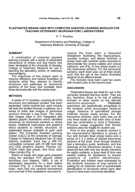
J Int Soc Plastination, Vol 5:16 -19, 1991 16PLASTINATED BRAINS USED WITH COMPUTER ASSISTED LEARNING MODULES FORTEACHING VETERINARY NEUROANATOMY LABORATORIESP. T. PurintonDepartment of Anatomy and Radiology, College ofVeterinary Medicine, University of GeorgiaSUMMARYA combination of <strong>computer</strong> <strong>assisted</strong><strong>learning</strong> <strong>modules</strong> <strong>with</strong> a series of plastinateddissections of sheep and dog <strong>brains</strong> hasbeen developed at the University of Georgia,College of Veterinary Medicine <strong>for</strong> use inteaching the laboratory portion of veterinaryneuroanatpmy.The objectives of this project were toimprove efficiency and reduce frustration ofstudents while they attempt to identifystructures and pathways on transversesections of the brain and correlate themthree-dimensionally <strong>with</strong> the whole brain.METHODSA series of 13 <strong>modules</strong>, covering all of thestructures and pathways studied, has beengenerated. Using minimal text, each modulewill lead the student through a pathway via aseries of video disk images of transversesections through a dog brain. The videodisk images were in turn integrated <strong>with</strong>labeled graphic illustrations which identifiedstructures on transverse sections or whole<strong>brains</strong>. Additional instructions and graphicsdirect students to identify structures onplastinated tissues available at each workstation. The Computer Assisted LearningCenter (CALC) at the University of Georgia,College of Veterinary Medicine has 16 workstations available. Each has a 286 PCcompatible <strong>computer</strong> <strong>with</strong> 20 MB hard diskand VGA graphics display, Pioneer LD-V4200 video disk player, and Sony colorVideo monitor (Fig. 1).Brains of sheep and dogs were preparedusing the standard S10 silicone rubbertechnique (von Hagens, 1985). A set ofplastinated specimens <strong>for</strong> a work stationincluded (Fig. 2): a sheep brain and dogbrain, each had one cerebral hemisphereand one-half of the cerebellum removed toexpose the brain stem; a dissectedsheepbrain illustrating the hippocampus,caudate nucleus, and lateral ventricle; asheep brain <strong>with</strong> cerebral cortex removed todemonstrate the corona radiata and corpuscallosum; and (Fig. 3) two sheep <strong>brains</strong> cutinto transverse sections. For the transversesections, each brain was cut at three levelssuch that the set of two <strong>brains</strong> illustratessections at six different levels.The <strong>modules</strong> have been <strong>used</strong> two years<strong>with</strong> revision prior to the second year.DISCUSSION<strong>Plastinated</strong> tissues are ideal <strong>for</strong> use in the<strong>computer</strong> <strong>assisted</strong> <strong>learning</strong> center. They aredry, there<strong>for</strong>e, there is no risk of spilling<strong>for</strong>malin or other liquids on the delicateelectronic equipment. <strong>Plastinated</strong>specimens are aesthetically acceptable tothe students which encourages hands-onusage. Absence of specialized storagemethods insures ready access. For thetransverse sections, each brain was cut atonly three levels so that each slice of brainwas two centimeters or more in thickness.Previous usage suggested that thinnersections were too fragile <strong>for</strong> student use.Prior to this project, students worked inthe neuroanatomy laboratory in groups offour <strong>with</strong> <strong>for</strong>malin fixed sheep <strong>brains</strong> and aseries of 2 X 2 slides of stained transversesections of the brain. Even though studentshad a laboratory guide, textbooks, and linedrawings of the transverse sections, it wasobserved that <strong>with</strong> only two faculty members<strong>for</strong> eighty students, a great deal of time waswasted waiting <strong>for</strong> assistance to answerquestions or confirm identification.Frustration and irritation were expressed bythe students because of long periods ofwaiting <strong>for</strong> an instructor and of questionableidentification of structures which were laterfound to have been incorrectly identified.
17 J Int Soc Plastination, Vol 5:16 -19, 1991Figure 1. Work station <strong>with</strong> 286 PC <strong>computer</strong>, laser video disk player, and dual monitors.The use of <strong>computer</strong> <strong>assisted</strong> <strong>learning</strong><strong>modules</strong> <strong>with</strong> plastinated tissues has severaladvantages <strong>for</strong> both the students andinstructors. For the students, there is littlewasted time in working through the <strong>modules</strong>.Errors in identification are reduced by havinga labeled drawing on the <strong>computer</strong> screenlinked <strong>with</strong> an unlabeled slide on the videodisk monitor. The combining of plastinatedtissues as an integral part of each <strong>computer</strong>lesson is important, because historicallystudents have tended to separate the studyof the gross brain from the study of thehistologic sections of the brain. This oftenresulted in failure to synthesize the two partsinto an integrated whole. By using onscreeninstructions and labeled graphics to directstudents to plastinated tissues, integration isfacilitated.Students generally work in groups of twoor three which encourages discussion andinteraction. Students have textbooks,lecture notes, illustrations of pathways, andunlabeled line drawings to use in conjunction<strong>with</strong> the <strong>computer</strong> and plastinated materialsthus allowing multiple modes of <strong>learning</strong>.Instructors also have noted that thenumber of questions has been dramaticallyreduced. Two instructors were unable tokeep up <strong>with</strong> student requests <strong>for</strong> help in theold system, but in the <strong>computer</strong> lab,questions are easily handled by the twoinstructors and often one faculty memberwould be sufficient. Questions that areasked tend to be more substantive ratherthan just asking <strong>for</strong> confirmation ofstructures which are being identified.Although the volume of in<strong>for</strong>mation has not
P. T. Purinton 18Figure 2. A. Dog brain; B. Sheep brain; C. Deep gray matter of sheep brain; D. Corpuscallosum and corona radiata of sheep brain.been reduced, fewer students haveexpressed a feeling of inability to master thesubject.In addition to subjective evaluation by theinstructors, students were asked to scorespecific statements evaluating the system ona six point scale, <strong>with</strong> a six being in agreement<strong>with</strong> the statement and a one beingdisagreement. Two of the statements and thepercentile of responses <strong>for</strong> each score aregiven in Table 1. In general, students foundthe <strong>computer</strong> programs and plastinated<strong>brains</strong> very helpful in <strong>learning</strong> neuroanatomy.The improvement in scores on bothstatements (especially the second statement)<strong>for</strong> the second year are a reflection of themodifications implemented. Improvementsconsisted of increasing the number ofplastinated specimens available at each workstation and adding on-screen instructions andlabeled illustrations specifically <strong>for</strong> theplastinated tissues. Additional dissections andmore labeled illustrations should furtherimprove student evaluation.The <strong>modules</strong> are installed on the hard driveof the CALC <strong>computer</strong>s and the plastinated<strong>brains</strong> are on library reserve, thus studentshave access to these <strong>learning</strong> materialsoutside of assigned class time, whenever thereading room is open and the <strong>computer</strong>center is not otherwise occupied. This alsoallows access by upper class studentswishing to review details of neuroanatomy <strong>for</strong>courses such as clinical neurology.The next major addition to this system is todevelop a series of interactive self-evaluation
19 P.T. PurintonFigure 3. Sheep <strong>brains</strong>, transverse sections.and quiz <strong>modules</strong>. This will allow students tomonitor their progress in mastering thesubject matter.REFERENCESvon HAGENS, G: Heidelberg PlastinationFolder: Collection of all technical leaflets<strong>for</strong> plastination. Anatomisches Institute 1,Universitat Heidelberg, 1985.Table 1. Student responses to statements of thecourse evaluation regrading usefulness ofNeuroanatomy teaching aids. A six (6) indicatescomplete agreement <strong>with</strong> the statement and a one (1)indicates complete disagreement.1.2.Score: 6 5 4 Moduleswere over all helpful:1990 52.8% 30.5% 5.5%1991 90.9% 9.1% 08.4%0<strong>Plastinated</strong> <strong>brains</strong> were helpfulin correlating three dimensional structure <strong>with</strong>transverse sections.1990 16.7% 30.6% 25.0% 19.4% 2.8% 5.5%1991 69.6% 17.4% 8.7% 4.3% 0 010 2.8%0