
You also want an ePaper? Increase the reach of your titles
YUMPU automatically turns print PDFs into web optimized ePapers that Google loves.
<strong>Research</strong> <strong>Report</strong> <strong>2002</strong><br />
<strong>MDC</strong><br />
Berlin-Buch<br />
Max Delbrück Center for Molecular Medicine
<strong>Research</strong> <strong>Report</strong> <strong>2002</strong><br />
(covers the period 2000-2001)<br />
Max Delbrück Center for<br />
Molecular Medicine (<strong>MDC</strong>)<br />
Berlin-Buch<br />
Robert-Rössle-Str. 10<br />
D-13125 Berlin<br />
Tel.: +49-30-9406-0<br />
Fax: + 49-30-949-4161<br />
e-mail: mdc@mdc-berlin.de<br />
This <strong>Research</strong> <strong>Report</strong> is also available<br />
on the World Wide Web<br />
http://www.mdc-berlin.de<br />
The <strong>MDC</strong> is a member of the<br />
HGF (Helmholtz Association<br />
of National <strong>Research</strong> Centers)<br />
The following <strong>Research</strong> <strong>Report</strong>s<br />
have been published previously:<br />
<strong>Research</strong> <strong>Report</strong> 1996<br />
(covers the period 1992-1995)<br />
<strong>Research</strong> <strong>Report</strong> 1996/97<br />
(covers the period 1996-1997)<br />
<strong>Research</strong> <strong>Report</strong> 2000<br />
(covers the period 1998-1999)<br />
Editorial board:<br />
Walter Birchmeier<br />
Carmen Birchmeier<br />
Thomas Blankenstein<br />
Udo Heinemann<br />
Helmut Kettenmann<br />
Friedrich C. Luft<br />
Peter M. Schlag<br />
Coordination:<br />
Barbara Bachtler<br />
Hans-Josef Linkens<br />
Volker Wunderlich<br />
Book design:<br />
Hoch Drei GmbH, 10963 Berlin<br />
Printing:<br />
Druckhaus Berlin-Mitte GmbH<br />
Printed in Germany <strong>2002</strong><br />
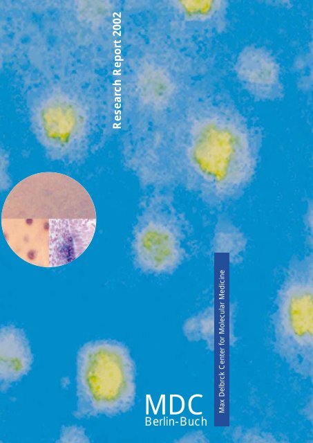
Legend to Cover Figures:<br />
In situ hybridization with the patched-1 cDNA<br />
reveals a block in embryonic hair placode<br />
formation in the absence of �-catenin/wnt<br />
signaling. In addition, tissue-specific ablation of<br />
the �-catenin gene in the skin of mice demonstrates<br />
the essential role of �-catenin signaling<br />
in the fate of skin stem cells in the adult: in the<br />
absence of �-catenin skin stem cells fail to<br />
differentiate into follicular keratinocytes, taking<br />
an epidermal route instead (from Huelsken et<br />
al., Cell (2001) 105, 533-545; this report p. 97)
<strong>Research</strong> <strong>Report</strong><br />
<strong>2002</strong><br />
Covers the period 2000/2001
Content<br />
Inhalt<br />
2<br />
Introduction<br />
Einführung .................................................................................................................................................................... 8<br />
Overview<br />
Überblick .................................................................................................................................................................... 21<br />
Genetics, Bioinformatics and Structural Biology<br />
Genetik, Bioinformatik und Strukturbiologie ........................................................................................................ 40<br />
Molecular Biology and Genetics of Cardiovascular Diseases<br />
Molekularbiologie und Genetik kardiovaskulärer Erkrankungen<br />
Detlev Ganten, Norbert Hübner ................................................................................ 43<br />
Molecular Biology of Peptide Hormones<br />
Molekularbiologie von Peptid-Hormonen<br />
Michael Bader .......................................................................................................... 45<br />
Genetics, Etiology, and Pathogenesis of Hypertension,<br />
Vascular Injury, and Renal Diseases<br />
Genetik, Ätiologie und Pathogenese von Bluthochdruck,<br />
Gefäßschädigung und Nierenerkrankungen<br />
Friedrich C. Luft ....................................................................................................... 47<br />
Gene Mapping and Identification in Monogenic and Complex Diseases<br />
Genkartierung und Identifizierung bei monogenen und komplexen Erkrankungen<br />
Peter Nürnberg ........................................................................................................ 49<br />
Cardiovascular Molecular Genetics<br />
Kardiovaskuläre Molekulargenetik<br />
Ludwig Thierfelder .................................................................................................... 52<br />
Obesity and Hypertension<br />
Fettleibigkeit und Bluthochdruck<br />
Arya M. Sharma ....................................................................................................... 54<br />
Disorders of the Autonomic Nervous System<br />
Erkrankungen des autonomen Nervensystems<br />
Jens Jordan (Helmholtz Fellow) ................................................................................ 56<br />
Characterization of newly identified human importin � proteins<br />
Charakterisierung von neuen humanen Importin-alpha Proteinen<br />
Matthias Köhler (Helmholtz Fellow) ........................................................................... 57<br />
Tumor Genetics<br />
Tumorgenetik<br />
Siegfried Scherneck ................................................................................................. 58<br />
Mouse Genetics<br />
Mausgenetik<br />
Carmen Birchmeier .................................................................................................. 61<br />
Developmental Genetics<br />
Entwicklungsgenetik<br />
Andreas Schedl ....................................................................................................... 64
3<br />
Lipids and Experimental Gene Therapy<br />
Lipide und experimentelle Gentherapie<br />
Thomas Willnow ....................................................................................................... 66<br />
Bioinformatics<br />
Bioinformatik<br />
Jens Reich, Peer Bork .............................................................................................. 68<br />
Protein Misfolding: From Basic Biopolymer Physics<br />
to Conformational Diseases<br />
Protein-Fehlfaltung: von der Biopolymerphysik zu Konformations-Erkrankungen<br />
Gregor Damaschun .................................................................................................. 70<br />
Structural Studies of Proteins and Nucleic Acids<br />
by X-ray Crystallography<br />
Strukturstudien an Proteinen und Nukleinsäuren mit Röntgenstrahl-Kristallographie<br />
Udo Heinemann ....................................................................................................... 72<br />
Role of Protein Dynamics in Enzyme Function<br />
Rolle der Proteindynamik bei der Enzymfunktion<br />
Christiane Jung ........................................................................................................ 75<br />
Computer Simulation of Nucleic Acid Structure and Interactions<br />
Computersimulation von Nukleinsäure-Strukturen und Wechselwirkungen<br />
Heinz Sklenar ........................................................................................................... 77<br />
Conformation, Stability and Interaction of Biological Macromolecules<br />
Konformation, Stabilität und Wechselwirkung von biologischen Makromolekülen<br />
Heinz Welfle ............................................................................................................. 79<br />
Bioethics and Science Communication<br />
Bioethik und Wissenschaftskommunikation<br />
Christof Tannert ....................................................................................................... 81<br />
Cell Growth and Differentiation<br />
Zellwachstum und Differenzierung ................................................................................................................. 84<br />
Growth Control and Gene Regulation in the Hematopoietic System<br />
Wachstumskontrolle und Genregulation im hämatopoetischen System<br />
Achim Leutz ............................................................................................................. 88<br />
Signal Transduction in Tumor Cells<br />
Signaltransduktion in Tumorzellen<br />
Claus Scheidereit ..................................................................................................... 90<br />
Differentiation and Growth Control in Lymphocyte Development<br />
and Immunopathogenesis<br />
Differenzierung und Wachstumskontrolle<br />
bei der Lymphozyten-Entwicklung und Immunpathogenese<br />
Martin Lipp .............................................................................................................. 92<br />
Initiation of DNA Replication<br />
Initiierung der DNA-Replikation<br />
Manfred Gossen ...................................................................................................... 95<br />
Epithelial Differentiation, Invasion and Metastasis<br />
Epitheliale Differenzierung, Invasion und Metastasierung<br />
Walter Birchmeier ..................................................................................................... 97
4<br />
Surgical Oncology<br />
Chirurgische Onkologie<br />
Peter M. Schlag ..................................................................................................... 100<br />
Molecular Muscle Physiology<br />
Molekulare Muskelphysiologie<br />
Ingo L. Morano ...................................................................................................... 103<br />
Cell Biology of Cardiovascular Diseases<br />
Zellbiologie kardiovaskulärer Erkrankungen<br />
Heinrich Leonhardt, M. Cristina Cardoso ................................................................ 105<br />
Intracellular Proteolysis<br />
Intrazelluläre Proteolyse<br />
Thomas Sommer ................................................................................................... 107<br />
Nucleocytoplasmic Transport in the Yeast Saccharomyces cerevisiae<br />
Nukleo-zytoplasmatischer Transport bei der Bäckerhefe<br />
Katrin Stade (Helmholtz Fellow) .............................................................................. 109<br />
Cytochrome P450 and Endoplasmic Reticulum<br />
Cytochrom P450 und endoplasmatisches Retikulum<br />
Wolf-Hagen Schunck ............................................................................................. 110<br />
Cell Polarity and Epithelial Formation in Development and Disease<br />
Zellpolarität und Epithelbildung in Entwicklung und Krankheit<br />
Salim Abdelilah-Seyfried ......................................................................................... 112<br />
Electron Microscopy<br />
Elektronenmikroskopie<br />
Gerd Kempermann, Bettina Erdmann ..................................................................... 114<br />
Molecular Therapy<br />
Molekulare Therapie ....................................................................................................................................... 118<br />
Myocardial Regeneration<br />
Regeneration des Myokards<br />
Rainer Dietz ............................................................................................................ 119<br />
Control of Smooth Muscle Cell Function<br />
Kontrolle der Funktion der glatten Muskelzelle<br />
Maik Gollasch (Helmholtz Fellow) ............................................................................ 121<br />
Immunology of Cardiovascular Diseases<br />
Immunologie kardiovaskulärer Erkrankungen<br />
Gerd Wallukat ........................................................................................................ 123<br />
Hematology, Oncology and Tumor Immunology<br />
Hämatologie, Onkologie und Tumorimmunologie<br />
Bernd Dörken ......................................................................................................... 125<br />
Molecular Immunotherapy<br />
Molekulare Immunotherapie<br />
Antonio Pezzutto .................................................................................................... 128<br />
Molecular Immunology and Gene Therapy<br />
Molekulare Immunologie und Gentherapie<br />
Thomas Blankenstein ............................................................................................. 130
5<br />
Cellular Immunology of Autoimmune Reactions<br />
Zelluläre Immunologie von Autoimmunreaktionen<br />
Kirsten Falk, Olaf Rötzschke ................................................................................... 132<br />
Molecular and Cell Biology of Hematopoietic Cells<br />
Molekular-und Zellbiologie hämatopoietischer Zellen<br />
Martin Zenke .......................................................................................................... 134<br />
Cell Cycle Regulation and Gene Therapy<br />
Zellzyklus-Regulation und Gentherapie<br />
Forschungsgruppe der Humboldt-Universität zu Berlin am <strong>MDC</strong> ............................. 136<br />
Evolution, Regulation and Genetic Applications of Transposable<br />
Elements in Vertebrates<br />
Evolution, Regulation und genetische Anwendungen von transposablen<br />
Elementen bei Vertebraten<br />
Zoltán Ivics ............................................................................................................. 138<br />
Experimental Pharmacology<br />
Experimentelle Pharmakologie<br />
Iduna Fichtner ........................................................................................................ 139<br />
Drug Targeting<br />
„Drug Targeting“ (Zielgenaue Medikamentenentwicklung)<br />
Regina Reszka ....................................................................................................... 141<br />
RNA Chemistry<br />
RNA Chemie<br />
Eckart Matthes ....................................................................................................... 143<br />
Molecular and Developmental Neurosciences<br />
Molekulare Neurowissenschaft und Entwicklungsneurobiologie ............................................................... 146<br />
Neurodegeneration<br />
Neurodegeneration<br />
Christiane Alexander .............................................................................................. 148<br />
Neuronal Stem Cells<br />
Neuronale Stammzellen<br />
Gerd Kempermann ................................................................................................ 150<br />
Cellular Neurosciences<br />
Zelluläre Neurowissenschaften<br />
Helmut Kettenmann ............................................................................................... 152<br />
Growth Factor and Regeneration Group<br />
Wachstumsfaktoren und Regeneration<br />
Gary R. Lewin ........................................................................................................ 154<br />
Synapse Formation and Function<br />
Bildung und Funktion von Synapsen<br />
Frank W. Pfrieger .................................................................................................... 156<br />
Developmental Neurobiology<br />
Entwicklungsneurobiologie<br />
Fritz G. Rathjen ...................................................................................................... 157<br />
Proteomics and Molecular Mechanisms of Neurodegenerative Disorders<br />
Proteomik und molekulare Mechanismen neurodegenerativer Erkrankungen<br />
Erich Wanker .......................................................................................................... 160
Structure and Organization<br />
Struktur und Organisation<br />
6<br />
Organizational Structure<br />
Stiftungsorgane ................................................................................................... 164<br />
The Board of Trustees ............................................................................................ 164<br />
Kuratorium<br />
Members of the Board of Trustees ............................................................... 164<br />
Mitglieder des Kuratoriums<br />
Members of the Scientific Committee .......................................................... 165<br />
Mitglieder des Wissenschaftlichen Ausschusses<br />
The Management Board ......................................................................................... 166<br />
Stiftungsvorstand<br />
Scientific Council .................................................................................................... 166<br />
Wissenschaftlicher Rat<br />
Staff Council .......................................................................................................... 167<br />
Personalrat<br />
Supporting Divisions ........................................................................................... 168<br />
Stabsstellen<br />
Safety ......................................................................................................... 168<br />
Sicherheit<br />
Building Coordination, Engineering and Reconstruction ............................... 168<br />
Bauangelegenheiten<br />
Auditing and Legal Affairs ............................................................................ 168<br />
Innenrevision und Recht<br />
Patents/Licences ........................................................................................ 169<br />
Patente und Lizenzen<br />
Technology Transfer .................................................................................... 169<br />
Technologietransfer<br />
Press and Public Relations ................................................................................ 170<br />
Presse- und Öffentlichkeitsarbeit<br />
Administration<br />
Verwaltung ........................................................................................................... 172<br />
Personnel ................................................................................................... 172<br />
Personal<br />
Finances .................................................................................................... 172<br />
Finanzen<br />
Purchasing and Materials Management ....................................................... 173<br />
Einkauf und Materialwirtschaft
7<br />
Central Facilities<br />
Zentrale Dienste .................................................................................................. 174<br />
Library ........................................................................................................ 174<br />
Bibliothek<br />
Animal Facilities ........................................................................................... 174<br />
Tierhaltung<br />
Campus Net Management ........................................................................... 174<br />
Campusnetz-Management<br />
Data and Image Processing ......................................................................... 175<br />
Daten- und Bildverarbeitung<br />
Technical Affairs .......................................................................................... 175<br />
Technik<br />
Meetings, Workshops and Symposia ................................................................ 176<br />
Wissenschaftliche Veranstaltungen<br />
Awards ................................................................................................................. 178<br />
Auszeichnungen<br />
Addresses of Scientific Journals at the Berlin-Buch Campus ......................... 179<br />
Adressen wissenschaftlicher Zeitschriften auf dem<br />
Campus Berlin-Buch<br />
Index .................................................................................................................... 180<br />
Index<br />
Organigram .......................................................................................................... 188<br />
Organigramm<br />
Campus Map ................................................................................. Inside Back Cover<br />
Campusplan ............................................................................. Innenumschlag hinten<br />
How to find your way to the <strong>MDC</strong> ................................................ Inside Back Cover<br />
Wie gelangen Sie zum <strong>MDC</strong> .................................................... Innenumschlag hinten
Introduction<br />
Molecular Medicine –<br />
A Concept and its Application at<br />
the <strong>MDC</strong> in Berlin-Buch<br />
History and Foundation<br />
On January 1, <strong>2002</strong>, the Max Delbrück Center for Molecular<br />
Medicine (<strong>MDC</strong>) Berlin-Buch celebrated its 10th birthday. It<br />
was founded on January 1, 1992, from the Institutes of the<br />
East German Academy of Sciences, the Central Institute for<br />
Cancer <strong>Research</strong>, the Central Institute for Cardiovascular<br />
<strong>Research</strong> and the Central Institute for Molecular Biology. The<br />
center is named after Max Delbrück, a native of Berlin, and<br />
one of the founding fathers of molecular biology who won the<br />
Nobel Prize in 1969. Delbrück collaborated at the Kaiser Wilhelm<br />
Institute for Brain <strong>Research</strong> in Berlin-Buch with the<br />
Russian geneticist, N. W. Timoféev-Ressovsky and the physicist,<br />
K.G. Zimmer. In 1935, their interdisciplinary research<br />
led to the publication of the epoch-making paper “The Nature<br />
of Gene Structure and Gene Mutation”.<br />
Anyone who experienced the reunification of Germany<br />
between November 9, 1989, and January 1, 1992 – involving<br />
the break up of the GDR, the unification of the two Germanies,<br />
with the evaluation of the entire scientific system of the<br />
GDR, the many plans for the restructuring of the scientific<br />
establishments and institutional and personal uncertainties of<br />
every kind – can imagine the problems people experienced,<br />
as well as the hope and expectations during this two-year period.<br />
In 1991, in Berlin-Buch, there were over 1,000 staff,<br />
physicians, researchers, and technicians working in the institutes<br />
of the Academy and the clinics. At its foundation, the<br />
<strong>MDC</strong> had 350 budgeted posts. In such circumstances it was<br />
no simple matter to set up a new organization to match the<br />
standard of the former institutes of the Academy and guarantee<br />
all qualified staff a future post. The details of this critical<br />
and exciting period of the restructuring of science on the<br />
Buch Campus has been comprehensively described else-<br />
8<br />
Einführung<br />
Molekulare Medizin –<br />
Ein Konzept und seine Umsetzung<br />
am <strong>MDC</strong> in Berlin-Buch<br />
Geschichte und Gründung<br />
Am 01. Januar <strong>2002</strong> beging das Max-Delbrück-Centrum für<br />
Molekulare Medizin (<strong>MDC</strong>) Berlin-Buch sein 10-jähriges<br />
Bestehen. Es ist am 01. Januar 1992 aus den Instituten der<br />
Akademie der Wissenschaften der DDR, dem Zentralinstitut<br />
für Krebsforschung, dem Zentralinstitut für Herz-Kreislaufforschung<br />
und dem Zentralinstitut für Molekularbiologie hervorgegangen.<br />
Die Benennung des Institutes erfolgte nach<br />
Max Delbrück, dem aus Berlin stammenden Wegbereiter der<br />
Molekularbiologie, der 1969 mit dem Nobelpreis ausgezeichnet<br />
worden war. Delbrück hatte am Kaiser-Wilhelm-Institut<br />
für Hirnforschung in Berlin-Buch zusammen mit dem russischen<br />
Genetiker N. W. Timoféeff-Ressovsky und dem Physiker<br />
K.G. Zimmer gearbeitet, und bei dieser interdisziplinären<br />
Forschung war 1935 die epochemachende Arbeit über die<br />
„Natur der Genstruktur und der Genmutation“ entstanden.<br />
Wer die Zeit der deutschen Wiedervereinigung zwischen dem<br />
09. November 1989 und dem 1. Januar 1992 persönlich miterlebt<br />
hat – mit dem Zusammenbruch der DDR, mit dem Prozess<br />
der Vereinigung der beiden deutschen Staaten, mit der<br />
Evaluation des gesamten Wissenschaftssystems der DDR, mit<br />
den vielen Plänen für die Neustrukturierung der wissenschaftlichen<br />
Einrichtungen und mit den Unsicherheiten in institutioneller<br />
und persönlicher Art –, kann sich vorstellen, welche Belastungen,<br />
aber auch welche Hoffnungen und Erwartungen in<br />
diesen zwei Jahren des Prozesses der deutschen Wiedervereinigung<br />
durchlebt wurden. In Berlin-Buch waren 1991 weit<br />
über 1.000 Mitarbeiter, Ärzte, Wissenschaftler, Technische<br />
Angestellte in den Akademieinstituten und Kliniken beschäftigt.<br />
Das <strong>MDC</strong> hatte bei der Gründung 350 budgetierte Stellen.<br />
Es war in dieser Situation nicht einfach, eine dem Leistungsniveau<br />
der Akademieinstitute entsprechende neue
where. It was only possible because everyone was sympathetic<br />
to the final aim and worked together to ensure that it<br />
was achieved.<br />
The significant and unavoidable problems at personal and institutional<br />
levels were made easier by the fact that everyone<br />
was aware of the great tradition of the Buch Institutes which<br />
had supported outstanding researchers like Oskar and Cécile<br />
Vogt, Walter Friedrich, Karl Lohmann, Arnold Graffi, Erwin<br />
Negelein, Albert Wollenberger and Hans Gummel. The real<br />
and positive role played by tradition in building new structures<br />
at a time of great change can be particularly well observed<br />
and experienced in Berlin. Attention to tradition was<br />
and still is important as far as setting up the <strong>MDC</strong> and the<br />
Berlin-Buch Campus are concerned.<br />
The clinics of the Academy of Sciences were incorporated<br />
into the university system – thanks to the energetic efforts of<br />
the then Senator for Science, Prof. Manfred Erhardt – as the<br />
Robert Rössle Cancer Clinic and the Franz Volhard Cardiovascular<br />
Clinic of the Charité, Berlin-Buch Campus. Thanks<br />
to particularly well defined cooperation contracts, they were<br />
linked to the <strong>MDC</strong> in both personnel and institutional terms.<br />
BBB Biotechnologie Berlin-Buch Management GmbH set up<br />
a biotechnology park. This has allowed the basic research<br />
activities at the <strong>MDC</strong>, and clinical research at the Charité<br />
clinics and the Buch Clinic as well as the Technology Park established<br />
by BBB Management GmbH to develop a synergy<br />
with respect to all research efforts that has subsequently been<br />
shown to be successful based on any number of criteria. The<br />
pure research carried out on the Berlin-Buch Campus was<br />
boosted in1999 by the arrival of the Forschungsinstitut für<br />
Molekulare Pharmakologie (FMP; <strong>Research</strong> Institute for<br />
Molecular Pharmacology), which is part of the Wilhelm Gottfried<br />
Leibniz Association. The institute collaborates closely<br />
with the <strong>MDC</strong> on structural research into biologically active<br />
molecules. The privatization of the clinics under the auspices<br />
of Helios Kliniken GmbH in summer 2001 has created even<br />
more opportunities for cooperation.<br />
Bundespräsident Johannes Rau (Mitte) im Gespräch mit Prof. Detlev Ganten (<strong>MDC</strong>-Stiftungsvorstand)<br />
und dem damals designierten und jetzigen Administrativen Vorstand des<br />
<strong>MDC</strong>, Dr. Waltraud Kreutz-Gers, anläßlich der Verleihung des Deutschen Zukunftspreises<br />
des Bundespräsidenten am 29. November 2001 im <strong>MDC</strong> Kommunikationszentrum.<br />
The President of the Federal Republic of Germany, Johannes Rau (in the middle), chats<br />
with the Scientific Director of the <strong>MDC</strong>, Detlev Ganten (MD. Ph.D), and the then<br />
Administrative Director designate, Dr. Waltraud Kreutz-Gers, after the awarding of the<br />
President's “Prize for Technology and Innovation” which took place at the <strong>MDC</strong>`s new<br />
Max Delbrück Communications Center (<strong>MDC</strong>.C) on November 29, 2001.<br />
Copyright: <strong>MDC</strong>; Annett Krause<br />
9<br />
Organisationsform zu finden und allen qualifizierten Mitarbeitern<br />
die ihnen zukommende Position zuzusichern. Über<br />
Einzelheiten dieser entscheidenden und aufregenden Monate<br />
und Jahre der Neuorientierung der Wissenschaft auf dem<br />
Campus Buch ist an anderer Stelle ausführlich berichtet und<br />
geschrieben worden. Sie konnten nur bewältigt werden, weil<br />
es von allen Seiten viel Verständnis und Unterstützung gab.<br />
Die großen und wohl unvermeidbaren Brüche in institutioneller<br />
und personeller Hinsicht wurden unter anderem dadurch<br />
erleichtert, dass ganz bewusst die große Tradition der<br />
Bucher Institute mit so herausragenden Wissenschaftlern wie<br />
Oskar und Cécile Vogt, Walter Friedrich, Karl Lohmann, Arnold<br />
Graffi, Erwin Negelein, Albert Wollenberger und Hans<br />
Gummel fortgeführt wurde. Die reale und konkrete positive<br />
Bedeutung der Tradition für die Zukunftsgestaltung in einer<br />
Zeit großer Umbrüche, kann in Berlin besonders gut beobachtet<br />
und erlebt werden. Traditionspflege wurde und wird<br />
bewußt für den Aufbau des <strong>MDC</strong> und des Campus Berlin-<br />
Buch eingesetzt.<br />
Die Kliniken der Akademie der Wissenschaften wurden mit<br />
tatkräftiger Unterstützung des damaligen Senators für Wissenschaft,<br />
Prof. Manfred Erhardt, in das universitäre System<br />
eingegliedert als Robert-Rössle-Krebsklinik und Franz-Volhard-Herz-Kreislaufklinik<br />
der Charité, Campus Berlin-Buch.<br />
Durch einen besonders engen Kooperationsvertrag wurden<br />
sie mit dem <strong>MDC</strong> personell und institutionell verbunden. Der<br />
Aufbau eines Biotechnologieparks wurde durch die BBB<br />
Biotechnologie Berlin-Buch Management GmbH begründet.<br />
Auf diese Weise wurde mit der Grundlagenforschung am<br />
<strong>MDC</strong>, der klinischen Forschung an den Charité-Kliniken und<br />
dem Klinikum Buch sowie mit dem von der BBB Management<br />
GmbH eingerichteten Technologiepark ein Synergien<br />
schaffendes komplettes Forschungssystem eingerichtet, das<br />
sich in den folgenden Jahren in jeder Hinsicht als erfolgreich<br />
erwiesen hat. 1999 wurde die Grundlagenforschung auf dem<br />
Campus Berlin-Buch durch den Zuzug des Forschungsinstituts<br />
für Molekulare Pharmakologie (FMP), das zur Wilhelm-<br />
Gottfried-Leibniz-Gemeinschaft gehört, verstärkt. Dieses<br />
Institut arbeitet mit dem <strong>MDC</strong> insbesondere auf dem Gebiet<br />
der Strukturforschung für biologisch aktive Moleküle intensiv<br />
zusammen. Die Privatisierung der Kliniken durch die<br />
Helios Kliniken GmbH im Sommer 2001 schafft erweiterte<br />
Möglichkeiten der Kooperation.<br />
Aufbruchstimmung in Berlin-Buch<br />
Auf dem Campus Berlin-Buch herrscht Aufbruchstimmung:<br />
Das Max Delbrück Communications Center (<strong>MDC</strong>.C) mit<br />
Hörsälen, Seminar- und Laborräumen für Kongresse, Fortbildung<br />
und Kursen ist im Jahre 2001 fertig gestellt und in Betrieb<br />
genommen worden. Der erste Bauabschnitt des Helmholtz<br />
Hauses für Tierlaboratorien und Büroräume wird noch im<br />
Jahre <strong>2002</strong> bezogen. Ein neues Zentrum für Medizinische Genomforschung<br />
des <strong>MDC</strong> ist im Entstehen. Vier neue Laborgebäude<br />
für Existenzgründer und Biotechnologiefirmen sind<br />
fertig gestellt bzw. befinden sich zur Zeit im Bau. Das neue<br />
„Helios Klinikum Berlin“ wird auch die universitären Kliniken<br />
der Charité in einem Neubau für 400 Millionen Mark mit aufnehmen,<br />
so dass eine der modernsten und größten Kliniken<br />
Berlins in unmittelbarer Nachbarschaft des Forschungscampus
Developments in Berlin-Buch<br />
Spirits on the Berlin-Buch Campus are high as the site develops:<br />
the Max Delbrück Communications Center (<strong>MDC</strong>.C),<br />
with its lecture theaters, seminar rooms and laboratory space<br />
for congresses, further education and training courses, was<br />
opened in 2001. The first section of the “Helmholtz Haus” for<br />
animal laboratory facilities and offices will be ready in <strong>2002</strong>.<br />
A new <strong>MDC</strong> Center for Medical Genome <strong>Research</strong> is being<br />
created. Four new laboratory buildings for start-up companies<br />
and biotechnology companies have been completed or<br />
are under construction. The new “Helios Klinikum Berlin”<br />
will also incorporate the university clinics of the Charité in a<br />
new setting costing approximately 200 million Euro so that<br />
one of the most modern and largest clinics in Berlin will be<br />
located right next to the research campus. The space occupied<br />
by the clinics previously represents an area of over 100 ha<br />
and this will allow the Technology Park to expand. The clinic<br />
buildings designated as being of historic interest will be used<br />
as a “Cité Universitaire”, and will be adopted by the European<br />
College of Liberal Art (ECLA). A new shopping center<br />
will be built right next to the S-Bahn station. It is also planned<br />
to build a hotel for visitors and congress delegates as well as<br />
relatives of the patients being treated in the clinics.<br />
The health theme, which has made Berlin-Buch, the “Health<br />
Region” is becoming more and more important as a part of<br />
the modern knowledge-based society. In the future, Berlin-<br />
Buch will be a center for molecular medicine where the latest<br />
advances in genome research and gene technology will be<br />
applied to the clinical setting and in the field of business,<br />
allowing biotech companies and service firms to develop and<br />
create new jobs.<br />
The Sculpture Park on the Berlin-Buch Campus with works by,<br />
among others, Ipousteguy, Szymanski and Kriester, reflects the<br />
close relationship between the Arts and Sciences, which is particularly<br />
encouraged in Berlin-Buch. In this way, Buch will be<br />
seen as an intellectual focal point where the development of<br />
molecular medicine and gene technology is being advanced<br />
alongside a continuing dialog with people from many different<br />
backgrounds. In this way Berlin-Buch will become a model of<br />
a future human knowledge-based society. On the <strong>MDC</strong> Campus<br />
over 2000 staff are currently working in the research institutes,<br />
clinics and biotech companies, many more than before<br />
reunification in 1989. During the last 10 years many new jobs<br />
have been created in this science-based environment.<br />
The rapid connections linking Berlin-Buch to the cultural<br />
center of central Berlin and the proximity of the Barnim Nature<br />
Conservation Park have made Berlin-Buch one of the<br />
most attractive and dynamic suburbs of the new Berlin.<br />
Many of the staff who work in the research institutes and the<br />
biotech companies live only 15 minutes away in Prenzlauer<br />
Berg, one of the favorite parts of Berlin with many theaters,<br />
bars and cinemas. Prenzlauer Berg is close to the cultural and<br />
spiritual heart of the city with the Humboldt University, the<br />
Museum Island, the main theaters, the Berlin Philharmonic<br />
and the Kulturforum.<br />
10<br />
entstehen wird. Die freiwerdenden Klinikgebäude in einem<br />
Areal von über 100 Hektar werden für den expandierenden<br />
Technologiepark zur Verfügung stehen. Denkmalgeschützte<br />
Klinikgebäude sollen für eine „Cité Universitaire“, die auch<br />
das European College of Liberal Art (ECLA) aufnehmen wird,<br />
genutzt werden. In unmittelbarer Nähe des S-Bahnhofes wird<br />
ein neues Einkaufszentrum gebaut. Dazu ist geplant, ein Hotel<br />
zu errichten für Besucher und Kongressteilnehmer sowie für<br />
Angehörige von Patienten der Kliniken.<br />
Das Thema Gesundheit, dem sich Berlin-Buch, die „Gesundheitsregion“,<br />
verschrieben hat, wird als Teil der modernen<br />
Wissensgesellschaft immer weiter an Bedeutung gewinnen.<br />
Berlin-Buch soll in Zukunft ein Zentrum für die Molekulare<br />
Medizin sein, in dem die Fortschritte der Genomforschung<br />
und Gentechnik verantwortungsvoll in Klinik und Wirtschaft<br />
angewendet werden und um das herum sich Biotechnologiefirmen<br />
und Serviceeinrichtungen entwickeln und neue Arbeitsplätze<br />
entstehen lassen. Der Skulpturenpark auf dem<br />
Campus Berlin-Buch mit Werken unter anderem von Ipousteguy,<br />
Szymanski und Kriester ist Ausdruck der engen Ver-<br />
Dr. Erwin Jost, Administrativer Vorstand des <strong>MDC</strong>, Dr. Arend Oetker (Vorsitzender des<br />
Kuratoriums Deutscher Zukunftspreis und Präsident des Stifterverbandes) und Prof.<br />
Detlev Ganten (<strong>MDC</strong>-Stiftungsvorstand) beim Empfang nach der Vergabe des Zukunftspreises<br />
des Bundespräsidenten am 29. November 2001 im neuen Max Delbrück Communications<br />
Center (<strong>MDC</strong>.C)<br />
Reception after the awarding of the “Federal President's Prize for Technology and Innovation”<br />
in the new Max Delbrück Communications Center (<strong>MDC</strong>.C) on November 29,<br />
2001 (from left): Dr. Erwin Jost (Administrative Director of the <strong>MDC</strong>), Dr. Arend Oetker<br />
(Chairman of the Board of Trustees of the President's “Prize for Technology and Innovation“<br />
and President of the Stifterverband für die Deutsche Wissenschaft – Donor´s Association<br />
for the Promotion of the Sciences and Humanities) and Detlev Ganten (MD.,<br />
Ph.D, <strong>MDC</strong>`s Scientific Director).<br />
Copyright: <strong>MDC</strong>; Siegfried Röpke
Buch is a place of synergies. As a link between town and<br />
country, between pure research and the clinics as well as<br />
scientific applications of this research, living, working and<br />
relaxing in Berlin-Buch are all equally attractive.<br />
The concept of molecular medicine<br />
As far as the success of any scientific institution is concerned,<br />
the key factors are the original idea upon which it is based,<br />
the efforts of its scientists and other key staff, their commitment,<br />
the quality of the research activities, projects and its<br />
publication record.<br />
Abschied nach acht Jahren <strong>MDC</strong>: Prof. Jutta Schnitzer, wissenschaftliche Referentin<br />
des <strong>MDC</strong> und jetzige Generalsekretärin der Leopoldina, Halle. <strong>MDC</strong>-Vorstand Prof.<br />
Detlev Ganten beim Überreichen des Blumenstrausses.<br />
Farewell after eight years at the <strong>MDC</strong>: Prof. Jutta Schnitzer, <strong>MDC</strong>`s former Scientific<br />
Coordinator and now Secretary General of the “Leopoldina” (Halle) receives a bouquet<br />
of flowers from Detlev Ganten (MD., PhD), <strong>MDC</strong>`s Scientific Director<br />
Copyright: <strong>MDC</strong>; Siegfried Endruweit<br />
When the <strong>MDC</strong> was founded, we took advice based on the<br />
recommendations of the Scientific Council and the Founders<br />
Committee regarding ideas about the long-term developments<br />
in medicine and the latest results of molecular genetics<br />
and genome research.<br />
There are two reasons for the extraordinary successful and<br />
sustained development of the modern life sciences. On the<br />
one hand, there were the scientists themselves, who highlighted<br />
the new, often physico-chemical, methods which<br />
could identify the macromolecules which had key biological<br />
functions in biology and medicine, thereby allowing a better<br />
understanding of the interplay between the form and function<br />
of cells. One the other hand, institutions like the Rockefeller<br />
Foundation play an important role. For example, in 1938, it<br />
supported a research program for the advancement of genetics<br />
giving it the name “molecular biology”. One aim of this<br />
project was to raise the level of biosciences at that time and<br />
turn it into a quantitative “Science of Man”, which went beyond<br />
simply describing events that happened in Nature. Over<br />
the period 1937–39, the person who gave his name to the<br />
<strong>MDC</strong>, Max Delbrück, held a fellowship from the Rockefeller<br />
Foundation and their support helped him become one of the<br />
11<br />
bindung von Kunst und Wissenschaft, die in Berlin-Buch besonders<br />
gepflegt wird. Buch soll sich auf diese Weise als intellektueller<br />
Kristallisationspunkt profilieren, in dem die Entwicklung<br />
der Molekularen Medizin und Gentechnologie<br />
vorangebracht und durch den Dialog mit breiten Kreisen der<br />
Gesellschaft kritisch begleitet wird. Auf diese Weise wird Berlin-Buch<br />
zu einem Modell für die zukünftige humane Wissensgesellschaft.<br />
Auf dem Campus Berlin-Buch arbeiten zur<br />
Zeit in den Forschungseinrichtungen, Kliniken und Biotechnologiefirmen<br />
über 2.000 Mitarbeiter, weit mehr als vor der<br />
Wiedervereinigung 1989. Im Umfeld der Wissenschaft sind<br />
damit in den vergangenen 10 Jahren viele neue anspruchsvolle<br />
und zukunftsfähige Arbeitsplätze geschaffen worden.<br />
Die schnelle Anbindung von Berlin-Buch an das kulturelle<br />
Zentrum in Berlin-Mitte und die direkte Nachbarschaft zum<br />
Naturschutzpark Barnim machen Berlin-Buch zu einem der<br />
attraktivsten und dynamischsten Vororte des neuen Berlin.<br />
Viele Mitarbeiter der Forschungseinrichtungen und Biotechnologiefirmen<br />
wohnen in dem 15 Minuten entfernten Prenzlauer<br />
Berg, einem der beliebtesten Stadtteile Berlins mit vielen<br />
Theatern, Kneipen und Kinos. Dem Prenzlauer Berg schließt<br />
sich direkt das kulturelle und geistige Zentrum der Stadt an mit<br />
der Humboldt Universität, der Museumsinsel, den großen<br />
Theatern, der Philharmonie und dem Kulturforum.<br />
Buch ist ein Ort der Synergien. Als Nahtstelle zwischen Stadt<br />
und Land, zwischen Forschung und klinischer sowie wirtschaftlicher<br />
Anwendung ist das Leben, das Arbeiten und das<br />
Erholen in Berlin-Buch gleichsam attraktiv.<br />
Das Konzept der Molekularen Medizin<br />
Entscheidend für den Erfolg einer wissenschaftlichen Institution<br />
ist das inhaltliche Konzept, die wissenschaftliche Leistung,<br />
die tragenden Personen, das Engagement der Wissenschaftlerinnen,<br />
Wissenschaftler und Mitarbeiter, die Qualität<br />
der Forschungstätigkeit, Projekte und Publikationen.<br />
Zur Zeit der Gründung des <strong>MDC</strong> haben wir uns auf der Basis<br />
der Empfehlungen des Wissenschaftsrates und des Gründungskomitees<br />
bei den Konzepten von langfristigen Entwicklungen<br />
der Medizin und neuesten Ergebnisse der molekularen<br />
Genetik und Genomforschung leiten lassen.<br />
Es gibt zwei Quellen für das außerordentlich erfolgreiche und<br />
nachhaltige Konzept der modernen Lebenswissenschaft. Zum<br />
einen waren dies die Wissenschaftler selbst, die mit neuen,<br />
über die Biologie und Medizin hinausreichenden, häufig physikalisch-chemischen<br />
Methoden die Strukturen von Makromolekülen<br />
mit wichtigen biologischen Funktionen erkunden<br />
konnten und sich anschließend daran machten, das Wechselspiel<br />
von Form und Funktion in der Zelle zu verstehen. Zum<br />
zweiten spielten Institutionen wie etwa die Rockefeller Stiftung<br />
eine wichtige Rolle, die im Jahre 1938 einem Forschungsprogramm<br />
zur Förderung der Genetik den Namen<br />
„Molekularbiologie“ gab. Ein Ziel dieser Initiative lag darin,<br />
das Niveau der damaligen Biowissenschaften anzuheben und<br />
zu einer quantitativ operierenden „Science of Man“ auszuweiten,<br />
die über die reine Beschreibung der Natur hinausging.<br />
Der Namenspatron des <strong>MDC</strong>, Max Delbrück, gehörte in
founding fathers of molecular biology; there is much that we<br />
can still learn from him today.<br />
When the <strong>MDC</strong> was established ten years ago in Berlin-Buch<br />
and raised a flag as far as the development of molecular medicine<br />
was concerned, there were only a few institutes around<br />
the world engaged in applying the latest results of pure research<br />
to medical research in a closely-knit institutional setting.<br />
At that time, no one could have known the kind of<br />
progress that would take place in genome analysis in the next<br />
ten years. The mission of the <strong>MDC</strong> is to combine modern<br />
medical and clinical research with the methods of molecular<br />
biology and cell biology as well as the expansion in gene<br />
technology, which systematically concentrates on the genome<br />
and the latest results arising from research into it. As far as<br />
pure research is concerned, key areas should be those that are<br />
of vital importance for the analysis of disease phenomena and<br />
which lead to new opportunities firmly based on the natural<br />
sciences for diagnosis, treatment and prevention.<br />
The multitalented molecules of molecular medicine<br />
Touch and Pain: two senses, one ion channel named DRASIC<br />
Our senses of touch and pain, opposites one might think, have something<br />
in common, an ion channel called DRASIC. DRASIC, which<br />
stands for dorsal root ganglion acid sensitive channel, has been<br />
shown to be critical for both these senses. The DRASIC protein forms<br />
a channel in the membrane that when opened conducts sodium ions<br />
which then leads to the electrical excitation of neurons. A first clue<br />
about the possible function of the DRASIC protein came from a genetic<br />
screen for mutations in the nematode worm C.elegans which<br />
showed that similar proteins in the worm are necessary for the worm<br />
to sense touch. These proteins including DRASIC are all members of<br />
the Deg/Enac channel superfamily. Thus arose the idea that mechanical<br />
forces on the membrane could open the channel: this would make<br />
it a mechanical sensor. Thus the same channel obviously helps sensory<br />
neuron to detect innocuous and painful stimuli in a completely different<br />
way. It is known that DRASIC can potentially form heteromeric<br />
ion channel complexes with other members of the Deg/Enac family of<br />
proteins.<br />
Another aspect of DRASIC channel function is the fact that acidification<br />
ie. protons are a very effective stimulus to open the channel. Acidification<br />
is a universal consequence of injury or inflammation of peripheral<br />
tissues. Mice lacking DRASIC were much less affected by muscle<br />
acidification, in other words the lack of the channel protein had an analgesic<br />
effect. The diversity of function for the DRASIC channel reported<br />
here for the first time shows directly that the channel does not<br />
work alone but in concert with other as yet unknown proteins. It is precisely<br />
this kind of in vivo analysis of protein function that highlights the<br />
principle that the function of a single protein can vary enormously depending<br />
on the cellular context. Thus products of single genes are not<br />
simple entities that work alone to accomplish a well-defined function<br />
but rather execute their function in a multitude of ways depending the<br />
other proteins that they can interact with. For further details see Gary<br />
Lewin´s research group.<br />
12<br />
den Jahren 1937–39 zu den Stipendiaten der Rockefeller Stiftung<br />
und ist mit ihrer Hilfe zu dem Wegbereiter der Molekularbiologie<br />
geworden, von dem wir heute noch lernen können.<br />
Als das <strong>MDC</strong> vor zehn Jahren in Berlin-Buch gegründet<br />
wurde und sich die Entwicklung einer Molekularen Medizin<br />
auf die Fahnen schrieb, gab es weltweit nur wenige Institute,<br />
die so konsequent die Anwendung neuester Methoden in der<br />
Grundlagenforschung für medizinische Forschung in enger<br />
institutioneller Verbindung verfolgten. Niemand konnte damals<br />
wissen, welche Fortschritte die Genomanalyse in diesem<br />
Zeitraum machen würde. Die Aufgabe des <strong>MDC</strong> bestand<br />
darin, moderne medizinische und klinische Forschung im<br />
Verband mit molekularbiologischen, zellbiologischen sowie<br />
zunehmend gentechnischen Methoden zu betreiben, die sich<br />
systematisch auf das Genom konzentrierten und von ihm ausgingen.<br />
Wegweisend für die Grundlagenforschung sollten<br />
Themen sein, die von prinzipieller Bedeutung für die Analyse<br />
von Krankheitsphänomenen sind und aus denen sich naturwissenschaftlich<br />
begründet neue Möglichkeiten für Diagnostik,<br />
Therapie und Prävention ableiten lassen.<br />
Die vielseitigen Moleküle der Molekularen Medizin<br />
Berührung und Schmerz: Zwei Sinne, ein Ionenkanal namens<br />
DRASIC<br />
Der Tastsinn und der Schmerz erscheinen zwar als gegenteilige Sinne,<br />
sie haben aber etwas gemeinsam, einen Ionenkanal mit Namen DRA-<br />
SIC. DRASIC kürzt den englischen Ausdruck „dorsal root ganglion<br />
acid sensitive channel“ ab, womit ein säureempfindlicher Ionenkanal<br />
im dorsalen Wurzelganglion gemeint ist. Es konnte gezeigt werden,<br />
dass DRASIC für die beiden genannten Sinne wesentlich ist. Das<br />
DRASIC Protein formt einen Kanal durch die Membran, durch den im<br />
geöffneten Zustand Natriumionen strömen, die dann zur elektrischen<br />
Erregung von Neuronen führen. Einen ersten Hinweis auf die mögliche<br />
Funktion des DRASIC Proteins ergab eine genetische Reihenuntersuchung<br />
von Mutationen in dem Fadenwurm C. elegans. Sie zeigte,<br />
dass ähnliche Proteine nötig sind, damit der Wurm berührungsempfindlich<br />
wird. Diese Proteine – einschließlich DRASIC – sind alle Mitglieder<br />
der Deg/Enac Kanal Großfamilie. Aus dieser Kenntnis leitet<br />
sich die Idee ab, dass mechanische Kräfte, die auf die Membran wirken,<br />
zur Öffnung des Kanals führen, der auf diese Weise ein mechanischer<br />
Sensor würde. Derselbe Kanal hilft offenbar sensorischen Neuronen,<br />
harmlose und schmerzhafte Reize auf vollständig verschiedene<br />
Weise zu registrieren. Es ist bekannt, dass DRASIC in der Lage ist,<br />
heteromere Kanalkomplexe mit anderen Mitgliedern der Deg/Enac<br />
Familie zu bilden.<br />
Ein weiterer Aspekt der Funktion des DRASIC Kanals steckt in der<br />
Tatsache, dass eine Zunahme des Säuregrads – also das Auftreten<br />
von Protonen – als effektiver Reiz zur Kanalöffnung funktioniert. Die<br />
Zunahme des Säuregrads ist die universelle Konsequenz einer Verletzung<br />
oder Entzündung des peripheren Gewebes. Mäuse, die nicht<br />
über DRASIC verfügten, waren jedoch viel weniger durch die Muskelsäuerung<br />
betroffen. Mit anderen Worten, die Abwesenheit des Kanalproteins<br />
hatte einen analgetischen Effekt. Die Verschiedenheit der<br />
Funktionen für den DRASIC Kanal, die hier zum ersten Mal vorgestellt<br />
wird, zeigt direkt, dass der Kanal nicht allein, sondern im Verbund mit<br />
anderen Proteinen funktioniert, die bislang unbekannt sind. Es ist nun<br />
genau diese Art der in vivo Analyse einer Proteinfunktion, die das Prinzip<br />
deutlich macht, demzufolge die Funktion eines einzelnen Proteins<br />
je nach dem zellulären Kontext enorm variieren kann. Produkte einzelner<br />
Gene sind also nicht bloß einfache Einheiten, die alleine operieren,<br />
um wohldefinierte Funktionen zu erfüllen. Sie üben ihre Aufgaben vielmehr<br />
in einer Vielzahl von Wegen aus, die von anderen Proteinen abhängen,<br />
mit denen sie in Wechselwirkung treten können. Weitere<br />
Details siehe Arbeitsgruppe von Gary Lewin.
<strong>MDC</strong>-Neujahrsveranstaltung am 28. Januar 2000: Prof. Fritz Melchers (Mitte), der<br />
frühere Vorsitzende des Wissenschaftlichen Ausschusses des <strong>MDC</strong> (Basel, Schweiz,<br />
Mitte) erhält für seine Verdienste um das <strong>MDC</strong> von Prof. Detlev Ganten (<strong>MDC</strong>-Stiftungsvorstand)<br />
(re.) die Max-Delbrück-Medaille. Interessierter Zuschauer: Wolf-Michael Catenhusen<br />
(Kuratoriumsvorsitzender des <strong>MDC</strong> und Parlamentarischer Staatssekretär im<br />
Bundesforschungsministerium).<br />
At the <strong>MDC</strong>`s New Year Reception on January 28, 2000: Prof Fritz Melchers (in the<br />
middle), former chairman of the Scientific Committee of the <strong>MDC</strong> from Basle (Switzerland)<br />
receives the Max Delbrück Medal for his contributions to the development of the<br />
<strong>MDC</strong> from <strong>MDC</strong>`s Scientific Director, Detlev Ganten (MD., Ph.D; right) as Wolf-Michael<br />
Catenhusen, chairman of the <strong>MDC</strong>`s Board of Trustees and Parliamentary State Secretary<br />
from the Federal Ministry for Education and <strong>Research</strong> looks on.<br />
Copyright: <strong>MDC</strong>; Siegfried Endruweit<br />
The interdisciplinary nature of pure research is reflected in<br />
the fact that its results are of importance and have applications<br />
in almost all classic clinical disciplines. The same molecules<br />
involved in cell differentiation, growth factors, and ion<br />
channels in membranes also play important roles in cancer,<br />
cardiovascular and nervous system diseases. Molecular medicine<br />
overcomes the traditional boundaries of these disciplines<br />
and provides conditions for true content-based and<br />
structural interdisciplinary activities.<br />
At the start of the 1980s, people began to understand for the<br />
first time how the tools of gene technology could be used to<br />
map the human genes, but for a long time many scientists<br />
were unable to make any sense of the immense efforts associated<br />
with the identification of the human gene sequence and<br />
the three billion or so building stones involved. The hope of a<br />
genetic analysis of diseases was based more on optimism<br />
than any empirical evidence. Only when clinical observations<br />
in combination with molecular biology research clearly<br />
showed that cancer, for example, is a genetic disease and the<br />
formation of tumors is affected by DNA variants, did the project<br />
really get underway, leading to the sequencing of the human<br />
genome and other model genomes. Due to these genome<br />
projects the ideas of molecular medicine became accepted by<br />
the general public with their promise of sweeping perspectives<br />
for the future.<br />
The 1990s saw the start of a new form of collaboration in<br />
medicine whereby scientists involved in molecular genetics<br />
and genome research, using genetic engineering, tried to answer<br />
clinically relevant questions and develop new products<br />
for the healthcare market. Today, it is perhaps possible to<br />
think of the past decade as the first period of biomedicine,<br />
during which the scope of gene diagnosis was enormously<br />
extended as well as the identification of many new targets as<br />
starting points for the development of drugs of the future.<br />
These developments are not only characterized by immense<br />
13<br />
Die Interdisziplinarität in der Grundlagenforschung spiegelte<br />
sich wider in der Tatsache, dass die Ergebnisse Bedeutung<br />
haben und Anwendung finden in fast allen klassischen klinischen<br />
Fächern. Die gleichen Moleküle der Zelldifferenzierung,<br />
Wachstumsfaktoren, Ionenkanäle in den Membranen<br />
sind für Krebs, Herzkreislauf- und Nervenerkrankungen in<br />
gleicher Weise wichtig. Die Molekulare Medizin überwindet<br />
damit die traditionellen Grenzen der Disziplinen und schafft<br />
die Voraussetzung für wahrhaft inhaltliche und strukturelle<br />
Interdisziplinarität.<br />
Zu Beginn der achtziger Jahre war zum ersten Mal verstanden<br />
worden, wie die Werkzeuge der Gentechnik es erlaubten, Genkarten<br />
des Menschen anzufertigen, aber lange Zeit hindurch<br />
konnten viele Wissenschaftler keinen Sinn in dem ungeheuren<br />
Aufwand erblicken, der mit der Erstellung der menschlichen<br />
Gensequenz und ihren rund drei Milliarden Bausteinen verbunden<br />
sein würde. Die Hoffnung auf genetische Analysen<br />
von Krankheiten beruhte mehr auf Optimismus als auf empirischer<br />
Evidenz. Erst als klinische Beobachtungen in Verbindung<br />
mit molekularbiologischen Forschungen deutlich vor<br />
Augen führten, dass zum Beispiel Krebs eine genetische<br />
Krankheit ist und die Bildung von Tumoren von DNA Varianten<br />
beeinflusst wird, kam das Projekt in Schwung, in dessen<br />
Verlauf das humane Genom und andere Modellgenome sequenziert<br />
wurden. Mit diesen Genomprojekten konnte sich die<br />
Idee der Molekularen Medizin öffentliche Anerkennung verschaffen<br />
und umfassende Perspektiven für die Zukunft bieten.<br />
In den neunziger Jahren hat dann zunehmend eine neue Form<br />
der Zusammenarbeit auf dem Gebiet der Medizin begonnen.<br />
Dabei versuchen molekulargenetisch und genomisch orientierte<br />
Wissenschaftler mit gentechnischen Methoden klinisch<br />
relevante Fragestellungen zu beantworten und ihre Lösungen<br />
in neue Produkte für den Gesundheitsmarkt umzusetzen.<br />
Es ist heute vielleicht möglich, vom vergangenen Jahrzehnt<br />
als einem ersten Zeitalter der Biomedizin zu sprechen, in dem<br />
nicht nur das Spektrum der Gendiagnostik enorm erweitert<br />
wurde, sondern in dem auch viele neue Zielstrukturen (Targets)<br />
erkannt wurden, die als Ausgangspunkt für die Entwicklung<br />
künftiger Medikamente dienen. Diese Entwicklung<br />
ist nicht nur durch ihre ungeheure Dynamik und das konsequent<br />
interdisziplinäre Vorgehen der Wissenschaft charakterisiert,<br />
sondern auch durch die nahtlose Zusammenarbeit von<br />
Grundlagenforschung, Klinik und Industrie, die – so lässt<br />
sich vorhersagen – in Zukunft noch intensiviert wird. Der<br />
Campus in Berlin-Buch hat von Anfang an diese Entwicklung<br />
wahrgenommen, mitgeprägt, zu ihr beigetragen und sein Vorgehen<br />
an diesem wissenschaftlichen, gesellschaftlichen und<br />
ökonomischen Verbund orientiert.<br />
Das <strong>MDC</strong> mit seinen mehr als 700 Mitarbeiterinnen und Mitarbeitern,<br />
von denen 300 wissenschaftlich tätig sind, konzentriert<br />
seine Forschungen auf insgesamt sechs Felder:<br />
• Herz-Kreislaufforschung,<br />
• Krebsforschung,<br />
• Neurowissenschaften,<br />
• Genetik und Genomforschung, Strukturbiologie, Bioinformatik,<br />
• Zellwachstum und Zelldifferenzierung<br />
• Molekulare Therapie.
The multitalented molecules of molecular medicine<br />
C/EBPs flip a switch in chromatin remodelling and gene activation<br />
The human genome contains over 30,000 different genes. Expression<br />
of various combinations of these genes gives rise to specific cell types,<br />
such as muscle, skin, blood or nerves, and also determines whether<br />
cells remain in a resting state, grow and proliferate or die and self-destruct.<br />
Inappropriate gene regulation causes autoimmune diseases, a<br />
variety of other genetic diseases and cancer. <strong>Research</strong>ers in the <strong>MDC</strong><br />
laboratory directed by Achim Leutz are interested in how cells make<br />
decisions to differentiate and how these processes are linked to the<br />
development of disease, especially leukemia. By focusing on a family<br />
of transcription factors called C/EBP proteins, which were originally<br />
identified as regulators of liver-specific genes but which are now<br />
known to be important regulators for many cell types, the <strong>MDC</strong> scientists<br />
have found that the ability of C/EBP proteins to influence various<br />
programs of differentiation depends on their ability to produce<br />
changes in chromatin structure. Chromatin is the higher order structure<br />
of chromosomes in a cell that helps keep some genes inaccessible<br />
and inactive, while others are exposed and activated. The C/EBP<br />
proteins work in concert with other transcription factors to change the<br />
structure of chromatin, in essence allowing some genes to switch from<br />
an inactive state to an activated one. The <strong>MDC</strong> research group has<br />
shown that abrogation of the interaction between C/EBP and collaborating<br />
transcription factors, such as c-Myb, is an important event during<br />
leukemogenesis. Meanwhile, workers in other laboratories have<br />
identified frequent mutations in C/EBP genes in patients with several<br />
hematological disorders. <strong>Research</strong>ers at the <strong>MDC</strong> believe that C/EBP<br />
proteins can integrate signals elicited by nutrients or growth factors,<br />
translating them into changes in chromatin structure, gene regulation<br />
and cell fate. As a key regulator of cellular differentiation, and an important<br />
contributor to oncogenesis, C/EBP proteins are ideal candidates<br />
for the development of novel therapeutic agents.<br />
dynamic and subsequent interdisciplinary scientific activities<br />
but also by the ready collaboration between pure research, the<br />
clinics and industry, which – as previously said – will be even<br />
more intense in the future. Right from the start, the Berlin-<br />
Buch Campus has recognized the importance of this and been<br />
an enthusiastic participant by adopting a perspective that combines<br />
scientific, social and economic attitudes.<br />
The <strong>MDC</strong>, with over 700 staff, 300 of whom are engaged in<br />
scientific work, is concentrating its research efforts in six<br />
areas:<br />
• Cardiovascular research,<br />
• Cancer research,<br />
• Neurosciences,<br />
• Genetics and Genome <strong>Research</strong>, Structural-Biology, Bioinformatics,<br />
• Cell Growth and Cell Differentiation<br />
• Molecular Therapy.<br />
These are described in detail in this <strong>Research</strong> <strong>Report</strong>. The<br />
Forschungsinstitut für Molekulare Pharmakologie with almost<br />
200 staff is collaborating closely with the <strong>MDC</strong> in its<br />
search for targets for new drugs. They are investigating the<br />
three-dimensional structure of biological macromolecules<br />
and using this information to characterize their pharmacolog-<br />
14<br />
Die vielseitigen Moleküle der Molekularen Medizin<br />
C/EBP Proteine: Schalter der Chromatinumordnung und<br />
Genaktivierung<br />
Das menschliche Genom enthält mehr als 30.000 verschiedene Gene.<br />
Die Expression von verschiedenen Kombinationen dieser Gene führt<br />
nicht nur zu spezifischen Zelltypen wie etwa Muskel-, Haut-, Blut- und<br />
Nervenzellen, sondern bestimmt auch, ob Zellen in einem Ruhezustand<br />
bleiben oder ob sie wachsen, sich teilen, sterben und selbst zerstören.<br />
Die ungeeignete Regulation von Genen kann zu Autoimmunkrankheiten<br />
und einer Reihe von anderen Genkrankheiten bis hin zu Krebs<br />
führen. Das von Achim Leutz geleitete <strong>MDC</strong>-Laboratorium ist an der<br />
Frage interessiert, wie Zellen Entscheidungen bei der Differenzierung<br />
treffen und wie diese Vorgänge mit dem Entstehen von Krankheiten<br />
verbunden sind, wobei das besondere Augenmerk der Leukämie gilt.<br />
Indem sie sich auf eine Familie von Transkriptionsfaktoren mit Namen<br />
C/EBP Proteinen konzentriert haben, die ursprünglich als Regulatoren<br />
von leberspezifischen Genen identifiziert worden waren und von denen<br />
man inzwischen weiß, dass sie wichtige Regulatoren für viele Zelltypen<br />
sind, haben die Wissenschaftler am <strong>MDC</strong> herausfinden können, dass<br />
die Fähigkeit der C/EBP Proteine, verschiedene Differenzierungsprogramme<br />
zu beeinflussen, auf ihrem Vermögen beruht, Änderungen der<br />
Chromatinstruktur zu bewirken. Chromatin heißt die höhere Ordnungsstruktur<br />
der Chromosomen in einer Zelle, mit deren Hilfe einige Gene<br />
unzugänglich und inaktiv gehalten werden, während andere exponiert<br />
und aktiviert werden. Die C/EBP Proteine arbeiten mit anderen Transkriptionsfaktoren<br />
zusammen, um die Struktur des Chromatins zu<br />
ändern, wobei vor allem einige Gene ihren inaktiven Zustand verlassen<br />
und aktiv werden. Die <strong>MDC</strong> Forschungsgruppe hat gezeigt, daß die<br />
Aufhebung der Wechselwirkung zwischen den C/EBP Proteinen und<br />
anderen Transkriptionsfaktoren wie etwa c-Myb ein wichtiger Schritt<br />
während der Entstehung der Leukämie ist. In der Zwischenzeit haben<br />
andere Laboratorien häufige Mutationen in C/EBP Genen in Patienten<br />
mit mehreren hämatologischen Störungen identifiziert. Forscher am<br />
<strong>MDC</strong> glauben, daß C/EBP Proteine Signale integrieren könnten, die<br />
von Nährstoffen oder Wachstumsfaktoren stammen, um sie in Änderungen<br />
der Chromatinstruktur, der Genregulation und des Schicksals<br />
einer Zelle umzusetzen. Als Schlüsselmoleküle der Zelldifferenzierung<br />
und als wichtige Faktoren der Onkogenese sind die C/EBP Proteine<br />
ideale Kandidaten für die Entwicklung neuartiger therapeutischer<br />
Substanzen.<br />
Die Schwerpunkte werden in diesem Forschungsbericht im<br />
Detail vorgestellt.<br />
Das Forschungsinstitut für Molekulare Pharmakologie (FMP)<br />
mit seinen knapp 200 Mitarbeiterinnen und Mitarbeitern, mit<br />
dem das <strong>MDC</strong> eng zusammenarbeitet, fahndet nach Zielpunkten<br />
für neue Medikamente. Man versucht, biologische<br />
Makromoleküle unter dieser Vorgabe in ihrer Raumstruktur<br />
aufzuklären und auf pharmakologische Wirkungen hin zu<br />
charakterisieren, ohne dabei die komplexe Biologie einer<br />
Zelle aus den Augen zu verlieren.<br />
Die Besonderheit des <strong>MDC</strong> besteht in der Orientierung seiner<br />
Forschungen an den klinischen Fragestellungen, und die beiden<br />
Kliniken, mit denen die Wissenschaftler kooperieren,<br />
sind die Robert-Rössle-Krebs-Klinik und die Franz-Volhard-<br />
Herz-Kreislauf-Klinik.<br />
Oben wurde der Hinweis gegeben, dass das Humane Genomprojekt<br />
unter anderem deshalb in die Wege geleitet wurde,<br />
weil erkannt worden war, dass zahlreiche genetische Komponenten<br />
zur Entwicklung von Krebs beitragen können. Inzwischen<br />
setzt sich die wissenschaftlich begründete Ansicht<br />
durch, dass es für eine Behandlung besser ist, einen Tumor<br />
(Krebsgeschwulst) nicht mehr nur durch das Organ zu
ical effects, without losing sight of the complex biological<br />
processes that take place within cells.<br />
The uniqueness of the <strong>MDC</strong> lies in the application of its research<br />
efforts to answer clinical questions, and the two clinics<br />
with which the researchers collaborate are the Robert<br />
Rössle Cancer Center and the Franz Volhard Clinic for Cardiovascular<br />
Diseases.<br />
It was mentioned earlier that the Human Genome Project, due<br />
to the way it was conducted, showed that many genetic components<br />
could contribute to the development of cancer. Since<br />
then, researchers have found that it is better for treatment if a<br />
tumor is no longer just characterized by the organ in which it<br />
occurs. It is much more desirable to carry out a precise analysis<br />
of as many genes as possible in the cancerous transformed<br />
cells and associated tissues, as is possible today with<br />
gene chips. Now scientists talk about the genetic profile of a<br />
tumor and they are able to use this to develop more specific<br />
treatments which are based on particular situations.<br />
The application of molecular medicine has a number of advantages,<br />
such as a better choice of available drugs, since a<br />
genetic analysis can show which drugs have too narrow a<br />
spectrum of activity or highlight cases where undesirable side<br />
effects are likely to occur. Similar developments are expected<br />
in the treatment of cardiovascular diseases, if researchers<br />
manage to obtain more detailed information on the genetic<br />
causes of cardiomyopathies or learn more about the molecular<br />
mechanisms which lead to high blood pressure or heart<br />
failure.<br />
These problems are the subject of intensive study at the <strong>MDC</strong><br />
where pure scientists have to rely on simplified systems,<br />
models and animal experiments because of the complexity of<br />
the problems. In our laboratories genes have been identified<br />
which in altered (mutated) form can trigger changes in heart<br />
function, blood pressure regulation or the nervous system<br />
which clinicians see everyday in their patients.<br />
A simple blood test may increase patient safety before gene<br />
therapy with adenoviruses<br />
Adenoviruses are the most widely used vectors in gene therapy studies<br />
and their role is to transport the therapeutic genes to their site of<br />
action. Viruses are particularly versatile and effective gene transporters.<br />
There are great expectations that they will useful in the treatrment<br />
of cancer. However, two years ago, the 18-year-old Jesse Gelsinger<br />
died suddenly in the USA from organ failure after he had received an<br />
injection of a genetically modified adenovirus, used as a gene taxi, directly<br />
into his bloodstream. The exact molecular biological cause of his<br />
death remains unknown. However, new information about a previously<br />
unknown immune reaction to adenoviruses, or as “Science” commented<br />
on January 25, <strong>2002</strong>, “a new piece in the puzzle”, has now come<br />
from a research team led by Günter Cichon from the Humboldt University,<br />
Berlin, at the Max Delbrück Center for Molecular Medicine<br />
(<strong>MDC</strong>) and Reinhard Burger from the Robert Koch Institute, Berlin,<br />
and published in the journal “Gene Therapy” (Vol. 8 (2001), pp. 1794-<br />
1800). Under laboratory conditions, high concentrations of the adenoviruses<br />
used in gene therapy produce an unexpected intense activation<br />
of the so-called complement system. The complement system<br />
consists of a group of proteins which circulate in the blood and act as<br />
an initial defense mechanism against an assault by infectious pathogens.<br />
In order to increase patient safety, the authors suggest carrying<br />
out a simple blood test to measure the degree of the complement<br />
reaction before gene therapy.<br />
More detailed information can be obtained in the report of the Humboldt<br />
University research group at the <strong>MDC</strong>.<br />
15<br />
charakterisieren, in dem er entstanden ist. Vielmehr ist es geboten,<br />
in den krebsartig transformierten Zellen und dem erkrankten<br />
Gewebe eine genaue Analyse von möglichst vielen<br />
Genen durchzuführen, wie sie heute mit sogenannten Genchips<br />
gelingt. Man spricht dabei von dem genetischen Profil<br />
eines Tumors und kann mit seiner Hilfe das Ziel einer Therapie<br />
in Angriff nehmen, die der individuellen Situation angemessen<br />
und insofern massgeschneidert ist.<br />
Der Ansatz der Molekularen Medizin erlaubt unter anderem<br />
eine bessere Auswahl unter den verfügbaren Medikamenten,<br />
da eine Genanalyse Hinweise gibt, welche Arzneistoffe über<br />
eine zu geringe Wirksamkeit verfügen bzw. in welchen Fällen<br />
unzumutbare Nebenwirkungen auftreten können. Ähnliche<br />
Entwicklungen sind für die Behandlung von Herz-Kreislauferkrankungen<br />
zu erwarten, wenn es gelingt, die genetischen<br />
Ursachen etwa von Kardiomyopathien genauer zu verstehen<br />
oder mehr von den molekularen Mechanismen zu erfahren, die<br />
Bluthochdruck- oder eine Herzinsuffizienz zur Folge haben.<br />
Am <strong>MDC</strong> wird intensiv an diesen Fragen gearbeitet, wobei<br />
die Grundlagenforschung wegen der Komplexität der Fragestellungen<br />
auf reduktionistische Systeme, Modelle und<br />
Tierversuche angewiesen bleibt. Dabei konnten in unseren<br />
Laboratorien Gene identifiziert werden, die in veränderter<br />
(mutierter) Form Störungen bei der Herzfunktion, Blutdruckregulation<br />
oder im Nervensystem auslösen, wie sie den Klinikern<br />
auch bei erkrankten Menschen bekannt sind.<br />
Wenn die Forscher des <strong>MDC</strong> in Zusammenarbeit mit den Kliniken<br />
erforschen, wie es möglich wird (und hoffentlich zu<br />
verhindern ist), dass aus einer Genvariante eine krankhafte<br />
Störung – Krebswachstum oder Herzkreislaufkrankheit –<br />
wird, dann legen sie ihren Überlegungen ein Konzept zugrunde,<br />
das sich zum ersten Mal bei Max Delbrück findet. Es geht<br />
um die Idee der Signalumwandlung bzw. der Signalkette und<br />
Ein einfacher Bluttest könnte die Sicherheit der Patienten vor<br />
einer Gentherapie mit Adenoviren erhöhen<br />
Adenoviren sind die am meisten benutzten Vektoren bei Gentherapie-<br />
Studien. Ihre Aufgabe ist es, therapeutische Gene an ihren Wirkort zu<br />
transportieren. Viren sind besonders vielseitige und wirksame Gentaxis.<br />
Große Hoffnungen, z. B. bei der Behandlung von Krebs, gründen<br />
sich auf deren Einsatz. Vor zwei Jahren starb jedoch plötzlich der<br />
18-jährige Jesse Gelsinger in den USA an Organversagen, nachdem er<br />
eine Injektion eines genetisch veränderten, als Gentaxi benutzten<br />
Adenovirus direkt in den Blutstrom erhalten hatte. Die genaue molekularbiologische<br />
Ursache seines Todes blieb bisher rätselhaft. Neue Informationen<br />
über eine zuvor unbekannte Immunreaktion gegenüber<br />
Adenoviren, oder wie die „Science“ am 25. Januar <strong>2002</strong> kommentierte<br />
„ein neues Stück des Puzzles“ haben nun ein Team um Günter Cichon<br />
von der Humboldt-Universität zu Berlin am Max-Delbrück-Centrum für<br />
Molekulare Medizin (<strong>MDC</strong>) und Reinhard Burger vom Robert-Koch-<br />
Institut Berlin vorgelegt und in der Zeitschrift „Gene Therapy“ (Band 8<br />
(2001), pp. 1794–1800) publiziert. Unter Laborbedingungen können<br />
die bei der Gentherapie verwendeten hohen Konzentrationen an<br />
Adenoviren eine unerwartet heftige Aktivierung des so genannten<br />
Komplementsystems hervorrufen. Dieses System besteht aus einer<br />
Gruppe von Proteinen, die im Blut zirkulieren und gemeinsam infektiöse<br />
Pathogene angreifen. Um die Sicherheit von Patienten zu erhöhen,<br />
schlagen die Autoren vor, den Grad der Komplementreaktion<br />
künftig vor einer Gentherapie mit einem einfachen Bluttest zu messen.<br />
Weitere Einzelheiten im Bericht der Forschungsgruppe der Humboldt<br />
Universität am <strong>MDC</strong>.
If <strong>MDC</strong> researchers work in close collaboration with the clinics<br />
to discover how it is possible (and hopefully how it can be<br />
prevented) that a gene variant can cause a serious change –<br />
the growth of a tumor or cardiovascular disease – then their<br />
conjectures will form the basis of a concept that was first proposed<br />
by Max Delbrück. This involves the idea of signal<br />
transformation or the signal chain and signal transduction. In<br />
this context, Delbrück meant the molecular route that led<br />
from an altered gene sequence via the changed gene product<br />
(protein), then via another intermediate information carrier to<br />
the final result which manifested itself as a disease. As far as<br />
Delbrück was concerned, a mechanism could only be understood<br />
if the route of signal transfer was completely known.<br />
This has often led to a number of major surprises, such as just<br />
how many different effects a signal molecule can exhibit.<br />
One example of this is “�-catenin”, which appears to have a<br />
key function in the life of cells.<br />
The multitalented molecules of molecular medicine<br />
One single signal molecule controls the programs responsible<br />
for the development of both hair and skin<br />
Adult skin stem cells are able to produce two different types of cells:<br />
they can develop into skin cells of the outer layer of the skin (epidermal<br />
cells) as well as hair follicles with the horn cells of the hair. <strong>MDC</strong> researchers<br />
Walter Birchmeier and Jörg Hülsken have now discovered<br />
the signal that drives the development potential of skin stem cells in<br />
the direction of the skin as well as the signal that drives them in the<br />
direction of hair. The answer is a surprising one in that the choice of<br />
direction involves a single signal molecule, �-catenin. This key molecule<br />
is well known to Walter Birchmeier´s group since it plays a central<br />
role in an important cell communication system, the Wnt/�-catenin<br />
signal pathway. For years the group has been studying this signal<br />
pathway which controls the transfer of signals from the cell surface to<br />
its nucleus. For some time researchers have believed that �-catenin<br />
must be involved in the formation of skin and hair. �-Catenin plays a<br />
role in a type of tissue related to hair, the feathers of birds. In addition,<br />
according to Birchmeier, mutations in other components of the Wntpathway<br />
in the mouse have provided “certain information” about defects<br />
in teeth, mammary glands and hair. However, absolutely nothing<br />
was known about which cells were involved and how �-catenin<br />
worked. It was mice, which had been modified by Jörg Hülsken so<br />
that only the �-catenin gene in their skin had been switched off (socalled<br />
conditional knock-out), that provided the explanation: these<br />
mice lost their hair 20 to 30 days after birth. They then remained<br />
naked, because they were unable to produce new hair follicles which<br />
would have led to the growth of new hair. In the adult, healthy animal<br />
the hair follicles undergo a continuous process of self-renewal, which<br />
is triggered by stem cells producing hair follicles. Only then can the<br />
hair continue to grow. In addition, it was found that knock-out mice<br />
were not just bald: the skin of these animals contained unusual cysts<br />
coated with stem cells. These cysts were produced by a defect in<br />
stem cell regulation, which resulted in stem cells losing their ability to<br />
form hair follicle cells. Therefore, the results obtained by the <strong>MDC</strong> researchers<br />
showed two things: 1. Skin stem cells require �-catenin, in<br />
order to produce hair follicles. 2. If there is no �–catenin available, then<br />
the stem cells cannot produce hair cells, only skin cells. The fact that<br />
�-catenin controls the development program for stem cells is a new<br />
discovery. Investigations by other groups have shown that the Wnt/�catenin<br />
signal pathway also plays an important role in the control of<br />
other stem cells (e.g. the Lieberkühn crypts in the small intestine).<br />
For further details see research group of Walter Birchmeier<br />
16<br />
Signaltransduktion. Delbrück meinte damit den molekularen<br />
Weg, der von einer veränderten Gensequenz über das dann<br />
veränderte Genprodukt (Protein) und weiter über andere Zwischenträger<br />
der Information bis zum Endergebnis führt, das<br />
sich als Krankheit zeigt. Für Delbrück war ein Mechanismus<br />
erst dann verstanden, wenn sich der Signalübertragungsweg<br />
lückenlos nachvollziehen ließ. Dabei treten oft große Überraschungen<br />
zutage, nämlich dann, wenn sich herausstellt, wie<br />
vielfältig einige Signalmoleküle sein können. Ein Beispiel<br />
hierfür ist das „Beta-Catenin“, das im Leben von Zellen eine<br />
Schlüsselfunktion zu haben scheint.<br />
Unter dem oben genannten Gesichtspunkt der Signalumwandlung<br />
lassen sich auch neurobiologische Arbeiten aus<br />
dem <strong>MDC</strong> anführen, in denen es nicht nur wie früher schon<br />
bei den Arbeiten von Timoféeff-Ressovsky um die Neurogenese<br />
– die Entwicklung des Nervensystems – geht, sondern<br />
mit denen die Mechanismen erkundet werden, die dem Ab-<br />
Die vielseitigen Moleküle der Molekularen Medizin<br />
Ein einziges Signalmolekül steuert die Entwicklungsprogramme<br />
für Haut und Haare<br />
Adulte Stammzellen der Haut haben die Fähigkeit, zwei verschiedene<br />
Zelltypen auszubilden: Aus ihnen können sowohl Hautzellen der Oberhaut<br />
(epidermale Zellen) als auch Haarfollikel mit den Hornzellen der<br />
Haare hervorgehen. Welche Signale dieses Entwicklungspotenzial<br />
der Hautstammzellen einmal in Richtung Haut und einmal in Richtung<br />
Haar regulieren, haben Wissenschaftler des <strong>MDC</strong> um Walter<br />
Birchmeier und Jörg Hülsken herausgefunden. Dabei stellte sich<br />
überraschenderweise heraus, dass für die Wahl der Richtung nur ein<br />
einziges Signalmolekül, das �-Catenin, ausschlaggebend ist. Dieses<br />
Schlüsselmolekül ist ein alter Bekannter von Walter Birchmeier´s<br />
Gruppe, da es eine zentrale Rolle in einem wichtigen Kommunikationssystem<br />
der Zelle, dem Wnt/�-Catenin-Signalweg einnimmt. Zur<br />
Kenntnis dieses Signalweges, der dafür sorgt, dass Signale von der<br />
Oberfläche der Zelle in den Zellkern gelangen, hat die Gruppe seit<br />
Jahren wesentlich beigetragen. Schon seit einiger Zeit hatte man daher<br />
vermutet, dass �-Catenin in die Haut- und Haarbildung eingebunden<br />
sein muss. So spielt �-Catenin bei einem dem Haar verwandten<br />
Organ, den Federn von Vögeln, eine Rolle. Auch Mutationen in anderen<br />
Komponenten des Wnt-Weges in der Maus haben, so Birchmeier,<br />
„gewisse Hinweise” auf Defekte in Zähnen, den Brustdrüsen und<br />
eben in den Haaren ergeben. Völlig unbekannt war jedoch, in welchen<br />
Zellen und auf welche Weise �-Catenin wirkt. Erst Mäuse, bei denen<br />
durch Jörg Hülsken das �-Catenin-Gen nur in der Haut ausgeschaltet<br />
wurde (sogenannter konditionaler knock-out), lieferten eine Erklärung:<br />
solche Mäuse verloren 20 bis 30 Tage nach der Geburt ihr Haarkleid.<br />
Danach blieben sie nackt, weil sich keine neuen Haarfollikel mehr bilden<br />
und damit auch kein Haar mehr nachwachsen konnte. Im erwachsenen,<br />
gesunden Organismus sind die Haarfollikel einem ständigen<br />
Selbsterneuerungsprozess unterworfen, der durch Stammzellen<br />
an den Seiten der Haarfollikel ausgelöst wird. Nur dann kann das Haar<br />
ständig nachwachsen. Weiterhin wurde festgestellt, dass Knock-out-<br />
Mäuse nicht bloß kahl waren: in der Haut der Tiere fanden sich ungewöhnliche,<br />
mit Hautzellen ausgekleidete Zysten. Diese Zysten<br />
entstehen aus fehlgeleiteten Stammzellen, die ihre Fähigkeit verloren<br />
haben, Haarfollikelzellen zu bilden. Mit ihren Forschungen konnten die<br />
<strong>MDC</strong>-Forscher demnach zweierlei zeigen: 1. Stammzellen der Haut<br />
benötigen �-Catenin, um Haarfollikel zu bilden. 2. Fehlt �-Catenin, so<br />
bilden sich aus den Stammzellen keine Haare sondern lediglich Hautzellen.<br />
Die Tatsache, dass �-Catenin die Entwicklungsprogramme von<br />
Stammzellen steuert, ist eine neue Erkenntnis. Untersuchungen anderer<br />
Gruppen haben ergeben, dass der Wnt/�-Catenin-Signalweg<br />
auch bei der Kontrolle anderer Stammzellen (z. B. der Lieberkühn-<br />
Krypten im Dünndarm) eine wichtige Rolle spielt.<br />
Weitere Details siehe Arbeitsgruppe Walter Birchmeier
From the above viewpoint of signal transfer, one can also cite<br />
neurobiological studies from the <strong>MDC</strong>, not only the early<br />
work involving the research of Timoféev-Ressovsky on neurogenesis<br />
– the development of the nervous system – but also<br />
on how those mechanisms are signaled, which are fundamental<br />
for the death of brain tissue. For example, at the <strong>MDC</strong>, researchers<br />
are investigating the signals that are responsible for<br />
the stability of the connections (synapses) between neurons<br />
or initiate their destruction. Scientists are also carrying out<br />
research on glial cells and neurons and the sites where the<br />
signals manifest their effects (receptors), as well as the genes<br />
that are involved in the production of the relevant proteins.<br />
The entrepreneurial side<br />
Alongside these research facilities and clinics, over forty biotech-oriented<br />
companies have set themselves up in Berlin-<br />
Buch using the Campus as a biotechnology park and, together<br />
with the associated service companies and supply firms, they<br />
employ over 550 staff. These entrepreneurial activities are<br />
coordinated by “BBB Management GmbH Campus Berlin<br />
Buch” which was set up in 1995. Since 1998, it has operated<br />
an Innovation and Start-up Center (German abbrev. IGZ), the<br />
second stage of which was completed in Spring 2001. With<br />
its help, it should be easier for scientists to transfer the results<br />
of their basic research into useful applications and assist in<br />
the commercial exploitation of their business ideas. The biotech<br />
companies, which are located on the Buch Campus offer<br />
a variety of new techniques, for example, to improve the diagnosis<br />
of myocardial infarction; they are also trying to link<br />
the vast synthetic ability of micro-organisms with modern<br />
The multitalented molecules of molecular medicine<br />
Cholesterol in the brain: Making or breaking synapses<br />
“The smooth operation of the nervous system depends on rapid communication<br />
between nerve cells at meeting areas called synapses. Although<br />
synapses were first identified 100 years ago, their formation, a<br />
process called synaptogenesis, has remained something of a mystery.<br />
For example, it is still not clear how many synapses a neuron can<br />
make with other nerve cells. Is the number of synapses rigidly preprogrammed<br />
or is it fluid, governed by interactions with neighboring cells?<br />
Thanks to a simple model system in which a defined type of neuron<br />
from the central nervous system is purified and cultured, two intriguing<br />
but unanticipated conclusions about synaptogenesis have been<br />
reached. The first is that neurons by themselves form few synapses<br />
unless they have help from other nerve cells called glial cells, the second<br />
is that the synapse-promoting signal released by glial cells is cholesterol.”<br />
Cholesterol, hitherto known as an essential neutral lipid constituent,<br />
makes its “nervous debut”.<br />
“These studies suggest that glial cells, long thought to be passive bystanders<br />
in the formation and operation of our neural circuitry, actively<br />
participate in the making and breaking of synapses.”<br />
Excerpted from a comment of B.A. Barres and S.J. Smith (Stanford<br />
University) to papers from Pfrieger´s group at the <strong>MDC</strong> - SCIENCE 294<br />
(2001), 1296-97<br />
So, cholesterol is not only of interest to cardiovascular researchers because<br />
of the way it coats arteries, it is also being studied by neurologists.<br />
Further details of this are given in the report of the work of Frank<br />
Pfrieger and his group.<br />
17<br />
sterben von Hirngewebe zugrunde liegen. Gefragt wird am<br />
<strong>MDC</strong> konkret zum Beispiel nach den Signalen, die für die<br />
Stabilität der Verbindungen (Synapsen) zwischen Neuronen<br />
sorgen oder ihren Abbau in die Wege leiten und wie sich<br />
Synapsen bei der Entwicklung des Gehirns entfalten. Erforscht<br />
wird auch die Funktion von Gliazellen und Neuronen<br />
und ihren Rezeptoren. Weiter wird erforscht, welche Gene an<br />
der Herstellung der entsprechenden Proteine beteiligt sind.<br />
Die unternehmerische Seite<br />
Neben den genannten Forschungseinrichtungen und den Kliniken<br />
haben sich in Berlin-Buch mehr als vierzig biotechnisch<br />
ausgerichtete Firmen eingerichtet, die den Campus als<br />
Biotechnologiepark nutzen und zusammen mit dazugehörigen<br />
Dienstleistungsunternehmen und Zulieferfirmen mehr als<br />
550 Angestellte haben. Koordiniert werden die unternehmerischen<br />
Aktivitäten durch die BBB Management GmbH, die<br />
1995 gegründet worden ist. Sie betreibt seit 1998 ein Innovations-<br />
und Gründerzentrum (IGZ), dessen zweite Baustufe im<br />
Frühjahr 2001 fertig geworden ist. Mit seiner Hilfe soll es für<br />
Wissenschaftler leichter werden, Ergebnisse aus der Grundlagenforschung<br />
in verwertbare Anwendungen zu überführen<br />
und ihrer Geschäftsidee den Weg auf den Markt zu ermöglichen<br />
(Existenzgründungen). Die Biotechfirmen, die auf dem<br />
Campus in Buch vertreten sind, bieten unter anderem neue<br />
Verfahren an, um Herzinfarkte besser diagnostizieren zu können;<br />
sie versuchen, die vielfältigen Syntheseleistungen von<br />
Mikroorganismen mit den modernen Methoden der Genomanalyse<br />
zu verbinden, um rascher Zugang zu bioaktiven<br />
Substanzen und damit zu neuen Medikamenten zu bekom-<br />
Die vielseitigen Moleküle der Molekularen Medizin<br />
Das Cholesterol im Gehirn: Aufbau oder Abbau der Nervenübertragung<br />
„Das reibungslose Operieren des Nervensystems hängt von der raschen<br />
Kommunikation zwischen Nervenzellen ab, die sich an Orten<br />
begegnen, die Synapsen heißen. Obwohl Synapsen zum ersten Mal<br />
vor 100 Jahren identifiziert wurden, stellen ihre Formation und der dazugehörige<br />
Prozeß der Synaptogenese nach wie vor ein Geheimnis<br />
dar. So ist zum Beispiel nicht klar, wie viele Synapsen ein Neuron mit<br />
anderen Nervenzellen ausbilden kann. Ist die Zahl der Synapsen starr<br />
programmiert, oder ist sie flexibel in Abhängigkeit von der Wechselwirkung<br />
mit benachbarten Zellen? Dank eines einfachen Modellsystems,<br />
in dem eine bestimmte Art von Neuron aus dem zentralen Nervensystem<br />
gereinigt und kultiviert wird, konnten gleich zwei verblüffende<br />
und unerwartete Schlüsse über die Synaptogenese gezogen<br />
werden. Der erste besteht darin, dass Neuronen selbst nur wenige<br />
neue Synapsen ausbilden, solange sie keine Hilfe von anderen Nervenzellen<br />
namens Gliazellen bekommen, und der zweite besteht<br />
darin, dass das Signal, das Gliazellen freisetzen, um die Bildung von<br />
Synapsen anzuregen, das Cholesterin ist, das auf diese Weise sein<br />
Debüt im Nervensystem macht.<br />
„Diese Experimente legen den Gedanken nah, daß die Gliazellen, die<br />
man lange als passive Zuschauer sowohl bei der Bildung von Synapsen<br />
als auch bei der Operation des Nervensystems angesehen hat,<br />
sich vielmehr aktiv am Auf- und Abbau der Synapsen beteiligen.“<br />
Auszüge aus einem Kommentar von B. A. Barres und S. J. Smith<br />
(Stanford Universität) zu Arbeiten der Pfrieger Gruppe vom <strong>MDC</strong>:<br />
SCIENCE 294 (2001), 1296–97<br />
Cholesterin interessiert also nicht nur die Herz-Kreislaufforscher bezüglich<br />
der Entstehung von Arterienverkalkung, sondern auch die Hirnforscher.<br />
Näheres im Bericht der Arbeitsgruppe von Frank Pfrieger.
methods of genome analysis in order to obtain more rapid access<br />
to bioactive agents, leading eventually to new drugs;<br />
they are also involved in the development of new types of<br />
agents which can be used in cancer therapy, and drugs which<br />
can modulate the immune system so that the body’s defensive<br />
reaction after organ transplantation is reduced to a minimum;<br />
in addition, they offer tools for the reliable sequencing of<br />
DNA-fragments on a large scale, in order to produce biomedically<br />
relevant genes and establish the associated risk profile<br />
or to predict both the beneficial and side effects of drugs<br />
(Pharmacogenetics).<br />
Many of the firms have been able to obtain venture capital in<br />
recent years in order to enter into an expansive phase of development.<br />
The resulting demand for laboratory and office<br />
space cannot be met in the long-term by the Campus itself.<br />
Dealing with requests from people from other regions at<br />
home and abroad to come to Berlin-Buch, is the responsibility<br />
of BBB Management GmbH, which is currently closely<br />
involved in expanding the Biotechnology Park beyond the<br />
Campus itself.<br />
When scientists become involved in medical problems, a discovery<br />
can be made in the laboratory at any time which can<br />
have long-term commercial implications. Scientific institutions<br />
must, therefore, think about exploiting their research<br />
findings (technology transfer). In collaboration with the other<br />
Helmholtz <strong>Research</strong> Centers, the GBF (Braunschweig), the<br />
GSF (Munich) and the DKFZ (Heidelberg), the <strong>MDC</strong> has set<br />
up a company called Ascenion, whose task it is to undertake<br />
the centralized marketing of the results of the biomedically<br />
oriented research institutions that belong to the Helmholtz-<br />
Association of German <strong>Research</strong> Centers. Any income from<br />
these activities will be ploughed back into the participating<br />
research institutes and research groups or to those responsible<br />
for the discoveries.<br />
The background to this coordinated activity is the increasing<br />
sensitivity of scientists regarding the potential economic<br />
applications of their research results and this is reflected in<br />
the increasing number of patent applications and start-up<br />
companies. This also includes the <strong>MDC</strong>.<br />
The public understanding of science<br />
The future of molecular medicine does not just depend on the<br />
results of research and their associated application in the clinics<br />
and in general practice. It is also determined by the response<br />
to the questions: “How much do the general public<br />
understand about modern biomedicine and genetics?” and<br />
“How well informed do individual citizens feel?” Patient education<br />
will be an important “drug” in the future.<br />
As far as these questions are concerned, the Campus is trying<br />
to help in two ways. On the one hand, a Life Science Learning<br />
Laboratory has been built where, on a daily basis, school children,<br />
their teachers and other groups are taught about the elementary<br />
methods which are needed to understand genetics,<br />
gene technology and genome research. The Life Science<br />
Learning Laboratory is currently being extended with financial<br />
support from the European Regional Development Fund (German<br />
abbrev. EFRE). On the other hand, the <strong>MDC</strong> – also within<br />
18<br />
men; sie bemühen sich um die Entwicklung von neuartigen<br />
Substanzen, die zur Krebstherapie eingesetzt werden können,<br />
und von Arzneien, die das Immunsystem so modulieren, dass<br />
die Abwehrreaktion nach Organtransplantationen minimal<br />
wird; sie bieten die verlässliche Sequenzierung von DNA-<br />
Abschnitten in großem Maßstab an, um biomedizinisch relevante<br />
Gene und dazugehörige Risikoprofile zu erstellen oder<br />
um die Wirkungen und Nebenwirkungen von Medikamenten<br />
vorherzusagen (Pharmakogenetik).<br />
Die zwischen 1998 und 2001 errichteten neuen Laborgebäude des Biotechnologieparks.<br />
The new laboratory buildings in the Biotechnology Park erected between 1998 and<br />
2001.<br />
Copyright: BBB GmbH/Dr. Ulrich Scheller<br />
Mehrere der Unternehmen konnten in den vergangenen Jahren<br />
erfolgreich Risikokapital („venture capital“) einwerben<br />
und in eine expansive Phase der Entwicklung eintreten. Die<br />
daraus resultierende Nachfrage nach Labor- und Bürofläche<br />
kann langfristig auf dem Campusareal selbst nicht mehr<br />
gelöst werden. Die Nachfrage nach Ansiedlungsmöglichkeiten,<br />
die aus anderen Bundesländern und dem Ausland in Berlin-Buch<br />
eintreffen, veranlasst die BBB Management GmbH,<br />
sich daher zur Zeit intensiv um eine Erweiterung des Biotechnologieparks<br />
über den Campus hinaus zu bemühen.<br />
Wenn sich Grundlagenforscher medizinischen Fragestellungen<br />
zuwenden, kann jederzeit im Laboratorium eine Entdeckung<br />
gemacht werden, die sich langfristig vermarkten<br />
lässt. Wissenschaftliche Institutionen müssen sich daher<br />
rechtzeitig Gedanken um die Verwertung von Forschungsergebnissen<br />
machen (Technologietransfer). Das <strong>MDC</strong> hat zusammen<br />
mit anderen, ebenfalls der Helmholtz-Gemeinschaft<br />
gehörenden Zentren, der GBF (Braunschweig), der GSF<br />
(München) und dem DKFZ (Heidelberg), die Firma Ascenion<br />
gegründet, deren Aufgabe die zentrale Vermarktung von<br />
Arbeitsresultaten aus biomedizinisch orientierten Forschungsinstitutionen<br />
ist, die zur Helmholtz-Gemeinschaft Deutscher<br />
Forschungszentren e. V. gehören. Eventuell erwirtschaftete<br />
Erträge werden dabei an die beteiligten Forschungseinrichtungen<br />
und Arbeitsgruppen bzw. die Erfinder zurückfließen.<br />
Hintergrund dieser koordinierten Aktivität ist die zunehmende<br />
Sensitivität von Wissenschaftlern für eine mögliche ökonomische<br />
Umsetzung ihrer Forschungsergebnisse, die sich<br />
unter anderem an der zunehmenden Zahl von Patentanmeldungen<br />
und Existenzgründungen auch am <strong>MDC</strong> zeigt.
the context of the infrastructure of EFRE support – has obtained<br />
funding for the construction of a communications center<br />
for science (<strong>MDC</strong>.C), which was opened in Fall 2001 by the<br />
Federal President Johannes Rau on the occasion of the awarding<br />
of the President’s “Prize for Technology and Innovation”.<br />
Starting in <strong>2002</strong>, the Communications Center in association<br />
with the Berlin company, K.I.T. GmbH, holds scientific conferences<br />
and other cultural activities which are compatible<br />
with the proclaimed aims of the <strong>MDC</strong> and try to give a new<br />
dimension to the “public understanding of science”. The<br />
<strong>MDC</strong>.C promotes communication both at a scientific level –<br />
by means of specialist conferences – as well as at a public<br />
level between interested groups and individual members of<br />
the public to demonstrate the role the scientific process plays<br />
in our culture and the close links between the sciences, the<br />
arts and culture. Ethical questions are also discussed. The<br />
German <strong>Research</strong> Organizations have designated 2001 as the<br />
“Year of the Life Sciences” and the <strong>MDC</strong> has been closely<br />
involved in many of the activities.<br />
The future of molecular medicine<br />
When, at the start of 2001, the first reliable total sequences of<br />
the human genome were presented, some scientists dared to<br />
predict that the future would be shaped by this new information<br />
in the context of a new molecular form of medicine. As<br />
far as the predictions for 2010 are concerned, these include<br />
the availability of genetic tests which can be used for the<br />
(“preventive”) prediction of about a dozen diseases. This will<br />
allow prevention of diseases on an individual basis. This<br />
would also be accompanied by effective legislation which<br />
would prevent any form of genetic discrimination. The year<br />
2020 will see the announcement of methods for the treatment<br />
of cancer which will be tailor-made to suit the genetic profile<br />
of a particular tumor and, in addition, scientists expect drugs<br />
to be tailor-made to suit individual patients, with their development<br />
and selection being based on genetic testing. Even 10<br />
years ago, anyone who had dared to make these predictions<br />
would not have been believed. Now, they are made by successful<br />
scientists and accepted as sensible. One can understand<br />
this and, thanks to molecular medicine, they can be<br />
Das neue Kommunikationszentrum des <strong>MDC</strong>, das Max Delbrück Communications<br />
Center (<strong>MDC</strong>.C)<br />
<strong>MDC</strong>`s new Max Delbrück Communications Center (<strong>MDC</strong>.C)<br />
Copyright: <strong>MDC</strong>/Uwe Eising<br />
19<br />
Das öffentliche Verständnis von Wissenschaft<br />
Die Zukunft der Molekularen Medizin wird nicht nur von<br />
Forschungsergebnissen und der dazugehörenden Umsetzung<br />
in die klinische oder allgemeine Praxis abhängen. Sie wird<br />
auch durch die Antwort auf die Frage bestimmt werden, wie<br />
groß das Verständnis in der Öffentlichkeit für die moderne<br />
Biomedizin und Genetik sein wird und wie gut informiert<br />
sich der einzelne Bürger fühlt. Die Bildung der Patienten<br />
wird ein wichtiges „Arzneimittel“ der Zukunft werden.<br />
Der Campus versucht, bei diesen Fragen auf zwei Weisen zu<br />
helfen. Auf der einen Seite wurde ein Gläsernes Labor eingerichtet,<br />
in dem täglich Schüler mit ihren Lehrern und andere<br />
Gruppen in die elementaren Methoden eingeführt werden, die<br />
zum Verständnis der Genetik, Gentechnologie und Genomforschung<br />
gehören. Das Gläserne Labor wird zur Zeit mit Unterstützung<br />
des Europäischen Fonds zur regionalen Entwicklung<br />
(EFRE) weiter ausgebaut. Auf der anderen Seite konnte<br />
das <strong>MDC</strong> – ebenfalls im Rahmen der EFRE-Infrastrukturförderung<br />
– Mittel zum Bau eines Kommunikationszentrums<br />
für die Wissenschaft einwerben (<strong>MDC</strong>.C), das im Herbst<br />
2001 von Bundespräsident Johannes Rau im Rahmen der Verleihung<br />
des „Deutschen Zukunftspreises“ eröffnet wurde.<br />
Das Kommunikationszentrum führt in Verbindung mit der in<br />
Berlin ansässigen K.I.T. GmbH wissenschaftliche Konferenzen<br />
und andere kulturelle Veranstaltungen durch, die mit den<br />
geschilderten Zielen des <strong>MDC</strong> vereinbar sind und den<br />
Bemühungen um ein „Public Understanding of Science“ eine<br />
neue Dimension geben. Das <strong>MDC</strong>.C fördert die Kommunikation<br />
sowohl auf der wissenschaftlichen Ebene – durch<br />
Fachkonferenzen – als auch im gesellschaftlichen Bereich<br />
zwischen interessierten Gruppen und einzelnen Menschen.<br />
Ethikdiskurse wird es ebenso geben wie Veranstaltungen, die<br />
zeigen, welchen Anteil das wissenschaftliche Vorgehen an<br />
unserer Kultur hat und wie eng Wissenschaft, Kunst und Kultur<br />
verbunden sind. Die Deutschen Forschungsorganisationen<br />
hatten 2001 zum „Jahr der Lebenswissenschaften“ ausgerufen<br />
und das <strong>MDC</strong> hatte sich intensiv daran beteiligt.<br />
Die Zukunft der Molekularen Medizin<br />
Als zu Beginn des Jahres 2001 die ersten zuverlässigen Gesamtsequenzen<br />
des menschlichen Genoms vorgelegt wurden,<br />
wagten einige Wissenschaftler vorherzusagen, welche Möglichkeiten<br />
sich mit den neu gewonnenen Informationen für<br />
die Molekulare Medizin eröffnen könnten. Zu den Vorhersagen<br />
für das Jahr 2010 gehört die Verfügbarkeit von genetischen<br />
Testverfahren, die zur („präventiven“) Vorhersage für<br />
rund ein Dutzend Krankheiten eingesetzt werden können.<br />
Dieses ermöglicht eine individuelle Vorbeugung (Prävention)<br />
von Krankheiten. Dazu wird begleitend zugleich eine wirksame<br />
Gesetzgebung kommen, die jedwede Form von genetischer<br />
Diskriminierung verhindert. Für das Jahr 2020 werden<br />
Methoden der Krebsbehandlung angekündigt, die an dem genetischen<br />
Profil des Tumorgewebes ausgerichtet sind, und<br />
außerdem erwarten die Wissenschaftler individualisierte – für<br />
einzelne Patienten massgeschneiderte – Medikamente, die<br />
nach genetischen Tests entwickelt und ausgewählt werden.<br />
Vor 10 Jahren wäre jeder ungläubig angesehen worden, der es
Prof. Eric S. Lander vom Whitehead Institute des Massachusetts Institute of Technology<br />
(MIT), Cambridge, USA bei seiner „Berlin Lecture“ über „The Human Genome and<br />
Beyond“ am 5. Dezember 2001 im neuen Kommunikationszentrum des <strong>MDC</strong>.<br />
Professor Eric S. Lander (Whitehead Institute, Massachusetts Institute of Technology<br />
MIT, Cambridge, USA) delivering his “Berlin Lecture” on “The Human Genome and<br />
Beyond” on December 5, 2001 in <strong>MDC</strong>`s new Max Delbrück Communications Center<br />
(<strong>MDC</strong>.C)<br />
Copyright: <strong>MDC</strong>; Dr. Ulrich Scheller<br />
regarded as realistic. On the Berlin-Buch Campus we are<br />
working to make these predictions come true. The next ten<br />
years will show just how successful we will be. One thing is<br />
certain, that the medical care and human investment in the future<br />
will be more important than ever.<br />
As far as we in Berlin-Buch are concerned, the term “molecular<br />
medicine” is more than just the application of the methods<br />
of molecular biology, gene technology and genome research<br />
to the understanding of the molecular biological<br />
mechanisms governing health and disease. We regard molecular<br />
medicine as a comprehensive interdisciplinary concept,<br />
which combines all the new methodological opportunities of<br />
genome research with its use in experimental research in the<br />
laboratory, the transfer of these results to the clinics and<br />
everyday general practice, addressing new questions in bioethics<br />
as well as the practical and commercial use of the results<br />
for the diagnosis, prevention and treatment of diseases<br />
affecting the cardiovascular and nervous systems as well as<br />
cancer.<br />
Over the last ten years we have established a research system<br />
to achieve exactly this on the Berlin-Buch Campus. In addition<br />
to the Max Delbrück Center for Molecular Medicine (<strong>MDC</strong>)<br />
Berlin-Buch and the basic research of the Forschungsinstitut<br />
für Molekulare Pharmakologie, there are clinical applications<br />
at the Robert Rössle Cancer Center (German abbrev. RRK)<br />
and the Franz Volhard Clinic for Cardiovascular Diseases<br />
(German abbrev. FVK) and the Buch Clinic as well as the<br />
commercial applications of our research results in the Biotechnology<br />
Park with its 40 companies. This system also includes<br />
university teaching. The Life Science Learning Laboratory<br />
and the <strong>MDC</strong>.C, the clinics and the <strong>MDC</strong> take actively<br />
part in the debate about the future of medicine and the role of<br />
bioethics. In this context, Berlin-Buch can be seen as a suitable<br />
model for a future knowledge-based world. Science will<br />
shape our future.<br />
Detlev Ganten<br />
20<br />
riskiert hätte, solche Prognosen abzugeben. Jetzt kommen sie<br />
von erfolgreichen Wissenschaftlern und machen Sinn. Man<br />
kann sie nachvollziehen und dank der Molekularen Medizin<br />
für realistisch halten. Auf dem Campus in Berlin-Buch arbeiten<br />
wir mit daran, sie in die Tat umzusetzen. Die nächsten<br />
zehn Jahre werden zeigen, wie weit wir dabei kommen. Sicher<br />
ist, dass die ärztliche Fürsorge und menschliche Zuwendung<br />
in Zukunft eher noch wichtiger wird.<br />
Mit dem Begriff „Molekulare Medizin“ verbinden wir in Berlin-Buch<br />
mehr als den Einsatz von Methoden der Molekularbiologie,<br />
Gentechnologie und Genomforschung für das Verständnis<br />
der molekularen biologischen Mechanismen bei<br />
Gesundheit und Krankheit. Wir verstehen die Molekulare<br />
Medizin als ein umfassendes interdisziplinäres Konzept, das<br />
die neuen methodischen Möglichkeiten der Genomforschung<br />
in ihrer ganzen Breite mit der Anwendung in der experimentellen<br />
Forschung im Labor und der Übertragung der Ergebnisse<br />
in die Klinik und in die ärztliche Praxis verbindet. Dazu<br />
gehört weiter die praktische und wirtschaftliche Nutzung der<br />
Ergebnisse für Diagnose, Vorbeugung und Therapie von<br />
Krankheiten im Bereich Herz-Kreislauf, Krebs und des Nervensystems<br />
sowie die Auseinandersetzung mit neuen Fragen<br />
der Bioethik.<br />
Wir haben auf dem Campus in Berlin-Buch dazu in den vergangenen<br />
10 Jahren ein Forschungssystem aufgebaut. Dieses<br />
umfasst neben dem Max-Delbrück-Centrum für Molekulare<br />
Medizin (<strong>MDC</strong>) Berlin-Buch und der Grundlagenforschung<br />
des Forschungsinstitutes für Molekulare Pharmakologie<br />
(FMP), die klinische Anwendung in der Robert-Rössle-<br />
Krebsklinik (RRK) und Franz-Volhard-Herz-Kreislauf-Klinik<br />
(FVK) und dem Klinikum Buch und die wirtschaftliche<br />
Nutzung der Ergebnisse im Biotechnologiepark mit seinen 40<br />
Firmen aber auch die Lehre an den Universitäten. Mit dem<br />
Gläsernen Labor und dem <strong>MDC</strong>.C sowie in verschiedenen<br />
nationalen und internationalen Gremien beteiligen wir uns<br />
aktiv an der Debatte über die Fragen zur Zukunft der Medizin<br />
und zur Rolle der Bioethik. Berlin-Buch kann auf diese Weise<br />
zu einer erlebbaren Modellregion einer zukünftigen humanen<br />
Wissensgesellschaft werden. Die Wissenschaft wird unsere<br />
Zukunft prägen.<br />
Detlev Ganten
Overview<br />
Clinical <strong>Research</strong><br />
The collaboration between the <strong>MDC</strong> and the two university<br />
clinics, the Franz Volhard Clinic for Cardiovascular Diseases<br />
(FVK) and Robert Rössle Cancer Center (RRK), have developed<br />
in an exceedingly satisfactory manner in recent years.<br />
Annually, the <strong>MDC</strong> makes available about 7 million Euro<br />
from its budget for joint research projects. The following procedures<br />
have been set up to allocate <strong>MDC</strong> research funds to<br />
clinics:<br />
The four heads of departments of the FVK and RRK also<br />
head research groups at the <strong>MDC</strong>. This means that the clinicians<br />
are part of the <strong>MDC</strong> research activities and members<br />
with equal rights on all committees of the <strong>MDC</strong>, including<br />
the management working party. This ensures that there is an<br />
unhindered institutional link between clinical and basic research.<br />
The same refers to teaching activities at the universities.<br />
Clinical <strong>Research</strong> Units (CRUs) form the basis of collaborative<br />
projects. These CRUs are not involved in routine patient<br />
care but are exclusively intended for diagnostic procedures<br />
and the scientific investigation of patients required as part of<br />
the collaborative projects. In this respect, they represent an<br />
unusual feature of the research structure of a university clinic.<br />
Examples of <strong>MDC</strong>-funded measures include the equipping of<br />
a center for the identification of genetic diseases and for establishing<br />
experimental protocols in the field of circulatory<br />
diseases at the FVK as well as setting up a tumor bank at the<br />
RRK. This is available to all researchers for their research<br />
projects and provides a useful service involving future highthroughput<br />
procedures based on DNA chip technology to<br />
help identify altered disease genes.<br />
All the research funding which the <strong>MDC</strong> provides for Collaborative<br />
Projects (“Twinning Grants”) between clinicians and<br />
basic research groups is monitored by internal and external<br />
experts.<br />
21<br />
Überblick<br />
Klinische Forschung<br />
Die Zusammenarbeit zwischen dem <strong>MDC</strong> und den beiden<br />
Universitätskliniken – der Franz-Volhard-Klinik für Herz-<br />
Kreislauferkrankungen (FVK) und der Robert-Rössle-Krebsklinik<br />
(RRK) hat sich in den vergangenen Jahren sehr gut entwickelt.<br />
Das <strong>MDC</strong> stellt jährlich etwa sieben Millionen Euro<br />
aus seinem Budget für gemeinsame Forschungsvorhaben zur<br />
Verfügung. Für die Vergabe von <strong>MDC</strong>-Forschungsgeldern in<br />
die Kliniken wurden folgende Verfahren eingerichtet:<br />
Die vier leitenden Ärzte der FVK und der RRK leiten gleichzeitig<br />
eine eigene Forschungsgruppe am <strong>MDC</strong>. Als <strong>MDC</strong>-<br />
Forschungsgruppenleiter sind sie zugleich Mitglieder in allen<br />
<strong>MDC</strong>-Gremien einschließlich des Leitungskollegiums. Damit<br />
wird eine institutionelle Verbindung zwischen Klinik und<br />
Grundlagenforschung sichergestellt.<br />
Klinische Forschungsgruppen bilden die Basis der gemeinsamen<br />
Projekte. Sie beteiligen sich nicht an der Routineversorgung<br />
der Patienten, sondern ausschließlich an diagnostischen<br />
Verfahren und wissenschaftlichen Untersuchungen von Patienten<br />
im Rahmen von gemeinsamen Forschungsprojekten.<br />
Sie stellen in dieser Hinsicht eine Besonderheit in der Forschungsstruktur<br />
einer Universitätsklinik dar.<br />
Zu den Beispielen der vom <strong>MDC</strong> geförderten Maßnahmen<br />
gehören die Ausstattung eines Zentrums für die Identifizierung<br />
genetisch bedingter Krankheiten sowie die Etablierung<br />
experimenteller Protokolle auf dem Gebiet der Herz-Kreislauferkrankungen<br />
an der FVK oder einer Tumorbank an der<br />
RRK. Diese Einrichtungen stehen allen Forschern für ihre<br />
Arbeiten zur Verfügung. Sie sind ein nützliches Instrument<br />
auch für die künftig verfügbaren Verfahren mit hohen Durchlaufkapazitäten,<br />
die auf der Chip-Technologie basieren und<br />
dabei helfen, veränderte Gene, die eine Bedeutung für die Entstehung<br />
von Krankheiten haben könnten, zu identifizieren.<br />
Die gesamte Forschungsförderung, die das <strong>MDC</strong> für die gemeinsamen<br />
Projekte („Twinning Grants“) von Klinikern und
The multitalented molecules of molecular medicine<br />
NF-�B: a protein that switches on genes<br />
NF-�B is the name for a small group of related proteins that are to be<br />
found in all cells in the body. They normally exist in their inactive form<br />
in the cytosol but, after the external effect of a variety of signaling<br />
agents, like cytokines, growth factors or bacterial pathogens, the<br />
NF-�Bs are transported from the cytosol into the cell nucleus where<br />
they bind to the genomic DNA. This triggers the copying of a group of<br />
genes which results in a subsequent increase in the proteins coded by<br />
these genes. These proteins, which are under the control of NF-�B, include<br />
a number of important switching molecules, some of which control<br />
cellular growth properties and programmed cell death, while others<br />
can trigger signal-regulated gene activation in other cells. A number<br />
of studies have shown that NF-�B- regulated signal-dependent<br />
gene regulation is a pre-requisite for the correct functioning of the cells<br />
of the immune system. No humoral or cellular immune reactions can<br />
take place without NF-�B. Since NF-�B activates the expression of<br />
certain cytokines, such as tumor necrosis factor and a number of<br />
interleukins, it also plays a key role in chronic inflammation. NF-�B-associated<br />
conditions include a wide spectrum of autoimmune and inflammatory<br />
conditions, such as rheumatoid arthritis, psoriasis and<br />
other skin conditions, asthma, Crohn’s disease and atherosclerosis.<br />
Due to its anti-apoptotic and cell-division-activating effects, NF-�B is<br />
of general importance as a potential therapeutic target as far as the resistance<br />
of tumors to chemo- or radiotherapy is concerned. Defective<br />
cellular control of NF-�B is considered to be a critical step in the pathogenesis<br />
of certain lymphomas (e.g. Hodgkin’s disease) and leukemia.<br />
Biochemical and biological investigations of NF-�B have provided fundamental<br />
information which is making an increasing impact on clinical<br />
research. Due to its widespread clinical significance, <strong>MDC</strong> researchers<br />
have been studying NF-�B for a long time at both a basic level and in<br />
collaboration with its two clinics.<br />
For further details see the report of Claus Scheidereit´s research group<br />
The clinical research supported by <strong>MDC</strong> funds is based exclusively<br />
on research projects. These projects obtain financial<br />
support for research-associated additional expenditure provided<br />
that it involves collaborative projects between the<br />
<strong>MDC</strong> and the clinics. These projects are proposed following<br />
close agreement with the coordination sectors of the <strong>MDC</strong><br />
and, as far as the topics are concerned, they are classified<br />
under the research specialities of molecular therapy, genetics,<br />
cell growth and differentiation as well as neurosciences. In<br />
general, such projects run for 2–3 years. The maximum period<br />
of funding is 5 years.<br />
This allows the application of research methods under quality-controlled<br />
conditions and the support of collaboration<br />
between basic research and the clinics. In addition, the two<br />
clinics have the opportunity to gain access to third-party<br />
funding, independently of the <strong>MDC</strong>, to help support their<br />
clinical research.<br />
The fundamental idea behind a special clinical training program<br />
for young doctors is to use targeted trainee sponsorship<br />
of doctors who have completed their clinical training to facilitate<br />
the switch to basic research at the <strong>MDC</strong> and to lay the<br />
foundation for their further scientific development. At present<br />
16 young physicians are taking part in this training program.<br />
These four <strong>MDC</strong>-sponsored programs for clinical research<br />
22<br />
Die vielseitigen Moleküle der Molekularen Medizin<br />
NF-�B: ein Eiweiß, das Gene anschaltet<br />
NF-�B bezeichnet eine kleine Gruppe verwandter Proteine, die in allen<br />
Zelltypen des Körpers vorkommen. Dort befinden sie sich in inaktiver<br />
Form im Zytosol. Nach äußerer Einwirkung verschiedener als Signale<br />
wirkender Substanzen, wie Zytokine, Wachstumsfaktoren oder bakterieller<br />
Pathogene, werden die NF-�Bs aus dem Zytosol in den Zellkern<br />
transportiert und binden dort an die genomische DNA. Hierdurch wird<br />
die Abschrift einer Gruppe von Genen aktiviert, was zu einer nachfolgenden<br />
Zunahme der von diesen Genen kodierten Proteine führt.<br />
Diese durch NF-�B kontrollierten Proteine umfassen eine Anzahl von<br />
wichtigen Schaltermolekülen, von denen einige das zelluläre Wachstumsverhalten<br />
und den programmierten Zelltod steuern, andere wiederum<br />
eine Signal-vermittelte Genaktivierung in anderen Zellen auslösen<br />
können. Eine Anzahl von Untersuchungen haben gezeigt, daß<br />
NF-�B vermittelte Signal-abhängige Genregulation eine Voraussetzung<br />
für die Funktionsweise von Zellen des Immunsystems ist. Weder<br />
humorale noch zelluläre Immunreaktionen können ohne NF-�B zustande<br />
kommen. Da NF-�B die Expression von bestimmten Zytokinen,<br />
wie Tumornekrosisfaktor und eine Reihe von Interleukinen aktiviert,<br />
spielt es auch eine Schlüsselrolle bei chronischen Entzündungen.<br />
NF-�B-assoziierte Erkrankungen schließen ein weites Spektrum<br />
von Autoimmun- und Entzündungserkrankungen ein, wie rheumatoide<br />
Arthritis, Psoriasis und andere Dermatosen, Asthma, Morbus<br />
Crohn und Artherosclerose. Wegen seiner anti-apoptotischen sowie<br />
zellteilungs-aktivierenden Wirkungen erlangt NF-�B bei der Resistenz<br />
von Tumoren gegen Chemo- oder Radiotherapie als mögliches therapeutisches<br />
Target eine generelle Bedeutung. Eine defekte zelluläre<br />
Kontrolle von NF-�B wird zudem bei bestimmten Lymphomen (z. B.<br />
Morbus Hodgkin) und Leukämien als einer der entscheidenden<br />
Schritte der Krankheitsentstehung angenommen.<br />
Biochemische und biologische Untersuchungen von NF-�B haben ein<br />
umfassendes Grundlagenwissen geschaffen, welches in zunehmendem<br />
Maße auf die klinische Forschung Einfluß nimmt. Wegen der breiten<br />
klinischen Signifikanz werden am <strong>MDC</strong> seit längerem NF-�B Forschungsarbeiten<br />
nicht nur im Grundlagenlabor, sondern auch in Kooperation<br />
mit den beiden Kliniken durchgeführt.<br />
Weitere Einzelheiten im Bericht der Arbeitsgruppe von Claus Scheidereit<br />
Gruppen der Grundlagenforschung aufbietet, wird von internen<br />
und externen Experten überwacht.<br />
Die Finanzierung der klinischen Forschung aus <strong>MDC</strong>-Mitteln<br />
erfolgt ausschließlich für gemeinsame Forschungsprojekte<br />
mit dem <strong>MDC</strong>. Diese Projekte werden in enger Übereinstimmung<br />
mit dem <strong>MDC</strong> vorgeschlagen und thematisch<br />
den Forschungsschwerpunkten des <strong>MDC</strong> zugeordnet, also<br />
der Molekularen Therapie, Genetik, Zellwachstum und Zelldifferenzierung<br />
und den Neurowissenschaften. Im allgemeinen<br />
haben diese Projekte eine Laufzeit von zwei bis drei Jahren.<br />
Die maximale Förderdauer liegt bei fünf Jahren.<br />
Damit wird die Unterstützung der Zusammenarbeit zwischen<br />
Grundlagenforschung und klinischer Forschung sowie der<br />
Einsatz von Forschungsmethoden und ihrer Qualitätskontrolle<br />
sichergestellt. Daneben haben die beiden Kliniken die<br />
Möglichkeit, sich Zugang zur Drittmittelförderung – unabhängig<br />
vom <strong>MDC</strong> – zu verschaffen.<br />
Grundidee eines speziellen Ausbildungsprogramms für junge<br />
Ärzte ist es, Medizinern mit abgeschlossener klinischer Ausbildung<br />
gezielt den Übergang in die Grundlagenforschung<br />
am <strong>MDC</strong> zu erleichtern und die Basis für ihre weitere wissenschaftliche<br />
Entwicklung zu legen. Zur Zeit nehmen 16<br />
junge Ärzte an diesem Ausbildungsprogramm teil.
form a unit and have been set up by agreed procedures to<br />
guarantee that outstanding clinical research projects can be<br />
supported for specific periods from <strong>MDC</strong> resources in the<br />
RRK and FVK.<br />
To bring together the two different worlds of basic and clinical<br />
research was quite unique in Germany in 1992. However,<br />
this model of close collaboration between these two worlds<br />
has turned out to be very promising.<br />
One example of this is the area of molecular therapy. The<br />
<strong>MDC</strong> and the clinics have developed various strategies for<br />
gene therapeutic approaches. The <strong>MDC</strong> and the clinics have<br />
come to an agreement with other working groups and institutions<br />
in conjunction with the Union of Clinical Pharmacology<br />
Berlin/Brandenburg and obtained substantial funding from<br />
the BMBF for a period of 8 years.<br />
Moreover, laboratories meeting the good manufacturing<br />
practice standards (GMP laboratories) have been set up in the<br />
<strong>MDC</strong> in conjunction with one partner of the pharmaceutical<br />
industry, Schering AG (Berlin). This GMP facility involves<br />
four laboratories for vector production and two for analysis<br />
and cell culture. It is also available for use in collaborative<br />
projects with other partners e.g. biotechnology companies<br />
located on the Campus.<br />
GTB GenTherapeutika Berlin-Buch GmbH<br />
GTB GenTherapeutika Berlin-Buch is a company specializing in the<br />
manufacture of clinical trial samples for clinical studies in phases I, II<br />
and III. The trial samples are viral and non-viral vectors as well as liposomal<br />
carrier systems for gene therapy, cellular products and oncolytic<br />
viruses. It was founded in August 2000 and is a cooperation between<br />
the pharmaceutical company Schering AG Berlin and the Max<br />
Delbrück Center for Molecular Medicine (<strong>MDC</strong>) in Berlin-Buch.<br />
GTB GenTherapeutika is a contract development and manufacturing<br />
organization. Besides manufacturing clinical trial samples, the company<br />
also offers manufacture of non-GMP material for pre-clinical<br />
studies and development of lab scale processes for GMP-compatible<br />
processes. As manufacturer of biopharmaceutical products, GTB<br />
GenTherapeutika has established a quality management and a quality<br />
assurance system according to German laws and regulations governing<br />
the manufacture of drugs.<br />
The manufacture facility has been established as a cleanroom facility.<br />
The facility consists of several cleanrooms which represent the cleanroom<br />
classification A, B and D. Cleanroom areas of categories A and<br />
B are equipped and devised for aseptic works, the areas of category D<br />
are for analytics (molecular-biological and cell-based analytics) and for<br />
product storage (cryo cell storage). The entire facility as well as the single<br />
processing steps are being monitored by a complex supervision<br />
and documentation system. The cleanroom facility and the operational<br />
equipment of the clean room areas are qualified, validated where necessary<br />
and in compliance with the current GMP guidelines of the EU<br />
and the FDA (USA). In addition, the facility is suitable for work with genetically<br />
modified organisms of Risk Group 2 (GenTG).<br />
23<br />
Diese vier Programme für die klinische Forschung bilden<br />
eine Einheit. Sie sind nach gemeinsam aufgestellten Kriterien<br />
eingerichtet und begutachtet worden. Damit wird sichergestellt,<br />
dass hervorragende klinische Forschungsprojekte in<br />
der RRK und FVK zeitlich befristet mit <strong>MDC</strong>-Mitteln gefördert<br />
werden können.<br />
Es war im Deutschland des Jahres 1992 einzigartig, die beiden<br />
so verschiedenen Welten von Grundlagenforschung und<br />
klinischer Forschung zusammenzubringen. Das Modell der<br />
engen Zusammenarbeit zwischen diesen beiden Bereichen<br />
hat sich jedoch viel versprechend entwickelt.<br />
Ein Beispiel betrifft dabei das Gebiet der Molekularen Therapie.<br />
Das <strong>MDC</strong> und die Kliniken haben verschiedene gentherapeutische<br />
Strategien entwickelt. Das <strong>MDC</strong> und die Kliniken<br />
konnten sich zu diesen Arbeiten mit anderen Gruppen<br />
und Institutionen abstimmen und zusammen mit dem Verbund<br />
für Klinische Pharmakologie Berlin/Brandenburg erhebliche<br />
Fördermittel des Bundes (BMBF) für den Zeitraum<br />
von acht Jahren einwerben.<br />
Darüber hinaus konnte das <strong>MDC</strong> mit einem Partner aus der<br />
pharmazeutischen Industrie – der Schering AG –, GMP-Laboratorien<br />
(GMP – Good Manufacturing Practice) einrichten.<br />
Diese GMP Einrichtung verfügt über vier Labore zur Vektorproduktion<br />
und zwei für Analyse und Zellkultur. Die Laboratorien<br />
stehen für Projekte mit anderen Partnern – wie etwa<br />
Biotechnologiefirmen auf dem Campus – zur Verfügung.<br />
GTB GenTherapeutika Berlin-Buch GmbH<br />
Die GTB GenTherapeutika Berlin-Buch GmbH ist ein Unternehmen,<br />
das sich auf die Herstellung von Proben für klinische Versuche spezialisiert<br />
hat, die für Arzneimittelstudien in den klinischen Phasen I, II und<br />
III eingesetzt werden. Die Versuchsproben enthalten neben viralen<br />
und nicht-viralen Vektoren auch liposomale Transportsysteme für die<br />
Gentherapie und darüber hinaus zelluläre Produkte und onkolytische<br />
Viren. Das Unternehmen ist im August 2000 gegründet worden, und<br />
zwar als Kooperation zwischen der Schering AG und dem <strong>MDC</strong>.<br />
Die GTB GenTherapeutika Berlin-Buch GmbH agiert als Organisation<br />
im Bereich der Kontraktentwicklung und der Herstellung. Neben der<br />
Anfertigung von klinischen Versuchsproben bietet das Unternehmen<br />
die Herstellung von Materialien für präklinische Studien an, die nicht<br />
den GMP Richtlinien unterliegen, und es entwickelt Abläufe im Labormaßstab<br />
für solche Verfahren, für die der GMP Standard verlangt<br />
wird. Als Hersteller von biopharmazeutischen Produkten hat die GTB<br />
GenTherapeutika Berlin-Buch GmbH ein Qualitätsmanagement und<br />
ein Qualitätssicherungssystem entwickelt, das den deutschen Arzneimittelgesetzen<br />
Rechnung trägt.<br />
Die Produktionsanlagen sind als „cleanroom facilities“ (Reinräume) angelegt.<br />
Sie bestehen aus mehreren Reinräumen entsprechend der<br />
Reinraum-Klassifikation A, B und D. Zonen der Kategorie A und B<br />
sind ausgerüstet für aseptische Arbeiten, während die Kategorie D für<br />
analytische Arbeiten (molekularbiologischer und zellanalytischer Art)<br />
und Produktlagerung (eingefrorene Zellen) vorgesehen ist. Sowohl die<br />
gesamte Anlage als auch die einzelnen Verarbeitungsschritte unterliegen<br />
einem komplexen Überwachungs- und Dokumentationssystem.<br />
Die Reinraum-Einrichtungen und die operationelle Ausrüstung sind<br />
qualifiziert, wenn nötig validiert und in Übereinstimmung mit den derzeitigen<br />
GMP Richtlinien der EU und der amerikanischen FDA. Daneben<br />
ist die Anlage auch für genetisch modifizierte Organismen der<br />
Risikogruppe 2 (GenTG) geeignet.
A new partner: the Helios-Kliniken GmbH<br />
Since June 2001, the Franz Volhard Clinic for Cardiovascular<br />
Diseases and Robert Rössle Cancer Clinic of the “Charité”<br />
Medical School of the Humboldt University of Berlin, have<br />
privatized their health care facilities which are now managed<br />
by Helios-Kliniken GmbH. This will ensure that, in the future,<br />
healthcare and research will be provided by means of<br />
close cooperation between the Humboldt University and the<br />
private sector. Helios-Kliniken GmbH will erect a new clinic<br />
costing approximately 200 million Euro in Berlin-Buch and<br />
incorporate the Berlin-Buch university clinics of the Charité,<br />
FVK and RRK, in this new building together with the former<br />
1000 bed Community Hospital. Thus, one of the most modern<br />
and largest clinics of Berlin will be located in the vicinity of<br />
the Berlin Buch campus.<br />
Plans for a new Medical Genomics Building at<br />
the <strong>MDC</strong><br />
The Max Delbrück Center is planning to set up a new research<br />
facility on the Berlin-Buch campus, a laboratory building for<br />
Medical Genomics. It will be devoted to the application of<br />
modern genomics for the analysis of gene functions in health<br />
and complex genetic diseases. The <strong>MDC</strong> staff has developed<br />
a concept which has been refined in expert meetings with<br />
scientists from various European countries and the USA. The<br />
concept has also been presented to the Federal Ministry of<br />
Education and <strong>Research</strong> and the Senate of Berlin. The Scientific<br />
Advisory Board of the <strong>MDC</strong> approved the concept in<br />
July 2000.<br />
The following research technologies were identified as of primary<br />
importance for future research in the center for medical<br />
genomics: (i) bioinformatics, (ii) structural biology, (iii) human<br />
genetics and epidemiology, (iv) genetics of animal models<br />
and (v) the genome-wide analysis of the expression of genetic<br />
information. These technologies are to be aimed at the<br />
analysis of normal physiological processes and complex dis-<br />
Bundesgesundheitsministerin Ulla Schmidt (Mitte) mit <strong>MDC</strong>-Stiftungsvorstand Prof.<br />
Detlev Ganten (2.v.re) sowie Klinikern und Wissenschaftlern bei ihrem Besuch des<br />
Campus Berlin-Buch am 8. Mai 2001.<br />
Federal Secretary of Health, Ulla Schmidt (in the middle), listening to <strong>MDC</strong>`s Scientific<br />
Director Detlev Ganten (2nd from right) during her visit to the Berlin-Buch Campus on<br />
May 8, 2001, as clinicians and researchers from Buch look on.<br />
Copyright: <strong>MDC</strong>/Uwe Eising<br />
24<br />
Ein neuer Partner – die Helios-Kliniken GmbH<br />
Seit Juni 2001 ist die Patientenversorgung der FVK und der<br />
RKK (beide gehören zur Charité, der Medizinischen Fakultät<br />
der Berliner Humboldt-Universität ) privatisiert und wird von<br />
der Helios-Kliniken GmbH gemanagt. Zukünftig ist damit<br />
sicher gestellt, dass Krankenversorgung und Forschung in<br />
enger Kooperation von Humboldt-Universität und dem priva-<br />
Der frühere Wissenschaftssenator Dr. Christoph Stölzl (2. v. l.) besuchte am 3. Juli 2000<br />
das <strong>MDC</strong>. Mit dabei (v.l): Prof. Bernd Dörken (damaliger Ärztlicher Leiter der Robert-<br />
Rössle-Klinik der Charité), Dr. Gudrun Erzgräber (Geschäftsführerin der BBB Management<br />
GmbH Campus Berlin-Buch), Prof. Rainer Dietz (Ärztlicher Leiter der Franz-<br />
Volhard-Herz-Kreislauf-Klinik) und Prof. Detlev Ganten (<strong>MDC</strong>-Stiftungsvorstand).<br />
Former Berlin Senator for Science, Dr. Christoph Stölzl (2nd from left) visited the <strong>MDC</strong><br />
on July 3rd, 2000. With him (from left) are: Prof. Bernd Dörken (former Medical Director<br />
of the Robert Rössle Cancer Center of the Charité), Dr. Gudrun Erzgräber (Managing<br />
Director of the BBB Management GmbH Campus Berlin-Buch), Prof. Rainer Dietz<br />
(Medical Director of the Franz Volhard Clinic for Cardiovascular Diseases of the Charité)<br />
and Detlev Ganten (MD, Ph.D, <strong>MDC</strong>`s Scientific Director).<br />
Copyright: <strong>MDC</strong>/Uwe Eising<br />
ten Träger erfolgen können. Helios-Kliniken GmbH wird in<br />
Berlin-Buch eine neue Klinik für rund 200 Millionen Euro<br />
errichten und die Bucher Universitätskliniken der Charité,<br />
der FVK und des RRK, zusammen mit dem Bucher Klinikum<br />
mit ehemals 1.000 Betten, in das neue Gebäude einbeziehen.<br />
Damit wird sich eine der modernsten und größten Kliniken<br />
von Berlin in der Nähe des Buches Campus befinden.<br />
Pläne für ein neues Gebäude für Medizinische<br />
Genomforschung am <strong>MDC</strong><br />
Das <strong>MDC</strong> plant, eine neue Forschungseinrichtung auf dem<br />
Campus Berlin-Buch zu bauen, und zwar ein Laborgebäude<br />
für Medizinische Genomforschung. Es wird sich der Anwendung<br />
der modernen Genomforschung durch die Analyse von<br />
Genfunktionen für die Gesundheit und für komplexe genetische<br />
Krankheiten widmen. Am <strong>MDC</strong> ist dafür ein Konzept<br />
entwickelt worden, das in Fachtagungen mit Wissenschaftlern<br />
aus verschiedenen europäischen Ländern und den USA<br />
weiter ausgearbeitet worden ist. Das Konzept ist ebenfalls<br />
dem Bundesministerium für Bildung und Forschung und dem
eases (neurological diseases, cancer, cardiovascular or metabolic<br />
diseases). A major financial contribution is to be provided<br />
by EFRE (European Fund for Regional Development).<br />
The architects have been chosen, and the laboratory building<br />
is planned to be ready for use in September 2004.<br />
Biotechnology Park with its Innovation and<br />
Start-up Center<br />
Since the beginning of the 1990s, right next to the basic research<br />
and clinical activities, there has been a technology<br />
park on the Berlin-Buch Campus with an Innovation and<br />
Start-up Center (German abbrev IGZ) where small and medium-sized<br />
companies can carry out research and production<br />
activities. At present, 39 companies are located on the campus.<br />
Of these, 31 are biotechnology companies and the remaining<br />
eight are involved in providing biotech support and<br />
services. The companies employ about 550 staff in all.<br />
Young and start-up companies are currently located in five<br />
modern buildings on the IGZ site covering an area of about<br />
14,100 square meters of attractive specially adapted space. In<br />
2001, BBB Management GmbH Campus Berlin-Buch, the<br />
company responsible for the management and development<br />
of the campus, took over the running of another two buildings,<br />
the Otto Warburg House (House 80) and the Karl Lohmann<br />
House (House 82). With their laboratory and office<br />
space, these buildings are able to meet the needs of a series of<br />
companies including three newcomers (Combinature Biopharm<br />
AG, Custos Biotech AG and Semaia GmbH).<br />
Die früheren Berliner Senatoren Wolfgang Branoner (re.) und Dr. Christoph Stölzl am<br />
13. Juli 2000 bei der Grundsteinlegung für das <strong>MDC</strong>-Kommunikationszentrums.<br />
Two former Berlin Senators, Wolfgang Branoner (right) and Dr. Christoph Stölzl laying<br />
the foundation stone of the Max Delbrück Communications Center (<strong>MDC</strong>.C).<br />
Copyright: BBB GmbH<br />
In September 2001, building was started on the third stage of<br />
the IGZ. BBB Management GmbH is providing support in<br />
the region of 21 million Euro. The extension with its 8,500 m 2<br />
working area will be available to future users by the first<br />
quarter of 2003. The building is designed so that future users,<br />
especially those engaged in bioinformatics, will have a suitable<br />
place to carry out their activities on the campus. Completion<br />
of this third and final stage of its building program means<br />
25<br />
Senat von Berlin vorgestellt worden. Der Wissenschaftliche<br />
Beirat des <strong>MDC</strong> hat dem Konzept im Juli 2000 zugestimmt.<br />
Die folgenden Forschungstechniken wurden als äußerst<br />
wichtig für künftige Forschungen im Zentrum für Medizinische<br />
Genomforschung angesehen. (1) Bioinformatik und (2)<br />
Strukturbiologie, (3) Humangenetik und Epidemiologie, (4)<br />
Genetik von Tiermodellen und (5) Genomweite Analyse der<br />
Genexpression. Diese Technologien sollen zur Analyse gesunder<br />
physiologischer Prozesse und von komplexen Krankheiten<br />
(neurologischen Erkrankungen, Krebs, Herz-Kreislaufoder<br />
Stoffwechselstörungen) genutzt werden. Ein maßgeblicher<br />
finanzieller Beitrag wird vom Europäischen Fonds für<br />
die Regionalförderung (EFRE) erwartet. Die Architekten sind<br />
ausgewählt, und das Laborgebäude könnte im September<br />
2004 bezugsfähig sein.<br />
Biotechnologiepark mit Innovations- und<br />
Gründerzentrum<br />
Im direkten Umfeld von Grundlagenforschung und klinischer<br />
Praxis ist auf dem Campus Berlin-Buch seit Beginn der<br />
1990er Jahre ein Technologiepark mit Innovations- und<br />
Gründerzentrum (IGZ) entstanden, in dem kleine und mittelständige<br />
Unternehmen forschen und produzieren. Gegenwärtig<br />
sind auf dem Campus 39 Unternehmen angesiedelt. Davon<br />
sind 31 Biotechnologieunternehmen, die verbleibenden<br />
acht Unternehmen sind in den Bereichen Support und Services<br />
für Biotechs tätig. Die Firmen beschäftigen insgesamt<br />
circa 550 Mitarbeiter.<br />
Junge Unternehmen und Existenzgründer finden im IGZ derzeit<br />
in fünf modernen Gebäuden auf 14.100 Quadratmetern<br />
branchenspezifische, attraktive Rahmenbedingungen. 2001<br />
nahm die BBB Management GmbH Campus Berlin-Buch,<br />
die Betreiber- und Entwicklungsgesellschaft des Campus, mit<br />
dem Otto-Warburg-Haus (Haus 80) und dem Karl-Lohmann-<br />
Haus (Haus 82) zwei weitere Gebäude in Betrieb. Mit diesen<br />
beiden Labor- und Bürogebäuden konnte zunächst der Bedarf<br />
einer Reihe von Unternehmen nach Erweiterung sowie von<br />
drei Neugründungen (Combinature Biopharm AG, Custos<br />
Biotech AG und Semaia GmbH) nach Ansiedlung auf dem<br />
Campus befriedigt werden.<br />
Im September 2001 wurde mit der Errichtung der 3. Ausbaustufe<br />
des IGZ begonnen. Der BBB Management GmbH stehen<br />
dafür Fördermittel in Höhe von insgesamt etwa 21 Millionen<br />
Euro zur Verfügung. Der Erweiterungsbau mit 8.500<br />
m 2 Nutzfläche wird bis zum I. Quartal 2003 den künftigen<br />
Nutzern zur Verfügung stehen. Das Gebäude ist so ausgelegt,<br />
dass künftig insbesondere auch Bioinformatik-Unternehmen<br />
auf dem Campus angesiedelt werden können. Mit dieser dritten<br />
Baustufe wird das IGZ mit insgesamt ca. 22.500 m 2 seine<br />
Endausbaustufe erreichen. Damit verfügt der Campus über<br />
eines der größten branchenspezifischen Gründerzentren in<br />
Deutschland.<br />
Die BBB Management GmbH verfolgt zielstrebig Pläne zur<br />
Erweiterung des Biotechnologieparks über den jetzigen Campus<br />
hinaus. Im Mittelpunkt stehen Firmenentwicklungen<br />
außerhalb des IGZ-Bereichs mit Flächenbedarf um oder über
Dr. Gudrun Erzgräber (l.) von der BBB Management GmbH Campus Berlin-Buch und<br />
die Berliner Bildhauerin Sabina Grzimek enthüllen die Büste von Otto Warburg bei der<br />
Eröffnung des Otto-Warburg-Hauses am 12. Oktober 2001.<br />
Dr. Gudrun Erzgräber (left), BBB Management GmbH Campus Berlin-Buch, and the<br />
Berlin sculptress, Sabina Grzimek, unveil the bust of Otto Warburg at the opening of the<br />
Otto Warburg House on October 12, 2001.<br />
Copyright: BBB GmbH; Stefan Kühl, Helios Klinikum Berlin<br />
that the IGZ will cover an area of ca. 22,500 m 2 . This will<br />
provide the campus with one of the largest specialized startup<br />
centers in Germany.<br />
BBB Management GmbH is determined to pursue its plans<br />
for the expansion of the Biotechnology Park beyond the limits<br />
of the present campus. At the heart of this are business developments<br />
over and above that of the IGZ requiring 2,000 m 2<br />
or more of space. BBB Management GmbH is working hard<br />
to convert the site occupied by the former Section V of the<br />
Clinic (Hobrechtsfelder Chaussee Campus). According to the<br />
renovation plans of the BBB, after renovation and modernization,<br />
there will be a 15 hectare facility which will provide<br />
ca. 14,000 m 2 office and laboratory space and this will be followed<br />
by new building work to provide a further 14,000 m 2 of<br />
working space.<br />
Due to the activities of BBB GmbH, the campus has taken on<br />
the character of a biomedical research and business park. The<br />
close collaboration between research, the clinics and the<br />
companies makes this a very lively campus.<br />
BBB GmbH manages the campus’s modern broad-band communications<br />
system, and is an active partner in Berlin, national<br />
and international networks and is responsible for targeted<br />
marketing for the Campus.<br />
Since it was set up in 1995, BBB GmbH has invested about<br />
65.3 million Euro in the expansion and modernization of the<br />
campus and its Biotechnology Park. This involves funding<br />
from the joint task force for the improvement of regional economic<br />
structures (German abbrev GA) and the European<br />
Regional Development Fund.<br />
26<br />
2.000 m 2 . Die BBB Management GmbH arbeitet intensiv an<br />
der Nachnutzung des ehemaligen örtlichen Bereiches V des<br />
Klinikums (Campus Hobrechtsfelder Chaussee). Das Nachnutzungskonzept<br />
der BBB sieht für das 15 ha große Areal vor,<br />
in Bestandsgebäuden durch Sanierung und Modernisierung<br />
ca. 14.000 m 2 Büro- und Laborfläche und sukzessive weitere<br />
Neubauten mit nochmals insgesamt 14.000 m 2 Nutzfläche zu<br />
schaffen.<br />
Durch die Tätigkeit der BBB GmbH hat der Campus den<br />
Charakter eines biomedizinischen Wissenschafts- und Wirtschaftsparks<br />
erhalten. Das enge Zusammenwirken von Forschung,<br />
Kliniken und Unternehmen prägt das Campusleben.<br />
Die BBB GmbH betreibt das moderne Breitbandkommunikationssystem<br />
des Campus, ist aktiver Partner in Berliner,<br />
nationalen und internationalen Netzwerken und führt das<br />
Standortmarketing für den Campus durch. Seit ihrer Gründung<br />
1995 hat die BBB GmbH rund 65,3 Millionen Euro in<br />
den Ausbau und die Modernisierung des Campus sowie seinen<br />
Biotechnologiepark investiert. Darin sind Mittel der<br />
Gemeinschaftsaufgabe zur Verbesserung der regionalen Wirtschaftsstruktur<br />
(GA) und des Europäischen Fonds für Regionale<br />
Entwicklung enthalten.<br />
Die BBB GmbH wurde durch das Max-Delbrück-Centrum<br />
für Molekulare Medizin (<strong>MDC</strong>) Berlin-Buch gegründet. Mitgesellschafter<br />
sind das Forschungsinstitut für Molekulare<br />
Pharmakologie (20 %) sowie die Schering AG (20 %).<br />
Das ehemalige Dr. Heim-Krankenhaus in Berlin-Buch – Standort für die Erweiterung des<br />
Biotechnologieparks<br />
The former Dr. Heim Hospital in Berlin-Buch – site for expansion of the Biotechnology<br />
Park<br />
Copyright: BBB GmbH/Annett Krause
BBB GmbH was set up by the Max Delbrück Center for Molecular<br />
Medicine (<strong>MDC</strong>) Berlin-Buch. Its other partners are<br />
the <strong>Research</strong> Institute for Molecular Pharmacology (20%)<br />
and Schering AG (20%).<br />
Life Science Learning Laboratory and activities<br />
involving the general public<br />
The activities involving the general public organized by the<br />
Campus Users Committee has been able to handle the growing<br />
interest in the Campus by politicians, management, professional<br />
bodies from business and industry as well as the<br />
public in the from of visits by delegations, on-site meetings,<br />
campus and company visits, inquiries and events. The Life<br />
Besucher experimentieren im Gläsernen Labor bei der „Langen Nacht der Wissenschaften“<br />
am 15. September 2001<br />
Visitors carry out experiments in the Life Science Learning Laboratory during the „Long<br />
Night of the Sciences“ on September 15, September 2001<br />
Copyright: BBB GmbH; Thomas Oberländer/Helios-Kliniken<br />
Science Learning Lab, headed by Dr. Ulrich Scheller, has fulfilled<br />
the increasing need for a centrally located Info-Center<br />
for the Campus. Since 1999, the Life Science Learning Lab<br />
has offered practical instruction in biology for children from<br />
highschools. Annually, about 3,500 pupils mainly from Berlin<br />
and Brandenburg visit the Life Science Learning Lab. In<br />
addition to school courses, the Life Science Learning Lab<br />
also plays an important role in teacher training and the development<br />
of new pupil experiments – which ideally should be<br />
able to be carried out in every school. A 5-day intensive<br />
course for pupils is offered during the vacation period to encourage<br />
pupils interested in the natural sciences.<br />
Since September 2001, the Life Science Learning Lab has<br />
been successfully associated with the Berlin model project<br />
“Life Science in Schools”. The aim of this project supported<br />
by the regional schools office, BIOTOP and the TSB is to allow<br />
over 5000 pupils a year to gain practical experience of<br />
working in a laboratory. Under the overall control of the Life<br />
Science Learning Lab, similar courses have also been held<br />
since Autumn 2001 at the Professional Education Chemistry<br />
Center in Adlershof as well as the “Lise Meitner” and “Emil<br />
Fischer” upper-school centers.<br />
27<br />
Gläsernes Labor und Öffentlichkeitsarbeit<br />
Die von der Nutzergemeinschaft des Campus gemeinsam getragene<br />
Öffentlichkeitsarbeit war in der Lage, das wachsende<br />
Interesse am Campus durch Politik, Verwaltung, Fachkreise<br />
aus Wirtschaft und Unternehmertum sowie Öffentlichkeit in<br />
Form von Delegationsbesuchen, Vorortterminen, Campusund<br />
Firmenbesichtigungen, Anfragen und Veranstaltungen zu<br />
bewältigen. Das Gläserne Labor (Life Science Learning Lab)<br />
unter Leitung von Dr. Ulrich Scheller erfüllt zunehmend Aufgaben<br />
eines zentralen Info-Center für den Campus. Es bietet<br />
seit 1999 praktischen Biologieuntericht für Schüler der gymnasialen<br />
Oberstufe an. Pro Jahr besuchen rund 3.500 Schüler<br />
vorwiegend aus Berlin und Brandenburg die Kurse im Gläsernen<br />
Labor. Über die Schülerkurse hinaus liegt ein Schwerpunkt<br />
der Arbeit des Gläsernen Labors in der Weiterbildung<br />
von Lehrern, der Entwicklung von neuen Schülerexperimenten<br />
– die idealer Weise auch in jeder Schule realisierbar sein<br />
sollten. Für die Förderung des naturwissenschaftlichen Nachwuchs<br />
wird ein 5-tägiger Schülerintensivkursen in den Ferien<br />
angeboten.<br />
Seit September 2001 ist das Gläserne Labor erfolgreich in das<br />
Berliner Modellprojekt „Life Science in die Schule“ eingebunden.<br />
Ziel dieses vom Landesschulamt, BIOTOP und<br />
der TSB geförderten Projektes ist es, über 5.000 Schülern pro<br />
Jahr das praktische Erlebnis eines Labortags zu ermöglichen.<br />
Unter Federführung des Gläsernen Labors werden ähnliche<br />
Kurse seit Herbst 2001 auch am Berufbildungszentrum<br />
Chemie in Adlershof sowie an den Oberstufenzentren „Lise-<br />
Meitner“ und „Emil-Fischer“ angeboten.<br />
Besucher in der Ausstellung des Gläsernen Labor während der „Langen Nacht der Wissenschaften“<br />
am 15. September 2001<br />
Visitors to the Life Science Learning Laboratory`s exhibition during the „Long Night of<br />
the Sciences“ on September 15, 2001<br />
Copyright: BBB GmbH; Thomas Oberländer/Helios-Kliniken
The Helmholtz Association<br />
The <strong>MDC</strong> belongs to the Helmholtz Association of German <strong>Research</strong><br />
Centres (Helmholtz-Gemeinschaft Deutscher Forschungszentren,<br />
HGF). The Helmholtz Association is Germany´s largest governmentfunded<br />
research organization consisting of 15 national research centers.<br />
A total of 10 Helmholtz Centers contribute to research in the<br />
health field. The major four Helmholtz Centers in health research are<br />
GSF-Forschungszentrum für Umwelt und Gesundheit GmbH, DKFZ-<br />
Deutsches Krebsforschungszentrum Heidelberg, GBF-Gesellschaft<br />
für Biotechnologische Forschung mbH and the Max Delbrück Center<br />
for Molecular Medicine (<strong>MDC</strong>) Berlin-Buch.<br />
The members of the Helmholtz Association have a high research profile<br />
in various fields of medical research, particularly in the areas of basic,<br />
clinical and public health research and they have key skills in research<br />
fields with complex, interdisciplinary and long-term scientific programs.<br />
Recently, the Helmholtz Association has been reorganized. Under the<br />
reforms, most of the Helmholtz Association´s annual budget will be<br />
earmarked for six research fields, among them health research. These<br />
activities and capacities are structured in programs which are of a<br />
long-term, but not permanent, nature. In future, the funding of the<br />
Helmholtz Centers will be program-oriented. This means that funding<br />
will take place through the provision of resources for programs aimed<br />
at long-term research objectives carried out on the basis of cooperation<br />
and competition with external evaluation. Program-oriented funding<br />
is intended to raise the collaboration between the legally autonomous<br />
Helmholtz Centers to an even higher standard.<br />
The <strong>MDC</strong> takes part in three out of seven programs in the research<br />
field, Health. These are the programs Cancer <strong>Research</strong>, Cardiovascular<br />
and Metabolic Disease <strong>Research</strong> and the program Function and<br />
Dysfunction of the Nervous System. The strategic evaluation of health<br />
research of the Helmholtz Centers is scheduled for summer <strong>2002</strong>.<br />
During the conduct of these programs, the four centres devoting more<br />
than 60 percent of their total budget to the research field, Health<br />
(DKFZ, <strong>MDC</strong>, GBF, GSF), will strive to harmonize their managerial<br />
structures. For example, by setting up a steering committee consisting<br />
of the managerial boards of the four centres in order to achieve very<br />
close coordination. Furthermore, it is being considered to establish<br />
one common scientific advisory board for future program evaluations.<br />
External Evaluation<br />
During the period November 1996 to April 1998, three external<br />
evaluations of its entire research activities were carried<br />
out at the <strong>MDC</strong> and, in early <strong>2002</strong>, there was an evaluation of<br />
the clinical research programs in which basic <strong>MDC</strong> researchers<br />
closely collaborated with the clinicians at the Robert<br />
Rössle and Franz Volhard Clinics. A comprehensive strategic<br />
evaluation as part of the HGF Health program took place in<br />
summer <strong>2002</strong>.<br />
The evaluation were conducted in accordance with the same<br />
basic principle as that employed by special research areas of<br />
the German <strong>Research</strong> Society (DFG). The evaluation committee<br />
spent two days on-site to allow them the opportunity to<br />
get to know the researchers who gave a number of presentations.<br />
Following this site visit, the evaluation committee prepared<br />
a report which included a number of recommendations.<br />
Following these recommendations, the <strong>MDC</strong> has undertaken<br />
a series of financial and structural measures following detailed<br />
discussions by the Scientific Management Board. The<br />
results of these measures are regularly monitored using a<br />
check-list.<br />
28<br />
Die Helmholtz Gemeinschaft<br />
Das <strong>MDC</strong> gehört zur Helmholtz Gemeinschaft Deutscher Forschungszentren<br />
(HGF). Die HGF ist Deutschlands größte staatlich finanzierte<br />
Forschungsorganisation, die aus 15 nationalen Zentren besteht.<br />
Insgesamt zehn Helmholtz Zentren arbeiten auf den Gebiet der<br />
Gesundheit. Die vier größten Helmholtz Zentren sind das GSF-Forschungszentrum<br />
für Umwelt und Gesundheit GmbH, das DKFZ<br />
Krebsforschungszentrum Heidelberg, die GBF-Gesellschaft für Biotechnologische<br />
Forschung mbH und das <strong>MDC</strong>. Die Mitglieder der<br />
HGF weisen hohe Forschungsprofile in verschiedenen Bereichen der<br />
medizinischen Wissenschaft auf, insbesondere bei der Erforschung<br />
der biologischen Grundlagen, der klinischen Anwendung und der allgemeinen,<br />
die Gesundheit fördernden Maßnahmen („public health<br />
research“). Sie verfügen über Schlüsselkompetenzen in Forschungsgebieten<br />
mit komplexen, interdisziplinären und langfristigen wissenschaftlichen<br />
Programmen.<br />
Vor kurzem ist die HGF restrukturiert worden. Als Konsequenz der<br />
Reform wird der größte Teil des jährlichen Budgets auf sechs Forschungsfelder<br />
verteilt werden, von denen eines die Gesundheit ist.<br />
Innerhalb dieser Forschungsfelder werden die Aktivitäten und Kapazitäten<br />
in Programmen strukturiert, die zwar langfristig, aber nicht auf<br />
Dauer angelegt sind. In Zukunft wird die Mittelvergabe bei der HGF<br />
programmorientiert sein. Dies bedeutet, daß die Förderung durch die<br />
Vergabe von Mitteln für Programme vor sich geht, die langfristige Forschungsziele<br />
haben, die auf der Basis von Kooperation und Kompetition<br />
mit externer Evaluierung durchgeführt werden.<br />
Das <strong>MDC</strong> nimmt an drei von sieben Programmen auf dem Gebiet der<br />
Gesundheit teil. Dies sind die Programme Krebsforschung, Erforschung<br />
von Herz-Kreislauferkrankungen und Stoffwechselkrankheiten<br />
sowie das Programm Funktion und Fehlfunktion des Nervensystems.<br />
Die strategische Bewertung der Gesundheitsforschung ist für den<br />
Sommer <strong>2002</strong> vorgesehen. Im Verlauf der Fortschritte in diesen Programmen<br />
werden die vier Zentren (DKFZ, <strong>MDC</strong>, GBF, GSF), die mehr<br />
als 60% ihres Gesamtbudgets dem Thema Gesundheit widmen, sich<br />
bemühen, ihre Managementstrukturen zu harmonisieren. So lassen<br />
sich zum Beispiel Lenkungsausschüsse einrichten, zu denen das<br />
Management der vier Zentren gehört, um eine möglichst enge Kooperation<br />
zu erreichen. Darüber hinaus ist vorgesehen, einen gemeinsamen<br />
Wissenschaftsausschuss für die kommenden wissenschaftlichen<br />
Evaluierungen einzurichten.<br />
Externe Evaluierung<br />
In der Zeit von November 1996 bis April 1998 ist am <strong>MDC</strong> in<br />
drei externen Begutachtungen die gesamte Forschung evaluiert<br />
worden. Im Frühjahr <strong>2002</strong> sind die klinischen Forschungsprogramme<br />
begutachtet worden, in denen Grundlagenwissenschaftler<br />
des <strong>MDC</strong> mit den Klinikern der<br />
Robert-Rössle-Klinik und der Franz-Volhard-Klinik eng zusammenarbeiten,<br />
Eine umfassende strategische Evaluation<br />
im Rahmen des Gesundheitsprogramms der HGF erfolgt im<br />
Sommer <strong>2002</strong>.<br />
Die bisherigen Begutachtungen verliefen nach dem gleichen<br />
Grundprinzip wie bei den Sonderforschungsbereichen der<br />
Deutschen Forschungsgemeinschaft (DFG). Die Gutachterkommissionen<br />
hatten auf der Grundlage ausführlicher Unterlagen<br />
vor Ort zwei Tage lang Gelegenheit, die Wissenschaftlerinnen<br />
und Wissenschaftler bei Vorträgen und Laborbesuchen<br />
kennenzulernen. Im Anschluss an die „site visit“ erstellten die<br />
Gutachterkommissionen einen Bericht, in dem Empfehlungen<br />
abgegeben wurden.
In addition, the <strong>MDC</strong> has carried out an evaluation of its<br />
management procedures. This involved all aspects of the<br />
structure, organization, quality and financial efficiency of the<br />
infrastructure and management of the <strong>MDC</strong>. In 1998, 1999<br />
and 2000 there were also three rounds of evaluations involving<br />
Staff and Social Affairs, Purchasing and Materials as well<br />
as Finance and Accounting Procedures.<br />
The Helmholtz Society centers were also evaluated by the<br />
Scientific Advisory Board as part of a system evaluation in<br />
2000. On March 16 and 17, 2000, the <strong>MDC</strong> had a visit from<br />
the Scientific Advisory Board. In January 2001 the results of<br />
the evaluation by the Scientific Advisory Board were published.<br />
Based on this expert report, a fundamental reorganization<br />
of the entire Helmholtz Association has taken place.<br />
Graduate student education – dean of graduate<br />
students<br />
The support and structure of graduate student education at the<br />
<strong>MDC</strong> is given extreme importance. We have established a<br />
graduate program to accommodate highly qualified candidates<br />
and prepare them for careers in research science. The program<br />
provides training and research opportunities at the highest<br />
level within existing resources of the <strong>MDC</strong>. Ph.D./M.D.<br />
students participate in lectures and seminars held at the <strong>MDC</strong><br />
and gain broad knowledge in biomedical sciences. A strong<br />
student-advisor relationship is essential for superior academic<br />
performance and is the basis for development of students<br />
into independent and creative researchers.<br />
The approximately 120 <strong>MDC</strong> graduate students elect representatives<br />
who negotiate graduate student affairs with the institute<br />
and interact closely with the elected dean of graduate<br />
students. In addition, the student representatives organize<br />
scientific and social meetings for their fellow graduate students<br />
such as the traditional annual Student Symposium.<br />
Helmholtz fellows<br />
Helmholtz Fellowships are intended to allow promising<br />
young scientists an early opportunity to carry out their own<br />
independent research. The Helmholtz Fellowships have been<br />
established for young scientists who have already demonstrated<br />
that they are capable of carrying out high quality research<br />
on their own but who do not yet qualify for C3-grade<br />
posts or equivalent positions with their own facilities and personal<br />
freedom. The fellowships support the positions for up<br />
to a maximum of five years. The fellows are given their own<br />
equipment and the cost of this is borne by the research group.<br />
Generally, working space and personal equipment is supplied<br />
by the research group of the institution they join. It is expected<br />
that support is provided by the research group to allow the<br />
research fellow full independence. The aim is to allow the<br />
Helmholtz Fellows during the period of their award to set<br />
themselves up as independent researchers and compete on an<br />
equal footing. They are also expected to apply for third-party<br />
funding in their own right. Fellows are chosen following a<br />
positive assessment on the basis of suggestions from the research<br />
groups involved.<br />
29<br />
Das <strong>MDC</strong> hat aufgrund der Empfehlungen der Gutachter finanzielle<br />
und strukturelle Maßnahmen ergriffen, die im Wissenschaftlichen<br />
Ausschuss des Kuratoriums detailliert diskutiert<br />
worden sind. Die Ergebnisse der Maßnahmen werden<br />
anhand einer Checkliste regelmäßig überprüft.<br />
Darüber hinaus hat das <strong>MDC</strong> eine Begutachtung der Verwaltung<br />
durchgeführt. Diese bezog sich auf alle Fragen der Gliederung,<br />
Organisation, Qualität und Wirtschaftlichkeit von Infrastruktur<br />
und Verwaltung des <strong>MDC</strong>. Sie fand 1998, 1999<br />
und 2000 in drei Runden, jeweils für die Abteilungen Personal-<br />
und Sozialwesen, Einkauf und Materialwirtschaft sowie<br />
Finanz- und Rechnungswesen statt.<br />
Alle Zentren der Helmholtz Gemeinschaft wurden im Rahmen<br />
einer Systemevaluation im Jahre 2000 durch den Wissenschaftsrat<br />
evaluiert. Am 16. und 17. März 2000 wurde das<br />
<strong>MDC</strong> durch den Wissenschaftsrat besucht. Im Januar 2001<br />
wurden die Ergebnisse der Begutachtung durch den Wissenschaftsrat<br />
veröffentlicht. Auf der Basis dieser Begutachtung<br />
erfolgte die grundlegende Reorganisation der gesamten<br />
Helmholtz Gemeinschaft.<br />
Ausbildung von Doktoranden –<br />
Der Vertrauensdozent<br />
Der Förderung und Struktur der Ausbildung von Doktoranden<br />
wird im <strong>MDC</strong> besondere Aufmerksamkeit geschenkt. Es<br />
wurde ein Graduiertenprogramm eingerichtet, um hochqualifizierte<br />
Kandidaten auf ihre Karriere in der Forschung vorzubereiten.<br />
Das Programm sieht Trainings- und Forschungsmöglichkeiten<br />
auf höchstem Niveau im Rahmen der vorhandenen<br />
<strong>MDC</strong> Ressourcen vor. Doktoranden nehmen an Vorlesungen<br />
und Seminaren des <strong>MDC</strong> teil und erlangen dabei ein breites<br />
Wissen auf dem Feld der biomedizinischen Wissenschaften.<br />
Eine enge Verbindung zwischen Student und Lehrer ist wesentlich<br />
für eine herausragende akademische Qualifikation,<br />
und sie stellt die Grundlage dafür dar, dass aus den Doktoranden<br />
unabhängige und kreative Forscher werden.<br />
Es gibt rund 120 Doktoranden am <strong>MDC</strong>. Sie wählen ihre Vertreter,<br />
welche die Belange der Doktoranden mit dem Institut<br />
verhandeln und die engen Kontakt mit dem gewählten Vertrauensdozent<br />
haben. Darüber hinaus organisieren die Studentenvertreter<br />
wissenschaftliche und soziale Zusammenkünfte<br />
für ihre Kommilitonen wie zum Beispiel das traditionelle jährliche<br />
Doktoranden-Symposium.<br />
Helmholtz-Stipendiaten<br />
Helmholtz-Stipendien sind vorgesehen, um die frühe Unabhängigkeit<br />
junger, erfolgversprechender Wissenschaftler zu<br />
ermöglichen. Die Helmholtz-Stipendien werden für junge<br />
Wissenschaftler eingerichtet, die bereits nachgewiesen haben,<br />
dass sie hervorragende eigenständige wissenschaftliche Arbeit<br />
leisten, die aber sich noch nicht für C3-Stellen oder andere<br />
äquivalente Positionen mit eigener Ausstattung und persönlicher<br />
Unabhängigkeit qualifizieren. Die Stipendien finanzieren<br />
die Stelle des Stipendiaten für einen Zeitraum bis zu maximal<br />
fünf Jahren. Sie werden mit eigenen Sachmitteln ausgestattet,
Scientific Journals<br />
The <strong>MDC</strong> is the site of several editorial offices.<br />
Journal of Molecular Medicine (JMM)<br />
The Journal of Molecular Medicine (JMM) is published<br />
monthly by Springer Verlag Heidelberg since 1995. The focus<br />
is in molecular medicine, a field which applies the methods<br />
and knowledge of molecular biology and gene technology<br />
to medical research, therapy, and disease prevention.<br />
JMM’s goal is to bring together basic science and clinical medicine<br />
in the field of molecular and gene technology research,<br />
which has become particularly important for the progress of<br />
medicine in all aspects. The journal’s editor-in-chief is Detlev<br />
Ganten, Scientific Director of the <strong>MDC</strong>.<br />
Neuroforum<br />
Neuroforum is the Newsletter of the German Neuroscience<br />
Society (Neurowissenschaftliche Gesellschaft). The journal<br />
was founded in 1994, Neuroforum gives an overview of the<br />
activities in the field of neuroscience research in Germany.<br />
The journal publishes review articles covering all aspects of<br />
neuroscience research. Besides that, Neuroforum publishes<br />
articles on key persons in the history of the neurosciences,<br />
meeting reports, methodological aspects, book reviews, opinions,<br />
portraits of industrial research institutions, information<br />
on educational and research programs, and news from the<br />
German Neuroscience Society. The journal is published by<br />
Spektrum Akademischer Verlag, Heidelberg, Germany. The<br />
editor-in-chief is Helmut Kettenmann.<br />
Glia<br />
Glia, founded in 1988, provides a dedicated forum for a broad<br />
range of experimental topics in the field of glial research and<br />
is an indispensible medium for scientific exchanges among<br />
researchers interested in neuroglial research. Original articles,<br />
short communications, review articles and Special Issues<br />
on the physiology, anatomy, pharmacology, chemistry, and<br />
pathology of glia are published. The publisher is WILEY-LISS,<br />
New York, USA, editors-in-chief are Bruce Ransom and<br />
Helmut Kettenmann.<br />
30<br />
die sich an den Kosten für die Arbeitsrichtung orientieren. Die<br />
räumliche und personelle Ausstattung soll im Allgemeinen<br />
durch die aufnehmende Arbeitsgruppe der Institution gewährleistet<br />
werden. Es wird erwartet, dass von dieser Arbeitsgruppe<br />
Unterstützung geleistet wird und die Unabhängigkeit<br />
des Wissenschaftlers gefördert wird. Ziel ist es, dass die<br />
Helmholtz-Stipendiaten während der Zeit ihres Stipendiums<br />
in unabhängige Positionen hineinwachsen und sich darum bewerben.<br />
Für selbst eingeworbene Drittmittel wird eine selbstständige<br />
Verantwortlichkeit vorgesehen. Das Auswahlverfahren<br />
erfolgt über geeignete Begutachtung auf Vorschlag der<br />
aufnehmenden Arbeitsgruppe.<br />
Wissenschaftliche Journale<br />
Das <strong>MDC</strong> ist Sitz mehrerer wissenschaftlicher Redaktionen.<br />
Journal of Molecular Medicine (JMM)<br />
Das JMM wird seit 1995 monatlich vom Springer Verlag publiziert.<br />
Die Zeitschrift konzentriert sich auf das Gebiet der<br />
molekularen Medizin, in dem die Methoden und die Ergebnisse<br />
der Molekularbiologie und Gentechnologie für die medizinische<br />
Forschung, Therapie und Prävention genutzt werden.<br />
Das Ziel des JMM besteht darin, Grundlagenforschung<br />
und klinische Medizin zusammenzubringen, weil das Zusammengehen<br />
von Molekularbiologie und Gentechnologie eine<br />
besondere Bedeutung für alle Aspekte des Fortschritt in der<br />
Medizin hat. Der verantwortliche Herausgeber der Zeitschrift<br />
ist Detlev Ganten, der wissenschaftliche Vorstand des <strong>MDC</strong>.<br />
Neuroforum<br />
Neuroforum ist das Mitteilungsblatt der deutschen Neurowissenschaftlichen<br />
Gesellschaft. Die Zeitschrift ist 1994 gegründet<br />
worden. Neuroforum bietet einen Überblick über die<br />
Aktivitäten auf dem Gebiet der Neurowissenschaften in<br />
Deutschland. Die Zeitschrift publiziert Übersichtsartikel zu<br />
allen Aspekten der neurowissenschaftlichen Forschung. Darüber<br />
hinaus publiziert Neuroforum Aufsätze über Schlüsselpersonen<br />
in der Geschichte der Neurobiologie, bringt Berichte<br />
über Konferenzen, stellt methodologische Aspekte vor, bringt<br />
Buchrezensionen, Porträts von Industrieforschungsinstitute,<br />
informiert über Ausbildungs- und Forschungsprogramme und<br />
meldet die Neuigkeiten der deutschen Neurowissenschaftlichen<br />
Gesellschaft. Die Zeitschrift erscheint im Spektrum<br />
Akademischer Verlag, Heidelberg. Der Herausgeber ist Helmut<br />
Kettenmann.<br />
Glia<br />
Glia wurde 1988 gegründet und stellt ein engagiertes Forum<br />
für einen weiten Bereich an experimentellen Themen aus der<br />
Gliazellenforschung dar. Es ist ein maßgebliches Medium für<br />
den wissenschaftlichen Austausch unter Forschern in diesem<br />
Feld. Glia publiziert Originalbeiträge, kurze Mitteilungen,<br />
Übersichtsartikel und Sonderhefte über die Physiologie, Anatomie,<br />
Chemie, Pharmakologie und Pathologie der Gliazellen.<br />
Die Zeitschrift erscheint im Verlag Wiley-Liss, New York<br />
(USA), die Herausgeber sind Bruce Ransom und Helmut<br />
Kettenmann.
Chinese-German Laboratory for Molecular<br />
Medicine in Beijing<br />
The identification of genes which are involved in causing cardiovascular<br />
and associated diseases is the aim of the Chinese-<br />
German Laboratory for Molecular Medicine which has recently<br />
been set up in Beijing by the Fu Wai Hospital of the<br />
Chinese Academy of Medical Sciences (CAMS) and the<br />
<strong>MDC</strong>. This laboratory includes a “Genome Screening Center”<br />
which was set up in 1999 with funds from the German<br />
Federal <strong>Research</strong> Ministry (German abbrev. BMBF). There,<br />
scientists are trying to identify the genes that are responsible<br />
for these diseases. This has led to more intensive collaboration<br />
between researchers at the CAMS and the <strong>MDC</strong>. From<br />
November 2000, the Beijing Laboratory for Molecular Medicine<br />
(headed by Hui Rutai and Liu Lisheng) has been assisted<br />
by the activities of two “Young Investigator Partnership<br />
Groups”. The laboratory in the Fu Wai Hospital is supported<br />
by the CAMS, the Chinese Ministry of <strong>Research</strong>, the <strong>MDC</strong><br />
and the Swiss pharmaceutical company, Hoffmann-La Roche,<br />
in Basle. A number of other groups are also taking part in<br />
this research including the Chinese Human Genome Project<br />
and the Franz Volhard Clinic for Cardiovascular Diseases<br />
(Charité University Clinic on the Berlin-Buch Campus). Fellowships<br />
to support guest scientists in Beijing and Berlin are<br />
provided by the Society of Friends and Sponsors of the <strong>MDC</strong>.<br />
The Fu Wai Hospital, a specialist facility for cardiovascular<br />
diseases on western lines, also acts as a reference center for<br />
these diseases in China. Since 1995, the <strong>MDC</strong> has provided a<br />
center for gene mapping. This specialist facility is headed by<br />
Norbert Hübner and Peter Nürnberg and is funded by the<br />
BMBF under its German Human Genome Project.<br />
Congresses<br />
In the years reported, two major conferences took place in<br />
Berlin which were organized by scientists from the <strong>MDC</strong>.<br />
International Conference on “Cell Migration in Development<br />
and Disease”<br />
Cell migration is an important process in development. Frequently<br />
precursor cells are generated at one site in the embryo<br />
and then migrate over long distances to targets where they<br />
differentiate. Migration processes in the embryo are well controlled,<br />
and migrating cells are generated during distinct stages<br />
and use characteristic and even invariant paths to reach<br />
their targets. Ordered migration of cells can also be observed<br />
in the adult, e.g. in the immune system. Such well controlled<br />
processes are in contrast to the aberrant migration of cells in<br />
pathological processes such as cancer.<br />
Important recent insight into these processes was provided at<br />
an international conference on “Cell Migration in Development<br />
and Disease” (November 29 to December 1, 2001),<br />
which was held by the <strong>MDC</strong> in Berlin. The conference was<br />
funded by the Deutsche Forschungsgemeinschaft (DFG; German<br />
<strong>Research</strong> Fondation) and the <strong>MDC</strong>. More than 200<br />
scientists from the USA, Australia, Israel, Japan, and many<br />
31<br />
Chinesisch-deutsches Labor für Molekulare<br />
Medizin in Peking<br />
Die Identifizierung von Genen, die an der Entstehung von<br />
Herz-Kreislauferkrankungen und ihren Folgekrankheiten beteiligt<br />
sind, ist das Ziel des chinesisch-deutschen Labors für<br />
Molekulare Medizin, welches das Fu Wai Krankenhaus der<br />
Chinesischen Akademie der Medizinischen Wissenschaften<br />
(CAMS) und das <strong>MDC</strong> in Peking in den vergangenen Jahren<br />
aufgebaut haben. Zu diesem Pekinger Labor gehört ein 1999<br />
mit Mitteln des Bundesforschungsministeriums (BMBF) eingerichtetes<br />
„Genom-Screening-Zentrum“, in dem die Forscher<br />
systematisch nach Genen fahnden, die Krankheiten verursachen.<br />
Damit konnte die Zusammenarbeit zwischen Wissenschaftlern<br />
der CAMS und dem <strong>MDC</strong> intensiviert werden. Seit<br />
November 2000 ist das Pekinger Labor für Molekulare Medizin<br />
(Leitung Hui Rutai und Liu Lisheng) am Fu Wai Hospital<br />
durch zwei junge Forschergruppen („Young Investigator<br />
Partnership Groups“) verstärkt worden. Das Labor im Fu Wai<br />
Hospital wird gefördert von der CAMS, dem chinesischen<br />
Forschungsministerium, dem <strong>MDC</strong> sowie dem Schweizer<br />
Pharmakonzern Hoffmann-La Roche in Basel. An den Forschungsprojekten<br />
beteiligt sind auch das Chinesische Humangenomprojekt<br />
und die Franz-Volhard-Klinik für Herz-Kreislauferkrankungen<br />
(Universitätsklinikum Charité, Campus<br />
Berlin-Buch). Mit Stipendien für Forschungsaufenthalte von<br />
Gastwissenschaftlern in Peking und Berlin unterstützt der<br />
Verein der Freunde und Förderer des <strong>MDC</strong> diese Initiative.<br />
Das Fu Wai Krankenhaus, eine nach westlicher Medizin ausgerichtete<br />
Spezialklinik für Herz-Kreislauf-Erkrankungen, ist<br />
zugleich ein Referenzzentrum für diese Erkrankungen in<br />
China. Das <strong>MDC</strong> verfügt seit 1995 über ein Zentrum für Genkartierung.<br />
Dieses von Norbert Hübner und Peter Nürnberg<br />
geleitete Speziallabor wird im Rahmen des Deutschen<br />
Humangenomprojekts vom BMBF gefördert.<br />
Kongresse<br />
In den vergangenenen zwei Jahren fanden zwei wichtige<br />
Konferenzen in Berlin statt, die von Wissenschaftlern des<br />
<strong>MDC</strong> organisiert wurden.<br />
Internationaler Kongress zu „Cell Migration in Development<br />
and Disease“<br />
Die Fähigkeit von Zellen, während der Entwicklung eines<br />
Organismus zu wandern, ist ein lebenswichtiger Prozess.<br />
Sehr häufig entstehen in einem Embryo so genannte Vorläuferzellen,<br />
die dann zu weit entfernten Zielorganen wandern<br />
müssen, wo sie differenzieren, das heißt, zu ganz spezifischen<br />
Körperzellen werden. Diese Wanderungsprozesse von Zellen<br />
im Embryo sind sehr genau gesteuert. Die Fähigkeit von Zellen,<br />
ihren Entstehungsort zu verlassen, wird in bestimmten<br />
Entwicklungsphasen des Organismus benötigt. Sie nutzen<br />
ganz charakteristische, vorgebene und sich niemals ändernde<br />
Pfade, um ihre Zielpunkte zu erreichen. Eine geordnete Zellwanderung<br />
wird auch im erwachsenen Organismus beobachtet,<br />
zum Beispiel im Abwehrsystem des Körpers, dem Immunsystem.<br />
Gerät die Zellwanderung aus dem Takt, dann können<br />
Krankheiten, wie zum Beispiel Krebs, entstehen.
European countries attended. The organisers were Walter<br />
Birchmeier (<strong>MDC</strong>), Doris D. Wedlich (Technical University<br />
of Karlsruhe, Germany), Carmen Birchmeier and Martin<br />
Lipp (both <strong>MDC</strong>).<br />
2nd International Symposium on Obesity and Hypertension<br />
Overweight and obesity currently affect approximately 100<br />
million adults in the countries of the European Union and in<br />
most European countries more than 20 per cent of the population<br />
are obese. In Germany, more than 16 million people are<br />
obese while more than 40 million, i.e. half the population, is<br />
overweight. Obesity is now recognized as an important risk<br />
factor for a number of medical problems including cardiovascular<br />
diseases, such as hypertension, and diabetes. Healthcare<br />
related costs are around 200 times higher for obese individuals,<br />
says Arya Sharma, physician and nephrologist at the<br />
Franz Volhard Clinic (FVK) for Cardiovascular Diseases of<br />
the Charité, Medical School of the Humboldt University of<br />
Berlin. In recent years, there have been remarkable advances<br />
in our understanding of the biology of fat (adipose) tissue. Fat<br />
cells (adipocytes) are now seen as an important source of a<br />
variety of molecules that can affect blood pressure regulation<br />
and kidney function. Scientists and physicians hope that these<br />
findings will eventually lead to new preventive strategies and<br />
therapies for managing obesity and related cardiovascular<br />
diseases. These diseases were the key topics of the 2nd International<br />
Symposium on Obesity and Hypertension held in<br />
Berlin from October 25 to October 27, 2001 at the <strong>MDC</strong>. 200<br />
scientists and physicians from the USA, Canada, Japan, Australia,<br />
Israel and Europe attended this symposium. The organisers<br />
were Arya Sharma, Friedrich Luft (FVK and <strong>MDC</strong>) and<br />
Achim Leutz (<strong>MDC</strong>). This symposium was officially endorsed<br />
by the European Society of Hypertension, the European<br />
Association for the Study of Obesity and the International<br />
Obesity Task Force. It was supported by the Deutsche Forschungsgemeinschaft,<br />
and by the “High-Level Scientific<br />
Meeting Programme” of the European Union.<br />
Academic Appointments<br />
Six new group leaders have been appointed during the last<br />
two years (2000 and 2001).<br />
The biologist Christiane Alexander was appointed head of<br />
the research group “Neurodegeneration” in the Neurosciences<br />
section of the <strong>MDC</strong> for a period of five years in July<br />
2001. She received her degree from the University of Düsseldorf<br />
in 1994 and her doctorate four years later. Her postdoctoral<br />
research involved molecular genetics, first one year at<br />
Tübingen University Clinic, followed by two years at University<br />
College, London, supported by a DFG-Postdoctoral<br />
scholarship.<br />
Gerd Kempermann was appointed head of the “Neuronal<br />
Stem Cells” research group at the <strong>MDC</strong> in September 2000.<br />
He completed his medical studies in Cologne and Freiburg im<br />
Breisgau with a practical year in New York in 1993. In the<br />
same year, he obtained his doctorate in the Department of<br />
Neuropathology, Pathology Institute, University of Freiburg.<br />
He spent a period of two years practical training as a physician.<br />
He then obtained a DFG scholarship for three years at<br />
32<br />
Mit diesen Prozessen befasste sich ein Internationaler Kongress<br />
„Cell Migration in Development and Disease“ (Zellwanderung<br />
in der Entwicklung von Organismen und bei der<br />
Entstehung von Krankheiten), den das <strong>MDC</strong> vom 29. November<br />
bis 1. Dezember 2001 in Berlin veranstaltet hatte. Der<br />
Kongress wurde unterstützt von der Deutschen Forschungsgemeinschaft<br />
(DFG) und dem <strong>MDC</strong>. Über 200 Wissenschaftler<br />
aus den USA, Australien, Israel, Japan und mehreren<br />
europäischen Ländern nahmen an dem Kongress teil. Organisatoren<br />
waren Walter Birchmeier (<strong>MDC</strong>), Doris D. Wedlich<br />
(Technische Universität Karlsruhe), Carmen Birchmeier und<br />
Martin Lipp (beide <strong>MDC</strong>).<br />
2. Internationales Symposium zu Fettsucht und<br />
Bluthochdruck<br />
Etwa 100 Millionen Erwachsene in den Ländern der Europäischen<br />
Union sind übergewichtig oder zu dick. Allein in der<br />
Bundesrepublik bringen 40 Millionen Menschen, und damit<br />
nahezu die Hälfte der Bevölkerung, nach Ansicht von Medizinern<br />
zuviel Gewicht auf die Waage. 20 Prozent, rund 16<br />
Millionen Menschen, gelten als adipös. Menschen mit Übergewicht<br />
und Fettsucht sind häufig auch chronisch krank. Sie<br />
sind anfällig für Herz-Kreislauf-Erkrankungen, zum Beispiel<br />
Bluthochdruck (Hypertonie), sowie für Diabetes. Sie verursachen<br />
nach Aussage von Arya Sharma (Franz-Volhard-Klinik<br />
(FVK) der Charité, Humboldt-Universität zu Berlin, Campus<br />
Berlin-Buch) darüber hinaus 200 mal höhere Gesundheitskosten<br />
als schlanke Menschen. In den vergangenen Jahren haben<br />
Grundlagenforscher sehr viele neue Erkenntnisse über<br />
die Biologie des Fettgewebes gewonnen. So wissen sie inzwischen,<br />
dass eine Vielzahl von Molekülen, die von Fettzellen<br />
produziert werden, direkt das Herz-Kreislaufsystem und<br />
die Nieren schädigen können. Fettsucht und Bluthochdruck<br />
standen im Mittelpunkt des 2. internationalen Symposiums<br />
für Grundlagenforscher und Kliniker, das Arya Sharma gemeinsam<br />
mit Friedrich Luft (FVK und <strong>MDC</strong>) und Achim<br />
Leutz (<strong>MDC</strong>) in Berlin-Buch organisiert hatte. An dem Symposium<br />
vom 25. Oktober bis 27. Oktober 2001 hatten rund<br />
200 Wissenschaftler aus den USA, Kanada, Japan, Australien,<br />
Israel und Europa teilgenommen. Es wurde unterstützt<br />
von der Europäischen Gesellschaft für Bluthochdruck, der<br />
Europäischen Vereinigung für die Erforschung der Adipositas,<br />
der Internationalen Adipositas Task Force sowie der<br />
Deutschen Forschungsgemeinschaft (DFG) und dem „High-<br />
Level Scientific Conference Programme“ der Europäischen<br />
Union.<br />
Akademische Berufungen<br />
Sechs neue Gruppenleiter sind in den beiden vergangenen<br />
Jahren (2000 und 2001) berufen worden :<br />
Die Biologin Christiane Alexander leitet seit Juli 2001 für<br />
fünf Jahre die Forschungsgruppe „Neurodegeneration“ im<br />
Bereich Neurowissenschaften am <strong>MDC</strong>. 1994 legte sie an der<br />
Universität Düsseldorf die Diplomprüfung ab und promovierte<br />
dort vier Jahre später. In ihrer Postdoc-Zeit beschäftigte<br />
sie sich mit molekularer Genetik, zuerst für ein Jahr am<br />
Universitätsklinikum Tübingen, anschließend zwei Jahre am<br />
University-College London/Großbritannien, finanziert mit<br />
einem DFG-Postdoc-Stipendium.
the Genetics Laboratory of the Salk Institute, La Jolla, California.<br />
In October 1998, he was appointed a scientific assistant<br />
at the Regensburg Neurological University Clinic before<br />
coming to the <strong>MDC</strong>.<br />
Peter Nürnberg was appointed head of the research group<br />
“Molecular Genetics and Center for Gene Mapping” at the<br />
<strong>MDC</strong> for a period of five years in February 2001. He studied<br />
biology until 1981 at the Humboldt University of Berlin. After<br />
carrying out research at the Institute of Biochemistry,<br />
Charité, and the Central Institute of Molecular Biology in<br />
Berlin-Buch, he obtained his doctorate in 1984. He has also<br />
worked at the Institute of Medical Genetics, Charité, where<br />
he was appointed head of the Molecular Genetics Department<br />
in 1989, and the Max Planck Institute of Psychiatry, Martinsried.<br />
In 1997, he qualified as a university lecturer in human<br />
genetics and was appointed senior assistant in September<br />
1998.<br />
Erich Wanker was appointed head of the “Proteome <strong>Research</strong><br />
and Molecular Mechanisms of Neurodegenerative Diseases”<br />
research group following a C4-grade appointment in “Molecular<br />
Medicine” at the Humboldt University of Berlin and the<br />
<strong>MDC</strong>, in September 2001. He is also responsible for the core<br />
facility “Proteomics” at the <strong>MDC</strong>. The Austrian-born scientist<br />
studied biochemistry at the Technical University in Graz,<br />
obtaining his doctorate in 1992. After a three-year post-doctoral<br />
fellowship at the University of California in Los Angeles,<br />
he led the “Huntington’s Disease Project” at the Max<br />
Planck Institute of Molecular Genetics in Berlin-Dahlem. In<br />
1999, he was appointed to a C3-grade post there before coming<br />
to the <strong>MDC</strong>.<br />
Salim Abdelilah-Seyfried has been head of the research group<br />
“Cell Polarity and Epithelium Formation” since September<br />
2001. After obtaining his high school leaving certificate in<br />
Düsseldorf and studying biochemistry, molecular biology and<br />
zoology in Manchester, Tübingen and Harvard Medical<br />
School, Boston/USA, the Algerian-born scientist graduated<br />
in biology at the University of Tübingen. He was interested in<br />
the Zebra fish as a model for genetic research and obtained<br />
his doctorate in 1996 from the Faculty of Biology, University<br />
of Tübingen. After a short period at the Max Planck Institute<br />
of Biochemistry in Martinsried, he undertook postdoctoral research<br />
at the University of California in San Francisco, before<br />
moving to the <strong>MDC</strong> for a period of 5 years.<br />
Christof Tannert is principal investigator in a project “Discourse<br />
to the Ethical Questions of Biomedicine”, funded by<br />
the Federal Ministry for Education and <strong>Research</strong>, and has<br />
headed a group “Bioethics and Scientific Communication” at<br />
the <strong>MDC</strong> since January <strong>2002</strong>. He studied biology at the University<br />
of Leipzig. He then carried out research at an Academy<br />
Institute in Leipzig, became an assistant at the Charité,<br />
Humboldt University, Berlin, studying the destruction of mitochondria<br />
and lipid peroxidation as well as the biochemistry<br />
of erythrocyte aging, and obtained his doctorate in biology in<br />
1979. From 1980 to 1984, he studied theology by enrolling in<br />
a distance-learning course in the diocese of Saxony and then<br />
worked freelance on questions involving scientific ethics. In<br />
1987, he started the Independent Institute for Environmental<br />
Problems, Berlin/Halle, which he headed from 1989 to 1993.<br />
33<br />
Gerd Kempermann ist im September 2000 für fünf Jahre als<br />
Leiter der Forschungsgruppe „Neuronale Stammzellen“ an<br />
das <strong>MDC</strong> berufen worden. Er studierte Medizin in Köln und<br />
Freiburg i. Br. und schloß es mit einem praktischen Jahr in<br />
New York 1993 ab. Im gleichen Jahr verteidigte er seine Promotion<br />
in der Abteilung Neuropathologie des Pathologischen<br />
Institutes der Universität Freiburg. Dort arbeitete er auch<br />
zwei Jahre als Arzt im Praktikum. Anschließend ging er mit<br />
einem Ausbildungsstipendium der DFG für drei Jahre an das<br />
Genetische Labor des Salk Instituts, La Jolla, Kalifornien/<br />
USA. Im Oktober 1998 hatte er die Stelle eines wissenschaftlichen<br />
Assistenten an der Neurologischen Universitätsklinik<br />
Regensburg erhalten, bevor er ans <strong>MDC</strong> kam.<br />
Peter Nürnberg leitet seit Februar 2001 für den Zeitraum von<br />
fünf Jahren die Forschungsgruppe „Molekulare Genetik und<br />
Zentrum für Genkartierung“. Er studierte bis 1981 an der<br />
Humboldt-Universität zu Berlin Biologie. Nach Forschungsarbeiten<br />
am Institut für Biochemie der Charité und am Zentralinstitut<br />
für Molekularbiologie in Berlin-Buch promovierte<br />
er 1984. Stationen seiner weiteren wissenschaftlichen Laufbahn<br />
waren neben Berlin-Buch, das Institut für Medizinische<br />
Genetik der Charité der Humboldt-Universität zu Berlin, an<br />
dem er ab 1989 die Abteilung „Molekulargenetik“ leitete, und<br />
das Max-Planck-Institut für Psychiatrie, Martinsried. 1997<br />
hatte er sich für das Fach Humangenetik habilitiert.<br />
Erich Wanker hat im September 2001 von der Humboldt-Universität<br />
zu Berlin und dem <strong>MDC</strong> einen Ruf als C4-Professor<br />
auf den Lehrstuhl „Molekulare Medizin“ sowie als Forschungsgruppenleiter<br />
„Proteomforschung und molekulare<br />
Mechanismen der neurodegenerativen Erkrankungen“ erhalten.<br />
Am <strong>MDC</strong> leitet er zudem die Core-facility „Proteomics“.<br />
Der in Österreich geborene Wissenschaftler studierte Biochemie<br />
an der Technischen Universität in Graz, wo er 1992 auch<br />
promovierte. Nach einer dreijährigen Postdoc-Zeit an der<br />
Universität von Kalifornien in Los Angeles leitete er am<br />
Max-Planck-Institut für Molekulare Genetik in Berlin-Dahlem<br />
das „Huntington’s Disease Project“. 1999 wurde er dort<br />
in eine C3-Position berufen.<br />
Salim Abdelilah-Seyfried leitet seit September 2001 die<br />
<strong>MDC</strong>-Forschungsgruppe „Zellpolarität und Epithelbildung“.<br />
Nach Abitur in Düsseldorf und Studien der Biochemie, Molekularen<br />
Biologie und Zoologie in Manchester/Großbritannien,<br />
Tübingen und an der Harvard Medical School in Boston/<br />
USA, legte der in Algerien geborene Wissenschaftler 1993<br />
seine Diplomprüfung in Biologie an der Universität Tübingen<br />
ab. Er beschäftigte sich mit dem Zebrafisch als Modell genetischer<br />
Untersuchungen und promovierte 1996 an der Fakultät<br />
für Biologie der Universität Tübingen. Als Postdoc ging er<br />
nach kurzem Aufenthalt am Max-Planck-Institut für Biochemie<br />
in Martinsried, an die Universität von Kalifornien/USA<br />
in San Francisco. Von dort wurde er für 5 Jahre an das <strong>MDC</strong><br />
berufen.<br />
Christof Tannert verantwortet ein vom BMBF gefördertes<br />
Projekt „Diskurs zu den ethischen Fragen der Biomedizin“<br />
und leitet seit Januar <strong>2002</strong> am <strong>MDC</strong> die Arbeitsgruppe<br />
„Bioethik und Wissenschaftskommunikation“. Er studierte<br />
an der Universität Leipzig Biologie und arbeitete danach in<br />
einem Leipziger Akademie-Institut und an der Charité der
From 1990 to 1994, he was a member of the Berlin State Parliament<br />
and, from 1994 to 1999, a member of the European<br />
Parliament in Strasbourg/France.<br />
Further appointments at the <strong>MDC</strong><br />
Thomas Willnow received a tenured position as research<br />
group leader at the <strong>MDC</strong> in February 2001. A joint C4-grade<br />
appointment with the Free University of Berlin in Molecular<br />
Cardiovascular <strong>Research</strong> was made in September of that<br />
same year. He studied biology in Munich and obtained his<br />
doctorate in 1991. He then worked at the Southwestern Medical<br />
Center in Dallas/USA. He received a Heisenberg-Stipend<br />
and has been research group leader at the <strong>MDC</strong> since 1996.<br />
He succeeded Ernst-Georg Krause, who had headed the research<br />
group “Intracellular Signal Transduction in the Cardiovascular<br />
System” until his retirement in December 2000.<br />
Wolfgang Uckert has been offered a joint C3-grade professorship<br />
in “Molecular Cell Biology and Gene Therapy” at the<br />
<strong>MDC</strong> and the Humboldt University, Berlin.<br />
Appointments of <strong>MDC</strong> scientists to other scientific<br />
Institutions<br />
Young-Ae Lee from the <strong>MDC</strong> Molecular Genetics and Gene<br />
Mapping Center has been appointed to a junior professorship<br />
in pediatrics for an initial period of 3 years at the Charité<br />
Medical School of the Humboldt-University of Berlin. She<br />
received her doctorate in 1999. Her research focusses on the<br />
analysis of complex genetic diseases.<br />
André Wiesmann Da Silva Reis, research group leader of<br />
<strong>MDC</strong>’s Molecular Genetics and Gene Mapping Center, was<br />
appointed to the Chair of Human Genetics at the Friedrich<br />
Alexander University Erlangen-Nürnberg in June 2000.<br />
Jürgen Behrens from the research group “Epithelial Differentiation,<br />
Invasion and Metastasis” accepted a C4-grade professorship<br />
in Experimental Medicine (Molecular Tumor<br />
<strong>Research</strong>) in September 2000 at the Friedrich Alexander University<br />
Erlangen-Nürnberg.<br />
Reinhold Förster from the research group “Molecular Tumor<br />
Genetics” was appointed to a grade C3 chair in Experimental<br />
Surgery at the University Erlangen-Nürnberg in January 2001.<br />
Frank W. Pfrieger, research group leader “Formation and<br />
Function of Synapses”, was appointed to a 5-year post to<br />
head a junior group at the Institute of Genetics and Molecular<br />
and Cellular Biology in Strasbourg/France. It is jointly funded<br />
by the French CNRS and the the Max Planck Society<br />
starting in January 2001.<br />
Andreas Schedl, research group leader “Developmental Genetics<br />
in the Cardiovascular System”, was appointed to a professorship<br />
in murine genetics at the Institute of Human Genetics<br />
at the International Centre for Life in Newcastle upon<br />
Tyne/United Kingdom in March 2001.<br />
Hans-Dieter Royer, research group leader “Cell Cycle Regulation”,<br />
was appointed group leader and chief physician at the<br />
Institute of Transplantation Diagnosis and Cell Therapeutics<br />
at the University of Düsseldorf in December 2000.<br />
34<br />
Humboldt-Universität zu Berlin. 1979 promovierte er im<br />
Fach Biologie. Von 1980 bis 1984 studierte er in einem Fernkurs<br />
der Kirchenprovinz Sachsen Theologie und arbeitete danach<br />
freiberuflich über Fragen der Wissenschaftsethik. 1987<br />
initiierte er das Unabhängige Institut für Umweltfragen Berlin/Halle,<br />
das er von 1989 bis 1993 leitete. Von 1990 bis 1994<br />
war er Mitglied des Berliner Abgeordnetenhauses sowie von<br />
1994 bis 1999 Mitglied des Europäischen Parlaments in<br />
Straßburg/Frankreich.<br />
Weitere Berufungen im <strong>MDC</strong><br />
Thomas Willnow ist im Februar 2001 als unbefristeter Forschungsgruppenleiter<br />
an das <strong>MDC</strong> berufen worden. Die gemeinsame<br />
Berufung mit der Freien Universität Berlin auf die<br />
C4-Position für das Fachgebiet Molekulare Herz-Kreislauf-<br />
Forschung erfolgte im September des gleichen Jahres. Er trat<br />
damit die Nachfolge von Ernst-Georg Krause an. Thomas<br />
Willnow studierte Biologie in München. Nach seiner Promotion<br />
1991 folgte ein mehrjähriger Forschungsaufenthalt am<br />
Southwestern Medical Center in Dallas/USA. Der Heisenberg<br />
Stipendiat hatte 1996 eine Forschungsgruppe am <strong>MDC</strong><br />
erhalten.<br />
Wolfgang Uckert von der <strong>MDC</strong>-Forschungsgruppe „Molekulare<br />
Immunologie und Gentherapie“ hat im Frühjahr <strong>2002</strong><br />
einen Ruf auf eine gemeinsame C 3-Professur „Molekulare<br />
Zellbiologie und Gentherapie“ am <strong>MDC</strong> und der Humboldt-<br />
Universität zu Berlin erhalten.<br />
Berufungen von <strong>MDC</strong>-Wissenschaftlern an andere wissenschaftliche<br />
Einrichtungen<br />
Young-Ae Lee vom Zentrum für Genkartierung des <strong>MDC</strong> und<br />
der Kinderklinik der Charité der Humboldt-Universität zu<br />
Berlin hat den Ruf auf eine vorerst auf 3 Jahre befristete<br />
Stelle der Juniorprofessur des Faches Kinderheilkunde an der<br />
Charite. Sie hatte 1999 an der Freien Universität (FU) Berlin<br />
promoviert. Ihr Forschungsschwerpunkt ist die genetische<br />
Analyse multifaktorieller Erkrankungen.<br />
André Wiesmann Da Silva Reis, Forschungsgruppenleiter des<br />
Zentrum für Genkartierung des <strong>MDC</strong> hat den Ruf auf den<br />
Lehrstuhl für Humangenetik der Friedrich-Alexander-Universität<br />
Erlangen-Nürnberg im Juni 2000 angenommen.<br />
Jürgen Behrens, Forschungsgruppe „Epitheliale Differenzierung,<br />
Invasivität und Metastasierung“, hat im September<br />
2000 den Ruf an die Friedrich-Alexander-Universität Erlangen-Nürnberg<br />
als C 4-Professor für Experimentelle Medizin<br />
(Molekulare Tumorforschung) angenommen.<br />
Reinhold Förster, Forschungsgruppe „Molekulare Tumorgenetik<br />
und Immungenetik“, hat im Januar 2001 den Ruf als<br />
C3-Professor auf den Lehrstuhl für Experimentelle Chirurgie,<br />
an die Universität Erlangen-Nürnberg angenommen .<br />
Frank W. Pfrieger, Forschungsgruppenleiter „Bildung und<br />
Funktion von Synapsen“, hat ab Januar 2001 eine auf 5 Jahre<br />
befristete Anstellung bei der Max-Planck-Gesellschaft erhalten,<br />
um im Rahmen eines Austauschprogramms eine Nachwuchsgruppe<br />
des CNRS am Institut de Genetique et de Biologie<br />
Moleculaire et Cellulaire in Strasbourg/Frankreich zu<br />
leiten.
Heinrich Leonhardt research group leader “Cell Biology of<br />
Cardiovascular Diseases” was appointed to a grade C3 university<br />
professorship in Molecular Human Genetics at the<br />
Ludwig Maximilian University, Munich, starting April 1,<br />
<strong>2002</strong>.<br />
Retirements<br />
Brigitte Wittmann-Liebold, research group leader “Protein<br />
Chemistry and Proteomic Analysis” since 1992 remained<br />
active although reaching retirement age until her group was<br />
taken over by Erich Wanker at the end of 2001. Her group<br />
collaborated closely with and received financial support from<br />
the Max Planck Society.<br />
Dietrich Arndt, research group leader “Phospholipids”, retired<br />
from his scientific activities at the <strong>MDC</strong> on reaching<br />
retirement age in December 2001. He had a PhD in chemistry<br />
and had worked at the Buch research institutes since 1972.<br />
Uwe Karsten, head of the research group “Tumor-associated<br />
Glycoconjugates” will have complete his scientific work in<br />
Berlin-Buch at the end of <strong>2002</strong>. He has a PhD in biology and<br />
worked in a number of the Academy’s institutes on the campus<br />
since 1967 .<br />
Ernst-Georg Krause retired in December 2000 as head of the<br />
<strong>MDC</strong> research group “Intracellular Signal Transduction in<br />
the Cardiovascular System”. His successor is Thomas Willnow.<br />
After obtaining his doctorate and qualifying as a university<br />
lecturer, he was appointed a professor in 1979 at the<br />
Humboldt University of Berlin. He had worked on the Berlin<br />
Campus since 1958 and headed the research group “Intracellular<br />
Signal Transduction in the Cardiovascular System” until<br />
his retirement. He is a member of the renowned Deutsche<br />
Akademie der Naturforscher Leopoldina in Halle (German<br />
Academy of Natural Scientists Leopoldina).<br />
Awards<br />
A number of prestigious prizes have been awarded to scientists<br />
of the <strong>MDC</strong> and clinicians of the collaborating university<br />
affiliated Robert Rössle Cancer Clinic and Franz Volhard<br />
Clinic for Cardiovascular Diseases in 2000 and 2001.<br />
Leibniz Prize awarded to Carmen Birchmeier<br />
Developmental biologist and gene researcher Carmen Birchmeier,<br />
research group leader at the <strong>MDC</strong>, has won the most<br />
valuable award of the German <strong>Research</strong> Society (Deutsche<br />
Forschungsgemeinschaft, DFG), the Gottfried Wilhelm Leibniz<br />
Prize. The prize is awarded for a period of five years. and<br />
is worth 1.55 million Euro (3 million Deutsch marks). Carmen<br />
Birchmeier’s main research interests center on molecular<br />
biology and mammalian embryo and organ development.<br />
She is particularly interested in signal transmission which is<br />
of critical importance for communication between cells during<br />
development. The ability of cells to maintain a continuous<br />
dialogue with each other and coordinate their functions is<br />
also fundamentally important for sustaining all the key functions<br />
that are essential to human and animal life. A breakdown<br />
in the communication pathways can lead to severe developmental<br />
problems in the embryo and result in serious<br />
35<br />
Andreas Schedl, Forschungsgruppenleiter „Entwicklungsgenetik<br />
des Herz-Kreislauf-Systems“, hat im März 2001 einen<br />
Ruf auf die Professur für Mausgenetik am Institute of Human<br />
Genetics des International Centre for Life in Newcastle upon<br />
Tyne/Grossbritannien angenommen.<br />
Hans-Dieter Royer, Forschungsgruppenleiter „Regulation<br />
des Zellzyklus“, hat im Dezember 2000 die Stelle eines<br />
Arbeitsgruppenleiters und leitenden Oberarztes am Institut<br />
für Transplantationsdiagnostik und Zelltherapeutika der Universität<br />
Düsseldorf angetreten.<br />
Heinrich Leonhardt, Forschungsgruppe „Zellbiologie kardiovaskulärer<br />
Erkrankungen“, hat im Juni 2001 einen Ruf als<br />
C3-Universitätsprofessor für Molekulare Humangenetik an<br />
die Ludwig-Maximilians-Universität München erhalten und<br />
ihn zum 1. April <strong>2002</strong> angenommen.<br />
Emeritierungen<br />
Brigitte Wittmann-Liebold leitete seit 1992 die Arbeitsgruppe<br />
„Proteinchemie und Proteomanalyse“, die in enger Kooperation<br />
und unter Förderung der Max-Planck-Gesellschaft tätig<br />
wurde. Auch nach ihrer Emeritierung war sie am <strong>MDC</strong> bis<br />
zur Übernahme der Arbeitsgruppe durch Erich Wanker, Ende<br />
des Jahres 2001, wissenschaftlich aktiv.<br />
Dietrich Arndt, Leiter der <strong>MDC</strong>-Arbeitsgruppe „Phospholipide“,<br />
hat nach Erreichen des Rentenalters seine wissenschaftliche<br />
Tätigkeit am <strong>MDC</strong> im Jahre <strong>2002</strong> beendet. Der<br />
promovierte Chemiker hatte seit 1972 in Bucher Forschungsinstituten<br />
gearbeitet.<br />
Uwe Karsten hat mit Ablauf des Jahres <strong>2002</strong> seine wissenschaftliche<br />
Tätigkeit in Berlin-Buch beendet. Er leitete die<br />
Arbeitsgruppe „Tumorassoziierte Glykokonjugate“. Als promovierter<br />
Biologe arbeitete er seit 1967 auf dem Wissenschaftscampus<br />
in verschiedenen Akademieinstituten und<br />
schließlich am <strong>MDC</strong> in der Krebsforschung.<br />
Ernst-Georg Krause, Leiter der Forschungsgruppe „Intrazelluläre<br />
Signalumsetzung im Herz-Kreislauf-System“, wurde<br />
am 31. Dezember 2000 emeriert. Er war seit 1958 auf dem<br />
Bucher Campus wissenschaftlich tätig. Nach Promotion und<br />
Habilitation an der Humboldt-Universität zu Berlin, wurde er<br />
1979 zum Professor berufen. Ernst-Georg Krause ist Mitglied<br />
der Deutschen Akademie der Naturforscher Leopoldina in<br />
Halle. Sein Nachfolger ist Thomas Willnow.<br />
Preise<br />
Eine Reihe angesehener Preise wurde in den Jahren 2000 und<br />
2001 Wissenschaftlern des <strong>MDC</strong> und der kooperierenden<br />
universitären Kliniken zuerkannt.<br />
Leibniz-Preis für Carmen Birchmeier<br />
Mit dem höchstdotierten Preis der deutschen Wissenschaft,<br />
dem Gottfried Wilhelm Leibniz-Preis der Deutschen Forschungsgemeinschaft<br />
(DFG), ist für das Jahr <strong>2002</strong> die Entwicklungsbiologin<br />
und Genforscherin Carmen Birchmeier<br />
vom <strong>MDC</strong> ausgezeichnet worden. Sie erhielt für die Dauer<br />
von fünf Jahren 1,55 Millionen Euro (drei Millionen Mark).
conditions in the fully developed animal. Carmen Birchmeier<br />
is engaged in the study of the function of a range of growth<br />
factors. The internationally renowned researcher was able to<br />
identify some of these factors as key role players in the development<br />
of the nervous system and heart.<br />
Berlin-Buch and Munich scientists win the Erwin Schrödinger<br />
Prize worth 100,000 Deutsch marks<br />
Martin Lipp (<strong>MDC</strong>), Reinhold Förster (formerly <strong>MDC</strong>, currently<br />
professor at the Medical University, Hannover), Elisabeth<br />
Kremmer (GSF-<strong>Research</strong> Center for Health and the<br />
Environment, Neuherberg) and Eckhard Wolf (Gene Center,<br />
Ludwig Maximilian University, Munich) have been awarded<br />
the Erwin Schrödinger Prize 2000 for their discovery of the<br />
fundamental mechanism that allows immune cells to track<br />
down and destroy pathogens and which may also play a role<br />
in autoimmune diseases and the rejection of transplanted organs.<br />
The prize was established by the Donors’ Association<br />
for German Science (Stifterverband für die Deutsche Wissenschaft)<br />
in 1999 and is awarded by the Helmholtz Association<br />
for interdisciplinary research.<br />
Friedrich Luft wins the Lingen Prize<br />
Friedrich Luft was awarded the Prize from the Helmut and<br />
Ruth-Lingen Foundation (Cologne), worth 100,000 Deutsch<br />
marks on November 30, 2001, for his “outstanding efforts in<br />
the pathophysiology, clinical aspects and treatment of renal<br />
and vascular disease”. Luft is an nephrologist at the Franz<br />
Volhard Clinic for Cardiovascular Diseases at the Charité,<br />
Humboldt University Berlin, and also leads a research group<br />
at the Max-Delbrück Center for Molecular Medicine (<strong>MDC</strong>)<br />
Berlin-Buch. He intends to use his prize money to support<br />
young researchers.<br />
Domagk Prize for Jürgen Behrens<br />
Jürgen Behrens (formerly of the <strong>MDC</strong>, currently professor at<br />
the University of Erlangen) has been awarded the Gerhard<br />
Domagk Prize for his “ground-breaking research” on the etiology<br />
of cancer. He received the prize worth 20,000 Deutsch<br />
marks on June 5, 2000, at the University of Münster. Since<br />
1963, the prize has been awarded biannually. Behrens and his<br />
group have discovered a number of control molecules which,<br />
when they undergo alteration, play an important role in the<br />
etiology of intestinal cancer because cellular signal cascades<br />
are not under proper control.<br />
Young-Ae Lee receives the SmithKline Beecham Prize<br />
The prize for clinical research, worth 25,000 Deutsch marks,<br />
donated by the SmithKline Beecham Foundation was<br />
awarded in 2001 to Young-Ae Lee who works at the <strong>MDC</strong><br />
and the Charité Children’s Clinic of the Humboldt University<br />
Berlin. The pediatrician and researcher received the prize on<br />
April 24, 2001, in Wiesbaden for her research into neurodermitis,<br />
one of the most common chronic diseases of childhood.<br />
In a study involving countries across Europe she collaborated<br />
with researchers from Germany, Italy, Sweden and the Netherlands<br />
and identified for the first time a region on chromosome<br />
3 which contains the neurodermitis gene. A significant<br />
system involved in a predisposition to allergies has been<br />
traced to this same site on chromosome 3.<br />
36<br />
Carmen Birchmeier befasst sich vor allem mit molekularbiologischen<br />
Fragen der Embryonal- und Organentwicklung der<br />
Säuger. Dabei erforscht sie insbesondere die Signalübertragung,<br />
die für ein geordnetes Zusammenspiel von Zellen<br />
während dieser Entwicklungsperioden eine wichtige Rolle<br />
spielen. Die Fähigkeit von Zellen, in ständigem Dialog miteinander<br />
zu stehen und ihre Funktionen aufeinander abzustimmen,<br />
ist auch für die Aufrechterhaltung der Lebensfunktionen<br />
von Mensch und Tier von grundlegender Bedeutung.<br />
Sind die fein aufeinander abgestimmten Signalwege gestört,<br />
kann es im Embryo zu Entwicklungsstörungen und im erwachsenen<br />
Organismus zur Entstehung schwerer Krankheiten<br />
kommen.<br />
Carmen Birchmeier und ihrer Forschungsgruppe ist es gelungen,<br />
die Rolle einer Reihe von Wachstumsfaktoren bei der<br />
Entwicklung des Organismus aufzuklären. Einige von ihnen<br />
sind entscheidend für die Entstehung von Brustkrebs oder lösen<br />
Fehlfunktionen des Nervensystems und des Herzens aus.<br />
Berlin-Bucher und Münchner Wissenschaftler erhalten den<br />
mit 100.000 Mark dotierten Erwin Schrödinger-Preis<br />
Für die Aufklärung eines grundlegenden Mechanismus,<br />
durch den Immunzellen ihre Fähigkeit erhalten, Krankheitserreger<br />
aufzuspüren und zu vernichten und der auch bei Autoimmunerkrankungen<br />
und der Abstoßung von Organtransplantaten<br />
eine Rolle spielen könnte, haben Martin Lipp<br />
(<strong>MDC</strong>), Reinhold Förster (früher <strong>MDC</strong>, jetzt Professor an der<br />
Medizinischen Hochschule Hannover), Elisabeth Kremmer<br />
(GSF-Forschungszentrum für Umwelt und Gesundheit, Neuherberg)<br />
und Eckhard Wolf (Gen-Zentrum der Ludwig-Maximilians-Universität<br />
München) den Erwin-Schrödinger-Preis<br />
2000 erhalten. Der vom Stifterverband für die Deutsche Wissenschaft<br />
initiierte Preis wird seit 1999 von der Helmholtz-<br />
Gemeinschaft für interdisziplinäre Forschung vergeben.<br />
Friedrich Luft erhält Lingen-Preis<br />
Für seine „herausragenden Leistungen auf dem Gebiet der<br />
Pathophysiologie, Klinik und Therapie der Nieren- und<br />
Gefäßerkrankungen“ hat Friedrich Luft am 30. November<br />
2001 den mit 100.000 Mark dotierten Preis der Helmut und<br />
Ruth-Lingen Stiftung (Köln) erhalten. Luft ist Nephrologe an<br />
der Franz-Volhard-Klinik für Herz-Kreislauf-Erkrankungen<br />
der Charité der Humboldt-Universität zu Berlin und leitet<br />
darüber hinaus eine Forschungsgruppe am Max-Delbrück-<br />
Centrum für Molekulare Medizin (<strong>MDC</strong>) Berlin-Buch. Mit<br />
dem Preisgeld will er junge Wissenschaftler fördern.<br />
Domagk-Preis an Jürgen Behrens<br />
Für seine „bahnbrechenden Forschungen“ über die Entstehung<br />
von Krebs ist Jürgen Behrens (damals <strong>MDC</strong>, jetzt<br />
Professor an der Universität Erlangen) mit dem Gerhard-<br />
Domagk-Preis ausgezeichnet worden. Er erhielt den mit<br />
20.000 Mark dotierten Preis am 5. Juni 2000 in der Universität<br />
Münster überreicht. Der Preis wird seit 1963 alle zwei<br />
Jahre verliehen. Behrens und Mitarbeiter entdeckten verschiedene<br />
Kontrollmoleküle, deren Veränderung bei der Entstehung<br />
von Darmkrebs eine wichtige Rolle spielt, weil zelluläre<br />
Signalkaskaden fehlreguliert werden.
SmithKline Beecham Prize for Thomas Willnow<br />
Thomas Willnow, a research group leader at the <strong>MDC</strong>, has<br />
won the Basic Medicine <strong>Research</strong> Prize 2000 awarded by the<br />
SmithKline Beecham Foundation for his research into the<br />
metabolism of Vitamin D. He has shown how Vitamin D, a<br />
steroid hormone which is essential for healthy bone growth,<br />
is taken up from the bloodstream and stored in the kidneys<br />
where it can be activated. The results of his research suggest<br />
how other steroid hormones in the body reach their target<br />
sites, including those that promote the growth of certain<br />
breast and prostate tumors. The prize is worth 25,000<br />
Deutsch marks and was presented to him on May 3, 2000, in<br />
Wiesbaden.<br />
Sofja Kovalevskaja Prize for Michael Gotthardt<br />
Physician Michael Gotthardt (32) is one of 29 trainee researchers<br />
who received the new and valuable Sofja Kovalevskaja<br />
Prize of the Alexander von Humboldt Foundation, to allow<br />
them to set up their own research groups in Germany.<br />
Several years ago he worked at the <strong>MDC</strong> in the group led by<br />
Prof. Michael Strauss, before going to the USA. There he<br />
worked at the Universities of Dallas (Texas) and Pullman<br />
(Washington). With the help of the prize fellowship worth 1.2<br />
million Euro, he returned to the <strong>MDC</strong> in <strong>2002</strong>. The program<br />
provides support for a period of three years.<br />
<strong>MDC</strong> and the Karlsruhe <strong>Research</strong> Center win the first technology<br />
transfer prize of the Federal <strong>Research</strong> Ministry<br />
The 400,000 Deutsch mark “Prize of the Federal Ministry for<br />
formation and research for start-up initiatives, awarded by the<br />
Karl Heinz Beckurts Foundation” is divided equally between<br />
the <strong>MDC</strong> and the Karlsruhe <strong>Research</strong> Center. This is the first<br />
time the prize has been awarded and the Federal <strong>Research</strong><br />
Ministry (Bundesministerium für Bildung und Forschung,<br />
BMBF) wishes to support initiatives by extra-university research<br />
institutions to develop new commercial ideas and encourage<br />
the setting up of technology-based companies. The<br />
prize was awarded to the two members of the Helmholtz<br />
Association of Helmholtz Association of National <strong>Research</strong><br />
Laboratories (Helmholtz-Gemeinschaft Deutscher Forschungszentren,<br />
HGF) on December 8, 2000, during a ceremony held<br />
by the Beckurts Foundation in Munich by the Parliamentary<br />
Secretary of State to the Federal <strong>Research</strong> Ministry, Wolf-<br />
Michael Catenhusen. The <strong>MDC</strong> received the prize for “Setting<br />
up a network of biomedical companies and research<br />
facilities for venture capital companies leading to a large<br />
number of companies establishing themselves on the campus”.<br />
37<br />
Young-Ae Lee erhielt SmithKline Beecham Preis<br />
Der mit 25 000 Mark dotierte Preis für klinische Forschung<br />
der SmithKline Beecham Stiftung ist im Jahr 2001 an Young-<br />
Ae Lee vom <strong>MDC</strong> und der Charité-Kinderklinik der Humboldt-Universität<br />
zu Berlin gegangen. Die Kinderärztin und<br />
Wissenschaftlerin erhielt den Preis am 24. April 2001 in<br />
Wiesbaden für ihre Forschungen über die Neurodermitis, eine<br />
der häufigsten chronischen Krankheiten im Kindesalter. In einer<br />
europaweiten Studie hatte sie gemeinsam mit Wissenschaftlern<br />
aus der Bundesrepublik, Italien, Schweden und<br />
den Niederlanden erstmals eine Genregion auf Chromosom 3<br />
identifiziert, die ein Krankheitsgen der Neurodermitis enthält.<br />
Eine erbliche Anlage zur Allergieneigung konnte an<br />
demselben Genort auf Chromosom 3 nachgewiesen werden.<br />
SmithKline Beecham Preis für Thomas Willnow<br />
Für die Erforschung des Vitamin D-Stoffwechsels ist Thomas<br />
Willnow, Forschungsgruppenleiter im <strong>MDC</strong>, mit dem Preis<br />
für medizinische Grundlagenforschung 2000 der SmithKline<br />
Beecham Stiftung ausgezeichnet worden. Er hatte nachgewiesen,<br />
auf welche Weise Vitamin D, ein Steroidhormon, das<br />
für gesundes Knochenwachstum unerlässlich ist, aus dem<br />
Blutkreislauf in die Niere geschleust wird, um dort aktiviert<br />
zu werden. Die Forschungsergebnisse geben möglicherweise<br />
auch einen Hinweis darauf, wie andere Steroidhormone im<br />
Körper an ihren Zielort gelangen, darunter auch solche, die<br />
das Wachstum bestimmter Brust- und Prostatatumoren fördern.<br />
Die mit 25.000 Mark dotierte Auszeichnung wurde ihm<br />
am 3. Mai 2000 in Wiesbaden überreicht.<br />
Sofja Kovalevskaja-Preis für Michael Gotthardt<br />
Der Mediziner Michael Gotthardt (32) ist einer von 29 jungen<br />
Forschern, die den neuen Sofja-Kovalevskaja-Preis der Alexander<br />
von Humboldt-Stiftung erhalten haben, um eine eigene<br />
Forschungsgruppe in Deutschland aufzubauen. Einige Jahre<br />
zuvor hatte er in der Gruppe von Prof. Michael Strauss am<br />
<strong>MDC</strong> gearbeitet, bevor er in die USA ging. Dort war er an<br />
den Universitäten in Dallas (Texas) und Pullman (Washington)<br />
tätig. Mit Hilfe des mit 1,2 Millionen Euro dotierten Stipendiums<br />
kehrte er <strong>2002</strong> für eine Förderperiode von 3 Jahren<br />
an das <strong>MDC</strong> zurück.<br />
<strong>MDC</strong> und Forschungszentrum Karlsruhe erhalten ersten<br />
Technologietransferpreis des Bundesforschungsministeriums<br />
Der mit insgesamt 400.000 Mark dotierte „Preis des Bundesministeriums<br />
für Bildung und Forschung für Gründungsinitiativen,<br />
vergeben durch die Karl Heinz Beckurts-Stiftung“<br />
ist zu gleichen Teilen an das <strong>MDC</strong> und das Forschungszentrum<br />
Karlsruhe gegangen. Mit dem zum ersten Mal verliehenen<br />
Preis will das Bundesforschungsministerium (BMBF)<br />
Initiativen außeruniversitärer Forschungseinrichtungen bei<br />
der Entwicklung von Geschäftsideen und bei der Gründung<br />
technologieorientierter Unternehmen unterstützen. Der Preis<br />
wurde den beiden Mitgliedern der Helmholtz-Gemeinschaft<br />
Deutscher Forschungszentren (HGF) am 8. Dezember 2000<br />
im Rahmen einer Festveranstaltung der Beckurts-Stiftung in<br />
München vom Parlamentarischen Staatssekretär im Bundesforschungsministerium,<br />
Wolf-Michael Catenhusen, überreicht.<br />
Das <strong>MDC</strong> erhält den Preis für den „Aufbau eines Netzwerkes<br />
von biomedizinischen Firmen und Forschungseinrichtungen<br />
bis hin zu Venture-Capital-Gesellschaften, aus dem eine<br />
große Zahl von Firmenansiedlungen resultiert“.
InnoRegio project launched in Berlin-Buch<br />
In the InnoRegio competition announced by the Federal Ministry<br />
of Education and <strong>Research</strong> (Bundesministerium für<br />
Bildung und Forschung, BMBF) for regional development in<br />
the new German states, Berlin-Buch, represented by the Gesundheitsregion<br />
(Health Region) Berlin-Buch e.V., was one<br />
of the few winners of the second round. Almost 5 million<br />
Euro was granted by the BMBF to establish a regional network<br />
of competence in order to encourage the development<br />
of novel therapeutic approaches, biomedical technologies<br />
and clinical applications that will have a decisive impact on<br />
regional development. As a first step in achieving this aim,<br />
the network management was set up at the beginning of <strong>2002</strong><br />
on the biomedical campus Berlin-Buch. The main task of the<br />
management team, consisting of a project manager and a project<br />
assistant, is to encourage and coordinate joint projects<br />
between science, biotechnology and clinical research. The<br />
key goal of InnoRegio and its management is to initiate a network<br />
of experts and technological platforms for the development<br />
and testing of new therapeutic agents, beginning with<br />
early studies of Human Genome Epidemiology, functional<br />
genomics, screening molecular targets for drugs, drug design,<br />
GMP-production of samples for clinical trials and finally<br />
testing new agents in Phase I–III clinical trials. All these<br />
activities involving local partners from diverse backgrounds<br />
will be supported by a company, which will be founded in the<br />
near future to provide site management of the activities of our<br />
partners in clinically oriented research and development. Projects<br />
concerned with education of study personnel and the<br />
improvement of the public understanding of genetics and<br />
clinical trials will complement the portfolio of InnoRegio.<br />
The first projects initiated in spring <strong>2002</strong> are focusing on the<br />
development of novel procedures for the screening of molecular<br />
drug targets and subsequent drug design as well as on<br />
the establishment of a new technological platform for gene<br />
assays to allow drug monitoring at a molecular level during<br />
clinical trials.<br />
38<br />
Ein InnoRegio Projekt für Berlin-Buch<br />
Im InnoRegio Wettbewerb, den das BMBF zur Förderung der<br />
regionalen Entwicklung in den neuen Bundesländern ausgerufen<br />
hatte, gehörte Berlin-Buch zu den wenigen Gewinnern der<br />
zweiten Runde. Buch wurde repräsentiert durch die Gesundheitsregion<br />
Berlin-Buch e.V. Das BMBF stellte einen Betrag<br />
von nahezu 5 Millionen Euro zur Verfügung, um ein regionales<br />
Kompetenz-Netzwerk zu etablieren, mit dessen Hilfe die<br />
Entwicklung neuartiger therapeutischer Ansätze, biomedizinischer<br />
Technologien und klinischer Anwendungen erleichtert<br />
werden soll, die einen entscheidenden Einfluss auf die regionale<br />
Entwicklung ausüben können. Als ersten Schritt zur Realisierung<br />
des Programms wurde zu Beginn des Jahres <strong>2002</strong> auf<br />
dem biomedizinischen Campus Berlin-Buch das Netzwerk<br />
Management etabliert. Die Hauptaufgabe dieses Management,<br />
zu dem ein Projektmanager und eine Projektassistentin<br />
gehören, liegt in der Stimulierung und Koordinierung gemeinschaftlicher<br />
Projekte zwischen der Wissenschaft, der Biotechnologie<br />
und der klinischen Forschung. Das oberste Ziel der<br />
InnoRegio und des dazugehörigen Managements besteht<br />
darin, ein Netzwerk von Experten und technologischen Plattformen<br />
zu knüpfen, das der Entwicklung und Prüfung von<br />
neuen therapeutischen Agenzien dient, beginnend mit frühen<br />
Studien der Humangenom-Epidemiologie, der funktionellen<br />
Genomik, der Reihenuntersuchung von molekularen Angriffspunkten<br />
für Medikamente, dem Design von Medikamenten,<br />
der Produktion von Proben für klinische Prüfungen nach den<br />
GMP-Richtlinien bis hin zu der Prüfung neuer Agenzien in<br />
der Klinik von Phase I bis Phase III. Alle diese Aktivitäten von<br />
verschiedenen Partnern mit jeweils verschiedenem Hintergrund<br />
werden von einer Firma unterstützt, die in nächster Zukunft<br />
gegründet wird, um die verstreuten Aktivitäten der Partner<br />
in der klinisch orientierten Forschung und Entwicklung<br />
von einer Stelle aus zu managen. Projekte, die der Ausbildung<br />
des Prüfungspersonals und dem verbesserten „public understanding“<br />
von Genetik und klinischen Prüfungen dienen, werden<br />
das Portfolio von InnoRegio ergänzen.<br />
Die ersten Projekte, die im Frühjahr <strong>2002</strong> in die Wege geleitet<br />
wurden, konzentrieren sich sowohl auf die Entwicklung<br />
neuartiger Verfahren für die Reihenuntersuchung von molekularen<br />
Angriffspunkten für Medikamente mit anschließendem<br />
„drug design“ als auch auf die Errichtung einer neuen<br />
technologischen Basis für Gentests, die auch eine Ermittlung<br />
der Medikamente auf der molekularen Ebene während klinischer<br />
Prüfungen erlauben.
Genetics,<br />
Bioinformatics and<br />
Structural Biology
Genetics, Bioinformatics and<br />
Structural Biology<br />
This <strong>Research</strong> Program combines groups that use approaches<br />
based on genetics, functional genomics, bioinformatics and<br />
structural biology to study the genetic and molecular basis of<br />
disease. Problems addressed in this context range from the<br />
genetic epidemiology of cardiovascular disease and cancer to<br />
developmental biology, signal transduction and macromolecular<br />
structure, function, and stability. Animal models are of<br />
particular importance for elucidating gene function during<br />
embryonic development and in the adult organism. Close collaboration<br />
with the Franz Volhard Clinic promotes research<br />
into the molecular basis of cardiovascular disease. Other<br />
research, carried out within the Genetics, Bioinformatics and<br />
Structural Biology Program, is directed at breast cancer. Indepth<br />
studies of the function of single genes and gene products<br />
in cells and tissues are complemented by systematic linkage<br />
analyses of genetic markers as carried out in the <strong>MDC</strong><br />
Gene Mapping Center. Along with various functional genomics<br />
approaches, gene mapping and genetic analyses will be<br />
important in the planned <strong>MDC</strong> Center of Medical Genomics<br />
that is a logical consequence of the work currently carried out<br />
in this <strong>Research</strong> Program. Another research discipline of<br />
great importance to the new Center is bioinformatics, which<br />
already supports and connects the genetic, genomic and<br />
structure-oriented research in the department.<br />
Carmen Birchmeier and colleagues are studying signal transduction<br />
processes that regulate cell migration and cell lineage<br />
determination during development. Using recently established<br />
mouse models with ubiquitous or conditional gene<br />
defects in the tyrosine kinase receptor ErbB2, its ligand neuregulin-1,<br />
and the transcription factor Sox10, they have identified<br />
signaling pathways controlling the migration of neural<br />
crest cells and their differentiation into the Schwann cell lineage.<br />
Furthermore, they identified crucial roles for the scatter<br />
factor/hepatocyte growth factor, its receptor c-Met, and the<br />
homeobox gene Lbx1 in the migration of myogenic precursors<br />
to the limb buds and in skeletal muscle formation.<br />
The group of Thomas Willnow also employs mouse models<br />
and conditional mutagenesis to elucidate the role of novel<br />
gene products. The focus of these studies is the functional<br />
40<br />
Genetik, Bioinformatik und<br />
Strukturbiologie<br />
Die in diesem Experimentellen Forschungsprogramm vereinten<br />
Gruppen untersuchen genetische und molekulare<br />
Grundlagen von Erkrankungen mit Ansätzen, die auf Genetik,<br />
funktioneller Genomforschung, Bioinformatik und Strukturbiologie<br />
beruhen. Die bearbeiteten Problemfelder reichen von<br />
der genetischen Epidemiologie kardiovaskulärer Erkrankungen<br />
und von Krebs zu Aspekten der Entwicklungsbiologie, zellulären<br />
Signalübertragung und makromolekularen Struktur,<br />
Funktion und Stabilität. Von besonderer Bedeutung für die<br />
Aufklärung genetischer Faktoren bei Embryonalentwicklung<br />
und im adulten Organismus sind Tiermodelle. In enger Kooperation<br />
mit der Franz-Volhard-Klinik der Charité werden molekulare<br />
Grundlagen kardiovaskulärer Erkrankungen erforscht.<br />
Daneben wird im Rahmen des Forschungsprogramms auch die<br />
Genetik von Brustkrebs untersucht. Vertiefte Untersuchungen<br />
der Funktion einzelner Gene oder Genprodukte in Zellen und<br />
Geweben werden im Genkartierungszentrum des <strong>MDC</strong>s durch<br />
systematische Analyse der Kopplung zwischen genetischen<br />
Markern ergänzt. So wie diverse Ansätze der funktionellen Genomforschung<br />
werden auch Genkartierungen und genetische<br />
Analysen für das geplante Zentrum für Medizinische Genomforschung<br />
des <strong>MDC</strong>s wichtig sein. Dieses Zentrum darf als<br />
logische Fortentwicklung der jetzt im Forschungsprogramm<br />
Genetik, Bioinformatik und Strukturbiologie durchgeführten<br />
Arbeiten angesehen werden. Die Forschungsdisziplin Bioinformatik<br />
wird für das neue Zentrum von ähnlich großer Bedeutung<br />
sein wie für das bestehende Forschungsprogramm, in dem<br />
sie genetische, genomische und strukturbiologische Forschung<br />
schon jetzt unterstützt und verbindet.<br />
Carmen Birchmeier und ihre Kollegen studieren Signalübertragungsvorgänge,<br />
welche die Zellmigration und Zelltyp-Spezifizierung<br />
während der Embryonalentwicklung regulieren.<br />
Ausgehend von unlängst etablierten Maus-Modellen mit ubiquitären<br />
oder konditionalen Defekten in den Genen des Tyrosinkinase-Rezeptors<br />
ErbB2, seines Liganden Neuregulin-1 und<br />
des Transkriptionsfaktors Sox10 konnten Signalübertragungswege<br />
aufgezeigt werden, die die Migration von Neuralleistenzellen<br />
und ihre Differenzierung in Schwann-Zellen steuern.<br />
Darüber hinaus konnte die entscheidende Bedeutung des Pro-
characterization of the LDL receptor gene family, a class of<br />
multifunctional endocytic receptors. Recent work has uncovered<br />
an important role of megalin, a member of this gene<br />
family, in the cellular uptake and metabolism of the steroid<br />
hormone vitamin D. Megalin defects have been identified as<br />
a possible cause of vitamin D deficiency in humans.<br />
Cardiovascular research depends heavily on model systems<br />
to elucidate genetic and non-genetic factors. Mouse, rat, and<br />
human disease models form the basis of a broad research<br />
spectrum targeted at unravelling critical pathways in cardiovascular<br />
pathology. Key molecules of the renin-angiotensin<br />
system and its counterplayer, the kallikrein-kinin system,<br />
have been modified in transgenic rodent models by Michael<br />
Bader’s group to define organ-specific effects. In addition to<br />
endocrine systems, unrecognized genetic factors also appear<br />
to play a major role in systemic hypertension. Detlev Ganten<br />
and his team are addressing the issue of genetic factors by<br />
mapping quantitative trait loci in congenic rat strains. Tools<br />
such as transgenic technologies, linkage maps, genomic libraries,<br />
and expression chips, will provide the necessary resources<br />
for rat genomics, and not only for hypertension research.<br />
Genetic mapping information is often limited in the<br />
case of human diseases. However, with the identification of<br />
large families, twins, and other disease populations, several<br />
groups (Friedrich Luft, Arya Sharma, Jens Jordan, Maik Gollasch,<br />
Matthias Köhler, and Ludwig Thierfelder) have successfully<br />
used these resources to map and identify genetic<br />
factors relevant to human hypertension, obesity, metabolic<br />
and vascular diseases, as well as heart failure.<br />
Genetic defects that predispose to the onset and progression<br />
of breast cancer in humans are the topics being addressed by<br />
Siegfried Scherneck and his coworkers. New studies include<br />
the detailed characterization of germ-line mutations in the<br />
breast cancer susceptibility genes BRCA1 and 2, and the fine<br />
mapping of novel tumor suppressor genes on chromosomes 6,<br />
8 and 17.<br />
The Gene Mapping Center of the <strong>MDC</strong> (Peter Nürnberg) provides<br />
techniques that permit the positional cloning of genes<br />
underlying genetic diseases. This facility provides highthroughput<br />
genetic linkage analysis with polymorphic microsatellite<br />
markers to identify the chromosomal location of candidate<br />
genes. Recently, gene mapping has been extended<br />
from the localization of genes underlying monogenic traits to<br />
identifying genetic factors involved in complex disease. The<br />
bioinformatics group of Jens Reich and Peer Bork is analyzing<br />
individual variations in the human genome and their role<br />
in lipid metabolism. By evaluating the importance of single<br />
nucleotide polymorphisms and splicing variants in the human<br />
genome, they are attempting to identify lipid traits as risk factors<br />
for arteriosclerosis.<br />
Within the structural biology program of the <strong>MDC</strong>, three-dimensional<br />
structure analysis of proteins and nucleic acids by<br />
X-ray diffraction is a central theme of Udo Heinemann’s<br />
research. His group has recently addressed problems of structure-<br />
and sequence-specific protein-nucleic-acid recognition,<br />
electron transfer in cytochrome P450 systems, the conformational<br />
stability of the ubiquitous cold shock domain, the function<br />
of LG domains in the sex-hormone binding globulin, and<br />
41<br />
teins Scatter Factor/Hepatozyten-Wachstumsfaktor, seines Rezeptors<br />
c-Met und des Homeobox-Gens Lbx1 für die Migration<br />
myogener Vorläufermoleküle zu den Extremitäten-Knospen<br />
und bei der Skelettmuskelbildung nachgewiesen werden.<br />
Die Gruppe um Thomas Willnow setzt ebenfalls Maus-Modelle<br />
und konditionelle Mutagenese ein, um die Funktion neuer<br />
Genprodukte zu untersuchen. Der Schwerpunkt ihrer Untersuchungen<br />
liegt in der funktionellen Beschreibung der LDL-<br />
Rezeptor-Genfamilie, einer Klasse multifunktioneller Transportrezeptoren.<br />
Unlängst konnte eine wichtige Rolle des<br />
Megalins, eines Mitglieds dieser Genfamilie, in der zellulären<br />
Aufnahme und dem Metabolismus des Steroidhormons Vitamin<br />
D beschrieben werden. Megalindefekte wurden als mögliche<br />
Ursache des Vitamin-D-Mangels beim Menschen entdeckt.<br />
Kardiovaskuläre Forschung bedarf zum Studium genetischer<br />
und nicht-genetischer Faktoren geeigneter Modellsysteme.<br />
Maus, Ratte und menschliche Erkrankungsmodelle bilden die<br />
Grundlage eines auf die Aufklärung kritischer Mechanismen<br />
kardiovaskulärer Pathologie abzielenden Forschungsspektrums.<br />
Schlüsselmoleküle des Renin-Angiotensin-Systems und<br />
seines Gegenparts, des Kallikrein-Kinin-Systems, wurden von<br />
Michael Baders Gruppe zur Beschreibung organspezifischer<br />
Effekte modifiziert. Zusätzlich zu endokrinen Systemen scheinen<br />
bei der Ausbildung des systemischen Bluthochdrucks noch<br />
unbekannte genetische Faktoren eine Rolle zu spielen. Detlev<br />
Ganten und seine Gruppe untersuchen genetische Faktoren<br />
kardiovaskulärer Erkrankungen durch Kartierung quantitativer<br />
Loci, die einen Phänotyp in congenen Rattenstämmen bestimmen.<br />
Analytische Werkzeuge wie transgene Techniken, genetische<br />
Kopplungs-analysen, genomische Bibliotheken und Expressions-Chips<br />
versprechen die bei der Genomanalyse der<br />
Ratte benötigten Ressourcen, nicht nur für die Erforschung des<br />
Bluthochdrucks, bereit zu stellen. Für Erkrankungen des Menschen<br />
liegen oft nur begrenzte Genkartierungsdaten vor. Mit<br />
der Identifizierung großer Familien, von Zwillingen und anderen<br />
krankheitsrelevanten Populationen haben einige Gruppen<br />
(Friedrich Luft, Arya Sharma, Jens Jordan, Maik Gollasch,<br />
Matthias Köhler und Ludwig Thierfelder) diese Ressourcen<br />
jedoch erfolgreich genutzt, um genetische Faktoren mit Bezug<br />
zu Bluthochdruck, Übergewicht, metabolischen und Gefäßerkrankungen,<br />
sowie Herzversagen beim Menschen zu identifizieren.<br />
Genetische Anlagen für das Auftreten und Fortschreiten von<br />
Brustkrebs in der Bevölkerung werden von Siegfried Scherneck<br />
und seinen Mitarbeitern untersucht. Neuere Studien<br />
beschäftigten sich mit eingehenden Analysen von Keimbahnmutationen<br />
in den Brustkrebs-Genen BRCA1 und 2, sowie der<br />
Feinkartierung neuer Tumorsuppressorgene auf den Chromosomen<br />
6, 8 und 17.<br />
Das Genkartierungszentrum des <strong>MDC</strong>s (Peter Nürnberg) stellt<br />
Techniken zur Verfügung, die letztendlich die Positions-<br />
Klonierung krankheitsauslösender Gene erlaubt. Diese Einrichtung<br />
ist für hohen Durchsatz bei genetischen Kopplungsanalysen<br />
mit polymorphen Mikrosatellitenmarkern zur<br />
Identifizierung der chromosomalen Lage eines Kandidatengens<br />
ausgelegt. Die Anwendbarkeit der Genkartierung wurde<br />
in jüngster Zeit von der Genanalyse bei monogenen Defekten<br />
auf die Identifizierung genetischer Faktoren, die an komplexen
the recognition of cell surface receptors, such as tissue factor,<br />
by monoclonal antibodies. The group also plays a leading<br />
role in the Protein Structure Factory, a Berlin-based structural<br />
genomics project devoted to establishing a technical basis for<br />
high-throughput protein structure analysis.<br />
Computer algorithms for the systematic conformational description<br />
of nucleic-acid helices and the treatment of electrostatics<br />
in simulations of nucleic-acid structure and ligand<br />
binding are being developed in the group led by Heinz<br />
Sklenar. These tools are being used to predict conformational<br />
properties of gene regulatory DNA sequences, non-canonical<br />
structural motifs in RNA, and singlet-oxygen generating dyes<br />
bound to DNA.<br />
Protein misfolding and non-native protein conformations related<br />
to amyloidoses, diseases characterized by deposits of<br />
�-stranded protein aggregates in tissue, are in the focus of<br />
Gregor Damaschun’s research. By combining several experimental<br />
techniques, his group is studying the folding pathways<br />
and kinetics of a number of polypeptides, including the Alzheimer<br />
A� peptides, to help understand the mechanisms of<br />
pathological protein misfolding.<br />
The group of Christiane Jung is interested in the dynamic behavior<br />
of the thiolate heme proteins, cytochrome P450 and<br />
NO synthase, during redox reactions. Using time-resolved<br />
Fourier-transform infrared spectroscopy discrete reaction<br />
intermediates associated with electron transfer have been<br />
identified for cytochrome P450, and structural features of the<br />
active site of NO synthase have been characterized. Antibody-peptide<br />
interactions and initiator-tRNA binding by the<br />
bacterial initiation factor IF2 have been investigated in the<br />
laboratory of Heinz Welfle. These studies used spectroscopic<br />
(circular dichroism, fluorescence, infrared, and Raman), as<br />
well as calorimetric methods.<br />
Carmen Birchmeier-Kohler, Udo Heinemann, Friedrich C.<br />
Luft, Jens G. Reich, Ludwig Thierfelder, Thomas Willnow<br />
42<br />
Erkrankungen beteiligt sind, erweitert. Die Bioinformatik-<br />
Gruppe von Jens Reich und Peer Bork beschäftigt sich mit der<br />
Analyse individueller genetischer Variationen beim Menschen<br />
und ihrer Rolle im Fettstoffwechsel. Durch Untersuchung der<br />
Bedeutung von Einzel-Nukleotid-Polymorphismen und Spleißvarianten<br />
im Humangenom wird versucht, Fettstoffwechsel-<br />
Anlagen als Risikofaktoren für Arteriosklerose zu identifizieren.<br />
Innerhalb der Strukturbiologie am <strong>MDC</strong> beschäftigt sich Udo<br />
Heinemanns Gruppe mit der Analyse dreidimensionaler<br />
Strukturen von Proteinen und Nukleinsäuren durch Röntgenbeugungsmethoden.<br />
In jüngerer Zeit hat sie Aspekte der struktur-<br />
und sequenzspezifischen Protein-Nukleinsäure-Erkennung,<br />
des Elektronentransfers im Cytochrom P450-System,<br />
der konformationellen Stabilität der ubiquitären Kälteschockdomäne,<br />
der Funktion von LG-Domänen, wie sie im Sexualhormonbindenden<br />
Globulin vorkommen, sowie der Erkennung<br />
von Zelloberflächen-Rezeptoren vom Typ des Gewebsfaktors<br />
durch monoklonale Antikörper untersucht. Darüber hinaus<br />
spielt die Gruppe eine führende Rolle in der „Proteinstrukturfabrik“,<br />
einem Berliner Gemeinschaftsprojekt der strukturellen<br />
Genomforschung mit dem Ziel der Etablierung einer technischen<br />
Infrastruktur für die Proteinstrukturanalyse bei hohem<br />
Durchsatz.<br />
Computeralgorithmen für die systematische Konformationsanalyse<br />
von Nukleinsäurehelices und die Behandlung der<br />
Elektrostatik in Simulationen der Nukleinsäurestruktur und der<br />
Ligandenbindung werden in Heinz Sklenars Gruppe entwickelt.<br />
Diese Werkzeuge dienen der Vorhersage von Konformationseigenschaften<br />
genregulatorischer DNA-Sequenzen,<br />
nicht-kanonischer Strukturmotive in RNA und der Bindung<br />
von Singulett-Sauerstoff freisetzenden Farbstoffen an DNA.<br />
Die Fehlfaltung von Proteinen und nicht-native Proteinkonformationen<br />
mit Bezug zu Amyloiderkrankungen, die durch die<br />
Ablagerung �-strukturierter Proteinaggregate in Gewebe charakterisiert<br />
sind, stehen im Mittelpunkt der Arbeit bei Gregor<br />
Damaschun. Durch Kombination verschiedener experimenteller<br />
Techniken untersucht seine Gruppe Faltungswege und<br />
-kinetik einer Reihe von Polypeptiden, darunter Alzheimer<br />
A�-Peptide, um Mechanismen pathologischer Proteinfehlfaltungen<br />
verstehen zu helfen.<br />
Christiane Jungs Gruppe interessiert sich für das dynamische<br />
Verhalten der Thiolat-Haem-Proteine Cytochrom P450 und<br />
NO-Synthase während Redoxreaktionen. Unter Anwendung<br />
zeitaufgelöster Fourier-Transform-Infrarotspektroskopie wurden<br />
diskrete Reaktionsintermediate beim Elektronentransfer<br />
bei Cytochrom P450, sowie strukturelle Eigenschaften des aktiven<br />
Zentrums der NO-Synthase beschrieben. Wechselwirkungen<br />
zwischen Antikörpern und Peptiden, sowie zwischen<br />
Initiator-tRNA und dem bakteriellen Initiationsfaktor IF2 wurden<br />
im Labor von Heinz Welfle untersucht. Dabei kamen spektroskopische<br />
(Circulardichroismus, Fluoreszenz, Infrarot und<br />
Raman) und kalorimetrische Methoden zum Einsatz.<br />
Carmen Birchmeier-Kohler, Udo Heinemann, Friedrich C.<br />
Luft, Jens G. Reich, Ludwig Thierfelder, Thomas Willnow
Molecular Biology and Genetics of<br />
Cardiovascular Disease<br />
Detlev Ganten<br />
Norbert Hübner<br />
Analysis of complex cardiovascular diseases<br />
in the rat<br />
The rat is one of the most important model systems for complex,<br />
polygenic diseases. Since all epidemiologically important<br />
human diseases belong to this category, the potential for<br />
major advances through systematic genetic investigation of<br />
the rat is substantial.<br />
Over the past years we have demonstrated that multiple chromosomal<br />
loci in rat models contribute to blood pressure regulation<br />
and hypertension. Independent of elevated blood pressure,<br />
additional genetic factors contribute to end-organ<br />
damage and stroke in these animals.<br />
Ongoing research in our laboratory is directed towards the<br />
identification of the underlying predisposing genes and the<br />
subsequent identification of their molecular variants which<br />
are causative for different cardiovascular disease phenotypes.<br />
To localize the disease genes within chromosomal regions<br />
linked to quantitative traits (e.g. blood pressure), we are establishing<br />
multiple congenic rat strains. These congenic strains<br />
are being developed by introgressing disease alleles encompassing<br />
the quantitative trait locus (QTL) into non-affected<br />
reference strains by successive backcrossing and molecular<br />
analysis. This strategy allows the observation of the effect and<br />
the genetic analysis of a single QTL. We are currently applying<br />
this strategy to a number of QTLs for blood pressure regulation,<br />
stroke, and kidney disease in the stroke-prone spontaneously<br />
hypertensive rat. A similar strategy is currently<br />
being adopted in collaboration with our Israeli partners to elucidate<br />
the genetic basis of salt-sensitive hypertension in the<br />
Sabra rat model.<br />
The combination of congenic experimentation with the development<br />
of subcongenic animals, with only a fraction of the<br />
initial congenic segment will allow the successive fine mapping<br />
within a QTL.<br />
43<br />
Production and high-throughput characterization<br />
of genomic resources for the rat genome<br />
The ultimate identification of disease-relevant genes within<br />
QTLs by positional cloning requires the availability of a variety<br />
of genomic tools, such as large-insert genomic library<br />
clones, cDNA libraries and mapping resources. As a partner<br />
of national and international rat genome research efforts our<br />
group has produced various tools for the rat genome, among<br />
them the first rat YAC library, a high-resolution mapping<br />
cross, and a hybridization based Interspersed Repetitive<br />
Sequence (IRS-)PCR marker system. A set of about 800 IRS<br />
markers has been assigned to the rat genetic and radiation<br />
hybrid (RH) maps. A preliminary physical framework map<br />
has been produced based on hybridization data from this set<br />
of markers against high density gridded filters representing<br />
about 90,000 YAC clones (corresponding to 20-fold coverage)<br />
of the rat genome.<br />
The mapping efforts of complex cardiovascular traits by congenic<br />
experimentation and positional cloning will be used in<br />
ongoing projects jointly with the establishment of gene expression<br />
signatures in target organs of congenic animals and<br />
their parental progenitors. High density arrays of cDNA clones<br />
or gene-specific oligonucleotides are used for this approach.<br />
A combinatorial approach of positional cloning and<br />
expression profiling will provide a powerful tool to identify<br />
positional candidate genes within chromosomal regions for<br />
genetically determined cardiovascular diseases (see Figure).<br />
Transgenic rat technology<br />
In order to study the functional relevance of genes linked to<br />
hypertension and stroke, transgenic rats have been produced<br />
with alterations in the expression of these genes. The power<br />
of this technology has been demonstrated in several transgenic<br />
rat models with modifications in the renin-angiotensin<br />
system. Rats expressing the mouse renin-2 gene have helped<br />
to reveal the physiological functions of local renin-angiotensin<br />
systems in tissues. Furthermore, transgenic rats carrying<br />
the human renin and angiotensinogen genes are excellent models<br />
to study pregnancy-induced hypertension and hypertension-induced<br />
end-organ damage, particularly in the kidney. In<br />
addition, numerous other transgenic rat models for the study<br />
of cardiovascular physiology have been generated and analyzed<br />
in collaboration with other groups. Furthermore, transgenic<br />
technology in the rat is being developed further by the<br />
generation of transgenic animals with large genomic constructs<br />
and the establishment of knockout technology for this<br />
species. Major efforts are directed towards the establishment<br />
of ES cells in rats.
Selected Publications<br />
Bohlender, J., Ganten, D., and Luft, F.C. (2000) Rats transgenic<br />
for human renin and human angiotensinogen as a model<br />
for gestational hypertension. J. Am. Soc. Nephrol. 11, 2056-<br />
2061.<br />
Fändrich F, Lin X, Chai GX, Schulze M, Ganten D, Bader M,<br />
Holle J, Huang DS, Parwaresch R, Zavazava N, Binas B.<br />
(<strong>2002</strong>). Preimplantation-stage stem cells induce long-term allogeneic<br />
graft acceptance without supplementary host conditioning.<br />
Nat. Med. 8, 171-178<br />
Gosele, C., Hong, L., Kreitler, T., Rossmann, M., Hieke, B.,<br />
Gross, U., Kramer, M., Himmelbauer, H., Bihoreau, M.T.,<br />
Kwitek-Black, A.E., Twigger, S., Tonellato, P.J., Jacob, H.J.,<br />
Schalkwyk, L.C., Lindpaintner, K., Ganten, D., Lehrach, H.,<br />
and Knoblauch, M. (2000). High-throughput scanning of the<br />
rat genome using interspersed repetitive sequence-PCR markers.<br />
Genomics 69, 287-294.<br />
Huang, B.S., Ganten, D., and Leenen, F.H. (2001) Responses<br />
to central Na(+) and ouabain are attenuated in transgenic rats<br />
deficient in brain angiotensinogen. Hypertension 37, 683-<br />
686.<br />
Monti, J., Gross, V., Luft, F.C., Franca Milia, A., Schulz, H.,<br />
Dietz, R., Sharma, A.M., and Hübner, N. (2001). Expression<br />
analysis using oligonucleotide microarrays in mice lacking<br />
bradykinin type 2 receptors. Hypertension. 38, E1-E3.<br />
Nadeau, J.H., Balling, R., Barsh, G., Beier, D., Brown, S.D.,<br />
Bucan, M., Camper, S., Carlson, G., Copeland, N., Eppig, J.,<br />
Fletcher, C., Frankel, W.N., Ganten, D., Goldowitz, D., Goodnow,<br />
C., Guenet, J.L., Hicks, G., Hrabé de Angelis, M.,<br />
Jackson, I., Jacob, H.J., Jenkins, N., Johnson, D., Justice, M.,<br />
Kay, S., Kingsley, D., Lehrach, H., Magnuson, T., Meisler,<br />
M., Poustka, A., Rinchik, E.M., Rossant, J., Russell, L.B.,<br />
Schimenti, J., Shiroishi, T., Skarnes, W.C., Soriano, P., Stan-<br />
44<br />
critical region<br />
Schematic representation of a congenic rat strain and a gene chip analysis. The transcriptional level of several thousand genes can be investigated simultaneously. Genes presenting<br />
evidence for allele specific transcriptional regulation are mapped to the rat genome. If they map to the identified critical region they represent a positional candidate gene.<br />
ford, W., Takahashi, J.S., Wurst, W., and Zimmer, A. (2001).<br />
Sequence interpretation. Functional annotation of mouse genome<br />
sequences. Science 291, 1251-1255.<br />
Structure of the Group<br />
Group leader<br />
Prof. Dr. Detlev Ganten<br />
Dr. Norbert Hübner<br />
Scientists<br />
Dr. Margit Knoblauch*<br />
Dr. Kathrin Meissner*<br />
Dr. Jan Monti<br />
Dr. Herbert Schulz*<br />
Dr. Yaxin Xu*<br />
Graduate and undergraduate students<br />
Claudia Gösele<br />
Maolian Gong<br />
Mathias Rickert*<br />
Yinyan Sun<br />
Heike Zimdahl<br />
Technical assistants<br />
Susanne Blachut<br />
Corinna Bräuner<br />
Anja Feldner<br />
Heide Kistel<br />
Anita Müller<br />
Sabine Schmidt<br />
* part of the time reported
Molecular Biology of Peptide<br />
Hormones<br />
Michael Bader<br />
Our group is interested in the molecular biology and function<br />
of hormone systems involved in cardiovascular regulation.<br />
Besides the cloning and characterization of genes for their<br />
components, the physiological functions of the systems are<br />
analyzed by the production and analysis of transgenic and<br />
gene-targeted animal models.<br />
Renin-angiotensin system<br />
The renin-angiotensin system (RAS) is central to blood pressure<br />
regulation and, therefore, has been studied in detail by<br />
transgenic techniques.<br />
A major focus of our research is the tissue RAS in the brain.<br />
Transgenic rats expressing an antisense RNA against angiotensinogen<br />
exclusively in brain astrocytes have been developed<br />
and show a decreased local concentration of this protein<br />
and lowered blood pressure and plasma vasopressin levels.<br />
These animals are suitable models for the study of the function<br />
of local angiotensin production in the brain. Using these<br />
rats, we showed that central angiotensin modulates circadian<br />
rhythms and the baroreceptor reflex. Furthermore, it is involved<br />
in the hypertensive and hypertrophic effects of circulating<br />
angiotensin.<br />
The role of the RAS in hypertension-induced end-organ damage<br />
is of major clinical importance. In a novel transgenic<br />
mouse model, we studied the function of locally produced angiotensin<br />
in the development of cardiac hypertrophy and<br />
nephrosclerosis. These mice carried a targeted disruption of<br />
the angiotensinogen gene which was compensated by a rat<br />
transgene exclusively expressed in liver and brain, but not in<br />
kidney and heart, where the angiotensinogen gene is expressed<br />
in normal mice. Because of elevated plasma angiotensinogen<br />
levels, the animals are hypertensive but they suffer less damage<br />
to the target organs as a result of the lack of local angiotensin<br />
synthesis.<br />
45<br />
Kallikrein-kinin system<br />
The kallikrein-kinin system (KKS) is an important hormone<br />
system for cardiovascular regulation mostly counteracting the<br />
effects of the RAS. As a model to study the functions of the<br />
KKS in an intact animal, transgenic rats were developed expressing<br />
the human tissue kallikrein gene under the control of<br />
the heavy-metal responsive metallothionein promoter. The<br />
animals express the transgene in all organs tested and excrete<br />
human tissue kallikrein in the urine. In these rats, blood pressure<br />
and its diurnal rhythm, as measured by telemetry, is significantly<br />
reduced compared with control rats. The hearts of<br />
the animals are protected against ischemic and hypertrophic<br />
injury.<br />
The functions of the kinin B1 receptor are an enigma. To investigate<br />
them, we developed mice lacking this subtype. The<br />
resulting animals exhibited analgesia and altered inflammatory<br />
reactions demonstrating the important role of the B1 receptor<br />
in pain transmission and inflammation.<br />
Embryonic stem cell technology<br />
Using embryonic stem cell technology, the gene for the<br />
smooth muscle myosin heavy chain has been deleted by homologous<br />
recombination. Lack of this gene still allowed<br />
smooth muscle contraction indicating redundancy of the contractile<br />
proteins. Furthermore, mice lacking tryptophan hydroxylase<br />
were developed and the genotype of these serotonin-deficient<br />
animals is under investigation.<br />
In order to also allow gene-targeting experiments in the rat,<br />
which is more suitable for research on cardiovascular diseases<br />
than the mouse, we are establishing embryonic stem<br />
cells and cloning technology for this species.<br />
Selected Publications<br />
Baltatu, O., Janssen, B.J., Bricca, G., Plehm, R., Monti, J.,<br />
Ganten, D., and Bader, M. (2001). Alterations in blood pressure<br />
and heart rate variability in transgenic rats with low brain<br />
angiotensinogen. Hypertension 37, 408-413.<br />
Fändrich F, Lin X, Chai GX, Schulze M, Ganten D, Bader M,<br />
Holle J, Huang DS, Parwaresch R, Zavazava N, Binas B.<br />
(<strong>2002</strong>). Preimplantation-stage stem cells induce long-term<br />
allogeneic graft acceptance without supplementary host conditioning.<br />
Nat. Med. 8, 171-178<br />
Krivokharchenko, A., Galat, V., Ganten, D., and Bader, M.<br />
(2001). In vitro formation of tetraploid rat blastocysts after<br />
fusion of two-cell embryos. Mol. Reprod. Dev., 61, 460-465.<br />
Morano, I.L., Chai, G.X., Baltas, L.G., Lamounier-Zepter, V.,<br />
Lutsch, G., Kott, M., Haase, H., and Bader, M. (2000).<br />
Smooth muscle contraction without smooth muscle myosin.<br />
Nature Cell Biol. 2, 371-375.
Pesquero, J.B., Araujo, R.C., Heppenstall, P.A., Stucky, C.L.,<br />
Silva, J.-A.Jr., Walther, T., Oliveira S.M., Pesquero, J.L.,<br />
Paiva, A.C., Calixto, J.B., Lewin, G.R., and Bader, M. (2000).<br />
Hypoalgesia and altered inflammatory responses in mice<br />
lacking kinin B1 receptors. Proc. Natl. Acad. Sci. USA 97,<br />
8140-8145.<br />
Silva, J.-A.Jr., Araujo, R.C., Baltatu, O., Oliveira, S.M.,<br />
Tschöpe, C., Fink, E., Hoffmann, S., Plehm, R., Chai, K.X.,<br />
Chao, L., Chao, J., Ganten, D., Pesquero, J.B., and Bader, M.<br />
(2000). Reduced cardiac hypertrophy and altered blood pressure<br />
control in transgenic rats with the human tissue kallikrein<br />
gene. FASEB J. 14, 1858-1860.<br />
Structure of the Group<br />
Group leader<br />
Dr. Michael Bader<br />
Scientists<br />
Dr. Ovidiu Baltatu<br />
Dr. Cécile Cayla<br />
Dr. Alexander Krivokharchenko<br />
Dr. Thomas Langenickel<br />
Dr. Elena Popowa<br />
Dr. Mihail Todiras<br />
Dr. Diego Walther<br />
Graduate and undergraduate students<br />
Natalia Alenina<br />
Heloisa Baptista Allegro*<br />
Saleh Bashamekh<br />
Luciana Aparecida Campos*<br />
Cibele Campos Cardoso<br />
Radu Iliescu<br />
Jens-Uwe Peter<br />
Claudia Wilhelm<br />
Ioulia Zaitseva<br />
Technical assistants<br />
Adelheid Böttger<br />
Monika Nitz<br />
Liselotte Winkler<br />
Secretariat<br />
Dana Lafuente<br />
* part of the period reported<br />
46
Genetics, Etiology, and<br />
Pathogenesis of Hypertension,<br />
Vascular Injury, and Renal Diseases<br />
Friedrich C. Luft<br />
Summary<br />
This group is interested in the genetics and pathogenesis of<br />
hypertension, vascular, and renal diseases. Sylvia Bähring<br />
leads a team concentrating on the molecular genetics of blood<br />
pressure regulation and lipid metabolism. Here, the aim is to<br />
find important regulatory genes by means of linkage, association,<br />
and positional cloning. Volkmar Gross focuses on sophisticated<br />
cardiovascular physiology in gene-targeted mice.<br />
He combines this technology with gene expression arrays.<br />
Dominik N. Müller concentrates his attention on the vascular<br />
wall. His team uses unique, transgenic rodent models to elucidate<br />
mechanisms responsible for hypertension-induced endorgan<br />
damage. Ralf Dechend is pursuing preeclampsia research<br />
in patients and in animal models. Ralf Kettritz is<br />
studying human vasculitis while Marek Drab has succeeded<br />
in disrupting the gene for caveolin-1.<br />
Molecular genetics<br />
Sylvia Bähring has been pursuing the gene responsible for autosomal-hypertension<br />
and brachydactyly. She, Hakan Toka,<br />
and Friedrich C. Luft have studied families from Turkey,<br />
France, Canada, and the United States with this problem. Affected<br />
persons have severe hypertension and die of stroke before<br />
the age of 50. The gene is mapped on chromosome 12p<br />
and attempts are being made to find the gene by positional<br />
cloning. Hussam Al-Kateb is working on autosomal-recessive<br />
hypercholesterolemia. His family has a mutation in the gene<br />
coding for an LDL receptor adapter protein on chromosome 1.<br />
Interestingly, the family also shows linkage to a site on chromosome<br />
13q that the team has previously identified as the site<br />
of a putative “lipid-lowering” gene. Andreas Busjahn has focused<br />
on monozygotic and dizygotic twins. He and his associates<br />
recently identified the PPAR� gene locus as the site of<br />
a gene involved in the process of dizygotic twinning.<br />
47<br />
Vascular research<br />
Volkmar Gross has succeeded in establishing blood pressure<br />
telemetry, cardiac catheterization, and sophisticated methods<br />
of renal function monitoring in gene disrupted mice. He has<br />
characterized mice lacking the angiotensin AT2 receptor and<br />
other mice lacking the bradykinin BK2 receptor. Other novel<br />
models are currently being studied. Coupling detailed physiology<br />
with microarray analyses promises to identify novel<br />
genes involved in cardiovascular disease. Dominik N. Müller<br />
has focused on angiotensin II-induced vascular damage. Reactive<br />
oxygen species generation, and activation of associated<br />
transcription factors, such as NF-�B and AP-1, are pivotal to<br />
this process. In collaboration with Claus Scheidereit, Müller<br />
has shown that aspirin treatment and other novel anti-inflammatory<br />
approaches can reduce vascular damage. A unique<br />
role for angiotensin II-induced adaptive immunity has been<br />
recently identified by the group.<br />
Ralf Dechend, a young cardiologist from the department of<br />
Rainer Dietz, has become interested in preeclampsia. The<br />
team had previously shown that women with preeclampsia<br />
develop autoantibodies capable of stimulating the angiotensin<br />
II AT1 receptor. Dechend has pursued this issue further and<br />
has shown that NADPH oxidase is activated by these antibodies<br />
in human placental tissue. Ralph Kettritz is an authority on<br />
vasculitis, particularly on Wegener’s granulomatosis and he<br />
recently observed that anticytoplasmic antibodies signal neutrophils<br />
via the MAP kinase pathway. The process has an important<br />
bearing on neutrophil apoptosis.<br />
Finally, Marek Drab spent four years in the laboratory of Teymuras<br />
Kurzchalia at the <strong>MDC</strong> where they succeeded in disrupting<br />
the caveolin-1 gene. The absence of caveolae in the<br />
mice impaired nitric oxide and calcium signaling in the cardiovascular<br />
system causing aberrations in endothelium-dependent<br />
relaxation, contractility, and maintenance of myogenic<br />
tone. In addition, the lungs of knockout animals displayed<br />
thickening of the alvaeolar septa caused by uncontrolled endothelial<br />
cell proliferation and fibrosis, resulting in severe<br />
physical limitations in caveolin-1-disrupted mice. The mice<br />
should prove invaluable for clarifying the importance of caveolae.<br />
Milestones<br />
Hermann Haller left the Nephrology Hypertension Department<br />
at the Franz Volhard Clinic to assume chairmanship of<br />
the Nephrology Department at the University of Hannover<br />
Medical School in Hannover, Germany. His position has been<br />
filled by Arya M. Sharma, whose research report is in this<br />
volume. Herbert Schuster founded the biotech company, IN-<br />
FOGEN, which specializes in genetic evaluations and risk assessment.<br />
He has been joined by Andreas Busjahn and Hans<br />
Knoblauch and close collaborations with the <strong>MDC</strong> have been<br />
maintained by these investigators. Ralf Dechend was awarded<br />
the Jan Brod prize (2000) for his preeclampsia work and Friedrich<br />
C. Luft was awarded the Arthur Corcoran Award of the<br />
American Heart Association (2000) and the Lingen Prize<br />
(2001) of the Lingen Foundation.
dTGR dTGR+DEXA SD<br />
Selected publications<br />
Busjahn, A., Knoblauch, H., Faulhaber, H.D., Aydin, A., Uhlmann,<br />
R., Tuomilehto, J., Kaprio, J., Jedrusik, P., Januszewicz,<br />
A., Strelau, J., Schuster, H., Luft, F.C., Muller-Myhsok,<br />
B. (2000) A region on chromosome 3 is linked to dizygotic<br />
twinning. Nat Genet. 26, 398-9.<br />
Milia, A.F., Gross, V., Plehm, R., De Silva, J.A. Jr., Bader, M.,<br />
Luft, F.C. (2001) Normal blood pressure and renal function in<br />
mice lacking the bradykinin B(2) receptor. Hypertension. 37,<br />
1473-9.<br />
Muller, D.N., Heissmeyer, V., Dechend, R., Hampich, F.,<br />
Park, J.K., Fiebeler, A., Shagdarsuren, E., Theuer, J., Elger,<br />
M., Pilz, B., Breu, V., Schroer, K., Ganten, D., Dietz, R.,<br />
Haller, H., Scheidereit, C., Luft, F.C. (2001) Aspirin inhibits<br />
NF-kappaB and protects from angiotensin II-induced organ<br />
damage. FASEB J. 15, 1822-4<br />
Dechend, R., Homuth, V., Wallukat, G., Kreuzer, J., Park,<br />
J.K., Theuer, J., Juepner, A., Gulba, D.C., Mackman, N.,<br />
Haller, H., Luft, F.C. (2000) AT(1) receptor agonistic antibodies<br />
from preeclamptic patients cause vascular cells to express<br />
tissue factor. Circulation. 101, 2382-7.<br />
Kettritz, R., Schreiber, A., Luft, F.C., Haller, H. (2001) Role<br />
of mitogen-activated protein kinases in activation of human<br />
neutrophils by antineutrophil cytoplasmic antibodies. J Am<br />
Soc Nephrol. 12, 37-46.<br />
48<br />
Hydroxy-ethidium method for determining reactive oxygen free radical production within the vascular wall (upper) and glomeruli (lower). Untreated double transgenic rats (dTGR)<br />
produce high amounts via local Ang II. Following dexamethasone treatment, this production is reduced to Sprague-Dawley (SD) levels.<br />
Drab, M., Verkade, P., Elger, M., Kasper, M., Lohn, M., Lauterbach,<br />
B., Menne, J., Lindschau, C., Mende, F., Luft, F.C.,<br />
Schedl, A., Haller, H., Kurzchalia, T.V. (2001) Loss of Caveolae,<br />
Vascular Dysfunction, and Pulmonary Defects in Caveolin-1<br />
Gene-Disrupted Mice. Science. 293, 2449-52<br />
Structure of the group<br />
Group leader<br />
Prof. Dr. Friedrich C. Luft<br />
Scientists<br />
Atakan Aydin<br />
Dr. Sylvia Bähring<br />
Dr. Marek Drab<br />
Dr. Volkmar Gross<br />
Dr. Volker Homuth<br />
Dr. Ralph Kettritz<br />
Dr. Dominik Müller<br />
Technicians<br />
Sabine Grüger<br />
Christine Junghans<br />
Eireen Klein<br />
Astrid Mühl<br />
Yvette Neuhaus<br />
Regina Uhlmann<br />
Doctoral students<br />
Hussam Al-Kateb<br />
Markus Bieringer<br />
Franziska Hampig<br />
Erdenechimeg Shagdarsuren
Gene Mapping and Identification in<br />
Monogenic and Complex Diseases<br />
Peter Nürnberg<br />
Positional cloning is now widely used for the identification of<br />
gene defects that are the basis of inherited diseases. A necessary<br />
first step for positional cloning is the mapping of the<br />
gene locus that co-segregates within families with a particular<br />
disease or trait, which allow allocation of a specific chromosomal<br />
position to the responsible gene. Although mapping<br />
was initially developed for monogenic traits, it has now become<br />
the most widely used strategy to locate genetic factors<br />
involved in the etiology of complex diseases. The most powerful<br />
technique currently available is linkage analysis with<br />
highly polymorphic microsatellite markers, which involves<br />
an examination of the entire genome with a set of evenly<br />
spaced markers. This type of study is usually referred to as a<br />
whole genome scan.<br />
The Gene Mapping Center is a specialized laboratory for such<br />
high-throughput genotyping for gene mapping in monogenic<br />
as well as complex diseases. We have developed various sets<br />
of well established markers from the Généthon and the CHLC<br />
genetic maps with different marker densities to accommodate<br />
the requirements of special study designs. The laboratory is<br />
mainly funded by grants from the German Federal Ministry of<br />
Education and <strong>Research</strong> (BMBF). Since January 1997, we<br />
have participated in the German Human Genome Project and,<br />
since 2001, in the National Genome <strong>Research</strong> Network as one<br />
of the major core facilities. The laboratory also undertakes<br />
mapping projects for researchers from Germany and abroad.<br />
Mapping of complex diseases<br />
The main focus of the Gene Mapping Center is mapping of<br />
genetic factors in multifactorial diseases. This type of study involves<br />
the analysis of large numbers of phenotypically well<br />
characterized families. Hundreds of markers are used for genotyping<br />
and sophisticated biostatistical analyses are subsequently<br />
required to identify genetic loci that contribute to a<br />
complex disease. For this purpose we have established the<br />
necessary techniques and, particularly, automation of the experimental<br />
procedures. Our annual capacity is about 2,000,000<br />
high-quality genotypes and we are working towards doubling<br />
49<br />
this capacity by the end of the year. Currently, mapping is<br />
based upon highly informative microsatellite markers but, in<br />
the future, analysis will shift more towards single-nucleotide<br />
polymorphisms (SNPs). Five scientists are involved in project<br />
management, genotyping and technology development while<br />
two scientists concentrate on laboratory information management<br />
(LIM) which involves the integration of genotype and<br />
phenotype data and the processing of these data for biostatistical<br />
analyses. This is done in close collaboration with the bioinformatics<br />
group (Dr. K. Rohde) and the University of Bonn<br />
(Prof. T. Wienker) who are also involved in data analysis.<br />
A total of seven genome scans for complex diseases have<br />
been completed and two further studies are underway. In a<br />
European collaborative study of the genetics of atopic dermatitis,<br />
199 families with two or more affected siblings each,<br />
totalling 839 probands, have been investigated and a major<br />
susceptibility locus identified on chromosome 3q21. In a second<br />
European collaborative study, 130 families comprising<br />
617 probands have been investigated to identify genetic factors<br />
involved in susceptibility to common idiopathic generalized<br />
epilepsies (IGE). The linkage results provided significant<br />
evidence for a novel IGE susceptibility locus on chromosome<br />
3q26 and suggestive evidence for two IGE loci on chromosomes<br />
14q23 and 2q36.1. The susceptibility loci of both diseases<br />
are currently being pursued further with refined mapping<br />
and testing of positional candidate genes. We expect to<br />
gain important insights into the etiology of both disease<br />
groups.<br />
Ongoing studies include an affected sib-pair study of type 2<br />
diabetes in about 400 affected sib pairs from Saxony (PI Dr.<br />
Tom Lindner, University Hospital Würzburg) and genotyping<br />
of a study of the genetic factors in hypertension in collaboration<br />
with the Franz-Volhard Clinic on the <strong>MDC</strong> campus (Prof.<br />
Friedrich Luft). The design of the latter study is based on isolated<br />
populations and takes advantage of the restricted genetic<br />
heterogeneity in these populations.<br />
Mapping of monogenic diseases<br />
In contrast to complex diseases, mapping of monogenic traits<br />
requires less genotyping. Usually, it is sufficient to analyze 30<br />
probands or less. The statistical evaluation is different and<br />
often requires skilled interpretation, e.g. haplotyping. In the<br />
six years the laboratory has been in existence, about 50 monogenic<br />
traits have been mapped in humans. The underlying<br />
gene defect has been identified for several of these, completing<br />
the process of positional cloning. For instance, an autosomal<br />
dominant bone dysplasia, craniometaphyseal dysplasia<br />
(CMD), was found to be caused by heterozygous mutations in<br />
ANKH, the human ortholog of the mouse progressive ankylosis<br />
gene. The gene product is a multipass transmembrane protein<br />
and seems to form a channel for inorganic pyrophosphate<br />
(PPi). These data are important since they link the PPi pathway<br />
with bone formation and remodelling. Further analysis of<br />
the molecular pathology of CMD and related conditions is in<br />
progress. Understanding this group of monogenic diseases<br />
will provide us with novel insights into more general processes<br />
controlling bone density and may offer new approaches<br />
to the therapy of osteoporosis.
Cosegregation of ANKH mutations with the CMD phenotype. Eight families with mutations in ANKH are shown. In family 6,<br />
no DNA samples from the unaffected parents were available for analysis. Presence or absence of ANKH mutations previously<br />
identified by genomic sequencing is demonstrated by restriction analysis (families 1, 2, 6-8) or fragment size determination of<br />
PCR products (families 3-5). Filled square, affected male; filled circle, affected female; open square, normal male; open circle,<br />
normal female; /, deceased; M, size marker.<br />
Another focus of the group is the molecular characterization<br />
of hereditary skin diseases. Congenital ichthyosis is a severe<br />
genodermatosis characterized by scaling of the skin over the<br />
entire body. Autosomal recessive congenital ichthyosis is<br />
both clinically and genetically heterogeneous. A new locus<br />
for this disorder has been identified on chromosome 17p by<br />
combination of linkage analysis and homozygosity mapping<br />
in families from Germany and Turkey.<br />
The etiology of chronic pancreatitis, a continuing or relapsing<br />
inflammatory disease of the pancreas, is still unclear in many<br />
cases. Mutations in the cationic trypsinogen gene were only<br />
found in 11 of 96 unrelated cases analyzed. However, we have<br />
identified a strong association between chronic pancreatitis<br />
and mutations in the pancreatic secretory trypsin inhibitor<br />
gene (SPINK1) in this group.<br />
50<br />
In addition, we have also initiated mapping of monogenic<br />
traits in animal models, mainly the mouse. Several spontaneous<br />
and ENU-induced mutants have been mapped and, in six<br />
cases, the underlying mutations have been identified. The majority<br />
of the projects originated in external laboratories and<br />
clinics in Germany as well as England, France, The Netherlands,<br />
Canada, the Republic of South Africa, the United Arab<br />
Emirates, Australia, and other countries.
Selected Publications<br />
Krebsová, A., Küster, W., Lestringant, G.G., Schulze, B.,<br />
Hinz, B., Frossard, P.M., Reis, A., and Hennies, H.C. (2001).<br />
Identification, by homozygosity mapping, of a novel locus for<br />
autosomal recessive congenital ichthyosis on chromosome<br />
17p, and evidence for further genetic heterogeneity. Am. J.<br />
Hum. Genet. 69, 216-222.<br />
Lee, Y.A., Wahn, U., Kehrt, R., Tarani, L., Businco, L.,<br />
Gustafsson, D., Andersson, F., Oranje, A.P., Wolkertstorfer,<br />
A., v. Berg, A., Hoffmann, U., Küster, W., Wienker, T.,<br />
Rüschendorf, F., and Reis, A. (2000). A major susceptibility<br />
locus for atopic dermatitis maps to chromosome 3q21. Nature<br />
Genet. 26, 470-473.<br />
Nürnberg, P., Thiele, H., Chandler, D., Hohne, W., Cunningham,<br />
M.L., Ritter, H., Leschik, G., Uhlmann, K., Mischung,<br />
C., Harrop, K., Goldblatt, J., Borochowitz, Z.U., Kotzot, D.,<br />
Westermann, F., Mundlos, S., Braun, H.S., Laing, N., and<br />
Tinschert, S. (2001). Heterozygous mutations in ANKH, the<br />
human ortholog of the mouse progressive ankylosis gene, result<br />
in craniometaphyseal dysplasia. Nature Genet. 28, 37-41.<br />
Sander, T., Schulz, H., Saar, K., Gennaro, E., Riggio, M.C.,<br />
Bianchi, A., Zara, F., Luna, D., Bulteau, C., Kaminska, A.,<br />
Ville, D., Cieuta, C., Picard, F., Prud’homme, J.F., Bate, L.,<br />
Sundquist, A., Gardiner, R.M., Janssen, G.A.M.A.J., de Haan,<br />
G.J., Kasteleijn-Nolst-Trenité, D.G.A., Bader, A., Lindhout,<br />
D., Riess, O., Wienker, T.F., Janz, D., and Reis, A. (2000). Genome<br />
search for susceptibility loci of common idiopathic generalised<br />
epilepsies. Hum. Mol. Genet. 9, 1465-1472.<br />
Witt, H., Luck, W., Hennies, H.C., Classen, M., Kage, A., Lass,<br />
U., Landt, O. and Becker, M. (2000). Mutations in the gene encoding<br />
the serine protease inhibitor, Kazal type 1 are associated<br />
with chronic pancreatitis. Nature Genet. 25, 213-216.<br />
Structure of the Group<br />
Group leader<br />
Dr. Peter Nürnberg<br />
Scientists<br />
Arif Bülent Ekici*<br />
Dr. Hans Christian Hennies<br />
Dr. Birgit Meyer*<br />
Dr. Holger Thiele*<br />
Mohammad Reza Toliat*<br />
Dr. Kathrin Saar*<br />
Guest Scientists<br />
Dr. Katrin Hoffmann<br />
Dr. Young-Ae Lee<br />
Dr. Franz Rüschendorf<br />
51<br />
Graduate and undergraduate students<br />
Silke Appel*<br />
Anja Brinckmann*<br />
Katja-Martina Eckl*<br />
Stefan Ey*<br />
Lisa Haucke*<br />
Julia Krause<br />
Inés Mácha*<br />
Dietlind Pachale<br />
Karen Uhlmann*<br />
Andreas Wollstein*<br />
Technical assistants<br />
Francoise André<br />
Christian Becker*<br />
Ingelore Bäßmann*<br />
Elisabeth Kirst*<br />
Barbara Lucke*<br />
Susanne Lützkendorf*<br />
Gudrun Nürnberg<br />
Regina Pospiech*<br />
Madeleine Skorna<br />
Inka Szangolies*<br />
Guest assistants<br />
Heike Fischer*<br />
Jenny Pech*<br />
Monika Schwarz<br />
Secretariat<br />
Kornelia Dokup<br />
* part of the period reported
Cardiovascular Molecular Genetics<br />
Ludwig Thierfelder<br />
Animal models for familial hypertrophic<br />
cardiomyopathy disease genes<br />
All disease genes for autosomal dominant familial hypertrophic<br />
cardiomyopathy (FHC) code for proteins of the contractile<br />
machinery of striated muscle. This led to the definition<br />
that FHC is a disease of the sarcomere. In order to better<br />
understand functional and molecular disease pathways in<br />
FHC caused by alpha-tropomyosin mutations, we overexpress<br />
two alpha-tropomyosin mutations (Asp175Asn;<br />
Glu180Gly) in a transgenic rat model. The phenotype differs<br />
for each mutation: mutation Glu180Gly is functionally silent,<br />
whereas mutation Asp175Asn shows decreased Ca++-sensitivity<br />
of cardiac myofilaments. Although no gross cardiac<br />
hypertrophy is observed, animals carrying either mutation<br />
develop myocyte disarray and reexpress fetal cardiac genes as<br />
early molecular markers of cardiac hypertrophy. This model<br />
is suited to the investigation of early functional and molecular<br />
events in the pathogenesis of alpha-tropomyosin mutations.<br />
Because the physiology of cardiac myosin binding protein C<br />
(MybP-C; another FHC disease gene), especially its N-terminus,<br />
is not well known, we have developed a knock-in<br />
mouse model to study functional aspects of mutated cardiac<br />
MybP-C.<br />
Molecular genetics of familial dilated<br />
cardiomyopathy<br />
Dilated cardiomyopathy (DCM) is a genetic disease caused<br />
by autosomal dominant mutations in 20-30% of cases. Only a<br />
few DCM disease genes are known and these belong to different<br />
classes of molecules (dystrophin; lamin A/C; beta myosin<br />
heavy chain, actin; desmin). In two large DCM families, positional<br />
cloning efforts in our laboratory have very recently led<br />
to the identification of mutations in titin (TTN), causing one<br />
form of non-syndromic DCM. Titin is the largest known<br />
molecule in mammals (3-3.7MDa) and encoded by up to<br />
114kb cDNA. A truncation mutation of A-band titin and a<br />
missense mutation in I-band titin cause a similar phenotype in<br />
52<br />
two unrelated families. Interestingly, although both mutations<br />
are expressed in cardiac and skeletal muscle, only the heart is<br />
clinically affected.<br />
Molecular genetics of arrhythmogenic right<br />
ventricular cardiomyopathy<br />
Arrhythmogenic right ventricular cardiomyopathy (ARVC) is<br />
cardiac condition and is associated with sudden death and<br />
heart failure. We have identified an autosomal dominant<br />
founder mutation on chromosome 3p25 in a large Canadian<br />
cardiomyopathy population of ~400 individuals at 50% risk<br />
of inheriting the condition. ARVC in these individuals is associated<br />
with a distinct electrocardiographic pattern and a life<br />
expectancy of
Witt, C.C., Gerull, B., Davies, M.J., Centner, T., Linke, W.A.,<br />
and Thierfelder, L. (2001). Hypercontractile properties of cardiac<br />
muscle fibers in a knock-in mouse model of cardiac myosin-binding<br />
protein-C. J. Biol. Chem. 16, 5353-5359.<br />
Cai L, Struk B, Adams MD, Ji W, Haaf T, Kang HL, Dho SH,<br />
Xu X, Ringpfeil F, Nancarrow J, Zach S, Schaen L, Stumm<br />
M, Niu T, Chung J, Lunze K, Verrecchia B, Goldsmith LA,<br />
Viljoen D, Figuera LE, Fuchs W, Lebwohl M, Uitto J, Richards<br />
R, Hohl D, Ramesar R. (2000) A 500-kb region on chromosome<br />
16p13.1 contains the pseudoxanthoma elasticum locus:<br />
high-resolution mapping and genomic structure. J Mol<br />
Med. 78:36-46.<br />
Struk B, Cai L, Zach S, Ji W, Chung J, Lumsden A, Stumm M,<br />
Huber M, Schaen L, Kim CA, Goldsmith LA, Viljoen D, Figuera<br />
LE, Fuchs W, Munier F, Ramesar R, Hohl D, Richards R,<br />
Neldner KH, Lindpaintner K. (2000) Mutations of the gene<br />
encoding the transmembrane transporter protein ABC-C6<br />
cause pseudoxanthoma elasticum. J Mol Med. 78:282-6.<br />
Gerull B, Gramlich M, Atherton J, McNabb M, Trombitas K,<br />
Sasse-Klaassen S, Seidman JG, Seidman C, Granzier H, Labeit<br />
S, Frenneaux M, Thierfelder L. (<strong>2002</strong>) Mutations of TTN,<br />
encoding the giant muscle filament titin, cause familial dilated<br />
cardiomyopathy. Nat. Genet. 30:201-4.<br />
53<br />
Modular structure of the titin filament (adapted from Gregorio et al., Curr. Opin. Cell Biol.11, 18, 1999). Arrowheads indicate the position of two titin mutations identified in large<br />
DCM pedigrees.<br />
Structure of the Group<br />
Group leader<br />
Prof. Dr. Ludwig Thierfelder<br />
Scientists<br />
Dr. Brenda Gerull<br />
Dr. Christine Kupprion<br />
Dr. Sabine Sasse-Klaassen<br />
Dr. Bertold Struk<br />
Dr. Dirk Wernicke*<br />
Graduate students<br />
Anke Geupel<br />
Michael Gramlich*<br />
Technical assistants<br />
Renate Kühn<br />
Sigrid Milan<br />
Ilona Trippmacher<br />
* part of the period reported
Obesity and Hypertension<br />
Arya M. Sharma<br />
Recent years have seen a world-wide increase in the prevalence<br />
of overweight and obesity, affecting more than 30-40%<br />
of the populations in many countries. Obesity has long been<br />
recognized as one of the main risk factors for hypertension,<br />
but the molecular mechanisms underlying this relationship<br />
have yet to be understood. Recent studies have shown that adipose<br />
tissue is an important source of a host of factors that can<br />
directly contribute to cardiovascular function. Furthermore,<br />
recent developments have led to the identification of a variety<br />
of pharmacological targets for obesity therapy. The main research<br />
focus of this group is a study of the importance of adipose<br />
tissue for cardiovascular regulation and to address the<br />
cardiovascular effects and efficacy of antiobesity medications.<br />
Expression of “cardiovascular” genes in human<br />
adipose tissue<br />
Adipose tissue is now recognized to express a variety of<br />
genes with well-known cardiovascular function. These include<br />
angiotensinogen, endothelin, nitric oxide, and leptin.<br />
The function of these genes in adipose tissue is not clear. In<br />
order to explore the expression pattern of cardiovascular and<br />
other genes in human adiposes tissue and their relationship to<br />
obesity, hypertension and the metabolic syndrome, we are<br />
performing adipose-tissue biopsies in a large number of clinically<br />
well-characterized obese and non-obese patients with<br />
and without hypertension. Gene-expression profiles are being<br />
examined using a glass – slide-based microarray technique.<br />
These studies are funded by the Deutsche Human-Genomprojekt<br />
and should provide important insights into the molecular<br />
mechanisms related to obesity and hypertension.<br />
54<br />
Role of the renin-angiotensin system in adipocyte<br />
growth and differentiation<br />
We have recently reported that all essential components of the<br />
renin-angiotensin system are widely expressed in human adipose<br />
tissue (Figure). We have now shown that this system is<br />
intimately involved in the adipogenic differentiation of human<br />
preadipocytes and also that the activity of this system is<br />
markedly elevated in patients with obesity, hypertension and<br />
insulin resistance. We hypothesize that increased activity of<br />
this system accounts for failure of adipocyte differentiation<br />
and may, therefore, contribute to the development of type 2<br />
diabetes and/or hypertension in obese individuals.<br />
Expression of AT1 receptors on a human fat cell<br />
(Confocal microscopy: Gorzelniak/Quass)<br />
Role of perivascular adipose tissue in vascular<br />
regulation<br />
Most blood vessels are intimately surrounded by substantial<br />
amounts of adipose tissue. As adipose tissue is now recognized<br />
as a source of a variety of vasoactive substances, we examined<br />
the potential role of perivascular adipose tissue in the<br />
regulation of arterial function. We found that perivascular adipose<br />
tissue markedly attenuated the contractile response to<br />
vasoactive substances (Ang II, serotonin, phenylephrine), an<br />
effect that appears to be mediated by one or more factors that<br />
inhibit the ATP-dependent K-channel in vascular smooth<br />
muscle cells and are not dependent on NO production. Based<br />
on these findings, we propose that adventitial adipose tissue is<br />
an important regulator of vascular tone and may serve as an<br />
important interface between tissue demand and blood flow.<br />
Following two decades of intensive research on endothelial<br />
dysfunction, we believe that it is now time to consider the<br />
role of “adventitial dysfunction” in vascular disease.<br />
Genetics of obesity and hypertension<br />
Both obesity and hypertension are complex disorders influenced<br />
both by environmental as well as genetic factors. Several<br />
genes have recently been identified in rare monogenic
forms of obesity and hypertension, but the importance of<br />
these genes for the development of non monogenic forms of<br />
hypertension remains to be explored. We are currently conducting<br />
several studies using both a population-based crosssectional<br />
as well as a family-based approach in an attempt to<br />
identify genetic variants that can account for the development<br />
of these disorders.<br />
Pharmacological management of obesity<br />
hypertension<br />
Although obesity accounts for a considerable proportion of<br />
cardiovascular morbidity and mortality, the efficacy and outcome<br />
of cardiovascular therapies have not been extensively<br />
studied in obese patients. Thus, whereas for non-obese patients<br />
the utility and efficacy of pharmacological intervention<br />
has been well established, these principles need to be reassessed<br />
regarding their utility and efficacy in obese patients.<br />
This is important, since obesity can affect both the pharmacokinetics<br />
and pharmacodynamics of various cardiovascular<br />
drug treatments. Furthermore, recent antiobesity drugs have<br />
been found to have a significant impact on cardiovascular<br />
regulation, but their role in the management of obesity hypertension<br />
remains to be defined. We are currently conducting<br />
several studies addressing these issues<br />
Selected Publications<br />
Boschmann, M., Ringel, J., Klaus, S., and Sharma, A.M.<br />
(2001). Metabolic and hemodynamic response of adipose tissue<br />
to angiotensin II. Obes. Res. 9, 486-491.<br />
Sharma, A.M., Pischon, T., Engeli, S., and Scholze, J. (2001).<br />
Choice of drug treatment for obesity-related hypertension:<br />
where is the evidence? J. Hypertens. 19, 123-134.<br />
Sharma, A.M., Pischon, T., Hardt, S., Kunz, I., and Luft, F.C.<br />
(2001). Beta-adrenergic receptor blockers and weight gain: A<br />
systematic analysis. Hypertension. 37, 250-254.<br />
Kunz, I., Klaus, S., Kallies, B., Schorr, U., and Sharma, A.M.<br />
(2000). Kinetic analysis of the thermic effect of food and its<br />
relationship to body composition in humans. Metabolism 49,<br />
1340-1345.<br />
Engeli, S., and Sharma, A.M. (2000). Role of adipose tissue<br />
for cardiovascular-renal regulation in health and disease.<br />
Horm. Metab. Res. 32, 485-499.<br />
Brand, E., Schorr, U., Kunz, I., Kertmen, E., Ringel, J., Distler,<br />
A., and Sharma, A.M. (2001). Tumor necrosis factoralpha-308<br />
G/A polymorphism in obese Caucasians.<br />
Int. J. Obes. Relat. Metab. Disord. 25, 581-585.<br />
55<br />
Structure of the Group<br />
Group leader<br />
Prof. Dr. Arya M. Sharma<br />
Scientists<br />
Stefan Engeli<br />
Dr. Jürgen Janke<br />
Dr. Margaret Wellner<br />
Kerstin Gorzelniak<br />
Dr. Heidrun Mehling<br />
Dr. Michael Stoeter<br />
Galina Dubrovska<br />
Dr. Carmen Kresse<br />
Graduate and undergraduate students<br />
Mareike Feldpausch<br />
Claudia Gössel<br />
Frauke Hartwig<br />
Dietician<br />
Jana Böhnke<br />
Study Nurse<br />
Iris Gottschalk<br />
Grit Stoffels<br />
Anke Strauß<br />
Technicians<br />
Bärbel Girresch<br />
Henning Damm<br />
Bettina Junker<br />
Ute Gerhardt<br />
Mathilde Schmidt<br />
Secretariat<br />
Sigrid Rehm
Disorders of the Autonomic<br />
Nervous System<br />
Jens Jordan (Helmholtz fellow)<br />
The main interest of the group is basic (mechanism-oriented)<br />
patient-oriented research in the field of clinical autonomic<br />
disorders, arterial hypertension, obesity, and genetics. One intention<br />
of our group is to combine patient-oriented research<br />
with basic science and genetics in the field of cardiovascular<br />
diseases. The purpose of our research is to develop new treatment<br />
strategies for patients with orthostatic hypotension, orthostatic<br />
intolerance, multiple system atrophy, pure autonomic<br />
failure, neurally-mediated syncope, and neurogenic hypertension<br />
based on a better understanding of the pathophysiology<br />
of these clinical syndromes. To elucidate the potential influence<br />
of candidate genes we participate in twin studies.<br />
Genetic influences on cardiovascular regulation in<br />
health and disease<br />
In a patient with orthostatic intolerance and her identical twin,<br />
we conducted extensive tests of autonomic nervous system<br />
regulation. We found evidence for impaired norepinephrine<br />
uptake mechanisms. Genetic testing revealed a functional mutation<br />
of the norepinephrine transporter gene causing the syndrome.<br />
We investigated blood pressure regulation in patients<br />
with monogenic hypertension, brachydactyly, and neurovascular<br />
brainstem contact. We found no difference in sympathetic<br />
nerve traffic or activation. However, blood pressure<br />
increases to phenylephrine were markedly augmented, compared<br />
with controls. Complete ganglionic blockade largely attenuated<br />
this difference, suggesting marked impairment of<br />
baroreflex buffering in these patients. In healthy dizygotic and<br />
monozygotic twins, we tested the hypothesis that baroreflex<br />
function, which has a central role in blood pressure regulation,<br />
is influenced by genetic factors. A large part of the variability<br />
in baroreflex function could be explained by genetic influences.<br />
In a subsequent association study, we found that part of<br />
the genetic influence on baroreflex function may be explained<br />
by a gene encoding a calcium-activated potassium channel.<br />
56<br />
Selected Publications<br />
Shannon, J. R., Flattern, N., Jordan, J., Jacob, G., Black, B.<br />
K., Biaggioni, I., Blakely, R. D., and Robertson, D. (2000).<br />
Orthostatic Intolerance intolerance and tachycardia associated<br />
with norepinephrine transporter deficiency. N. Engl. J.<br />
Med. 342, 541-548.<br />
Jordan, J., Shannon, J. R., Black, B. K., Ali, Y., Farley, M.,<br />
Costa, F., Diedrich, A., Robertson R. M., Biaggioni, I., and<br />
Robertson, D. (2000). The pressor response to water drinking<br />
in humans: a sympathetic reflex? Circulation. 101, 504-509.<br />
Jordan, J., Toka, H., Heusser, K., Toka, O., Shannon, J. R.,<br />
Tank, J., Diedrich, A., Stabroth, C., Stoffels, M., Oelkers, W.,<br />
Schuster, H., Schobel, H. P., Haller, H., and Luft, F. C. (2000).<br />
Severely impaired baroreflex-buffering in patients with<br />
monogenic hypertension and neurovascular contact. Circulation.<br />
102, 2611-2618.<br />
Jordan, J., Tank, J., Stoffels, M., Franke, G., Luft, F. C., and<br />
Boschmann, M. (2001). Interaction of beta-adrenoreceptor<br />
stimulation and nitric oxide release on tissue perfusion and<br />
metabolism. J. Clin. Endocrinol. Metab. 86, 2803-2810.<br />
Tank, J., Jordan, J., Diedrich, A., Stoffels, M., Franke, G.,<br />
Faulhaber, H. D., Luft, F. C., and Busjahn, A. (2001). Genetic<br />
influences on baroreflex function in normal twins. Hypertension.<br />
37, 907-910.<br />
Structure of the Group<br />
Group leader<br />
Dr. Jens Jordan<br />
Scientists<br />
Frauke Adams<br />
Marina Brinsuk<br />
Dr. Christoph Schröder<br />
Dr. Jens Tank<br />
Graduate and undergraduate students<br />
Götz Krupp<br />
Andreas Birkenfeld<br />
Study Nurses<br />
Mandy Stoffels<br />
Nadine Krüger<br />
Technicians<br />
Gabriele Franke
Characterization of newly identified<br />
human importin � proteins<br />
Matthias Köhler (Helmholtz fellow)<br />
Matthias Köhler and his group are working on nuclear protein<br />
import. In collaboration with Enno Hartmann and Dirk<br />
Görlich, he has identified four novel human importin � isoforms<br />
and demonstrated that they function as nuclear import<br />
factors. Using an in vitro import assay, he has shown that the<br />
� importins differ in their substrate specific import activity.<br />
He and his colleagues are trying to further characterize the<br />
function of the six known � importins in vitro and in vivo. Together<br />
with Jacqueline Franke, who is a doctoral fellow in his<br />
laboratory, he has recently demonstrated that nuclear import<br />
of an adenoviral E1A-NLS-BSA fusion protein is preferably<br />
mediated via importin �3 in vitro. Jacqueline Franke has been<br />
focusing on the analysis of the mechanisms mediating the nuclear<br />
import of nascent polypetide-associated complex (NAC).<br />
In collaboration with Brigitte Wiedmann, she has shown that<br />
Nuclear import of adenoviral E1A-NLS-BSA fusion proteins is mediated via importin �3<br />
in vitro.<br />
57<br />
non-ribosome-associated yeast NAC subunits can indeed<br />
translocate into the nucleus in vivo. Using in vitro assays and<br />
in vivo experiments, she has demonstrated that this transport<br />
is specific and can be mediated by several of the known import<br />
factors. Jacqueline Franke is now trying to find new<br />
import substrates via GST-pulldown assays using immobilized<br />
importin � proteins. Christina Quensel recently started<br />
to work as a postdoc in this group and she is trying to establish<br />
a model for inhibition of distinct � importins in cultured<br />
cells. The aim of this project is to test the hypothesis that the<br />
importin � proteins play a functional role during cellular<br />
differentiation and proliferation. Since the � importins are<br />
differentially expressed in various tissues, the group is also<br />
investigating if they are differentially regulated in chronic<br />
diseases. Together with Hermann Haller’s group in Hannover<br />
and Eero Mervaala in Helsinki, Matthias Köhler and his team<br />
recently found that the newly identified importin �7 is<br />
strongly upregulated in two different rat models of diabetic<br />
nephropathy. This finding supports a role for importin � proteins<br />
during the development of this common renal disease.<br />
Selected Publications:<br />
Köhler, M., Görlich, D., Hartmann, E., and Franke, J. (2001).<br />
Adenoviral E1A protein nuclear import is preferentially mediated<br />
by importin �3 in vitro. Virology 289, 186-191<br />
Köhler, M., Buchwalow, I.B., Alexander, G., Christiansen,<br />
M., Shagdarsuren, E., Samoilova, V., Hartmann, E., Meervala,<br />
E.M.A., and Haller, H. (2001). Increased Importin �<br />
Proteins Expression in Diabetic Nephropathy. Kidney Int. 60,<br />
2263-2273<br />
Franke, J., Reimann, B., Hartmann, E., Köhler, M., and Wiedmann,<br />
B. (2001). Evidence for a nuclear passage of nascent<br />
polypeptide-associated complex subunits in yeast. J. Cell Sci.<br />
114, 2641-2648.<br />
Structure of the group<br />
Group leader<br />
Dr. Matthias Köhler<br />
Scientists<br />
Dr. Christina Quensel<br />
Graduate students<br />
Jacqueline Franke<br />
Technical assistants<br />
Brigitte Nentwig
Tumor Genetics<br />
Siegfried Scherneck<br />
The research program of this group is aimed at obtaining a<br />
better understanding of the genetic basis of cancers, particularly<br />
human breast cancers (BC). BC, one of the most common<br />
cancers affecting women, has been demonstrated to arise<br />
through a multi-step process in which a number of oncogenes<br />
and tumor suppressor genes (TSG) contribute to the cancer<br />
when their function is altered. Although about 90% of BC are<br />
sporadic, the remaining cases are heritable and caused by mutations<br />
of at least two TSG, BRCA1 and BRCA2. It is reasonably<br />
likely that at least one more highly penetrant susceptibility<br />
gene will emerge. However, attention is now shifting<br />
toward more common low-penetrance mutations and their<br />
possible contribution to BC. Therefore, multiple approaches,<br />
such as linkage in high-risk families and association studies<br />
in large BC case-control studies, are being used to identify additional<br />
high- and low penetrance genes.<br />
Furthermore, identification and characterization of a number<br />
of genes that are somatically altered in sporadic BC as well as<br />
subsequent analysis of respective alterations in the germline<br />
of individuals of BC families (predisposing genes) will lead<br />
to a better understanding of the development and progression<br />
of BC. Advances in these areas may provide information<br />
about the clinical uses of these genes for predisposition testing,<br />
early detection, prognostication, and therapy selection.<br />
58<br />
Comprehensive analysis of German breast/ovarian<br />
cancer families for BRCA1/BRCA2 germline<br />
mutations: mutation profiles and frequencies in<br />
the German population<br />
W. Hofmann, E. Claßen, D. Horn, B. Jandrig, S. Seitz, A.<br />
Nothnagel, I. Sümnich, H. Zeidler, K. Krause, the “Berlin Center<br />
for Hereditary Breast and Ovarian Cancer” in cooperation<br />
with the “German Consortium for Hereditary Breast and<br />
Ovarian Cancer (GCHBOC)” and M. Schwab (DKFZ, Heidelberg)<br />
Germline mutations in the BC susceptibility genes, BRCA1/<br />
BRCA2, jointly explain the most significant part of the familial<br />
breast/ovarian cancer syndrome. Within the GCHBOC,<br />
which was initiated and supported by the “Deutsche Krebshilfe”,<br />
we have participated in a comprehensive study to analyze<br />
the entire coding sequence of the BRCA1 and BRCA2<br />
genes in about 1000 patients from German breast/ovarian<br />
cancer families. A total of about 80 BRCA1 and about 70<br />
BRCA2 distinct deleterious mutants in about 300 patients<br />
have been identified by the GCHBOC. More than one third of<br />
these mutations are novel and might be specific for the German<br />
population. The mutation study has defined groups of<br />
high-risk families. Mutation rates of only 50% and lower in<br />
the higher-risk groups provide evidence for further predisposing<br />
genes. At present, identified mutations and unclassified<br />
variants in the BRCA genes, as well as specific rearrangements<br />
in mutation carriers have been characterized for clarification<br />
of genotype-phenotype correlations by extended control<br />
studies and investigation of the corresponding tumors.<br />
The identification of BRCA1 and BRCA2 genes has led to<br />
changes in the management and treatment of high-risk<br />
women.<br />
Systematic search for genes contributing to the<br />
genesis and progression of sporadic and hereditary<br />
breast cancer<br />
B. Jandrig, S. Seitz, I. Lapidous, M. Plaumann, A. Schwartz,<br />
K. Wenzel, C. Zeller, R. Frege, K. Poppe, S. Werner, in cooperation<br />
with A. Meindl (Munich), N. Arnold (Kiel), D. Niederacher<br />
(Düsseldorf), K. Schmutzler (Bonn), B.M. Jockusch<br />
(Braunschweig), I. Petersen (Berlin), B. Hinzmann, A. Rosenthal<br />
(Berlin)<br />
Over the last few years, the increasing resolution of genetic<br />
and physical maps has allowed the identification of recurrent<br />
genetic alterations in BC development. We have focused our<br />
research on genes whose function is impaired or lost during<br />
BC development, with particular emphasis on chromosome<br />
regions 6q23-q25, 8p12-p21 and 17p13.3. Chromosome<br />
transfer studies using cell lines as target cells derived from<br />
breast carcinoma suggest the existence of TSG within these<br />
regions. Deletion fine mapping in combination with sequencing,<br />
sequence database searches and microarray technology<br />
have uncovered sequences of several candidate genes on 6q<br />
(i.e. <strong>MDC</strong> 11 and 12 genes), 8p (i.e. TRAIL receptor genes,<br />
EXTL3, LOXL2, BZF) and 17p13 (i.e. profilin 1) which are<br />
currently being tested. To validate breast cancer associated<br />
genes, several positional and functional approaches are being<br />
used in combination: identification of differentially expressed<br />
ESTs by electronic- and real Northern blotting and RT-PCR;
fine mapping of LOH hotspots; microarray-technology; mutation<br />
analysis. In addition, transfection assays and functional<br />
complementation tests have been applied.<br />
Somatic genetic alterations in breast cancer:<br />
correlation of genetic data with clinicopathological<br />
parameters<br />
S. Seitz, A. Nothnagel, K. Poppe, S. Werner in cooperation<br />
with P.M. Schlag (RRK, Berlin), J. Fischer (RRK, Berlin)<br />
Specific genetic alterations could become new diagnostic<br />
markers for BC prognosis, providing defined individualized<br />
information about clinical outcome and response to therapy.<br />
In particular, LOH has been shown to occur commonly<br />
among various alleles at specific loci. A significant correlation<br />
was found between loss of 17p13 and parameters associated<br />
with more aggressive tumor behaviour, including large<br />
tumor size, grade, proliferative activity and estrogen receptor<br />
status. At present we are examining the contribution and<br />
prognostic relevance of genetic alterations on chromosome<br />
regions 8p, 8q, 6q and 17p at the genomic and transcriptional<br />
level.<br />
Molecular pathology of solid tumors<br />
K. Kölble, B. Barthel, H. Fischer, M. Simon<br />
We have investigated the patterns of chromosomal and microsatellite<br />
instability in human tumors of the breast, gastrointestinal<br />
and urogenital tracts and have correlated these with the<br />
prevalence of germline and somatic mutations in a range of<br />
genes implicated in tumorigenesis (APC, CTNNB1, AXIN2).<br />
The development of a novel instrument and advanced methodologies<br />
for laser-assisted microdissection of human tissues<br />
and their integration with microscale real-time PCR and immunohistological<br />
expression analyses allows to detect quantitative<br />
expression patterns in morphologically selected small<br />
numbers of normal and neoplastic cells. Such multi-dimensional<br />
assays reflecting the various physiological and pathological<br />
differentiation states of tissues may help to refine the<br />
concepts and practice of molecular diagnostics.<br />
Antibody engineering<br />
B. Micheel, J. Schenk, G. Scharte<br />
Antibody technology has been used to produce and modify<br />
antibodies against different antigens, including tumor-associated<br />
gene products. Several monoclonal antibodies have<br />
been produced against epitopes of the BRCA1 gene product.<br />
The antibodies have been tested by immunohistochemical<br />
techniques for their value as diagnostic reagents. Metabolic<br />
labelling experiments have been performed to screen for<br />
those antibodies reacting with the intracellular BRCA1 gene<br />
product that is biologically active. Binding partners of the<br />
BRCA1 gene product will be identified with the help of these<br />
antibodies. Monoclonal antibodies have also been produced<br />
against the products of newly identified tumor associated<br />
genes and these will be used to study the biological function<br />
of the gene products.<br />
59<br />
Selected Publications<br />
Janke, J., Schlüter, K., Jandrig, B., Theile, M., Kölble, K., Arnold,<br />
W., Grinstein, E., Schwartz, A., Estevéz-Schwarz, L.,<br />
Schlag, P.M., Jockusch, B., and Scherneck, S. (2000). Suppression<br />
of tumorigenicity in breast cancer cells by microfilament<br />
protein profilin 1.<br />
J. Exp. Med. 191, 1675-1685.<br />
Seitz, S., Werner, S., Fischer, J., Nothnagel, A., Schlag, P.M.,<br />
and Scherneck, S. (2000). Refined deletion mapping in sporadic<br />
breast cancer at chromosomal region 8p12-p21 and association<br />
with clinicopathological parameters. Eur. J. Cancer<br />
36, 1507-1513.<br />
Zhang, Y., Siebert, R., Matthiesen P., Harder, S., Theile, M.,<br />
Scherneck, S., and Schlegelberger, B. (2000). Feasibility of<br />
simultaneous fluorescence immunophenotyping and fluorescence<br />
in situ hybridization study for the detection of estrogen<br />
receptor expression and deletions of the estrogen receptor<br />
gene in breast carcinoma cell lines. Virchows Arch 436, 271-<br />
275.<br />
Seitz, S., Poppe, K., Fischer, J., Nothnagel, A., Estevéz-<br />
Schwarz, L., Haensch, W., Schlag, P.M., and Scherneck, S.<br />
(2001). Detailed deletion mapping in sporadic breast cancer<br />
at chromosomal region 17p13 distal to the TP53 gene: association<br />
with clinicopathological parameters. J. Pathol. 194,<br />
318-326.<br />
Salvelyeva, L., Claas, A., Matzner, I., Schlag, P.M., Hofmann,<br />
W., Scherneck, S., Weber, B., and Schwab, M. (2001). Constitutional<br />
genomic instability with inversions, duplications, and<br />
amplifications in 9p23-24 in BRCA2 mutation carriers. Cancer<br />
Res. 61, 5179-5185.<br />
Structure of the Group<br />
Group leader<br />
Prof. Dr. Siegfried Scherneck<br />
Scientists<br />
Dr. Burkhard Jandrig<br />
Dr. Helmuth Prokoph*<br />
Dr. Arnfried Schwartz<br />
Dr. Susanne Seitz<br />
Dr. Elke Burghardt-Göttges*<br />
Dr. Denise Horn<br />
Dr. Irina Lapidous<br />
Dr. Marlies Plaumann*<br />
Dr. Katrin Wenzel*<br />
Elvira Claßen<br />
Graduate and undergraduate students<br />
Unn-Hilde Grasmo-Wendler*<br />
Wera Hofmann<br />
Constanze Zeller*<br />
Peter Waßmuth*<br />
Friedrich Wiedemann*
Technical assistants<br />
Renate Frege<br />
Konstanze Poppe<br />
Karin Rücker<br />
Ingeborg Sümnich<br />
Sabine Werner<br />
Helga Zeidler<br />
Kerstin Krause*<br />
Guest scientists<br />
Dr. Dr. Konrad Kölble<br />
Dr. Beatrix Barthel*<br />
Heike Fischer<br />
Dr. Lope Estevez-Schwarz*<br />
Secretariat<br />
Renate Galle<br />
Associated <strong>Research</strong> Group<br />
Group leader<br />
Prof. Dr. Burkhard Micheel<br />
Graduate student<br />
Jörg Schenk<br />
Technical assistant<br />
Gudrun Scharte<br />
* part of the time reported<br />
60
Mouse Genetics<br />
Carmen Birchmeier<br />
We are using mice as a model organism for the functional<br />
analysis of genes important for development and disease.<br />
Tools for the molecular genetics are well established in mice,<br />
and homologous recombination and embryonal stem cell<br />
technology make it possible to introduce targeted deletions or<br />
insertions into the mouse genome. A further development of<br />
the technique, the Cre-LoxP technology, now allows us to introduce<br />
subtle alterations like point mutations, or conditional<br />
mutations that are restricted to a particular cell lineage.<br />
The role of Sox10 and the Neuregulin signaling<br />
system in development of neural crest cells and the<br />
peripheral nervous system<br />
S. Britsch, M. Woldeyesus, A. Garratt, L. Li, M. Sieber<br />
Neuregulin-1 is an EGF-like growth and differentiation factor<br />
that signals via tyrosine kinase receptors of the ErbB family.<br />
Two receptors, ErbB3 and ErbB4, bind Neuregulin-1 with<br />
high affinity, and are expressed in distinct patterns during development:<br />
ErbB3 is expressed in neural crest and glial cells,<br />
whereas ErbB4 is expressed in the heart. The third receptor,<br />
ErbB2, is present ubiquitously and acts as an essential co-receptor.<br />
The functional receptor complexes in vivo are thus either<br />
ErbB2/3 or ErbB2/4 heteromers, depending on the organ<br />
or cell type. We have introduced null-mutations into mouse<br />
Neuregulin-1, ErbB2 and ErbB3 genes. All these mutations<br />
cause embryonic or postnatal lethality. We, therefore, also introduced<br />
more subtle mutations, for instance point mutations<br />
that eliminate either the alpha or beta isoform of Neuregulin,<br />
or conditional mutations in the ErbB2 receptor. Together, the<br />
analysis of these mutants revealed the important role of this<br />
signaling system in the development of neural crest cells and<br />
of the peripheral glial lineage.<br />
Animals with mutations in the Neuregulin/ErbB signaling<br />
system lack Schwann cells in the late developmental stages.<br />
The first changes observed in the development of the peripheral<br />
glial lineage appear very early, i.e. before differentiation<br />
of neural crest cells into early glial precursors. Since the sym-<br />
61<br />
pathogenic neural crest sub-populations require Neuregulin/<br />
ErbB2 for migration, we have concluded that a deficit in migration<br />
of neural crest cells along the developing axons is the<br />
cause of this phenotype. Interestingly, work on the conditional<br />
mutants demonstrated that Neuregulin/ErbB2 has additional<br />
roles during subsequent stages of development of peripheral<br />
glial cells and that a single signaling system takes<br />
over multiple roles during the development of the lineage.<br />
Firstly, the early function in migration of neural crest cells,<br />
the precursors of the glial lineage; secondly, an essential role<br />
for growth and survival of early glial precursors, that allows<br />
an adjustment of cell numbers in the precursor pool. Upon terminal<br />
differentiation of the lineage, the glia becomes independent<br />
of Neuregulin/ErbB2 signals in terms of growth and<br />
survival. However, this is when the third role in myelination<br />
of Schwann cells becomes apparent.<br />
Sox10 and ErbB3 have very similar expression patterns in<br />
neural crest cells and their derivatives. This prompted us to<br />
investigate a genetic interaction between the two genes. Following<br />
the analysis of Sox10 mutant mice, we found that appropriate<br />
ErbB3 expression in neural crest cells requires<br />
Sox10. Accordingly, Sox10 and ErbB3 mutant mice share<br />
phenotypes, for instance reduced numbers of neural crest<br />
cells that migrate along the outgrowing peripheral axons.<br />
However, analysis of the Sox10 mutants demonstrated that<br />
Sox10 has additional roles in development of the glial lineage,<br />
that are not mediated by ErbB3. Neuronal cells form in<br />
dorsal root ganglia in mice that carry a spontaneous or a targeted<br />
mutation of Sox10 but early Schwann cells or satellite<br />
cells are not generated. Sox10 is thus a key regulator of the<br />
differentiation of peripheral glial cells.<br />
The role of the Neuregulin signaling system in heart<br />
development and function<br />
C. Özcelik and A. Garratt<br />
Mice with null-mutations in Neuregulin/ErbB2 display deficits<br />
in heart development, that cause embryonic lethality<br />
at mid-gestation. This genetic analysis revealed for the first<br />
time a role of Neuregulin/ErbB2 in cardiomyocytes, which<br />
was also of interest for subsequent observations of side-effects<br />
of ErbB2 antibody therapy used now in patients for tumor<br />
treatment: a proportion of such patients develop cardiomyopathies.<br />
To analyze whether ErbB2 has an essential role in<br />
adult heart function, we used the Cre-loxP technology to mutate<br />
ErbB2 specifically in cardiomyocytes. Such conditional<br />
mutant mice develop a severe dilated cardiomyopathy, with<br />
signs of cardiac dysfunction appearing by the second postnatal<br />
month. We infer that signaling from the ErbB2 receptor,<br />
which is enriched in T-tubules in cardiomyocytes, is crucial<br />
not only during heart development, but also for the correct<br />
functioning of the adult heart. As in the development of the<br />
peripheral glial lineage, the signaling system is thus essential<br />
at distinct stages in the life of a cardiomyocyte. Conditional<br />
ErbB2 mutants provide a novel animal model of dilated cardiomyopathy,<br />
and will allow a rigorous assessment of the adverse<br />
effects of anti-ErbB2 antibodies on cardiac function.
Sox10 is essential for the differentiation of peripheral glial cells. Shown are cross<br />
sections through the spinal cord, dorsal root ganglia and spinal nerves of wild-type<br />
(upper image) and homozygous Sox10 mutant mice (lower image). Peripheral glial cells<br />
and neuronal cells are visualized with antibodies directed against B-FABP (red) and<br />
TuJ-1 (green), respectively. On the left side of each image only the red signal is shown;<br />
an overlay of the red and green signals is shown on the mirrored right side. Note that in<br />
homozygous Sox10 mutant animals, neuronal cells within the dorsal root ganglia form,<br />
but glial cells in the dorsal root ganglia and along the spinal nerves are not generated.<br />
62<br />
Genes that control migration of muscle precursor<br />
cells<br />
H. Brohmann, M. Strehle, E. Vassioutina<br />
Skeletal muscles in vertebrates, despite their functional and<br />
biochemical similarities, are generated by diverse developmental<br />
mechanisms. A major subclass of hypaxial muscle<br />
groups are derived from long-range migrating progenitor<br />
cells that delaminate from the dermomyotome. Our previous<br />
experiments have demonstrated that SF/HGF, its receptor<br />
c-Met and the adaptor, Gab1, are all essential for delamination<br />
of migrating myogenic precursors. A hypomorphic c-Met mutation<br />
and the mutation in Gab1 (the Gab1 gene encodes an<br />
adaptor important in c-Met signaling) reduce, but do not completely<br />
eliminate, c-Met signaling. Compound mutants in<br />
c-Met and Gab1 are able to “titrate” the signaling efficiency of<br />
the receptor further, without eliminating the signal completely.<br />
In such mutants, delamination of myogenic precursor cells occurs<br />
and, on occipital levels, precursors begin to migrate in the<br />
ventral direction. Instead of turning at a particular point along<br />
their route in an anterior direction, they stop migrating at this<br />
turning point, and appear to aggregate. Similarly, we have observed<br />
that myogenic precursor cells delaminate from somites<br />
at the forelimb level but they do not move distally and, instead,<br />
appear to aggregate in the proximal limb bud. We conclude<br />
from this that c-Met is not only essential for delamination, but<br />
also for migration of muscle precursor cells.<br />
The homeobox gene Lbx1 has been identified as a mouse homologue<br />
of the Drosophila ladybird gene. During muscle development,<br />
expression of the Lbx1 gene is restricted to the<br />
migratory lineage. To analyze Lbx1 function during development,<br />
we have developed mice with a targeted mutation in the<br />
gene. In such Lbx1 mutants, migratory precursors form and<br />
delaminate from the dermomyotome, but migrate in an abnormal<br />
manner. Most strongly affected are those cells destined to<br />
move to the extremities, which fail to move laterally towards<br />
the limb buds and migrate ventrally instead. These misrouted<br />
cells accumulate in the mesoderm of the ventral body wall,<br />
and their migratory path is similar to that taken by cells that<br />
move towards the septum transversum, the analog of the diaphragm.<br />
Selected Publications<br />
Britsch, S., Goerich, D., Riethmacher, D., Peirano, R., Rossner,<br />
M., Nave, K. A., Birchmeier, C., and Wegner, M. (2001).<br />
The transcription factor Sox10 is a key regulator of peripheral<br />
glial development. Genes & Dev. 15, 66-78.<br />
Garratt, A. N., Voiculescu, O., Topilko, P., Charnay, P., and<br />
Birchmeier, C. (2000). A dual role of erbB2 in myelination<br />
and in expansion of the Schwann cell precursor pool. J. Cell<br />
Biol. 148, 1035-1046.<br />
Woldeyesus, M. T., Britsch, S., Riethmacher, D. Xu, L., Sonnenberg-Riethmacher,<br />
E., Harvey, R., Caroni, P., and Birchmeier,<br />
C. (1999). Genetic rescue of cardiac morphogenesis in<br />
erbB2 mutant mice reveals functions of the ErbB2 receptor in<br />
development of the peripheral nervous system. Genes & Dev.<br />
19, 2538-2548.
Birchmeier, C., and Brohmann, H. (2000). Genes that control<br />
development of migrating myogenic precursors in the embryo.<br />
Curr. Opin. Cell Biol. 12, 725-731.<br />
Brohmann, H., Jagla, K., and Birchmeier, C. (2000). The<br />
Lbx1 gene is essential for migration of muscle precursor cells<br />
into the limb buds. Development 127, 437-445.<br />
Structure of the Group<br />
Group leader<br />
Dr. Carmen Birchmeier<br />
Scientists<br />
Dr. Stefan Britsch<br />
Dr. Ursula Gaio*<br />
Dr. Alistair Garratt<br />
Dr. Thomas Müller<br />
Dr. Cemil Özcelik*<br />
Dr. Masresha Woldeyesus*<br />
Graduate and undergraduate students<br />
Malgorzata Borowiak*<br />
Henning Brohmann<br />
Li Li<br />
Martin Sieber<br />
Michael Strehle<br />
Elena Vassioutina*<br />
Hendrik Wildner *<br />
Technical Assistants<br />
Sven Buchert<br />
Karin Gottschling<br />
Andrea Leschke<br />
Cathrin Rudolph<br />
Verena Sasse<br />
Tanja Schalow*<br />
Secretariat<br />
Brigitta Wedekind<br />
* part of the period reported<br />
63
Developmental Genetics<br />
Andreas Schedl<br />
Studying the molecular mechanisms underlying organ development<br />
is one of the key approaches to understanding the<br />
pathways leading to disease. We are particularly interested in<br />
two genes, the Wilms’ Tumor suppressor WT1 and the HMG<br />
box-containing gene SOX9, both of which are involved in a<br />
number of genetic disorders in humans. Using transgenic and<br />
knock-out strategies in mice, we are analysing the molecular<br />
function of these transcription factors, developing model<br />
systems for human diseases and trying to understand the cellular<br />
and pathophysiological processes leading to the disease<br />
phenotype in mouse and man.<br />
WT1 function in development and disease<br />
WT1 is required for development of the adrenal gland and<br />
nephrogenesis.<br />
Wilms’ tumour is a paediatric kidney tumour occurring in 1 in<br />
every 10000 children. A proportion of patients show mutations<br />
in the Wilms’ tumour suppressor WT1, which encodes a<br />
zinc finger protein with the ability to bind to both DNA and<br />
RNA. WT1 has also been implicated in embryonic development.<br />
Mice carrying homozygous mutations for WT1 fail to<br />
develop kidneys and gonads and die at day 13.5 in utero. We<br />
performed complementation experiments involving the<br />
mouse mutation by introducing human WT1 YAC constructs<br />
of 280kb or 470kb into the mouse genome. Both constructs<br />
rescued the knock-out mutation to different extents, which allowed<br />
us to look at WT1 function during later stages of development.<br />
Mice complemented with a 280kb construct survived<br />
until birth, but died within 48 hours due to defects in kidney<br />
development. Initial steps of nephrogenesis were completed,<br />
but mature epithelium never formed indicating a second role<br />
for WT1 during later stages of kidney development. Surprisingly,<br />
adrenal glands were also affected demonstrating the requirement<br />
of WT1 for this organ.<br />
64<br />
A crucial role for WT1 in the kidney podocyte: reduced expression<br />
levels cause glomerular disease<br />
As described above, complementation experiments with the<br />
human 280kb construct were only partially successful, possibly<br />
because of the lack of a regulatory region on the construct<br />
used for our rescue experiments. Indeed, when using<br />
the 470kb human transgene, all developmental defects were<br />
complemented. Interestingly, rescued mice developed progressive<br />
glomerulosclerosis and died of renal failure a few<br />
months after birth. Using quantitative analysis we demonstrated<br />
that the observed phenotype is caused by too low levels<br />
of WT1 expression within the podocytes of the kidney.<br />
Moreover, we showed that nephrin and podocalyxin, two major<br />
components of the slit diaphragm of the podocyte, are dramatically<br />
downregulated in rescued animals, offering an attractive<br />
hypothesis for the molecular mechanism leading to<br />
renal failure. Our data have demonstrated for the first time an<br />
essential role for WT1 in the maintenance of podocyte function<br />
in adult life (Guo et al., in press).<br />
Two splice variants of WT1 gene have distinct functions during<br />
sex determination and nephron formation<br />
Alternative splicing of WT1 results in the production of various<br />
isoforms. Of particular interest is an alternative splice donor<br />
site at the end of exon 9, which introduces the three aminoacids<br />
KTS between zinc fingers 3 and 4. In vitro analyses<br />
indicated that + and -KTS isoforms fulfil distinct functions.<br />
Whereas –KTS isoforms are likely to act as transcriptional<br />
regulators, proteins harbouring the +KTS sequence have been<br />
proposed to act at the posttranscriptional level. To investigate<br />
these functions in vivo we have introduced subtle mutations<br />
into the endogenous Wt1 locus using ES cell technology. The<br />
point mutations were designed to interfere with splice donor<br />
site selection and, hence, result in the production of only the<br />
+KTS or –KTS allele from the targeted allele. Heterozygous<br />
mice carrying +KTS ablated ES-cells developed kidney abnormalities<br />
reminiscent of the Frasier/Denys-Drash syndrome<br />
(progressive mesangial sclerosis). When crossed to homozygosity<br />
+KTS mice survived embryonic development, but died<br />
perinatally possibly due to kidney failure. Podocytes completely<br />
failed to develop footprocesses demonstrating the importance<br />
of +KTS variants for the terminal differentiation of<br />
this cell type. Excitingly, these mice show male-to- female<br />
sex reversal and completely lack Sox9 expression in the developing<br />
gonad. In contrast, mice lacking –KTS isoforms<br />
(production of +KTS only) show streak gonads with a limited<br />
amount of Sox9. These data demonstrate a different role for<br />
the + and –KTS isoforms in vivo and place +KTS forms before<br />
SRY in the sex determination cascade.<br />
SOX9 - a sex-determining gene<br />
Mutations in SOX9 have been found in patients suffering<br />
from the semidominant campomelic dysplasia syndrome<br />
which is characterised by bowing of the long bones, XY sex<br />
reversal and perinatal death. The observed sex reversal suggests<br />
that SOX9 is required for male sex determination and,<br />
indeed, it is the first gene with a male-specific expression pattern<br />
after the expression of the sex-determining gene Sry. To<br />
address whether Sox9 is not only required, but also sufficient,<br />
to induce the sex determination pathway, we performed ectopic<br />
expression studies in which the mouse Sox9 locus was<br />
brought under control of Wt1 regulatory regions. Transgenic
mice generated with this construct expressed Sox9 in both<br />
male and female gonads. Interestingly, XX transgenic mice<br />
developed as phenotypical males and expressed male-specific<br />
markers such as MIS. Our results demonstrate that Sox9 is<br />
sufficient to induce the male sex determination pathway and<br />
suggests that Sry acts only as a molecular switch (Vidal et al.,<br />
2001).<br />
Selected Publications<br />
Hammes, A., Guo, J. K., Lutsch, G., Leheste, J. R., Landrock,<br />
D., Ziegler, U., Gubler, M. C., and Schedl, A. (2001). Two<br />
splice variants of the Wilms' tumor 1 gene have distinct functions<br />
during sex determination and nephron formation. Cell<br />
106, 319-329.<br />
Vidal, V. P., Chaboissier, M. C., de Rooij, D. G., and Schedl,<br />
A. (2001). Sox9 induces testis development in XX transgenic<br />
mice. Nat. Genet. 28, 216-217.<br />
Drab, M., Verkade, P., Elger, M., Kasper, M., Lohn, M., Lauterbach,<br />
B., Menne, J., Lindschau, C., Mende, F., Luft, F. C.,<br />
Schedl, A., Haller, H., and Kurzchalia, T. V. (2001). Loss of<br />
caveolae, vascular dysfunction, and pulmonary defects in caveolin-1<br />
gene-disrupted mice. Science 293, 2449-2452.<br />
Kleinjan, D. A., Seawright, A., Schedl, A., Quinlan, R. A.,<br />
Danes, S., and van, H., V (2001). Aniridia-associated translocations,<br />
DNase hypersensitivity, sequence comparison and<br />
transgenic analysis redefine the functional domain of PAX6.<br />
Hum. Mol. Genet. 10, 2049-2059.<br />
Schedl, A. and Hastie, N.D. (2000). Cross-talk in kidney development.<br />
Curr. Opin. Genet. Dev. 10, 543-549.<br />
Moore, A.W., McInnes, L., Kreidberg, J., Hastie, N.D., and<br />
Schedl, A. (1999). YAC complementation shows a requirement<br />
for Wt1 in the development of epicardium, adrenal<br />
gland and throughout nephrogenesis. Development 126,<br />
1845-1857.<br />
65<br />
Structure of the Group<br />
Group leader<br />
Dr. Andreas Schedl<br />
Scientists<br />
Dr. Marie-Christine Chaboissier<br />
Dr. Annette Hammes<br />
Dr. Christian Mrowka<br />
Dr. Valerie Vidal<br />
Graduate and undergraduate students<br />
Jiankan Guo<br />
Yiming Xing<br />
Frances Wong<br />
Technical assistants<br />
Danilo Landrock<br />
Sabine Schmidt<br />
Ulrike Ziegler<br />
Present address<br />
Prof. Dr. Andreas Schedl, Institute of Human Genetics, International<br />
Centre for Life Sciences, Newcastle upon Tyne, NE1<br />
3BZ, Central Parkway, e-mail Andreas.Schedl@ncl.ac.uk
Lipids and Experimental Gene<br />
Therapy<br />
Thomas E. Willnow<br />
Introduction<br />
The low-density lipoprotein (LDL) receptor is a 150-kDa endocytic<br />
receptor that mediates the cellular uptake of lipoprotein<br />
particles and plays a central role in the removal of lipids<br />
from the systemic circulation. In patients with a genetic defect<br />
of the LDL receptor (Familial Hypercholesterolemia,<br />
FH), massive increase in the concentration of plasma lipoproteins<br />
results in hyperlipidemia and, as a consequence, in atherosclerosis<br />
and coronary artery disease. In recent years, a<br />
number of novel receptors have been identified that are structurally<br />
related to the LDL receptor and are designated members<br />
of the LDL receptor gene family (figure 1). Given the<br />
central role of the LDL receptor in the cardiovascular system,<br />
equally important roles for other receptors in this gene family<br />
are anticipated. Focus of our studies is the elucidation of the<br />
various functions that receptors of the LDL receptor gene<br />
family play in the (patho)physiology of the lipid metabolism.<br />
Towards this goal, we are using gene targeting approaches to<br />
generate mouse models with deficiencies in LDL receptor-related<br />
receptors and study the consequences of the receptor<br />
gene defects in vivo.<br />
Analysis of LDL receptor-related receptors<br />
In recent studies, we have focused on the functional characterization<br />
of megalin, a member of the LDL receptor gene<br />
family predominantly expressed in the proximal tubules of<br />
the kidney. Experimental evidence suggested that this receptor<br />
is involved in the clearance of macromolecules from the<br />
glomerular filtrate. To test this hypothesis and to identify its<br />
endogenous ligands, we generated mice genetically deficient<br />
for the receptor and analyzed their tubular resorptive function.<br />
These studies identified megalin as a receptor for vitamin<br />
D binding protein (DBP), the plasma carrier for the steroid<br />
25-(OH) vitamin D 3, and demonstrated that the receptor<br />
mediates the tubular retrieval of vitamin D 3-DBP-complexes<br />
filtered through the glomerulus. This receptor-mediated uptake<br />
is required to prevent the loss of vitamin D 3 metabolites<br />
66<br />
by glomerular filtration. Furthermore, it delivers 25-(OH)<br />
vitamin D 3 to tubular epithelial cells for conversion into 1,<br />
25-(OH) 2 vitamin D 3, the active form of the vitamin and a potent<br />
regulator of the systemic calcium and bone metabolism.<br />
Urinary excretion of 25-(OH) vitamin D 3 in megalin -/- mice<br />
results in vitamin D deficiency and impaired bone calcification.<br />
Thus, megalin acts as an endocytic receptor for uptake of<br />
lipophilic vitamins and regulates a crucial step in the transport<br />
and renal conversion of vitamin D 3 metabolites. In parallel<br />
studies, we were able to identify patients that exhibit<br />
pathological symptoms highly reminiscent of murine megalin<br />
deficiency including urinary excretion of DBP, plasma vitamin<br />
D 3 deficiency and impaired calcification of bones. This<br />
finding suggests that megalin dysfunction may be the underlying<br />
cause of a human disease.<br />
Ongoing research in our laboratory is directed towards the<br />
generation of new mouse models with obligate and conditional<br />
gene defects of LDL receptor-related receptors and the<br />
elucidation of their roles in the (patho)physiology of the cellular<br />
and systemic lipid metabolism.<br />
Identification of endocytic receptors for lipophilic<br />
vitamins and steroid hormones<br />
Mechanistically, the cellular uptake of 25-OH vitamin D 3-<br />
DBP-complexes by megalin resembles the endocytosis of<br />
lipoproteins by the LDL receptor and other lipoprotein receptors<br />
(figure 2). This observation suggests a common evolutionary<br />
origin of both lipid uptake pathways. In both cases,<br />
target cells express endocytic receptors that recognize a protein<br />
moiety, either a carrier protein, such as DBP, or an apoprotein<br />
embedded in a larger lipoprotein particle. After internalization<br />
of these complexes, the proteins are degraded in<br />
lysosomes while the lipid/vitamin moieties are transported<br />
into the cytosol where they are further metabolized. The identification<br />
of an endocytic pathway (megalin) for the steroid<br />
25-OH vitamin D 3 may have important implications for our<br />
Figure 1 Structural organization of some members of the LDL receptor gene family<br />
and related receptors.<br />
Structural elements found in the various receptor species are depicted. These elements<br />
include ligand binding type (filled dots) and epidermal growth factor (EGF) precursor<br />
type repeats (open dots), transmembrane domain (filled square), fibronectin type<br />
III repeat (open square) and Vps10p homology domain (filled oval).<br />
LDL Receptor<br />
Gene Family<br />
NH 2<br />
Ligand binding type repeat<br />
EGF precursor type repeat<br />
Membrane anchor<br />
Fibronectin type III domain<br />
Vps 10p homology domain<br />
LDL<br />
Receptor<br />
VLDL<br />
Receptor<br />
ApoE<br />
Receptor<br />
NH 2<br />
NH 2<br />
LRP-6<br />
NH 2<br />
LRP<br />
NH 2<br />
Megalin<br />
NH 2<br />
Related Vps 10p<br />
Receptors<br />
SorLA-1<br />
COOH COOH COOH COOH COOH COOH COOH COOH COOH<br />
NH 2<br />
Sortilin<br />
NH 2<br />
SorCS<br />
NH 2
understanding of the cellular metabolism of other steroid hormones.<br />
Because all steroid hormones are transported by specific<br />
plasma carrier proteins, similar endocytic uptake pathways<br />
may exist whereby bound steroids such as androgens<br />
and estrogens are specifically delivered to target cells. In particular,<br />
tissues that require large amounts of steroids, such as<br />
reproductive organs or steroid-responsive tumors, may use<br />
endocytosis to fulfill their needs for these essential regulators.<br />
In recent studies, we were able to identify additional endocytic<br />
receptors for DBP and for uteroglobin (Clara cell secretory<br />
protein), a proposed carrier for progesterone in the uterus<br />
and the lung, supporting a role of endocytic receptors in cellular<br />
steroid hormone uptake. In collaboration with industrial<br />
partners we are currently investigating the existence of additional<br />
pathways for the endocytic uptake of other steroid carrier<br />
proteins, and the role of the LDL receptor gene family in<br />
this process.<br />
Functional characterization of cellular sorting<br />
receptors<br />
Previously identified members of the LDL receptor gene family<br />
solely exhibit structural motifs found in the LDL receptor<br />
(figure 1). This observation suggests a role of these receptors<br />
in endocytosis of extracellular ligands, a hypothesis supported<br />
by our findings in receptor-deficient mouse models.<br />
Recently, a novel receptor sorLA-1 was uncovered that combines<br />
motifs of the LDL receptor gene family with structural<br />
elements found in the yeast vacuolar sorting receptor Vps10p<br />
and in the mannose 6-phosphate receptor (figure 1). SorLA-1<br />
in turn is highly homologous to a novel class of mammalian<br />
Vps10p-related receptors designated sortilins or sorCS (figure<br />
1). The physiological role of these sorting receptors and<br />
their relevance for the activities of the LDL receptor gene<br />
family is unclear at present. However, expression of the<br />
receptors in kidney, adipose tissue and heart suggests important<br />
functions in the cardiovascular systems. We have generated<br />
knockout mouse models lacking functional expression of<br />
the various members of this gene family and are currently<br />
working on the phenotypic analysis of these animal models.<br />
Selected Publications<br />
Nykjaer, A., Dragun, D., Walther, D., Vorum, H., Jacobsen,<br />
C., Herz, J., Melsen, F., Christensen, E.I. and Willnow, T.E.<br />
(1999). An endocytic pathway essential for renal uptake and<br />
activation of the steroid 25-(OH) vitamin D3. Cell 96, 507-<br />
515.<br />
Willnow, T. E., Nykjaer, A. and J. Herz. (1999). Lipoprotein<br />
receptors: new roles for ancient proteins. Nature Cell Biol. 1,<br />
E157-E162.<br />
Herz, J., Gotthard, M. and T. E. Willnow. 2000. Cellular signaling<br />
by lipoprotein receptors. Curr. Opin. Lipid. 11: 161-<br />
166.<br />
Schmitz, C., Gotthardt, M., Hinderlich, S., Leheste, R.-J.,<br />
Gross, V., Vorum, H., Christensen, E.I., Luft, F.C., Takahashi,<br />
S. and T. E. Willnow. 2000. Normal blood pressure and<br />
67<br />
Figure 2 Endocytosis of lipoproteins and lipid-carrier-complexes by the LDL receptor<br />
gene family.<br />
(A) Lipoproteins are taken up by the LDL receptor and other lipoprotein receptors via<br />
binding of the proteinaceous components of the lipoprotein particle, the apoproteins.<br />
The apoproteins are degraded in lysosomes, while cholesterol enters the cellular membrane<br />
pool or is stored as cholesterol esters. (B) Lipid-carrier-complexes are internalized<br />
via megalin through recognition of the carrier protein DBP. The carrier is degraded<br />
while the bound steroid 25-OH vitamin D 3 is converted into 1,25-(OH) 2 vitamin D 3 and<br />
re-secreted.<br />
plasma renin activity in mice lacking the renin-binding protein,<br />
a cellular renin inhibitor. J. Biol. Chem. 275: 15357-<br />
15362.<br />
Burmeister, R., Boe, I.-M., Nykjaer, A., Jacobsen, C., Moestrup,<br />
S. K., Verroust, P., Christensen, E.I., Lund, J. and T.E.<br />
Willnow. 2001. A two receptor pathway for catabolism of<br />
clara cell secretory protein in the kidney. J. Biol. Chem. 276:<br />
13295-13301.<br />
Structure of the Group<br />
Group leader<br />
Prof. Dr. Thomas E. Willnow<br />
Scientists<br />
Dr. Christian Bönsch*<br />
Dr. Jan Hilpert<br />
Dr. Jörg-Robert Leheste<br />
Dr. Helle Heibroch Petersen*<br />
Dr. Christian Schmitz*<br />
Graduate and undergraduate students<br />
Regina Burmeister<br />
Robert Spoelgen*<br />
Technical Assistants<br />
Marc Eigen*<br />
Charlotte Räder<br />
Hannelore Schulz<br />
Donate Vetter*<br />
Secretariat<br />
Verona Kuhle*<br />
* part of the period reported
Bioinformatics<br />
Jens Reich<br />
Peer Bork<br />
(P. B. is jointly appointed as guest researcher at EMBL)<br />
Individual variation of the human genome and its<br />
role in lipid metabolism: genetic-epidemiological<br />
study of risk factors for arteriosclerosis<br />
Two subgroups of our team (genome analysis, P.B., metabolic<br />
pathway analysis, S. Schuster, and lipid network, J.R.) continued<br />
in a joint endeavour to evaluate the importance of SNPs<br />
and splicing variants in the human genome. This was combined<br />
with an application study on 1250 normal probands (recruited<br />
by INFOGEN-VALIGEN Medical Genetics GmbH<br />
[H. Schuster], and Franz-Volhard-Clinic, FVK [F. Luft]), in<br />
which 30 SNP variants at 6 relevant gene loci (for names and<br />
abbreviations see figure legend) were measured together with<br />
5 clinically important indicators of human lipoprotein metabolism.<br />
This is the first time that the individual genotype of<br />
common SNPs (i.e. > 3% population frequency in a German<br />
population sample) relevant for a metabolic pathway were<br />
correlated with the physiological level of the resultant phenotype.<br />
Lipid traits are the major risk factor for arteriosclerosis<br />
and its severe complications (myocardial infarction, stroke,<br />
a.o.). Physiologically valid phenotypic values (plasma levels<br />
of total cholesterol, TC; triglyceride, TG; low density lipoprotein,<br />
LDL, high density lipoprotein, HDL; and the important<br />
clinical risk factor LDL/HDL) were measured at FVK under<br />
standardized conditions and SNP genotyping was done by<br />
VALIGEN´s genomic center in Paris. Earlier comparative<br />
studies on homozygotic and dizygotic twins (Busjahn & Luft)<br />
had established that a global heritability component of between<br />
30 and 40% contributes to the individual lipid level in<br />
humans. As SNPs are the most common genetic variants we<br />
wanted to learn which of them change the phenotype levels to<br />
a statistically significant extent. This genotype-phenotype<br />
correlation was to be integrated into a pathway model that we<br />
have developed on the basis of theoretical studies of metabolic<br />
models (S. Schuster et al. 2000, H. Knoblauch et al.,<br />
2000). The data obtained were subjected to an exhaustive<br />
mathematical-statistical analysis using advanced computer<br />
techniques.<br />
68<br />
The main result is that 25 of the studied polymorphisms<br />
showed common variation in the sample. The allelic association<br />
of these SNPs (expressed as score of linkage disequilibrium,<br />
LD) varied with their distance on the genome. The full<br />
genomic structure of all loci could be established for all gene<br />
loci by reference to the NCBI data base of the human genome<br />
(incidentally: Peer Bork and two further members of our<br />
group are the only German theorists co-authoring the milestone<br />
NATURE paper of Lander et al. that published the full<br />
reference sequence of the human genome on February 15 th ,<br />
see publication list). About half of the SNPs were in the coding<br />
region (non-synonymous and synonymous) the others in<br />
promoter and intron segments.<br />
We found that within 1 kbp distance (= 1000 nucleotide letters)<br />
all SNP pairs showed complete LD, since only 2 or 3 of<br />
the 4 possible 2-SNP haplotypes were found, pointing to the<br />
absence of recombination effects in the population at such<br />
small inter-SNP distance. At larger distances (our gene loci<br />
span between 30 and 140 kbp on the genome) the LD levels<br />
off and reaches full equilibrium due to recombination at about<br />
50 kbp.<br />
These data are in agreement with population-genetic models<br />
for a fully outbred panmictic population in Germany, and also<br />
with published data on other genome sections. Such a situation<br />
allows the mapping of functional gene alleles only at<br />
small distances from the marker SNP. However, we could establish<br />
the SNP-haplotype structure of all loci by way of analysis<br />
of the family pedigrees of the 1250 subjects, since they<br />
were recruited as nuclear families in a systematic field working<br />
project of INFOGEN. Haplotyping was achieved with a<br />
newly developed computer program (Rohde & Fürst, 2001)<br />
that permits the establishment, with high confidence, of the<br />
chromosomal phase of SNP positions when diploid genotype<br />
data from nuclear families are available, as was our case. It<br />
was found that in 4 of the studied gene loci only a few (4-5)<br />
haplotypes accounted for the genotype of about 80-90% of<br />
the population sample (see fig.). This means that chromosomal<br />
haplotypes of common SNPs are very “old”genomic<br />
structures (i.e. many tens of thousands generations old, not<br />
dissolved in the whole population by extensive recombination)<br />
and may therefore be reliable markers of functional alleles<br />
that explain the variation and risk status of the phenotype.<br />
This warrants a genotype-phenotype association study if<br />
controlled for stratification into subpopulations.<br />
The association was evaluated in combination with linkage<br />
tests according to modern models of biometric genetics, which<br />
partition the total variance into additive genetic, polygenic<br />
and environmental components. Comparison of genotypephenotype<br />
correlation between families and within families<br />
permit to control for stratification (sample inhomogeneity) effects.<br />
We found that at 5 of the loci there are chromosomal haplotypes<br />
in the population which change the mean lipid levels of<br />
their carriers in a significant way. Most impressive is the variation<br />
effect of apolipoprotein E (ApoE), which confirms results<br />
published for Sweden, America and other countries.<br />
ApoE, whose variants play a major role as risk or protection<br />
factors in lipid disorders as well as in the causation of Alzhei-
mer disease, explains about 5-10% of the global variance of<br />
the lipid traits in normal people. The contribution of SNPs at<br />
the other loci, notably lipoprotein lipase, hepatic lipase, cholesteryl<br />
ester transfer protein and the famous LDL receptor,<br />
was demonstrated for the first time. Their effect is less pronounced,<br />
but still significant. The SNP haplotype variation of<br />
all 5 variant loci studied by us explains between 10 and 20%<br />
of the whole variance and amounts therefore to one third to<br />
one half of the genetic inheritance that was established in the<br />
twin studies.<br />
The results (to be submitted) are a “proof of principle” of the<br />
“common-variant-explains-common-trait”-hypothesis (Collins<br />
& Chakravarti) for complex pathways. This is of high relevance<br />
for the prediction of the genetic contribution to the risk<br />
status of individuals. Before publication we will have to enlarge<br />
the sample and include some relevant loci (apoproteinA,<br />
apoproteinB) for which hitherto no SNP variants were obtained<br />
simultaneously with lipid values.<br />
Genetic diversity of lipid genes in the German population<br />
SNP-haplotype frequencies from 616 chromosomes of unrelated persons, coded 1<br />
(= more frequent SNP-variant) and 2 (= less frequent SNP-variant) from 5’ to 3’ on the<br />
genome: a) apolipoprotein E (ApoE), b) low density lipoprotein cholesterol receptor<br />
(LDLR), c) cholesteryl ester transfer protein (CETP), d) lipoprotein lipase (LPL), e) hepatic<br />
lipase (HL).<br />
69<br />
Selected Publications<br />
Knoblauch, H., Schuster, H., Luft, J. C., and Reich, J. (2000).<br />
A pathway model of lipid metabolism to predict the effect of<br />
genetic variability on lipid levels. J.Mol.Med. 78,<br />
507-515.<br />
Schultz, J., Doerks, T., Ponting, C.P., Copley, R., and Bork, P.<br />
(2000). More than 1,000 putative new human signalling proteins<br />
revealed by EST data mining. Nature Genet. 25, 201-<br />
204.<br />
Schuster, S., Fell, D., and Dandekar, T. (2000). A general definition<br />
of metabolic pathways useful for systematic organization<br />
and analysis of complex metabolic networks. Nature Biotechnol.<br />
18, 326-332.<br />
Lander, E. S., et al. (International human genome sequencing<br />
consortium, 246 authors, including Bork, P., Doerks, T.,<br />
Schultz, J.) (2001). Initial sequencing and analysis of the human<br />
genome. Nature 409, 860-921,<br />
see also Bork, P. and Copley, R. (2001). Nature 409 (same<br />
issue), 818-820.<br />
Rohde, K., and Fuerst, R. (2001). Haplotyping and estimation<br />
of haplotype frequencies for closely linked biallelic multilocus<br />
genetic phenotypes including nuclear family information.<br />
Human Mutation 17, 289-295.<br />
Structure of the Group<br />
Group leader<br />
Prof. Dr. Jens G. Reich<br />
Scientists<br />
Dr. Miguel Andrade*<br />
Dr. Peer Bork*<br />
Dr. David Brett*<br />
Dr. Thomas Dandekar<br />
Tobias Doerks*<br />
Eva Eicher*<br />
Dr. Wilfried Gunia<br />
Alexander Herrmann*<br />
Dr. Martijn Huijnen<br />
Dr. Ina Koch*<br />
Jan Korbel*<br />
Dr. Warren Lathe*<br />
Gerrit Lehmann*<br />
Dr. Ferdinand Moldenhauer<br />
Harald Pankow<br />
Dr. Carolina Perez-Iracheta*<br />
Dr. Heike Pospisil*<br />
Dr. Klaus Rohde<br />
Raun Rune Linding*<br />
Dr. Mauro Santibanez-Koref*<br />
Steffen Schmidt<br />
Dr. Willy Schmidt*<br />
Daniel Schober*<br />
Dr. Stefan Schuster<br />
Bernd Simon*<br />
Dr. Chamil Siouniaev*<br />
Dr. Imre Törö*<br />
Alexej Tschapek<br />
Graduate and undergraduate<br />
students<br />
Birgit Eisenhaber<br />
Joerg Schultz*<br />
Inga Zastrow<br />
Technical assistants<br />
Anja Bauerfeind*<br />
Anita Nothnagel<br />
Gudrun Nürnberg<br />
Brunhilde Poppe<br />
Verena Thiele<br />
Edelgard Wolf<br />
* part of the period reported
Protein Misfolding: From Basic<br />
Biopolymer Physics<br />
to Conformational Diseases<br />
Gregor Damaschun<br />
Amyloid fibers are the characteristic common to a number of<br />
so-called protein conformational disorders. Systemic or organspecific<br />
amyloidoses, including Alzheimer’s disease, and the<br />
transmissible spongiform encephalopathies belong to this<br />
class of disorders. More than twenty of these human and animal<br />
diseases are known where a specific protein is misfolded<br />
to form amyloid fibers in each case. In spite of the very different<br />
proteins involved, the amyloid fibers have very similar<br />
conformations. Slow kinetics of progression is typical of all<br />
amyloidoses and the process of fiber formation is only poorly<br />
understood up to now.<br />
Protein conformational disorders can be promoted by point<br />
mutations but appear also spontaneously or are caused by infectious<br />
protein conformations (prions).<br />
In recent years, it has been observed that about thirty proteins<br />
unrelated to so far known diseases can form amyloid fibers.<br />
This observation has led to the suggestion that the ability to<br />
form amyloid fibers is a common phenomenon and a generic<br />
property of polypeptide chains. The ability to form amyloid<br />
fibrils from a wide range of proteins allows a large number of<br />
model systems to be used to study the process of fibril formation<br />
in more detail.<br />
We have recognized that the adage “one sequence, one conformation”<br />
is not strictly true. Depending on solvent conditions<br />
at folding, probably any protein chain can adopt a variety<br />
of conformations in which there is a degree of periodic<br />
order, i.e., extensive-regions of secondary structure. A recent<br />
systematic study of the conformations adopted by the glycolytic<br />
enzyme, phosphoglycerate kinase, shows that in different<br />
media the chain can adopt five distinct states in vitro.<br />
However, such alternative states do not have the precisely and<br />
tightly packed side-chains, which are the hallmark of the native<br />
state of orthodox globular proteins.<br />
One of these alternatives is the amyloid state, the result of<br />
protein conformational disorders in vivo.<br />
70<br />
A�-Amyloids<br />
(in cooperation with E. Krause, FMP)<br />
A critical event in Alzheimer’s disease is the transition of A�<br />
peptides from their soluble forms into disease-associated<br />
�-sheet-rich conformers. Structural analysis of a complete<br />
D-amino acid replacement set of A�(1-42) has enabled us to<br />
localize in the full-length 42-mer peptide the region responsible<br />
for the conformational switch into a �-sheet structure.<br />
Although NMR spectroscopy of trifluoroethanol-stabilized<br />
monomeric A�(1-42) revealed two separated helical domains,<br />
only the destabilization of helix I, comprising residues 11-24,<br />
caused a transition to a �-sheet structure. This conformational<br />
�-to-� switch was directly accompanied by an aggregation<br />
process leading to the formation of amyloid fibrils.<br />
Kinetics of amyloid formation<br />
(in cooperation with H. Fabian, <strong>MDC</strong>)<br />
We have studied the transformation of different model proteins<br />
into the amyloid conformation by time-resolved simultaneous<br />
measurements by means of dynamic and static light<br />
scattering, X-ray scattering, circular dichroism and Fouriertransform<br />
infrared spectroscopy. This combination of methods<br />
allows us to determine simultaneously changes in particle<br />
dimensions and mass as well as changes in the secondary<br />
structure.<br />
The main results are the following. Monomers having the typical<br />
amyloid-like cross-� structure do not exist as was postulated<br />
by Prusiner for the prion. The conformational change<br />
from �-helices into �-sheets is triggered by the onset of<br />
monomer aggregation. The amount of �-structure grows until<br />
the formation of an ensemble of critical oligomers is finished.<br />
These critical oligomers assemble into wormlike amyloid fibers<br />
in a second step. The fractal dimensions of the two<br />
growth mechanisms are different.<br />
Protein misfolding and structure transformation<br />
Protein molecules adopt their native conformation in solution<br />
and within the cell only under specific environmental conditions.<br />
Alcohols are suitable for modulating the interactions<br />
between the polypeptide chain and the solvent as well as the<br />
interaction between different segments of the polypeptide<br />
chain. Recent investigations have dealt with the influence of<br />
cosolvents on the folding kinetics of proteins, the tuning of<br />
solvent conditions for studies of the amyloid formation of<br />
peptides and proteins, the dissection and reassembly of amyloid<br />
fibrils, and the �-�-transition of proteins, which plays an<br />
essential role in various conformational diseases.<br />
The conformational transitions of bovine �-lactoglobulin A<br />
and phosphoglycerate kinase from yeast induced by hexafluoroisopropanol<br />
(HFIP) and trifluoroethanol (TFE) have been<br />
studied by dynamic light scattering and circular dichroism<br />
spectroscopy in order to elucidate the potential of fluoroalcohols<br />
to bring about structural changes in proteins. Moreover,<br />
pure fluoroalcohol-water mixed solvents were investigated to<br />
prove the relationship between hydrophobic cluster formation
and the effects on proteins. The results demonstrate that cluster<br />
formation is mostly an accompanying phenomenon because<br />
important structural changes in the proteins occur well<br />
below the critical concentration of fluoroalcohol at which the<br />
formation of clusters occurs. According to our light scattering<br />
experiments, the remarkable potential of HFIP is a consequence<br />
of extensive preferential binding. Surprisingly, preferential<br />
binding seems to play a vanishing minor role in the<br />
case of TFE. However, the comparable Stokes radii of both<br />
proteins in the highly helical state induced by either HFIP or<br />
TFE point to a similar degree of solvation in both mixed solvents.<br />
This shows that both direct binding and an indirect<br />
mechanism must be taken into consideration to explain the<br />
effects of alcohols on proteins.<br />
Selected Publications<br />
Damaschun, G., Damaschun, H., Gast, K., and Zirwer, D.<br />
(1999). Proteins can adopt totally different folded conformations.<br />
J. Mol. Biol. 291, 715-725.<br />
Damaschun, G., Damaschun, H., Fabian, H., Gast, K.,<br />
Kröber, R., Wieske, M., and Zirwer, D. (2000). Conversion of<br />
yeast phosphoglycerate kinase into amyloid-like structure.<br />
Proteins Struct. Funct. Genet. 39, 204-211.<br />
Gast, K., Zirwer, D., and Damaschun, G. (2000). Time-resolved<br />
dynamic light scattering as a method to monitor compaction<br />
during protein folding. Macromol. Symp. 162, 205-<br />
220.<br />
71<br />
Amyloid Fibrils<br />
Critical Oligomer(s)<br />
Coagulation model for the formation of amyloid fibers from misfolded protein molecules. The blue bars and circular areas represent the increasing amount of newly formed cross-<br />
� structure of the polypeptide chains. Cross-� structure is typical of amyloid fibers. The conformational transformation is caused by aggregation.<br />
Gast, K., Siemer, A., Zirwer, D., and Damaschun, G. (2001).<br />
Fluoralcohol-induced structural changes of proteins: some aspects<br />
of cosolvent-protein interactions. Eur. Biophys. J. 30,<br />
273-283.<br />
Janek, K., Rothemund, S., Gast, K., Beyermann, M., Zipper,<br />
J., Fabian, H., Bienert, M., and Krause, E. (2001). Study of<br />
the conformational transition of A beta (1-42) using D-amino<br />
acid replacement analogues. Biochemistry 40, 5457-5463<br />
Structure of the Group<br />
Group leader<br />
Prof. Dr. Gregor Damaschun<br />
Scientists<br />
Hilde Damaschun<br />
Dr. Klaus Gast<br />
Dr. Dietrich Zirwer<br />
Graduate and undergraduate students<br />
Andreas Modler*<br />
Ansgar Siemer*<br />
Technical Assistant<br />
Reinhard Kröber<br />
* part of the period reported
Structural Studies of Proteins<br />
and Nucleic Acids by X-ray<br />
Crystallography<br />
Udo Heinemann<br />
Macromolecular crystallography is a uniquely powerful tool<br />
to study the three-dimensional structures of proteins, nucleic<br />
acids and their complexes. It permits to determine the precise<br />
arrangement of all atoms and the shape and property of all<br />
surfaces in small or medium-sized molecules as well as huge<br />
molecular complexes. This knowledge can be used to explain<br />
biochemical observations, to predict biological functions and<br />
to design ligands specific to a given protein molecule. We<br />
combine X-ray diffraction studies with biochemical and biophysical<br />
experiments of proteins involved in a variety of biological<br />
processes. Crucial to this work is the ability to prepare<br />
crystallizable samples of nucleic acids, proteins and protein<br />
domains by chemical, biochemical and gene-technological<br />
means. The scientific infra-structure at our disposal is currently<br />
being extended with the completion of a state-of-theart<br />
protein crystallography station at the electron storage ring<br />
BESSY II in Berlin-Adlershof.<br />
Below, selected examples from the different areas of our research<br />
are presented. They include work by Yves Muller, who<br />
has recently accepted an offer to join the faculty of the University<br />
of Sussex, UK, and his colleagues.<br />
Nucleic acid-interacting proteins<br />
R. Bienert, H. Delbrück, D. Khare, Y. Roske, T. Schwartz,<br />
E. Werner<br />
Recent work in the group of A. Rich (MIT, Cambridge, USA)<br />
had shown that the unusual left-handed Z-form of DNA can<br />
be induced and stabilized by binding of the Z� domain of the<br />
RNA-editing enzyme ADAR1. To examine whether ADAR1<br />
is part of a larger family of Z-DNA-binding proteins, the<br />
weakly sequence-related N-terminal domain of the tumor-associated<br />
protein DLM-1 from mouse was bound to a selfcomplementary<br />
six-base-pair oligonucleotide and crystallized.<br />
The crystal structure of this Z� DLM-DNA complex<br />
clearly showed the DNA to adopt the Z-conformation. Z� DLM,<br />
as well as Z� ADAR bind the DNA conformation- rather than<br />
sequence-specifically by contacting mainly the uniquely<br />
72<br />
shaped sugar-phosphate backbone. The single base contact<br />
made is with the C8 atom of a guanine which is exposed for<br />
intermolecular contact only in the Z-form. The crystal structure<br />
of the Z� DLM-Z-DNA complex thus proves the existence<br />
of a family of Z-DNA-binding proteins which includes further<br />
molecules such as the vaccinia virus protein E3L. In<br />
which way these proteins use the B↔Z transition of DNA to<br />
modulate biological processes remains to be established.<br />
Electron transport in cytochrome P450 systems<br />
J. J. Müller<br />
To continue our studies of electron transport to cytochromes<br />
P450, carried out in collaboration with K. Ruckpaul (<strong>MDC</strong>)<br />
and R. Bernhardt (Universität des Saarlandes), a complex of<br />
the [2Fe-2S] ferredoxin adrenodoxin cross-linked to its cognate<br />
adrenodoxin reductase was crystallized. The structure<br />
analysis of this complex was a challenge, since the crystals<br />
displayed an unusually long primitive axis of >600 Å. The<br />
structure (see figure) confirmed biochemical data regarding<br />
residues of adrenodoxin involved in reductase binding and<br />
identified two additional interaction regions of the ferredoxin.<br />
A probable route for the electron transfer from the FAD of<br />
adrenodoxin reductase to the iron cluster of adrenodoxin could<br />
be delineated based on the crystal structure.<br />
Structural basis of protein stability<br />
H. Delbrück, U. Mueller<br />
The small, bacterial cold shock proteins (Csp's) are extremely<br />
well suited for studies of the structural basis of protein stability,<br />
since they fold in a reversible two-state process and their<br />
analysis is not complicated by disulfide bonds, bound cofactors<br />
or multimerization. In collaboration with F.X. Schmid<br />
(Universität Bayreuth) we have studied two Csp's from different<br />
Bacillus strains that differ in only a small number of<br />
amino-acid side chains but show a large difference in thermal<br />
stability. We could show that the backbone structure of these<br />
proteins is virtually unchanged and the stability difference is<br />
mainly caused by electro-static interactions between side<br />
chains. Studying a set of Csp mutants it could be shown by biophysical<br />
experiments that only two surface residues are responsible<br />
for the entire stability difference and by crystal<br />
structure analysis that these side chains do not engage in defined<br />
pairwise salt bridges but rather help establish the global<br />
surace charge balance of the proteins. This work permits the<br />
important conclusion that proteins may be easier stabilized by<br />
the removal of electrostatic repulsion between side chains<br />
than the engineering of salt bridges.
LG domains and their functions<br />
I. Grishkovskaya, G. Sklenar, Y. A. Muller<br />
Laminin G-like domains (LG domains), also called “LNS”<br />
domains, are commonly found building blocks in extracellular<br />
proteins, where they bind a diverse range of ligands and<br />
are involved in a host of biological functions. The comparison<br />
of the crystal structure of sex hormone-binding globulin<br />
(SHBG) solved at <strong>MDC</strong> with the LG/LNS domains from neurexin<br />
and laminin �2 chain revealed a close structural relationship<br />
between LG domains and legume lectins. Of these<br />
proteins, SHBG appears to be a rather complex molecule. The<br />
crystal structures of various steroid complexes reveal that androgens<br />
and estrogens are bound in different orientations in<br />
the steroid-binding pocket. Steroid binding is modulated by<br />
zinc. The crystal structures of SHBG in presence and absence<br />
of zinc show how the binding of zinc to a site near the steroid<br />
site induces conformational disorder in a loop segment that<br />
covers the steroid.<br />
The cell surface receptors tissue factor and sky<br />
K. Faelber, C. Heiring, Y. A. Muller<br />
The outstanding importance of the antigen-antibody recognition<br />
process for the survival and defence strategy of higher organisms<br />
is in sharp contrast to the limited number of high-resolution<br />
structures available on antibody-antigen pairs with<br />
antigenic proteins. We solved the crystal structure of the complex<br />
between tissue factor (TF) and the humanized Fab fragment<br />
D3h44 at high resolution as well as the structure of un-<br />
73<br />
Crystal structure of the electron-transfer complex formed between adrenodoxin (Adx) and adrenodoxin reductase (AR). The redox-active groups of the two proteins and the aminoacid<br />
side chains involved in complex formation are shown in a schematic drawing of the molecules. From Müller et al. (2001).<br />
complexed D3h44. This gave us a unique opportunity to study<br />
the recognition process in detail. We observed that upon complex<br />
formation conformational changes are very small and almost<br />
exclusively limited to the reorientation of the side<br />
chains. Presently our focus has shifted to structural studies on<br />
the sky receptor. The sky receptor belongs to the Tyro-3 receptor<br />
tyrosine kinase family and is activated by the ligand<br />
Gas6.<br />
Structural genomics<br />
T. Niedenzu<br />
A recently established international initiative to determine the<br />
three-dimensional structures of a set of protein molecules representing<br />
all known sequence families has been given the<br />
name structural genomics. Our group has assumed a leading<br />
role in the Berlin-based structural genomics project, the Protein<br />
Structure Factory, which is at the forefront of research in<br />
this area. The Factory is setting up an infrastructure for highthroughput<br />
protein structure analysis by X-ray crystallography<br />
and NMR spectroscopy. Important parts of the Factory<br />
are centralized expression cloning and protein purification facilities,<br />
a large robotic station for protein crystallization and<br />
protein crystallography beamlines at the electron storage ring<br />
BESSY II in Berlin-Adlershof.
Selected Publications<br />
Avvakumov, G.V., Grishkovskaya, I., Muller, Y.A., and Hammond,<br />
G.L. (2001). Resolution of the human sex hormonebinding<br />
globulin dimer interface and evidence for two steroid-binding<br />
sites per homodimer J. Biol. Chem. 276,<br />
34453-34457.<br />
Faelber, K., Kirchhofer, D., Presta, L., Kelley, R., and Muller,<br />
Y.A. (2001). The 1.85 Å resolution crystal structures of tissue<br />
factor in complex with humanized Fab D3h44 and of free humanized<br />
Fab D3h44; revisiting the solvation of antigen combining<br />
sites. J. Mol. Biol. 313, 83-97.<br />
Heinemann, U., Frevert, J., Hofmann, K.-P., Illing, G.,<br />
Maurer, C., Oschkinat, H., and Saenger, W. (2001). An integrated<br />
approach to structural genomics. Prog. Biophys. Mol.<br />
Biol. 73, 347-362.<br />
Müller, J.J., Lapko, A., Bourenkov, G., Ruckpaul, K., and<br />
Heinemann, U. (2001). Adrenodoxin reductase-adrenodoxin<br />
complex structure suggests electron transfer path in steroid<br />
biosynthesis. J. Biol. Chem. 276, 2786-2789.<br />
Perl, D., Mueller, U., Heinemann, U., and Schmid, F.X.<br />
(2001). Two exposed amino acid residues confer thermostability<br />
on a cold shock protein. Nature Struct. Biol. 7, 380-<br />
383.<br />
Schwartz, T., Behlke, J., Lowenhaupt, K., Heinemann, U.,<br />
and Rich, A. (2001). Structure of the DLM-1–Z-DNA complex<br />
reveals a conserved family of Z-DNA-binding proteins.<br />
Nature Struct. Biol. 8, 761-765.<br />
74<br />
Structure of the Group<br />
Group leader<br />
Prof. Dr. Udo Heinemann<br />
Scientists<br />
Dr. Michael Kolbe*<br />
Dr. Jürgen J. Müller<br />
Dr. Yves A. Muller*<br />
Timo Niedenzu*<br />
Dr. Thomas Schwartz*<br />
Graduate and undergraduate students<br />
Ralf Bienert<br />
Heinrich Delbrück<br />
Katja Faelber<br />
Olaf Gaiser<br />
Irina Grishkovskaya<br />
Dheeraj Khare<br />
Uwe Mueller*<br />
Yvette Roske<br />
Gisela Tünnemann<br />
Erik Werner<br />
Technical Assistants<br />
Anette Feske<br />
Andreas Knespel<br />
Gisela Sklenar<br />
Secretariat<br />
Birgit Cloos
Role of Protein Dynamics in<br />
Enzyme Function<br />
Christiane Jung<br />
The dynamic behavior of protein structures and its relationship<br />
to protein folding and function are the main focus of our<br />
research group. The thiolate heme proteins, cytochrome P450<br />
and NO synthase, are the main subjects studied. While P450s<br />
are involved in several metabolic processes in animal and human<br />
organs, e.g. the biotransformation of drugs and the biosynthesis<br />
of steroid hormones, the NO synthases play a critical<br />
role in the production of the important signal molecule<br />
NO. Both enzymes have a very similar heme-iron coordination<br />
sphere and analogous intermediate steps in the reaction<br />
cycle, although their function, protein structure and interaction<br />
with redox partners are very different. Uncovering the<br />
fundamental structural requirements for this different behavior<br />
will contribute to a better understanding of the reaction<br />
mechanisms and to the development of new strategies for the<br />
design of enzyme inhibitors of great medical significance.<br />
In the last two years we have continued our studies of the<br />
structural analysis of cytochrome P450s from various sources<br />
and of inducible NO synthase using various techniques.<br />
Structural changes implicated in the reduction<br />
of Cytochrome P450<br />
There are two important steps in the reaction cycle which are<br />
involved in the reduction process - these are redox-partner<br />
complex formation and electron transfer to the heme iron.<br />
Using Fourier-transform infrared spectroscopy (FTIR) we<br />
have resolved changes in the protein structure occuring in<br />
both steps. In the first step, the iron-sulfur protein putidaredoxin<br />
(Pdx) is the natural redox partner for cytochrome P450cam.<br />
If Pdx is titrated by P450cam and analyzed by FTIR and<br />
principal-component analysis, an infrared difference spectrum<br />
is obtained which clearly indicates structural changes in<br />
the protein and amino-acid side-chains. We found that there<br />
was a salt-link formation between an arginine and an aspartate.<br />
In addition, there was deprotonation of a glutamate and<br />
changes in turn-regions of the secondary structure. This example<br />
shows that we have successfully established a new ap-<br />
75<br />
proach to studying protein-protein interactions by FTIR. For<br />
the second step, we previously showed that photoreduction is<br />
an appropriate tool to uncover structural differences between<br />
the oxidized and reduced form of cytochrome P450. Using ruthenium<br />
complexes covalently bound to amino acid sidechains<br />
on the protein surface, a long-range intramolecular<br />
electron transfer over 27 Å to the heme iron was detected by<br />
FTIR spectroscopy. The structural changes connected with<br />
this process are similar to those observed for photoreduction<br />
with unbound ruthenium complexes.<br />
Reaction intermediates of cytochrome P450<br />
The reaction cycle of thiolate heme proteins procedes via an<br />
unstable intermediate, called compound I or [Fe-O] species.<br />
For many years it was assumed that this intermediate was<br />
similar to the corresponding intermediates of peroxidases. In<br />
collaboration with the group of A.X. Trautwein, Medical<br />
University of Lübeck, we have shown for the first time that<br />
chloroperoxidase and cytochrome P450cam have a different<br />
intermediate. This intermediate has been produced and stabilized<br />
by freeze-quench experiments and characterized by<br />
Mössbauer and EPR spectroscopy. It turns out that the heme<br />
iron is in the Fe(IV) state. In addition, a tyrosine radical has<br />
been found for P450cam in contrast to the porphyrin �-cation<br />
radical in chloroperoxidase. These results have made an important<br />
impact on our understanding of the complex reaction<br />
mechanism for thiolate heme proteins.<br />
Active-site structure of iNO synthase indicating the hydrogen bond between the<br />
guanidinium nitrogen atom of the arginine substrate and the heme iron CO ligand (PDB<br />
entry code 1nod, the CO ligand is sketched).<br />
Active-site structure of NO synthase<br />
In cooperation with D.K. Ghosh, Duke University Medical<br />
Center, Durham, USA, and G. Guillemette, Dept. of Chemistry,<br />
University of Waterloo, Canada, we have analyzed the effect<br />
of arginine and tetrahydrobiopterin on the active-site<br />
structure of inducible NO synthase (oxygenase domain) from<br />
mouse and human full-length iNOS using FTIR. The stretch<br />
mode of the heme iron CO ligand was used as a spectroscopic
probe. We found that arginine forms a hydrogen bond with the<br />
CO ligand (Figure). High-pressure FTIR using a membranedriven<br />
sapphire-anvil cell allowed us to study the pressure-induced<br />
shift of the CO stretch mode and the compressibility of<br />
the active site was determined. It turns out that the active-site<br />
structure in arginine-bound iNOSox is less compressible than<br />
in camphor-bound P450cam.<br />
Selected Publications<br />
Jung, C. (2000). Insight into protein structure and proteinligand<br />
recognition by Fourier transform infrared spectroscopy.<br />
J. Mol. Recogn. 13, 325-351.<br />
Schünemann, V., Jung, C., Trautwein, A.X., Mandon, D., and<br />
Weiss, R. (2000). Intermediates in the reaction of substratefree<br />
cytochrome P450cam with peroxyacetic acid. FEBS Letters<br />
479, 149-154.<br />
Jung, C., Stuehr, D., and Ghosh, D.K. (2000). FT-infrared<br />
spectroscopic studies of the iron ligand CO stretch mode of<br />
iNOS oxygenase domain: Effect of arginine and tetrahydrobiopterin.<br />
Biochemistry 39, 10163-10171.<br />
Lei, C., Wollenberger, U., Jung, C., and Scheller, F.W. (2000).<br />
Clay-bridged electron transfer between cytochrome P450cam<br />
and electrode. Biochem. Biophys. Res. Commun. 268, 740-<br />
744.<br />
Simgen, B., Contzen, J., Schwarzer, R., Bernhardt, R., and<br />
Jung, C. (2000). Substrate binding to 15�-hydroxylase<br />
(CYP106A2) probed by FT infrared spectroscopic studies of<br />
the iron ligand CO stretch vibration. Biochem. Biophys. Res.<br />
Commun. 269, 737-742.<br />
Structure of the Group<br />
Group leader<br />
Dr. Christiane Jung<br />
Graduate and undergraduate students<br />
Jörg Contzen<br />
Andrei Kariakine<br />
Rolf Schwarzer*<br />
Technical Assistant<br />
Ursula Klamm<br />
* part of the time reported<br />
76
Computer Simulation of Nucleic<br />
Acid Structure and Interactions<br />
Heinz Sklenar<br />
Understanding nucleic acid structures is important due to the<br />
variety of biological functions fulfilled by DNA and different<br />
classes of RNA molecules. To contribute to the functional annotation<br />
of regulatory regions in genomic sequences we have<br />
focussed on the development of both structural analysis tools<br />
and new simulation techniques. Computer simulations are<br />
based on physical models that describe the driving forces for<br />
the formation of molecular structures. The results lead to a<br />
better understanding of biomolecular structures in terms of<br />
their physical properties, help to predict what structures are<br />
formed and how these structures interact in living systems.<br />
The computational approach complements high-resolution<br />
structure determination using X-ray crystallography and<br />
NMR spectroscopy, with the advantage that the full sequence<br />
space can be readily explored.<br />
Structural analysis tools<br />
The accumulation of structural data on nucleic acid fragments<br />
and nucleic acid complexes with proteins and other molecules<br />
has emphasized the need for clear and standardized analysis<br />
tools. In the case of helical structures, helical parameters<br />
make important geometrical features easily readable, provided<br />
they faithfully quantify sequence-dependent helical<br />
deformations which are generally present. The CURVES approach<br />
satisfies this goal by providing a full set of helical parameters<br />
and by defining an unique, generally curvilinear,<br />
helical axis.<br />
The revised version (CURVES 2000) takes into account the<br />
decisions from the 1999 Tsukuba meeting on DNA structural<br />
analysis and the new IUPAC-IUBMB approved base coordinate<br />
system (Olson et al. (2001) J. Mol. Biol. 313, 229-237).<br />
The new program is both simpler from a mathematical point<br />
of view and easier to use, and should avoid confusion<br />
between local and global helical parameters. It has also been<br />
extended to include a detailed analysis of groove geometry,<br />
accompanied by new graphical representations.<br />
77<br />
Functional annotation of regulatory regions in<br />
genomic sequences<br />
Subtle sequence effects on the helical geometry of DNA have<br />
been found to be critically important for selective recognition<br />
of specific base sequences by regulatory proteins. Structural<br />
libraries, derived from the analysis of experimentally solved<br />
structures and modeling results, allow for a structural description<br />
of binding sites for specific transcription factors and help<br />
in the search for sites with characteristic and common features<br />
in long sequences with unknown function. We are participating<br />
in an international initiative started during the expert<br />
meeting in Ascona 2001, with the goal to improve the<br />
underlying data by using large-scale Molecular Dynamics<br />
simulations on the current state-of-the-art level.<br />
Dynamics of DNA and RNA structures<br />
Besides the detailed atomic resolution structure an understanding<br />
of the mobility and conformational deformability of<br />
DNA and RNA molecules is important for interpreting their<br />
function. Currently, only some aspects of the dynamic behavior<br />
of nucleic acids can be measured experimentally. In a detailed<br />
study on the conformational deformability of Watson-<br />
Crick paired, mismatch and bulge containing RNA fragments,<br />
the harmonic-mode analysis method has been used. To overcome<br />
the limitations of this method we have now focussed on<br />
the development of a practicable simulation technique using<br />
the Monte Carlo Metropolis algorithm. In contrast to the more<br />
rigorous Molecular Dynamics approach, the new technique is<br />
computationally very fast. First encouraging results show<br />
experimentally known conformational conversions of the<br />
sugar-phosphate backbone as frequently observed events, and<br />
B-A-B transitions of DNA can easily be induced by changing<br />
solvent conditions. Thus, even folding simulations of small<br />
RNA fragments could become feasible by means of this<br />
method.<br />
DNA-ligand interactions<br />
Methylene blue, an efficient singlet oxygen generating dye,<br />
binds to DNA and allows photosensitized reactions to be used<br />
for sequence-specific cleavage of the DNA backbone. Intercalation<br />
and groove binding are possible binding modes of the<br />
dye, depending on base sequences and environmental conditions.<br />
The relative stability of six alternative structures, derived<br />
from our former modeling study of methylene blue<br />
binding to a DNA decamer with alternating GC base sequence,<br />
has been analyzed as a function of salt concentration.<br />
The results of finite difference numerical solutions of the nonlinear<br />
Poisson-Boltzmann equation show that the stabilizing<br />
effect of salt is larger for free DNA than for the modeled complexes.<br />
Accordingly, the estimated binding energies decrease<br />
with increasing ionic strength. A slightly higher stabilization<br />
of the groove binding complexes results in comparable binding<br />
energies for symmetric intercalation and minor groove<br />
binding at high salt concentration. Both results are in agreement<br />
with experimental data.
Conservation and diversification of C2H2 zinc<br />
finger proteins in eukaryotic genomes<br />
C2H2 zinc fingers, short repetitive sequence modules in zinc<br />
finger proteins, are the most frequent nucleic acid binding<br />
motifs in eukaryotic genomes. Their high sequence variability,<br />
combined with different arrangements of a variable number<br />
of fingers, results in rather diverse functions of the zinc<br />
finger proteins, ranging from sequence-specific binding to<br />
DNA or DNA/RNA hybrids, binding to RNA or non B-form<br />
DNA, to their involvement in protein-protein interactions.<br />
Using a substantially improved Pfam search pattern, developed<br />
in joint work with the Sanger Centre (UK), the complete<br />
sets of zinc finger proteins have been identified in the genomes<br />
of Arabidopsis, yeast, C. elegans, Drosophila and<br />
man. Comparative sequence analysis of these five sets has<br />
revealed a new level of zinc finger protein complexity due to<br />
their diversification in evolution. 90% of the zinc finger proteins<br />
encoded by the Arabidopsis and yeast genomes are species<br />
specific and not found in the three other genomes. 75% of<br />
C. elegans and Drosophila zinc finger proteins are species<br />
specific as well and not conserved in man. This diversification<br />
is mainly caused by independent expansion of the zinc<br />
finger proteins in each genome of the major eukaryotic lineages.<br />
Most of the zinc finger proteins conserved between<br />
invertebrates and man belong to subfamilies of DNA binding<br />
transcription factors. However, several of these are also expanded<br />
independently in the different species, with an<br />
increasing number of family members in the order C. elegans<br />
< Drosophila < man.<br />
Selected Publications<br />
Zacharias, M., and Sklenar, H. (1999). Conformational analysis<br />
of single base bulges in A-DNA and RNA using a hierarchical<br />
approach with a continuum solvent model. J. Mol.<br />
Biol. 289, 261-275.<br />
Zacharias, M., and Sklenar, H. (2000). Conformational deformability<br />
of RNA: A harmonic mode analysis. Biophys. J.<br />
78, 2528-2542.<br />
Rohs, R., Sklenar, H., Lavery, R., and Röder, B. (2000).<br />
Methylene Blue binding to DNA with alternating GC base sequence:<br />
A modeling study. J. Am. Chem. Soc. 122, 2860-<br />
2866.<br />
Rohs, R., and Sklenar, H. (2001). Methylene Blue binding to<br />
DNA with alternating GC Base sequence: Continuum treatment<br />
of salt effects. Ind. J. Biochem. Biophys. 38, 1-6.<br />
Olson, W. K., Bansal, M., Burley, S. K., Dickerson, R. E.,<br />
Gerstein, M., Harvey, S. C., Heinemann, U., Lu, X. J., Neidle,<br />
S., Shakked, Z., Sklenar, H., Suzuki, M., Tung, C. S., Westhof,<br />
E., Wolberger, C., Berman, H. M. (2001). A standard reference<br />
frame for the description of nucleic acid base-pair<br />
geometry. J. Mol. Biol. 313, 229-237.<br />
78<br />
Graphical representation of the CURVES analysis for a DNA fragment bound to the<br />
TATA box binding protein, showing the global curvilinear helical axis (green) and the<br />
calculated major (red) and minor (blue) groove geometry.<br />
Structure of the Group<br />
Group leader<br />
Dr. Heinz Sklenar<br />
Scientists<br />
Dr. Siegfried Boehm<br />
Dr. Wolfgang Schulz*<br />
Dr. Martin Zacharias*<br />
Graduate Students<br />
Alexandra Maier*<br />
Annekathrin Müller*<br />
Remo Rohs<br />
Technical assistent<br />
Werner Leistner<br />
* part of the period reported
Conformation, Stability<br />
and Interaction of Biological<br />
Macromolecules<br />
Heinz Welfle<br />
The main areas of our research involve studies of the physicochemical<br />
properties of nucleic acid-binding proteins and<br />
their complexes with RNA or DNA, and of antibodies and<br />
antibody-peptide complexes. The main tools used are circular<br />
dichroism, fluorescence, infrared and Raman spectroscopy,<br />
and calorimetric methods.<br />
Antibody-peptide interactions<br />
Essentially the same features determine the recognition of<br />
proteins and peptides by antibodies. However, the usually unfolded<br />
and flexible structure of peptide ligands in solution<br />
complicates the first steps of recognition; large changes in the<br />
conformational entropy contribute to the energetics of the<br />
binding reaction. Isothermal titration calorimetry provides an<br />
experimental tool for the investigation of the free energy of<br />
binding, �G°, separating it into enthalpic and entropic components.<br />
In collaboration with Wolfgang Höhne and Jens<br />
Schneider-Mergener, Humboldt-Universität Berlin, we have<br />
studied in detail the binding of peptides by high affinity antibodies<br />
CB/RS/1 (anti-interleukin 10 monoclonal antibody),<br />
CB4-1 (anti-p24 (HIV-1) monoclonal antibody), and tAB2<br />
(anti-TGF� monoclonal antibody). Although monospecific<br />
interaction of an endogeneous epitope is expected for the<br />
physiological function of antibodies, antibodies can bind to<br />
homologous and non-homologous peptides; high affinity<br />
binders were identified by screening of large combinatorial<br />
peptide libraries. For example, the murine anti-p24 (HIV-1)<br />
antibody CB4-1, binds to a linear peptide epitope of the capsid<br />
protein and also to several unrelated peptides. X-ray crystal<br />
structure analyses show that these peptides assume different<br />
backbone conformations and utilize different interactions<br />
in CB4-1 Fab fragment complexes with epitope-homologous<br />
and non-homologous peptides (T. Keitel, A. Kramer, H.<br />
Wessner, C. Scholz, J. Schneider-Mergener, Cell 91, 1997,<br />
811-820). Binding constants, binding stoichiometry and binding<br />
enthalpies were obtained experimentally to allow the calculation<br />
of binding free energies and binding entropies. For<br />
the antibody-peptide interactions studied so far, enthalpy and<br />
79<br />
entropy contributions to the free energy differ significantly,<br />
but in each system the complex formation is enthalpically<br />
driven.<br />
Translational initiation factor IF2 from Bacillus<br />
stearothermophilus<br />
Initiation factor IF2 is involved in the initiation step of eubacterial<br />
translation, and its main known function is the correct<br />
positioning of initiator fMet-tRNA fMet in the ribosomal P site.<br />
In collaboration with C.O. Gualerzi, University of Camerino,<br />
Italy, and U. Heinemann, <strong>MDC</strong>, we have provided clear evidence<br />
for the localization of the entire fMet-tRNA fMet binding<br />
site in a small C-terminal subdomain, IF2 C-2, from the Bacillus<br />
stearothermophilus factor. Our earlier protein unfolding<br />
studies had suggested that IF2 C, the 24.5-kDa fMet-tRNA<br />
binding domain of IF2, may consist of two subdomains. Replacement<br />
of the four Phe residues of IF2 C yielded four variant<br />
proteins having intrinsic fluorescence markers in different<br />
positions of the molecule. Comparison of the circular<br />
dichroism and Trp fluorescence changes induced by increasing<br />
concentrations of guanidine hydrochloride demonstrated<br />
that IF2 C indeed consists of two subdomains: the more stable<br />
N-terminal (IF2 C-1) subdomain and the less stable C-terminal<br />
(IF2 C-2) subdomain. Isolated subdomain C-2, which<br />
consists of just 110 amino acids, was found to bind fMettRNA<br />
fMet with the same specificity and affinity as native IF2 or<br />
IF2 C-domain. Characterization of IF2 C-2 by circular dichroism,<br />
by urea-, guanidine hydrochloride-, and temperatureinduced<br />
unfolding, and by differential scanning calorimetry<br />
indicates the properties of a globular molecule containing<br />
predominantly � structures (33%) and turns (19%). Complex<br />
formation between fMet-tRNA fMet and IF2 C or IF2 C-2 is accompanied<br />
by barely detectable spectral changes as demonstrated<br />
by a comparison of the Raman spectra of the complexes<br />
with the calculated sum of the spectra of the individual<br />
components. These results and the temperature-dependence<br />
of the Kd of the protein-RNA complexes, determined by analytical<br />
ultracentrifugation studies, indicate that complex formation<br />
is not accompanied by obvious conformational<br />
changes in the components and, possibly, depends on a rather<br />
small binding site comprising only a few interacting residues<br />
of the two components.<br />
Plasmid pSM19035-encoded proteins<br />
Because of the central role of the pSM19035-encoded �-protein<br />
in regulating the maintenance of low-copy-number plasmids,<br />
we initiated a detailed analysis of this protein. The<br />
longterm goal of this project is to elucidate, in collaboration<br />
with W. Saenger, FU Berlin, and J. Alonso, C.S.I.C. Madrid,<br />
the cellular systems that ensure stable inheritance of lowcopy-number<br />
plasmids within bacterial cells by an accurate<br />
control of DNA replication and an ordered partition at cell division.<br />
pSM19035-encoded products that avoid the appearance<br />
of plasmid-free cells, map within the SegA and SegB regions.<br />
The SegB region, that makes plasmids ~1000 times<br />
more stable than expected for random segregation, includes<br />
the �, �, � and � genes. At the transcriptional level, � protein,<br />
the product of the � gene, coordinates the expression of pro-
Titration of monoclonal anti-p24 (HIV-1) antibody CB4-1 with peptide ATPQDLNTn-<br />
LeuL. Titration was performed at 25°C in 50 mM sodium phosphate, pH 7.2, injecting<br />
100 µl of 0.53 mM peptide solution in 5 µl steps into 0.016 mM antibody solution. The<br />
binding isotherm is calculated from the experimental data and provides the number of<br />
binding sites n, the binding constant K a , the free energy �G°, the entropy T�S° and the<br />
enthalpy �H° of binding.<br />
teins required to control the copy number and maintainance of<br />
plasmids. Using circular dichroism, fluorescence spectroscopy,<br />
analytical ultracentrifugation and an electrophoretic<br />
mobility shift assay, the wild type � protein and a variant with<br />
a C-terminal hexa-histidine tag (�-H6) were characterized.<br />
The �-protein is mainly �-helical (42%), occurs as a dimer in<br />
solution, unfolds thermally with half transition temperatures,<br />
Tm, between ~43 and ~78°C depending on the ionic strength<br />
of the buffer, and binds PcopS DNA with high affinity. Further<br />
studies will focus on DNA binding properties of the � protein.<br />
Selected Publications<br />
Spurio, R., Brandi, L., Caserta, E., Pon, C.L., Gualerzi, C.O.,<br />
Misselwitz, R., Krafft, C., Welfle, K., and Welfle, H. (2000).<br />
The C-terminal sub-domain (IF2 C-2) contains the entire<br />
fMet-tRNA binding site of initiation factor IF2. J. Biol.<br />
Chem. 275, 2447-2454.<br />
80<br />
Krafft, C., Diehl, A., Laettig, S., Behlke, J., Heinemann, U.,<br />
Pon, C., Gualerzi, C.O., and Welfle, H. (2000). Interaction of<br />
fMet-tRNA fMet with the C-terminal domain of translational initiation<br />
factor IF2 from Bacillus stearothermophilus. FEBS<br />
Letters 471, 128-132.<br />
Hahn, M., Winkler, D., Welfle, K., Misselwitz, R., Welfle, H.,<br />
Wessner, H., Zahn, G., Seiffert, M., Schneider-Mergener, J.,<br />
and Höhne, W. (2001). Crossreactive binding of a cyclic peptide<br />
to an anti-TGF� antibody Fab fragment. J. Mol. Biol.,<br />
314, 293-309<br />
Misselwitz, R., de la Hoz, A.B., Ayora, S., Welfle, K., Murayama,<br />
K., Saenger, W., Alonso, J.C., and Welfle, H. (2001).<br />
Stability and DNA binding properties of the � regulator protein<br />
from the broad-host range Streptococcus pyogenes plasmid<br />
pSM19035. FEBS Letters, 505, 436-440.<br />
Welfle, K., Misselwitz, R., Sabat, R., Volk, H.-D., Schneider-<br />
Mergener, J., Reineke, U., and Welfle, H. (2001). Interaction<br />
of a designed interleukin-10 epitope mimic with an antibody<br />
studied by isothermal titration microcalorimetry. J. Mol. Recogn.<br />
14, 89-98.<br />
Structure of the Group<br />
Group leader<br />
Prof. Dr. Heinz Welfle<br />
Scientists<br />
Dr. Karin Welfle<br />
Dr. Rolf Misselwitz<br />
Graduate students<br />
Stefan Lättig<br />
Lubomir Dostal<br />
Technical assistant<br />
Brunhilde Kannen
Bioethics and Science<br />
Communication<br />
Christof Tannert<br />
At the beginning of <strong>2002</strong> a new research unit was established<br />
at <strong>MDC</strong> to collaborate closely with a sociological research<br />
group from the <strong>Research</strong> Center Juelich (Forschungszentrum<br />
Jülich, FZJ) on the project “Discourse on the Ethical Questions<br />
of Biomedicine”. The project is funded by the Federal<br />
Ministry for Education and <strong>Research</strong>.<br />
Brief description of the project<br />
• A public initiative designed to serve as a model of a biomedical<br />
research centre in close collaboration with a sociological<br />
unit of the FZJ on matters involving public information,<br />
dialog and consensus processes concerning<br />
ethically relevant biomedical questions of today<br />
• Testing and exploitation of a variety of structured discourse<br />
options to assist in the formation of public opinion/consensus<br />
building over a wide range of issues and at<br />
various levels<br />
• The testing of new communication media for discussions<br />
(Internet discussion)<br />
• A one-year scientific and interdisciplinary discussion process<br />
accompanied and moderated by experts and “advisers”<br />
from specific social groups on the subject of “Ethical<br />
questions regarding research on human stem cells and<br />
their medical exploitation”<br />
• Implementation of a conference of laymen on the same<br />
subject<br />
• Scientific evaluation and further development of the applied<br />
procedures to help resolve value conflicts in society<br />
Target discussion groups<br />
• The general public throughout the Federal Republic<br />
• Decision-makers from politics and business<br />
• Scientists and other experts<br />
81<br />
Principles<br />
The guiding principle of the project is a permanent, informed<br />
discourse between researchers and the public which is structured<br />
towards creating consensus processes. In this connection,<br />
the group aims to develop an interface for German biomedicine<br />
that will serve as an example in the future. This<br />
complex and long-term project will take place in close association<br />
with the <strong>MDC</strong> and its staff and take direct advantage<br />
of their expertise. However, it will not be thematically limited<br />
to research matters of interest to the <strong>MDC</strong>, but will extend beyond<br />
this and deal with the issues in a complex and interlinked<br />
manner and in close collaboration with the social sciences.<br />
It is of fundamental importance for achieving a consensus<br />
opinion of experts and the general public that the discourse is<br />
not designed to be simply an instrument for gaining acceptance,<br />
but to be more a form for identifying common social<br />
concerns. Declarations of ethical dissent based on technically<br />
argumentative discussion must, therefore, be considered as<br />
being completely valid.<br />
Finally, the essential objectives of the project are<br />
as follows:<br />
• gathering experience by means of an interdisciplinary discourse<br />
in a controlled environment, which will be recorded<br />
in a report and scientifically evaluated<br />
• introducing scientific and methodological evaluation and<br />
further development of interactive media forms for the<br />
public discussion of ethical matters (Internet discussion)<br />
• evaluating and further developing all the discussion options<br />
used with regard to their suitability for resolving<br />
value conflicts in society<br />
Structure of the Interdisciplinary Group<br />
Group leader<br />
Dr. Christof Tannert (Biologist)<br />
Sub-group leader FZJ<br />
Dr. Peter Wiedemann (Psychologist)*<br />
Scientists<br />
Dr. Susanne Reif (Biologist)<br />
Dr. Silke Schicktanz (Philosopher)*<br />
Graduate student<br />
Silke Domasch (Linguist)*<br />
Management assistant<br />
Ali ben Salem<br />
* Guest Scientists (FZJ)
Cell Growth and<br />
Differentation<br />
83
Cell Growth and Differentiation<br />
Approximately 35,000 genes make up the human genome.<br />
Although we know the DNA sequence of all these genes, we<br />
have only a basic idea of how they are regulated and the functions<br />
they exhibit, in particular those that can induce disease.<br />
Genes encode structural and regulatory proteins that balance<br />
cell growth, differentiation and homeostasis through the control<br />
of cell-cell interactions, signal transduction, cell cycle<br />
progression, and gene regulation. In many diseases, critical<br />
genes are mutated or misexpressed so that cell growth and<br />
differentiation are distorted. Thus, it is a formidable task to<br />
identify the critical disease genes and to find out how they<br />
communicate with each other and with the rest of the genome.<br />
This is essential for developing future rational therapies<br />
for almost all diseases, including cancer and cardiovascular<br />
disease, Alzheimer disease, cystic fibrosis, or diseases<br />
affecting bone and muscle development.<br />
In the Cell Growth and Differentiation Programme of the<br />
<strong>MDC</strong>, we study how genes and their products interact to control<br />
normal tissue development and function, and how disruption<br />
of their interactions leads to disease. A major focus of the<br />
Programme is to unravel all the features of cancer and cardiovascular<br />
disease at the molecular level using methods of<br />
molecular genetics, biochemistry and cell biology.<br />
A reductionistic approach is often required to track the function<br />
of a gene. Accordingly, we use model systems, such as<br />
Drosophila or baker’s yeast, to analyze basic gene functions.<br />
However, highly sophisticated tissue functions developed<br />
later during evolution and, so, we also need to study higher<br />
organisms that are amenable to genetic analysis such as the<br />
zebrafish or the laboratory mouse, to understand complex genetic<br />
functions. Such a combination of approaches has helped<br />
us to uncover several important mechanisms of disease development<br />
and major advances have been made in our Programme<br />
during the last two years.<br />
For example, an important role is played by chemokines and<br />
their receptors in the process of dissemination and metastasis<br />
of solid and of haematopoietic tumors. It was found that the<br />
84<br />
Zellwachstum und Differenzierung<br />
Das menschliche Genom umfaßt rund 35.000 Gene. Obwohl<br />
uns die DNA Sequenz aller Gene bekannt ist, wissen wir nur<br />
wenig über ihre Funktion und Regulation, insbesondere auch<br />
ihre Rolle bei der Entstehung von Krankheiten. Gene kodieren<br />
für Strukturproteine und Regulatorproteine, die Zellwachstum,<br />
Differenzierung und Homöostase durch Kontrolle<br />
von Zell-Zell-Interaktionen, Signaltransduktionen und Zellteilung<br />
in eine Balance bringen. In vielen Krankheiten sind<br />
kritische Gene mutiert oder fehlerhaft exprimiert, so dass<br />
Zellwachstum und Differenzierung gestört ablaufen. Unsere<br />
Aufgabe besteht somit darin, kritische Gene mit Krankheitswert<br />
zu ermitteln und herauszufinden, wie sie untereinander<br />
und mit dem Rest des Genoms kommunizieren. Dies ist die<br />
primäre Aufgabe, die es zu lösen gilt, um in Zukunft rationale<br />
Therapien für alle Krankheiten zu entwickeln, einschließlich<br />
Krebs, kardiovaskulären und neurodegenerativen Krankheiten,<br />
für die Zystische Fibrose oder für Krankheiten, die die<br />
Entwicklung der Knochen und Muskeln betreffen.<br />
Im <strong>MDC</strong>-Programm für Zellwachstum und Differenzierung<br />
untersuchen wir, wie Gene und ihre Produkte miteinander interagieren,<br />
um die Entwicklung und die Funktion von normalem<br />
Gewebe zu kontrollieren, und wie Veränderungen dieser<br />
Wechselwirkung zu Krankheiten führen. Ein wesentlicher<br />
Programmpunkt besteht darin, die molekulare Basis von<br />
Krebs und kardiovaskulären Krankheiten mit den Methoden<br />
der Molekulargenetik, der Biochemie und der Zellbiologie<br />
aufzudecken.<br />
Um der Funktion eines Gens auf die Spur zu kommen, muß<br />
man sich oft reduktionistischer Methoden bedienen. Demgemäß<br />
setzen wir einfache Modellsysteme wie z. B. die<br />
Fruchtfliege oder die Bäckerhefe ein, um grundlegende Genfunktionen<br />
zu analysieren. Jedoch haben sich komplexere<br />
Gewebefunktionen erst sehr spät im Verlauf der Evolution<br />
gebildet. Wir müssen deshalb auch höhere Organismen wie<br />
den Zebrafisch oder die Labormaus genetisch analysieren,<br />
wenn wir komplexe genetische Funktionen verstehen wollen.<br />
Durch die Kombination dieser Ansätze ist es in unserem Programm<br />
in den letzten zwei Jahren gelungen, mehrere wich-
chemokine receptors CCR7 and CXCR4 are expressed on<br />
Reed-Sternberg cells of classical Hodgkin’s disease. Upregulation<br />
of CCR7 correlates with the dissemination of neoplastic<br />
cells to the interfollicular T cell zone of lymph nodes<br />
where chemokine ligands are expressed. In Hodgkin-derived<br />
cell lines, CCR7 upregulation depends on constitutive NF�B<br />
activity (Höpken et al., Blood 99, 1109-1116, <strong>2002</strong>).<br />
The cell adhesion and signalling protein, �-catenin, is important<br />
in embryonic development and in the formation of carcinomas.<br />
Using Cre/loxP technology, a conditional deletion of<br />
the �-catenin gene was introduced in the epidermis and hair<br />
follicles. Hair is completely lost in mutants after the first hair<br />
cycle, and regressing hair follicles are transformed into epidermal<br />
cysts. It has been demonstrated that �-catenin is essential<br />
for the ultimate fate of stem cells in the skin: in the absence<br />
of �-catenin, stem cells do not differentiate into<br />
follicular keratinocytes, but instead have an epidermal fate<br />
(Huelsken et al., Cell 105, 533-545, 2001).<br />
Using a gene targeting approach in mice, NF-�B was shown<br />
to be required for the early morphogenesis of hair follicles,<br />
exocrine glands and teeth. The phenotype of NF-�B inhibition<br />
closely resembles hypohydrotic ectodermal dysplasia<br />
(HED) in humans and is identical to that in mice deficient in<br />
signalling molecules, called EDA or EDAR. Our data show<br />
that defective signalling through the EDA/NF-�B pathway<br />
causes HED (Schmidt-Ullrich et al., Development 128, 3843-<br />
3853, 2001).<br />
Novel genes that are associated with metastatic tumor progression<br />
have been identified. The expression of sialyl transferase<br />
ST6Gal-II is related to poor survival of patients with<br />
colorectal carcinomas. ST6Gal-I also plays a crucial role in<br />
the adhesion and metastasis of carcinoma cells. Breast carcinoma<br />
cells that express sialyltransferase display reduced cellcell<br />
adhesion and an enhanced invasion capacity, whereas<br />
cell clones that express an antisense-construct exhibit strong<br />
homotypic cell-cell adhesion (Schneider et al., Cancer Res.<br />
61, 4605-4611, 2001).<br />
C/EBP transcription factors are involved in many developmental<br />
processes that regulate proliferation and terminal<br />
differentiation in a wide variety of cell types. C/EBP� and<br />
C/EBP� proteins occur in different isoforms that display entirely<br />
different biological activities. It has been found that<br />
regulation of translational initiation is responsible for the<br />
generation of C/EBP isoforms. Dysregulation of translation<br />
initiation pathways or misexpression of C/EBP isoforms disrupting<br />
the coupling between proliferation and differentiation<br />
programs has direct implications for cancer research, haematopoiesis,<br />
adipogenesis, lung function and female reproductive<br />
biology (Calkhoven et al., Genes & Development 14,<br />
1920-1932, 2000).<br />
Ubiquitin-conjugation during endoplasmic reticulum-associated<br />
degradation (ERAD) is essential for the elimination of<br />
misfolded proteins from the secretory pathway. It has been<br />
shown that the basic ERAD activity is sufficient to eliminate<br />
unfolded proteins under normal growth conditions. However,<br />
under stress, ERAD activity is increased by the UPR (Unfolded<br />
Protein Response) through transcriptional induction of<br />
85<br />
tige Mechanismen der Krankheitsentstehung aufzuklären und<br />
dabei die folgenden, spürbaren Fortschritte zu erzielen.<br />
Eine wichtige Rolle bei der Verbreitung und Metastasierung<br />
von soliden und hämatopoietischen Tumoren spielen die Chemokine<br />
und ihre Rezeptoren. Es konnte gezeigt werden, dass<br />
die Chemokinrezeptoren CCR7 und CXCR4 von Reed-Sternberg-Zellen<br />
der klassischen Hodgkin-Krankheit exprimiert<br />
werden. Die Erhöhung von CCR7 korreliert mit der Verbreitung<br />
neoplastischer Zellen in der interfollikulären T-Zell-<br />
Zone der Lymphknoten, in denen Chemokin-Liganden exprimiert<br />
werden. In Zelllinien aus Hodgkin-Tumoren wiederum<br />
hängt die CCR7-Hochregulierung an der konstitutiven Aktivität<br />
von NF-�B (Höpken et al., Blood 99, 1109-1116, <strong>2002</strong>).<br />
Das Zelladhäsions- und Signalprotein �-Catenin spielt eine<br />
wichtige Rolle in der Embryonalentwicklung und bei der Bildung<br />
von Karzinomen. Mit Hilfe der Cre/lox-Technologie<br />
konnte eine konditionale Deletion des �-Catenin-Gens in die<br />
Epidermis und in die Haarfollikel der Maus eingefügt werden.<br />
Die Mutanten verlieren sämtliche Haare, und die sich<br />
rückbildenden Haarfollikel werden in epidermale Zysten<br />
transformiert. Es konnte gezeigt werden, dass �-Catenin entscheidend<br />
für das Schicksal der Stammzellen in der Haut ist:<br />
In der Abwesenheit von �-Catenin diffenzieren sich die<br />
Stammzellen nicht zu follikulären Keratinozyten und nehmen<br />
statt dessen epidermale Eigenschaften an (Huelsken et al.,<br />
Cell 105, 533-545, 2001).<br />
Mit Hilfe der Methode des „Gene-Targeting“ konnte in Mäusen<br />
gezeigt werden, dass NF-�B für die frühe Morphogenese<br />
der Haarfollikel, der exokrinen Drüsen und der Zähne<br />
benötigt wird. Der Phänotyp der Inhibition von NF-�B ähnelt<br />
sehr stark der hypohydrotischen ektodermalen Dysplasie<br />
(HED) beim Menschen. Außerdem wird der gleiche Phänotyp<br />
in Mäusen beobachtet, denen die als EDA und EDAR bekannten<br />
Signalproteine fehlen. Die Daten machen deutlich,<br />
dass Signaldefekte in der Synthese von EDA/NF-�B die Ursache<br />
von HED sind (Schmidt-Ullrich et al., Development<br />
128, 3843-3853, 2001).<br />
Neue Gene konnten identifiziert werden, die mit Tumorprogression<br />
und Metastasierung assoziiert sind. Die Expression<br />
der Sialyl-Transferase STGal-II steht in Beziehung zu geringer<br />
Überlebensrate von Patienten mit kolorektalen Tumoren.<br />
STGal-I spielt auch eine wichtige Rolle bei der Adhäsion und<br />
Metastasierung von Karzinomzellen. Brustkarzinomzellen,<br />
die Sialyltransferase exprimieren, zeigten eine verminderte<br />
Zell-Zell-Adhäsion und eine erhöhte Invasivität, während<br />
Zellklone, die ein Antisense-Konstrukt enthielten, starke<br />
homotypische Zell-Zell-Adhäsion zeigen (Schneider et al.,<br />
Cancer Res, 61, 4605-4611, 2001).<br />
C/EBP-Transkriptionsfaktoren spielen in vielen Entwicklungsprozessen<br />
und Zelltypen bei der Regulation der Proliferation<br />
und der terminalen Differenzierung eine ausschlaggebende<br />
Rolle. C/EBP-Proteine existieren in verschiedenen<br />
Isoformen, die vollkommen unterschiedliche biologische<br />
Aktivitäten aufweisen. Es konnte gezeigt werden, dass die<br />
Regulation des Translationsbeginns für die Generierung der<br />
C/EBP-Isoformen verantwortlich ist. Fehlregulation des<br />
Translationsbeginns oder fehlerhafte Expression der C/EBP-
the involved genes. This demonstrates for the first time that a<br />
regulatory loop between ERAD and UPR is essential for the<br />
normal growth of eukaryotic cells (Friedlander et al., Nature<br />
Cell Biol. 2, 379-384, 2000).<br />
Through large-scale screening of embryonic lethal mutations<br />
in zebrafish, the genes heart and soul have been identified,<br />
based on a defective heart tube morphology and a disrupted<br />
pigmented epithelium surrounding the neural retina. It has<br />
been shown that heart and soul encode a component of the<br />
apical Par-protein complex, termed aPKC�. The role of<br />
aPKC� in epithelial formation is best understood in Drosophila<br />
where Par-3, Par-6, and aPKC localize to the apico-lateral<br />
membrane of embryonic epithelia. During the early stages of<br />
epithelial differentiation, heart and soul appears to regulate<br />
the apical clustering and maintenance of adherens junctions.<br />
In addition to the epithelial defects, heart and soul affects the<br />
morphogenesis of the heart tube and the gut with its associated<br />
organs (Horne-Badovinac et al., Current Biol. 11,<br />
1492-1502, 2001).<br />
Smooth muscle cells (SMC) express different isoforms of the<br />
molecular motor type-II myosin. The expression of smooth<br />
muscle MyHC (SM-MyHC) can be eliminated by gene targeting<br />
technology. Smooth muscle from knock-out neonatal<br />
mice lacked phasic contraction while tonic contraction remained<br />
normal. Accordingly, sustained force generation in<br />
the absence of SM-MyHC was sufficient for normal fetal development.<br />
However, the strong phasic contraction becomes<br />
indispensable for survival and normal growth soon after<br />
birth. In addition, both contractile systems are associated<br />
with different second messenger pathways in smooth muscle:<br />
the SM-MyHC and NM-MyHC systems appear to be involved<br />
in electromechanical and pharmacomechanical<br />
coupling, respectively (Morano et al., Nature Cell Biol. 2,<br />
371-375, 2000).<br />
A fundamental process in every cell division cycle is faithful<br />
duplication of genetic and epigenetic information. Construction<br />
of a chimeric green fluorescent protein fused to a central<br />
component of the DNA replication machinery (proliferating<br />
cell nuclear antigen, PCNA) permitted visualization and dynamic<br />
studies of replication sites in living cells using realtime<br />
microscopy. DNA replication in the nucleus occurs at<br />
discrete sites (replication foci) that change throughout the<br />
S-phase of the cell cycle. These changes are not due to a directional<br />
movement of replication foci but rather to asynchronous<br />
disassembly, reassembly and activation of replication at<br />
new subnuclear sites (Leonhardt et al., J. Cell Biol. 149, 271-<br />
280, 2000).<br />
Members of our Programme have been invited speakers at<br />
many international scientific meetings during the last two<br />
years, among them Keystone Symposia, FASEB Conferences,<br />
EMBO Meetings and Workshops, ELSO Meeting,<br />
American Association of Cancer Res. Conferences, and the<br />
Banbury Meeting (Cold Spring Harbor).<br />
The revolution in molecular biology has begun to permeate<br />
cancer and cardiovascular medicine, and molecular methods<br />
are already being used in clinical diagnosis and disease prognosis.<br />
Moreover, better understanding of cell-type specific<br />
regulatory mechanisms may eventually allow us to selectively<br />
control proliferation and differentiation processes and<br />
to find new therapeutic interventions. Potential new com-<br />
86<br />
Isoformen unterbricht die Kopplung zwischen den Proliferations-<br />
und Differenzierungsprogrammen und hat unmittelbare<br />
Implikationen für die Krebsentstehung, die Hämatopoese,<br />
die Adipogenese, die Lungenfunktion und die Ovulation<br />
(Calkhoven et al., Genes & Development 14, 1920-1932,<br />
2000).<br />
Für die Eliminierung falsch gefalteter sekretorischer Proteine<br />
ist die Ubiquitin-Konjugation im endoplasmatischen Reticulum<br />
(ERAD) wesentlich. Es konnte gezeigt werden, dass die<br />
grundlegende ERAD-Aktivität ausreicht, um fehlgefaltete<br />
Proteine unter normalen Wachstumsbedingungen zu eliminieren.<br />
Unter Stressbedingungen nimmt die ERAD-Aktivität<br />
durch die UPR („Unfolded Protein Response“) zu, und zwar<br />
durch Transkriptionsinduktion der betroffenen Gene. Diese<br />
Beobachtung zeigt zum ersten Mal eine regulatorische Kopplung<br />
zwischen ERAD und UPD, die für das normale Wachstum<br />
von eukaryontischen Zellen essentiell ist (Friedlander et<br />
al., Nature Cell Biol. 2, 379-384, 2000).<br />
Durch „Screenen“ auf lethale Mutationen im Zebrafischembryo<br />
konnten die Gene heart and soul identifiziert werden,<br />
die mit einer defekten Morphologie des Herzens und einem<br />
zerstörten pigmentierten Epithel im Umkreis der neuralen<br />
Retina verbunden sind. Es konnte gezeigt werden, dass heart<br />
and soul eine Komponente des apikalen Par-Protein-<br />
Komplexes darstellt, der aPKC� genannt wird. Eine Rolle von<br />
aPKC� bei der Epithelbildung wird am besten in Drosophila<br />
verstanden, wo Par-3, Par-6 und aPKC an der apico-lateralen<br />
Membran embryonaler Epithelien sitzt. Zusätzlich zu den<br />
Epitheldefekten beeinflußt heart and soul die Morphogenese<br />
des Herzens und des Darms mit seinen assoziierten Organen<br />
(Horne-Badovinac et al., Current Biol. 11, 1492-1502, 2001).<br />
Glatte Muskelzellen (SMC) exprimieren unterschiedliche<br />
Isoformen des molekularen Typ-II-Myosins. Die Expression<br />
von glattem Muskel-MyHC (SM-MyHC) konnte durch die<br />
Technik des „gene targeting“ ausgeschaltet werden. Glatte<br />
Muskeln in neonatalen Knock-out-Mäusen ließen die phasische<br />
Kontraktion vermissen, während die tonische Kontraktion<br />
normal blieb. Entsprechend war die nachhaltige Kraftgenerierung<br />
in Abwesenheit von SM-MyHC für die normale<br />
fetale Entwicklung ausreichend. Die starke phasische Kontraktion<br />
wird jedoch unentbehrlich für das Überleben und das<br />
normale Wachstum nach der Geburt. Zusätzlich sind beide<br />
kontraktilen Systeme mit verschiedenen „second messenger<br />
pathways“ im glatten Muskel assoziiert: Während das SM-<br />
MyHC-System mit der elektromechanischen Ankopplung<br />
verbunden ist, hat das NM-MyHC-System mit der pharmakomechanischen<br />
Ankopplung zu tun (Morano et al., Nature Cell<br />
Biol. 2, 371-375, 2000).<br />
Ein grundlegender Vorgang bei jeder Zellteilung ist die genaue<br />
Duplikation der genetischen und der epigenetischen<br />
Information. Die Konstruktion eines chimärischen Grünfluoreszierenden<br />
Proteins, das mit einer zentralen Komponente<br />
der DNA Replikationsmaschinerie fusioniert war (dem proliferating<br />
cell nuclear antigen PCNA), erlaubte neben der direkten<br />
Beobachtung auch dynamische Studien der Replikationsorte<br />
in lebenden Zellen durch Echtzeitmikroskopie. Die<br />
Replikation der DNA beginnt in den Zellen an umgrenzten<br />
Replikationsstellen, die sich im Verlauf der S-Phase des
pounds for the treatment of diseases are presently being identified<br />
worldwide through so-called molecular screening.<br />
These procedures use molecular targets which operate in the<br />
signalling cascades of growth and differentiation processes of<br />
cells. Several groups in our Programme are involved in such<br />
research, mostly in collaboration with pharmaceutical companies.<br />
Moreover, together with the Molecular Therapy Program<br />
and the <strong>MDC</strong> clinics, gene therapy procedures are being<br />
developed based on an understanding of how genes influence<br />
growth differentiation of cells.<br />
Walter Birchmeier<br />
Achim Leutz<br />
Cited References<br />
Calkhoven, C.F., Müller, C., and Leutz, A. (2000) Translational<br />
control of C/EBP alpha and C/EBP beta isoform expression.<br />
Genes Dev., 1920-1932.<br />
Friedlander, R., Jarosch, E., Urban, J., Volkwein, C., and<br />
Sommer, T. (2000). A regulatory link between ER-associated<br />
protein degradation and the unfolded protein response. Nature<br />
Cell Biology 2, 379-384.<br />
Höpken, U.E., H.D. Foss, H.D., Meyer, D., Hinze, M., Leder,<br />
K., Stein, H., and Lipp, M. (<strong>2002</strong>). Upregulation of the chemokine<br />
receptor CCR7 in classical but not in lymphocyte predominant<br />
Hodgkin disease correlates with distinct dissemination<br />
of neoplastic cells in lymphoid organs. Blood 99,<br />
1109-1116<br />
Horne-Badovinac, S., Lin, D., Waldron, S., Schwarz, M.,<br />
Mbamalu, G., Pawson, T., Jan, Y.N., Stainier, D.Y.R., and Abdelilah-Seyfried,<br />
S. (2001). Positional cloning of heart and<br />
soul reveals multiple roles for PKC� in zebrafish organogenesis.<br />
Current Biology 11, 1492-1502.<br />
Huelsken, J., Vogel, R., Erdmann, B., Cotsarelis, G., and<br />
Birchmeier, W. (2001). Beta-catenin controls hair follicle<br />
morphogenesis and stem cell differentiation in the skin. Cell<br />
105, 533-545.<br />
Leonhardt, H., Rahn, H.-P., Weinzierl, P., Sporbert, A.,<br />
Cremer, T., Zink, D., and Cardoso, M. C. (2000). Dynamics<br />
of DNA replication factories in living cells. J. Cell Biol. 149,<br />
271-280.<br />
Morano, I., Chai, G.X., Baltas, L.G., Lamounier-Zepter, V.,<br />
Kott, M., Haase, E.H., Walther, T., and Bader, M. (2000).<br />
Smooth muscle contraction without smooth muscle myosin.<br />
Nature Cell Biology, 2, 371-375.<br />
Schmidt-Ullrich, R., Aebischer, T, Hülsken, J., Birchmeier,<br />
W., Klemm, U., and Scheidereit, C. (2001). Requirement of<br />
NF-kB/Rel for the development of hair follicles and other<br />
epidermal appendices. Development 128, 3843-3853.<br />
Schneider F, Kemmner W, Haensch W, Franke G, Gretschel<br />
S, Karsten U, and Schlag P.M. (2001). Overexpression of sialyltransferase<br />
ST6GalNAc-II is related to poor patient survival<br />
in human colorectal carcinomas. Cancer Res, 61, 4605-4611.<br />
87<br />
Zellzyklus verändern. Diese Änderungen kommen nicht<br />
durch eine gerichtete Bewegung der Replikationsorte zustande,<br />
sondern durch asynchrones Zerlegen, Zusammensetzen<br />
und Aktivieren der Replikation an neuen subnuklearen Stellen<br />
(Leonhardt et al., J. Cell Biol. 149, 271-280, 2000).<br />
In den vergangenen zwei Jahren sind Mitglieder unseres Programms<br />
zu Vorträgen auf zahlreichen internationalen wissenschaftlichen<br />
Tagungen eingeladen worden, unter anderem auf<br />
die Keystone Symposia, die FASEB-Konferenzen, EMBO-<br />
Tagungen und Workshops, ELSO-Meetings, Konferenzen der<br />
American Association of Cancer <strong>Research</strong> und zu Banbury-<br />
Meetings (in Cold Spring Harbor).<br />
Die Revolution der Molekularbiologie durchdringt inzwischen<br />
die Krebsmedizin und die Medizin kardiovaskulärer<br />
Erkrankungen. Längst sind molekularbiologische Methoden<br />
in der klinischen Diagnostik und für prognostische Einschätzungen<br />
im Einsatz. Darüber hinaus wird uns das Verständnis<br />
der Zelltyp-spezifischen Regulationsmechanismen helfen,<br />
schließlich Prozesse der Proliferation und Differenzierung<br />
selektiv zu kontrollieren und somit neue therapeutische Eingriffsmöglichkeiten<br />
zu finden. Neue Substanzen für die Behandlung<br />
von Krankheiten werden derzeit weltweit mit Hilfe<br />
des sogenannten molekularen Screenings identifiziert. Diese<br />
Verfahren beruhen auf molekularen Zielstrukturen, die in<br />
Signalkaskaden auftreten, welche Wachstums- und Differenzierungsprozesse<br />
der Zellen steuern. Mehrere Gruppen unseres<br />
Programms sind an solchen Untersuchungen beteiligt, zumeist<br />
in Kooperation mit pharmazeutischen Unternehmen.<br />
Darüber hinaus werden mit dem Molekulare-Therapie-Programm<br />
und den Kliniken des <strong>MDC</strong> Verfahren für die Gentherapie<br />
entwickelt, die auf dem Verständnis des Einflusses der<br />
Gene auf Wachstum und Differenzierung von Zellen basieren.<br />
Walter Birchmeier<br />
Achim Leutz
Growth Control and Gene Regulation<br />
in the Hematopoietic System<br />
Achim Leutz<br />
Cells of the blood originate from stem cells that are located in<br />
the bone marrow. Stem cells give rise to proliferating progenitors<br />
that differentiate into mature, growth arrested cell<br />
types such as, erythrocytes, granulocytes or macrophages.<br />
Dysregulation of pathways that control proliferation and differentiation<br />
may cause various diseases, such as immune defects<br />
and leukemia. Thus, hematopoiesis provides striking examples<br />
to address fundamental biological and clinically<br />
relevant questions such as: How is cell identity achieved during<br />
lineage commitment? How are cell proliferation and differentiation<br />
regulated and how are they intertwined?<br />
Hematopoiesis and leukemogenesis are regulated by a number<br />
of key transcription factors that control developmental<br />
programs and lineage specific gene expression. Therefore,<br />
understanding the molecular basis of growth control, lineage<br />
commitment, differentiation and leukemogenesis requires the<br />
identification of regulated genes and elucidation of how these<br />
genes are regulated. Several years ago, we identified the first<br />
molecular switch that controls myeloid differentiation. The<br />
switch consists of two types of transcription factors both of<br />
which may turn into oncogenes by mutation. Transcription<br />
factors of the CCAAT/Enhancer Binding Protein family<br />
(C/EBP) regulate myeloid differentiation and proliferation<br />
arrest. The other transcription factor is derived from the Myb<br />
protooncogene and is essential for development of all hematopoietic<br />
lineages. Both types of transcription factors collaborate<br />
in a concerted action to induce myeloid gene expression.<br />
Mutations in either group of transcription factors that abrogate<br />
their collaboration may induce leukemia. This concept<br />
has now been confirmed by many research groups and has<br />
been extended to other co-operating hematopoietic transcription<br />
factors in different cell lineages. Combinatorial gene<br />
switches permit plasticity of regulation and limit the number<br />
of regulators and pathways required for cell type specification.<br />
88<br />
Chromatin remodeling and lineage specific gene<br />
expression<br />
A prerequisite for activation of silent genes is to overcome the<br />
repressive effects of chromatin. Using an assay that monitors<br />
activation of endogenous, chromatin embedded genes, we<br />
have unraveled the mechanism of the collaboration between<br />
Myb and C/EBP. C/EBP� interacts with the chromatin remodeling<br />
SWI/SNF complex through an N-terminal domain.<br />
This domain is contained only in one particular isoform of<br />
C/EBP� that is generated by selective translation initiation<br />
control (see below). The interaction between SWI/SNF and<br />
C/EBP is required to activate several myeloid genes together<br />
with Myb. Grafting the N-terminus of C/EBP� onto Myb<br />
generates a chimeric transcription factor that no longer requires<br />
C/EBP� because it may recruit SWI/SNF all on its<br />
own. This was the first demonstration that transcription factors<br />
recruit the SWI/SNF complex to remodel chromatin during<br />
cell type specific gene expression in vertebrates. As<br />
C/EBPs participate in many cell specification events recruitment<br />
of SWI/SNF may represent a major determinant of cell<br />
lineage commitment.<br />
Translational regulation of C/EBP<br />
Individual C/EBP isoforms arise by differential initiation of<br />
translation. Individual isoforms mediate different functions.<br />
Full length C/EBP proteins are transactivators and inhibit proliferation<br />
while internally initiated, truncated proteins are repressors<br />
of gene activation that permit or even induce growth.<br />
Hence, regulation of translation initiation that determines the<br />
C/EBP isoform ratio plays a crucial role in the control of proliferation<br />
and differentiation.<br />
Site directed mutagenesis has shown that a small upstream<br />
open reading frame (uORF) mediates translation control of<br />
C/EBPs. Translation regulation through the uORF is downstream<br />
of the signal transduction pathways that modulate the<br />
activity of translation initiation factors and thus the generation<br />
of C/EBP isoforms. We found that differentially initiated<br />
C/EBP isoforms display striking differences in the recruitment<br />
of chromatin remodeling complexes and in transactivation.<br />
In addition, truncated C/EBP isoforms abrogated growth<br />
arrest and uncouple proliferation and differentiation.<br />
Differential selection of translation initiation sites is a conserved<br />
mechanism of gene expression control that allows<br />
cells to rapidly adjust to environmental, nutritional or hormonal<br />
changes. Proteins involved in translational regulation,<br />
such as PI3 kinase, AKT-kinase, PTEN-phosphatase or the initiation<br />
factors eIF-2� or eIF-4E, can also exhibit oncogenic<br />
properties. This suggests an important role of translation initiation<br />
control in cell proliferation, cell type specification and<br />
tumorigenesis. It is anticipated that pathways and factors involved<br />
in the control of translational initiation are important<br />
regulators of hematopoiesis and may represent novel targets<br />
for innovative drug therapies. Accordingly, we are searching<br />
for connections between dysregulated translation initiation<br />
control and tumorigenesis and we are developing screening<br />
systems to discover interfering drugs.
Activation of the notch pathway by E1A<br />
oncoprotein<br />
Signaling through the Notch pathway controls cell proliferation<br />
and differentiation in many metazoan cell lineages. Following<br />
binding of its ligands, the intracellular part of the cell<br />
surface Notch1 receptor (Notch1-IC) is released and translocates<br />
to the nucleus where it alters the function of the DNA<br />
binding transcription factor CBF1/RBP-J�. As a result,<br />
CBF1/RBP-J� is converted from a repressor to an activator of<br />
gene transcription that controls expression of cell fate genes.<br />
The Notch-CBF1 growth control pathway is exploited by the<br />
Epstein-Barr viral EBNA2 oncoprotein for viral replication.<br />
EBNA2 activates cellular and viral genes through CBF1/<br />
RBP-J� sites. The fact that functional CBF1/RBP-J� binding<br />
sites have been identified in various adenoviral promoters let<br />
us explore whether the adenoviral E1A proteins may also target<br />
CBF1/RBP-J�. We found that the adenoviral onco-protein<br />
13SE1A binds to CBF1/RBP-J�, displaces associated co-repressor<br />
complexes, and activates CBF1/RBP-J�-dependent<br />
gene expression. Our results suggest that the central role of<br />
the Notch-CBF1/RBP-J� signaling pathway in cell fate decisions<br />
renders it susceptible to various pathways of viral replication<br />
and oncogenic conversion. Moreover, our results suggest<br />
a central role of the transcription factor CBF1/RBP-J�<br />
that, like the Rb and p53 tumor-suppressors, is modulated by<br />
various oncoproteins of DNA tumor viruses.<br />
Selected Publications<br />
Calkhoven, C.F., Müller, C., and Leutz, A. (2000). Translational<br />
control of C/EBP alpha and C/EBP beta isoform expression.<br />
Genes & Development 14, 1920-1932.<br />
Ansieau, S., Strobl, L., and Leutz, A. (2001). Activation of the<br />
Notch regulated transcription factor CBF1/RBP-Jk through<br />
the 13SE1A oncoprotein. Genes & Development 15. 380-380.<br />
Müller, C., and Leutz, A. (2001). Development and Chromatin<br />
remodeling. Curr. Op. Gen. & Dev. 11, 167-174.<br />
Oelgeschläger, M., Kowenz-Leutz, E., Schreek, S., Leutz, A.,<br />
and Lüscher, B. (2001). Tumorigenic N-terminal deletions of<br />
c-Myb modulate DNA binding, transactivation, and cooperativity<br />
with C/EBP. Oncogene, 20, 7420-7424.<br />
Pedersen, T.A.; Kowenz-Leutz, E.; Leutz, A. and Nerlov, C.<br />
(2001) Cooperation between C/EBP� TBP/TFIIB and SWI/<br />
SNF recruiting domains is required for adipocyte differentiation.<br />
Genes & Development, 15, 3208-3216.<br />
89<br />
Structure of the <strong>Research</strong> Group<br />
Group Leader<br />
Dr. Achim Leutz<br />
Scientists<br />
Dr. Stephane Ansieau<br />
Dr. Valerie Begay-Müller<br />
Dr. Cor Calkhoven<br />
Dr. Christine Müller<br />
Dr. Elisabeth Kowenz-Leutz<br />
Dr. Yves Laumonnier<br />
Dr. Hubertus Pietsch<br />
Graduate Students<br />
Simone Joschko<br />
Volker Wiesenthal<br />
Technical Assistants<br />
Christiane Calließ<br />
Kerstin Klingner<br />
Secretariat<br />
Henriette Mödig
Signal Transduction in Tumor Cells<br />
Claus Scheidereit<br />
Cell growth and differentiation largely depends on the expression<br />
of sets of genes which are controlled essentially at the<br />
level of transcription. Gene expression patterns are programmed<br />
by transcription factors, whose activity in turn is<br />
modulated by complex networks of signal transducing molecules.<br />
The main aim of our laboratory is to understand how<br />
signal transduction processes are coupled to transcription. A<br />
model system with wide physiological and medical relevance<br />
is nuclear factor kappaB (NF-�B) and its co-regulators and<br />
accessory proteins. A major goal of our research is to understand<br />
the structures and mechanisms underlying gene regulation<br />
by this complex system and its implications in disease<br />
development.<br />
Pathways and structures that regulate<br />
NF-�B activity<br />
The pleiotropic transcription regulator nuclear factor kappaB<br />
(NF-�B) plays a central role in the inducible expression of a<br />
large number of genes which encode cytokines, surface receptors,<br />
adhesion molecules, transcription factors and other<br />
molecules controlling various immune functions as well as<br />
cell proliferation and programmed cell death. In its inactive<br />
latent form, NF-�B is retained in the cytoplasm by association<br />
with I�B molecules, which inhibit nuclear translocation<br />
and DNA binding activity of NF-�B. Stimulation of cells with<br />
a variety of agents, such as bacterial lipopolysaccharides,<br />
tumor necrosis factor � (TNF�), or interleukin-1� (IL-1�) results<br />
in the proteolysis of I�B molecules and liberation of active<br />
NF-�B into the nucleus. Induced I�B proteolysis is triggered<br />
by I�B phosphorylation mediated by an I�B kinase<br />
(IKK) complex, which is activated by many NF-�B-stimulating<br />
pathways. The composition and regulation of the IKK<br />
complex is under investigation.<br />
90<br />
Differential regulation of NF-�B activity by I�B�,<br />
I�B�, p105 and Bcl-3<br />
The mammalian NF-�B family consists of five members,<br />
p50, p65, p52, c-Rel and RelB. These proteins form heteroand<br />
homodimers and are bound by I�B molecules I�B�, �<br />
and �, the precursor proteins for p50 and p52, p105 and p100,<br />
respectively, or by the nuclear I�B homologue Bcl-3.<br />
The IKK complex phosphorylates I�B�, � and � at a conserved<br />
signal response domain and this sequence, containing<br />
also lysines for phosphorylation-dependent ubiquitin-conjugation,<br />
is sufficient to confer inducible degradation. The signal<br />
response domain of I�B�, when fused to other proteins,<br />
triggers degradation of these proteins when cells are activated<br />
by TNF� or other agents which activate IKKs. We have also<br />
found that the NF-�B precursor proteins, p105 and p100,<br />
which on processing give rise to p50 and p52, sequester other<br />
NF-�B subunits including their processing products in the cytoplasm<br />
and so act like I�B molecules. On stimulation with<br />
NF-�B activating agents, cellular p105 is phosphorylated by<br />
IKKs at two serines close to the carboxyterminal end of p105.<br />
To bind the IKK complex, p105 contains an IKK docking site<br />
located in a death domain, which is separate from the substrate<br />
site. Upon phosphorylation by IKK, p105 attracts the<br />
SCF E3 ubiquitin ligase substrate recognition molecule<br />
�TrCP, resulting in polyubiquitination and complete degradation<br />
by the proteasome. p105-associated NF-�B subunits,<br />
such as p50, which is formed by processing of p105, are liberated<br />
and transported to the nucleus. Thus, in parallel with<br />
the release of NF-�B dimers by IKK-induced degradation of<br />
I�B�, � or �, other NF-�B subunits, including p50 homodimers,<br />
are released by p105 degradation. In the nucleus, p50 homodimers<br />
then associate with Bcl-3 and act as transcriptional<br />
activators.<br />
Requirement of NF-�B for embryonic development<br />
of epidermal appendices and for secondary<br />
lymphoid organ formation<br />
Gene ablation studies of NF-�B transcription factors and I�B<br />
kinases (IKK) have shown essential functions of these proteins<br />
for immune response and inflammation, but also for<br />
bone-morphogenesis and keratinocyte differentiation. Other<br />
functions were inaccessible due to the embryonic lethality of<br />
NF-�B or IKK-deficient animals. With a conditional gene targeting<br />
approach, we have ubiquitously expressed an NF-�B<br />
superrepressor I�B��N to investigate NF-�B functions in the<br />
adult. Mice with suppressed NF-�B survive to adulthood and<br />
display macrophage dysfunction and alymphoplasia, the lack<br />
of secondary lymphoid organs. NF-�B inhibition causes severe<br />
defects in the early developmental steps of epidermal appendages,<br />
including hair follicles, tear and sweat glands. Normally,<br />
these structures display strong NF-�B transcriptional<br />
activity, as we demonstrated with �-galactosidase reporter<br />
mice. This includes the stem cell-containing bulge region in<br />
hair follicles, which responds to morphogenic signals for hair<br />
follicle generation. The epidermal phenotype is analogous to<br />
hypohidrotic (anhydrotic) ectodermal dysplasia (HED) in humans,<br />
and identical to phenotypes of eda, edar or crinkled<br />
mice. The eda and edar genes belong to the TNF family of
ligands and receptors, respectively. Therefore, our data indicate<br />
that NF-�B is required in epidermal development for<br />
edar to transmit eda signals.<br />
Requirement of NF-�B for cell proliferation and survival<br />
of lymphoma and leukemia cells<br />
In collaboration with the research group of B. Dörken, we<br />
have discovered an essential role of aberrant constitutive NF-<br />
�B activity in the viability of Hodgkin's disease (HD) tumor<br />
cells. The NF-�B/I�B system is dysregulated in a cell-autonomous<br />
manner, involving both mutations of I�B genes and<br />
persistent activation of the IKK complex. Constitutive NF-�B<br />
blocks apoptosis and promotes proliferation and tumorigenicity<br />
of the malignant cells. A similar constitutive NF-�B and<br />
IKK activation has been found in acute lymphoblastic leukemia,<br />
and also occurs in subsets of various cancers. We have<br />
investigated NF-�B target genes in HD cells by using high<br />
density cDNA membranes and DNA chip technology. NF-�B<br />
accounts for the high expression of a gene network encoding<br />
the cell-cycle regulatory protein cyclin D2, the antiapoptotic<br />
proteins Bfl-1/A1, c-IAP2, TRAF1, and Bcl-x(L), and other<br />
pathogenetically significant molecules. Many of the identified<br />
NF-�B-dependent genes are known to exhibit frequent,<br />
marker-like expression in primary HD cells, which underscores<br />
the crucial role of NF-�B in lymphoma etiology. Future<br />
studies aim at identifying the mechanism of constitutive<br />
NF-�B and IKK activation.<br />
Selected publications<br />
Hatada, E.N., Krappmann, D., and Scheidereit, C. (2000).<br />
NF-�B and the innate immune response. Curr. Opin. Immunol.<br />
12, 52-8.<br />
Krappmann, D., Hatada, E.N., Tegethoff, S., Li, J., Klippel,<br />
A., Giese, K., Baeuerle, P.A., and Scheidereit, C. (2000). The<br />
I�B kinase complex is tripartite and contains IKK� but not<br />
IKAP as a regular component. J. Biol. Chem. 275, 29779-<br />
29787.<br />
Heissmeyer, V., Krappmann, D., Hatada, E.N., and Scheidereit,<br />
C. (2001). Shared pathways of IKK induced SCF �TrCP mediated<br />
ubiquitination and degradation for the NF-�B precursor<br />
p105 and I�B�. Mol. Cell. Biol. 21, 1024-1035.<br />
Hinz, M., Löser, P., Mathas, S., Krappmann, D., Dörken, B.,<br />
and Scheidereit, C. (2001). Constitutive NF-�B maintains<br />
high expression of a characteristic gene network, including<br />
CD40, CD86 and a set of anti-apoptotic genes in Hodgkin/<br />
Reed-Sternberg cells. Blood 97, 2798-2807.<br />
Krappmann, D., Patke, A., Heissmeyer, V., and Scheidereit,<br />
C. (2001). B cell receptor and phorbolester-induced NF-�B<br />
and JNK activation in B cells requires novel PKCs. Mol. Cell.<br />
Biol. 21, 6640-6650.<br />
91<br />
Schmidt-Ullrich, R., Aebischer, T., Hülsken, J., Birchmeier,<br />
W., Klemm, U., and Scheidereit, C. (2001). Requirement of<br />
NF-�B/Rel for the development of hair follicles and other<br />
epidermal appendices. Development 128, 128, 3843-3853.<br />
Structure of the group<br />
Group leader<br />
Dr. Claus Scheidereit<br />
Scientists<br />
Dr. Annette Ahlers<br />
Dr. Michael Hinz<br />
Dr. Vigo Heißmeyer*<br />
Dr. Daniel Krappmann<br />
Dr. Stefan Mathas<br />
Dr. Felix Mehrhof*<br />
Dr. Benjamin Mordmüller<br />
Dr. Ruth Schmidt-Ullrich<br />
Graduate and undergraduate students<br />
Meike Bröhmer*<br />
Christian Freund*<br />
Petra Lemke*<br />
Alina Patke*<br />
Sebastian Tegethoff<br />
Elmar Wegener*<br />
Technical Assistents<br />
Erika Scharschmidt<br />
Sabine Jungmann<br />
Rudolf Dettmer<br />
Karin Ganzel<br />
Secretariat<br />
Daniela Keyner<br />
* part of the time reported
Differentiation and Growth Control<br />
in Lymphocyte Development and<br />
Immunopathogenesis<br />
Martin Lipp<br />
The identification and functional analysis of differentiation<br />
and growth control genes in lymphocyte development will<br />
improve our understanding how these genes are involved in<br />
the multistep process of tumorigenesis and immunopathogenesis.<br />
Many of these genes may also represent potential targets<br />
for novel therapeutical strategies. In this context, our investigations<br />
are focussing on the following research topics: i) role<br />
of chemokines and chemokine receptors in lymphocyte<br />
migration, lymphoid organogenesis and systemic immune responses;<br />
ii) immune modulatory and growth-inducing functions<br />
of chemokine receptors encoded by human herpesviruses;<br />
iii) role of lysosphingophospholipid receptors in the<br />
immune system; iv) regulation and function of CD155/polio<br />
virus receptor; v) cell cycle-dependent control of transcription.<br />
Organization of secondary lymphoid organs by<br />
CCR7 and CXCR5 - Lessons from knock-out mice<br />
The adaptive immune response relies on a precise temporal<br />
and spatial positioning of lymphocytes within lymphoid and<br />
nonlymphoid tissues. Chemokines, either constitutively expressed<br />
or induced during inflammation, provide a flexible<br />
navigation system directing lymphocytes into specific microenvironments.<br />
Chemokines are small basic proteins, which<br />
exert their chemoattractive activities via binding to seventransmembrane-domain<br />
receptors signalling via heterotrimeric<br />
G proteins. Precision and specificity in this process are<br />
achieved by varying the patterns of chemokine receptors expressed<br />
on the cell surface of lymphocytes in the course of<br />
cell differentiation. The chemokine receptors CXCR5 and<br />
CCR7 are principal regulators for targeting T cells, B cells<br />
and dendritic cells into secondary lymphoid organs, thereby<br />
promoting the encounter of cells that need to interact to generate<br />
an effective immune response. The analyses of knockout<br />
mice have been instrumental in exploring the crucial role<br />
of these receptors for the compartmentalization of secondary<br />
lymphoid organs into functionally separated T and B cell<br />
zones.<br />
92<br />
The impaired migration of lymphocytes and dendritic cells in<br />
CCR7 knockout mice causes a profound disorganization of<br />
secondary lymphoid tissues and a significant delay in mounting<br />
antibody responses and primary T cell responses. Interestingly,<br />
the recently recognized function of the lymphotoxin<br />
(LT)/TNF family and their receptors on regular lymphoid organ<br />
architecture can be attributed, at least in part, to their ability<br />
to induce expression of chemokines, CXCL13 (BLC) as<br />
well as CCL21 (SLC) and CCL19 (ELC), the ligands for<br />
chemokine receptors CXCR5 and CCR7, respectively. In<br />
cooperation with J. Cyster, UCSF, we have found that BLCdeficient<br />
mice exhibit a similar phenotype to the previously<br />
described CXCR5-deficient mice, and both molecules are required<br />
for the development of most lymph nodes and Peyer`s<br />
patches. In addition, BLC induces B cells to upregulate membrane<br />
lymphotoxin LT�1�2, which itself promotes BLC expression<br />
in follicular dendritic cells, establishing a positive<br />
feedback loop that is likely to be important in follicle development<br />
and homeostasis. This observation suggests the involvement<br />
of homeostatic chemokines in the formation of the<br />
organized ectopic lymphoid structures observed in cases of<br />
gastric lymphoma, rheumatoid arthritis, and autoimmune diseases.<br />
CCR7 in Hodgkin's disease<br />
(in cooperation with H.D. Foss and H. Stein, FU; M. Hinz,<br />
<strong>MDC</strong>)<br />
An important role has recently emerged for chemokines and<br />
their receptors involving their participation in the process of<br />
dissemination and metastasis of solid and haematopoietic tumor<br />
cells. Hodgkin's disease (HD) consists of a group of lymphomas<br />
characterized by the presence of neoplastic Reed-<br />
Sternberg cells and their variant forms most commonly<br />
located within lymphoid tissue. We have shown that chemokine<br />
receptors CCR7 and CXCR4 are expressed consistently<br />
on Reed-Sternberg cells of classic Hodgkin disease (cHD)<br />
and upregulation of CCR7 correlates with the dissemination<br />
of neoplastic cells to the interfollicular T cell zone of lymph<br />
nodes where both chemokine ligands for CCR7, namely<br />
CCL19 and CCL21, have been shown to be expressed. Interestingly,<br />
in Hodgkin-derived cell lines, CCR7 upregulation is<br />
maintained and depends on constitutive NF�B activity. In<br />
contrast to classic HD, in the so called lymphocyte predominant<br />
form of Hodgkin disease located within the B cell rich<br />
follicular structures of lymph nodes, the Hodgkin cells lack<br />
CCR7 expression.<br />
Systemic immunoregulatory functions of homeostatic<br />
chemokine receptors<br />
Based on the expression of human chemokine receptors it has<br />
been possible to define functionally separable memory/effector<br />
T cell populations. Previously, we have shown that the<br />
memory response is mediated by two distinct CD4+ T cell<br />
subsets: CCR7-negative effector memory T cells (TEM) able to<br />
migrate to inflamed tissue, and CCR7-positive central memory<br />
T cells (TCM) with the potential to home into lymphoid<br />
ogans. Recently, we have identified a novel subpopulation<br />
within the CCR7-positive central memory T cell pool that co-
Chemokine receptor-encoded differentiation of CD4+ T helper cells into functionally<br />
distinct subpopulations. Characteristic patterns of cell surface markers of the T helper<br />
subsets are indicated. Some cell populations may also be capable of self-renewal.<br />
express CXCR5. This T cell subset, which downmodulates<br />
CCR7 upon activation, can localize via CXCR5 to B cell<br />
follicles and supports immunoglobulin (IgG, IgM and IgA)<br />
production; they have, therefore, be named follicular B-helper<br />
T cells (TFH).<br />
Immune modulatory and growth-inducing functions<br />
of viral chemokine receptors<br />
(in cooperation with E. Kremmer, GSF; I. Anagnostopoulos,<br />
and H. Stein, FU)<br />
We have previously shown that Epstein-Barr-Virus (EBV)<br />
specifically transactivate expression of the cellular chemokine<br />
receptor CCR7 by its regulatory nuclear factor EBNA2.<br />
In contrast to EBV, several other human herpes viruses, like<br />
cytomegalovirus (CMV) or the lymphotropic human herpes<br />
viruses type 6 (HHV-6) and Kaposi’s sarcoma-associated<br />
herpes virus (KSHV), also termed HHV-8, encode viral<br />
chemokine receptors and chemokines in their genome suggesting<br />
that herpes viruses use the chemokine system to interfere<br />
with the growth and differentiation program of the host<br />
and subvert specific immune responses. Infection with KSHV<br />
has been linked by epidemiological and molecular evidence<br />
to the pathogenesis of all forms of Kaposi's sarcoma, a non-<br />
Hodgkin’s B cell lymphoma, and multicentric Castleman’s<br />
disease (MCD). This research project aims to establish whether<br />
the KSHV-encoded chemokine receptor (KSHV-GPCR),<br />
which is known to be constitutively activated and able to induce<br />
proliferation, plays a role in the development of human<br />
herpes virus 8-associated diseases and malignancies as an essential<br />
oncogenic or paracrine factor, or both. Generation of<br />
monoclonal antibodies specific for KSHV-GPCR revealed<br />
high expression of the viral chemokine receptor in all virusassociated<br />
diseases. Murine tumor models and KSHV-specific<br />
vaccines based on recombinant vaccinia viruses have<br />
been developed to prove whether the viral chemokine receptor<br />
induces an effective immune response.<br />
93<br />
Function and signalling of CMV encoded US28<br />
(in cooperation with A. Rehm, J. Droese and B. Dörken)<br />
The CMV encoded receptor US28 may play an important role<br />
in the pathogenesis of herpes virus infections through binding<br />
and sequestering of extracellular �-chemokines. This project<br />
is aimed at the elucidation of US28 signalling pathways<br />
underlying chemotaxis and chemokinesis in CMV infected<br />
cells. US28 displays constitutive activation of both phospholipase<br />
C and NF-kappaB signaling and its surface expression<br />
undergoes constitutive ligand-independent endocytosis. We<br />
have shown that US28 exhibits a high basal level of phosphorylation<br />
independently of ligand binding, whereas downstream<br />
phosphorylation kinetics of different mitogen-activated<br />
protein kinases is discernable upon RANTES<br />
stimulation. Currently, we have been investigating the relevance<br />
of the observed signalling events for the high internalization<br />
and recycling rate of US28 and its ability to mediate<br />
cell migration.<br />
Role of lysosphingophospholipid receptors in the<br />
immune system<br />
EDG receptors represent a novel family of G-protein-coupled<br />
receptors binding either lysophosphatidic acid (LPA) or<br />
sphingosine 1-phosphate (S1P). Although their in vivo functions<br />
remain largely unknown, in vitro extracellular application<br />
of LPA and S1P induces distinct EDG receptor-dependent<br />
cellular responses including proliferation, differentiation and<br />
migration. Our laboratory has identified and characterized<br />
EDG6, which is expressed specifically in cells and tissues of<br />
the lymphoid system. In collaboration with S. Spiegel, Washington,<br />
we have shown that EDG6 is a high affinity receptor<br />
for S1P. Recently, we have analyzed signalling pathways mediated<br />
via EDG6 and shown that EDG6 couples directly to<br />
G�i and very potently to G�12/13-subunits of trimeric G-proteins.<br />
Consequently, EDG6 induces pertussis toxin-sensitive<br />
PLC activation and Rho-GTPase-dependent cytoskeletal rearrangements<br />
like peripheral stress fiber formation and cell<br />
rounding upon S1P stimulation. The capacity of EDG receptors<br />
to mediate fundamental responses such as cell motility<br />
and shape change, via G�i- and G�12/13-coupled signalling<br />
pathways suggests an important in vivo role for the S1P-EDG<br />
system in the control of cell migration in the context of the<br />
tissue microenvironment during injury and acute and chronic<br />
inflammatory responses.<br />
Differentiation-specific regulation and function of<br />
CD155/poliovirus receptor<br />
CD155, a transmembrane protein possessing an Ig-like architecture,<br />
was discovered originally due to its ability to serve as<br />
the cellular receptor for poliovirus (PV). However there is no<br />
information regarding the natural function of CD155. We<br />
have now provided evidence that CD155 binds specifically to<br />
vitronectin with a dissociation constant of 72 nM as determined<br />
by surface plasmon resonance. Based on sequence homology<br />
with the CD155 gene, three poliovirus receptor-related<br />
genes (PRR1, PRR2, and PRR3) were cloned recently.<br />
PRR proteins were reported by others to mediate homophilic
cell adhesion. Neither PRR1 nor PRR2 binds poliovirus and it<br />
is assumed that their physiological functions differ from that<br />
of CD155. Indeed, we found that mPRR2 binds to vitronectin<br />
only weakly, but exhibits significant self-adhesion activity,<br />
whereas there is no evidence for CD155 self-adhesion. Both<br />
CD155 and vitronectin colocalize to follicular dendritic cells<br />
and B cells inside the germinal centers of secondary lymphoid<br />
tissue (tonsils). This novel observation suggests that the<br />
CD155/vitronectin interaction might be required for the establishment<br />
of a proper immune response in this particular<br />
context.<br />
Cell cycle-dependent transcriptional control via E2F<br />
Cell proliferation is controlled by a network of extracellular<br />
and intracellular signalling pathways leading either to initiation<br />
and maintenance, or arrest of cell cycle progression.<br />
Transitions between certain cell cycle stages are regulated at<br />
checkpoints monitored by coordinately regulated kinase cascades<br />
turning genes on and off. Recent evidence suggests that<br />
transcription factors of the E2F-family and the tumor suppressor<br />
protein RB do not only control genes necessary for<br />
cell cycle progression, but also induce growth arrest and<br />
apoptosis upon oncogenic and hyperproliferative signals by<br />
activating p53, a tumor suppressor protein known to be phosphorylated<br />
and govern checkpoint arrest in response to DNAdamaging<br />
agents. It is further supposed that phosphorylation<br />
of p53 occurs through a DNA-dependent kinase (DNA-PK)<br />
composed of a large catalytic subunit and two DNA-targeting<br />
proteins, Ku70 and Ku80. DNA-PK is also involved in DNA<br />
double-strand break repair and recombination of immunoglobulin<br />
genes. Based on our recent finding that E2F factors<br />
physically interact via a conserved domain with Ku70 and can<br />
be phosphorylated by the DNA-PK holoenzyme, this research<br />
project proposes that functional interaction of E2F and DNA-<br />
PK abrogates E2F-dependent transcription, thereby congregating<br />
the antiproliferative and apoptotic signals induced by<br />
DNA-damaging agents.<br />
Selected Publications<br />
Ansel, K. M., V. N. Ngo, P. L. Hyman, S. A . Luther, R.<br />
Förster, J. D. Sedgwick, J. L. Browning, M. Lipp, and J. G.<br />
Cyster (2000) A chemokine-driven positive feedback loop organizes<br />
lymphoid follicles. Nature, 406, 309-314<br />
Breitfeld, D., L. Ohl, E. Kremmer J. Ellwart, F. Sallusto, M.<br />
Lipp, and R. Förster (2000) Follicular B helper T cells express<br />
CXC chemokine receptor 5, localize to B cell follicles and<br />
support immunoglobulin production, J. Exp. Med., 192,<br />
1545-1552<br />
Müller, G., and M. Lipp (2001) Signal transduction by the<br />
chemokine receptor CXCR5: Structural requirements for G<br />
protein activation analyzed by chimeric CXCR1/CXCR5<br />
molecules. Biol. Chem. 382,1387-1397.<br />
94<br />
Höpken, U. E., H.-D. Foss, D. Meyer, M. Hinz, K. Leder, H.<br />
Stein, and M. Lipp (<strong>2002</strong>) Upregulation of the chemokine receptor<br />
CCR7 in classical but not in lymphocyte predominant<br />
Hodgkin disease correlates with distinct dissemination of<br />
neoplastic cells in lymphoid organs, Blood 99, 1109-1116<br />
Van Brocklyn#, J. R., M. H. Gräler#, G. Bernhardt, M. Lipp,<br />
and S. Spiegel (2000). Sphingosine-1-phosphate is a ligand<br />
for the G protein-coupled receptor EDG6, Blood 95, 2624-<br />
2629 (# Equal first authorship)<br />
Lange, R., E. Wimmer, M. Lipp, and G. Bernhardt (2001) The<br />
poliovirus receptor CD155 displays cell to matrix interaction<br />
by binding to vitronectin. Virology 285, 218-227.<br />
Structure of the Group<br />
Group leader<br />
Dr. Martin Lipp<br />
Scientists<br />
Dr. Günter Bernhardt<br />
Dr. Felix Cifire<br />
Dr. Uta Höpken<br />
Dr. Gerd Müller<br />
Dr. Christian Ried<br />
Dr. Reinhold Förster*<br />
Dr. Angelika Kusch*<br />
Graduate and undergraduate students<br />
Jemina Benga<br />
Sven Golfier<br />
Markus Gräler*<br />
Frank Jeblonski<br />
Heiko Johnen<br />
Rudolf Gabor Kaba*<br />
Robert Lange<br />
Thilo Mokros<br />
Lars Ohl*<br />
Ata-Ur Rasheed<br />
Steffen Posner*<br />
Philipp Reiterer<br />
Nanthakumar Thirunarayanan<br />
Frank Wilde<br />
Technical Assistants<br />
Dagmar Breitfeld<br />
Peter Graßhoff<br />
Dagmar Meyer<br />
Secretariat<br />
Daniela Keyner<br />
* Part of the period reported.
Initiation of DNA Replication<br />
Manfred Gossen<br />
The research group is interested in the mechanisms controlling<br />
the initiation of DNA replication in multicellular eukaryotes.<br />
Apart from bacteria, viral systems or yeast, in metazoans<br />
both the cis and trans acting elements contributing to<br />
the initation at replication are poorly characterized. This is,<br />
however, a prerequisite for a detailed understanding of those<br />
processes controlling cellular proliferation. It would also allow<br />
new insights in the way the cell safeguards one aspect of<br />
its genomic integrity. To this end, we are investigating the architecture<br />
of chromosomal replication origins as well as analyzing<br />
the binding proteins involved. This work is being conducted<br />
with mammalian tissue cultures as well as with<br />
Drosophila embryos or cultured cells.<br />
Analyzing the functional architecture<br />
of a Drosophila replication origin<br />
The chorion gene region of Drosophila melanogaster encompasses<br />
an origin of DNA replication, which by now is probably<br />
the best characterized of all metazoa. Its activity has<br />
been demonstrated in specialized cells of the ovary (the follicle<br />
cells), where it directs the developmentally controlled amplification<br />
of its neighbouring chromosomal regions. In recent<br />
years we have identified and analysed the origin<br />
recognition complex (ORC) of Drosophila. It is expected to<br />
interact with origins in concert with other replication initiation<br />
factors. Recent data show this interaction also occurs in<br />
chorion origins. However, it remains unclear if this origin is<br />
active in cells other than follicle cells and, if so, which of its<br />
sequences directs the tissue-specific amplification program.<br />
Alternatively, the chromatin structure might play a decisive<br />
role in determining origin specificity. We plan to investigate<br />
these questions in Drosophila embryos as well as in insect tissue<br />
cultures. Such studies are needed to decide if the chorion<br />
replication origin can serve as a model system for origin architecture<br />
in metazoans.<br />
95<br />
Expression profiling of human replication genes<br />
The proteins participating in the initiation of DNA replication<br />
are only poorly characterized at the biochemical level, which<br />
is largely due to the lack of a suitable in vitro assay for replication.<br />
As an alternative approach to gain insight into their<br />
function and regulation, we are undertaking quantitative expression<br />
profiling both at the RNA as well as the protein level.<br />
This analysis is performed by comparing cycling vs. resting<br />
and/or differentiated cells. We hope to learn which of the replication<br />
initiation factors are downregulated in nonproliferating<br />
cells and if this is actually one of the mechanisms by<br />
which unscheduled replication in these cells is prevented.<br />
Initiation proteins as diagnostic and prognostic<br />
markers for cancer cells<br />
in collaboration with R. C. Bargou, K. Bommert and B.<br />
Dörken, <strong>MDC</strong><br />
Unlike the downregulation of at least some of the replication<br />
initiation genes in differentiated cells, a reactivation of these<br />
genes has to take place in cells which reenter the cell cycle.<br />
Thus, one has to postulate that tumor cells express the complete<br />
set of DNA replication initiation genes described above,<br />
We are now analyzing quantitative and qualitative parameters<br />
of this reactivation and hope to learn if the expression of these<br />
genes can also serve as a diagnostic and prognostic marker for<br />
various malignant diseases. This part of our work relies on the<br />
immunohistochemical detection of the replication proteins.<br />
We plan to test the feasibilty of such an approach by comparing<br />
expression levels in biopsies from undiseased tissue with<br />
those from tissue representing various stages of Multiple<br />
Myeloma.<br />
Selected Publications<br />
Chesnokov, I., Gossen, M., Remus, D., and Botchan, M.<br />
(1999). Assembly of functionally active Drosophila origin<br />
recognition complex from recombinant proteins. Genes &<br />
Development 13, 1289-96.<br />
Parker, L., Harris, S., Gossen, M., and Botchan, M. (2000).<br />
The bovine papillomavirus E2 transactivator is stimulated by<br />
the E1 initiator through the E2 activation domain. Virology<br />
270, 430-43.<br />
Santarelli, L., Gobbi, G., Debs, P., Sibille, E., Blier, P., Hen,<br />
R., and Heath, M. (2001). Genetic and pharmacological disruption<br />
of neurokinin 1 receptor function decreases anxietyrelated<br />
behaviors and increases serotonergic function. Proc.<br />
Natl. Acad. Sci. USA 98, 1912-7.
Structure of the Group<br />
Group leader<br />
Dr. Manfred Gossen<br />
Scientists<br />
Dr. Katrin Engel<br />
Graduate and undergraduate students<br />
Tina Baldinger<br />
Pierre Debs<br />
Anand Ranjan<br />
Technical Assistants<br />
Dr. Maren Mieth<br />
Marion Papst<br />
96
Epithelial Differentiation, Invasion,<br />
and Metastasis<br />
Walter Birchmeier<br />
Our laboratory concentrates on the molecular analysis of epithelial<br />
morphogenesis and differentiation. In previous years,<br />
we have defined the adhesion and signaling capacities of the<br />
E-cadherin/catenin/Wnt system. Moreover, we have investigated<br />
the role of scatter factor/hepatocyte growth factor (SF/<br />
HGF) and its receptor, the c-met tyrosine kinase, in morphogenesis<br />
of epithelial cells. Components of the Wnt and c-met<br />
pathways are mutated in a variety of human tumors.<br />
Epithelial cells can loose expression of E-cadherin during tumor<br />
progression and this loss correlates with the appearance<br />
of highly invasive carcinoma cells. The function of cadherins<br />
depends directly on cytoplasmic linkage molecules, �-catenin,<br />
plakoglobin, p120, which mediate interaction of cadherins<br />
with the cytoskeleton. We have shown that �-catenin also<br />
binds to the transcription factors LEF-1/TCF and that this<br />
interaction translocates �-catenin to the cell nucleus and regulates<br />
gene expression (Behrens et al., 1996). This provides a<br />
molecular mechanism for transmission of signals from cell<br />
adhesion components and the Wnt signalling pathway to the<br />
cell nucleus.<br />
The scatter factor/c-met system transduces various signals in<br />
epithelial cells, such as scattering, differentiation and proliferation.<br />
A unique activity of SF/HGF and c-met on epithelial<br />
cells in culture is the ability to induce branching or other morphogenic<br />
events. We have recently identified a new substrate<br />
of c-met, Gab1, which mediates the signal responsible for<br />
branching morphogenesis (Weidner et al., 1996). Gab1 is a<br />
member of the family of membrane-bound multiadapter proteins<br />
which transmits signaling of tyrosine kinase receptors.<br />
97<br />
Requirement of �-catenin in anterior-posterior axis<br />
formation and hair development in mice<br />
Jörg Hülsken, Markus Morkel, and Regina Vogel. In cooperation<br />
with Carmen Birchmeier (<strong>MDC</strong>) and Bettina Erdmann<br />
(<strong>MDC</strong>)<br />
We recently developed �-catenin-deficient mice and observed<br />
a defect in anterior-posterior axis formation at embryonic day<br />
5.5, as visualized by the absence of the markers Hex and<br />
Hesx1 and the mislocation of Cerberus-like and Lim1 expression.<br />
Subsequently, no mesoderm and head structures are generated.<br />
Intercellular adhesion is maintained since plakoglobin<br />
substitutes for �-catenin. Our data demonstrate that �-catenin<br />
function is essential in anterior-posterior axis formation in the<br />
mouse, and experiments with chimeric embryos show that<br />
this function is required in the embryonic ectoderm (Huelsken<br />
et al., 2000).<br />
To study the role of �-catenin in development of the skin and<br />
its appendages, we introduced a conditional mutation of the<br />
gene in the epidermis and hair follicles using Cre/loxP technology.<br />
When �-catenin is mutated in the ectoderm during<br />
embryogenesis, formation of the epithelial placodes that generate<br />
hair follicles is blocked. We have shown that �-catenin<br />
is required genetically downstream of tabby/downless and<br />
upstream of bmp and shh for the formation of hair follicles. If<br />
�-catenin is mutated after hair follicles have formed, hair is<br />
completely lost after the first hair cycle, and the regressing<br />
hair follicles are transformed into epidermal cysts (see Figure).<br />
Further analysis demonstrates that �-catenin is essential<br />
for fate decisions of stem cells in the skin: in the absence of<br />
�-catenin, stem cells do not differentiate into follicular keratinocytes<br />
but, instead, adopt an epidermal fate (see Figure and<br />
Huelsken et al., 2001).<br />
A new member of the Wnt pathway, Diversin, has been discovered<br />
and is being studied by Thomas Schwarz-Romond<br />
and Christian Asbrand. A new binding partner of tyrosinephosphorylated<br />
E-cadherin, Hakai, has been identified by<br />
Yasuyuki Fujita. Hakai is a c-cbl-like E3 enzyme involved in<br />
turnover of E-cadherin. A gene ablation of the catenin p120<br />
has been performed by Michael Tönjes. This results in embryonic<br />
death and defects in the axial skeleton.<br />
Coupling of Gab1 to c-Met and downstream<br />
effectors mediate biological responses<br />
Martin Sachs, Ute Schaeper, Dietmar Zechner, Renate Franke<br />
and Ingrid Walther. In collaboration with Henning Brohmann,<br />
Thomas Müller and Carmen Birchmeier (<strong>MDC</strong>)<br />
The docking protein Gab1 binds phosphorylated c-Met receptor<br />
tyrosine kinase directly and mediates signals of c-Met in<br />
cell culture. Gab1 is phosphorylated by c-Met and by other receptor<br />
and non-receptor tyrosine kinases (Schaeper et al.,<br />
2000). We have performed the functional analysis of Gab1 by<br />
targeted mutagenesis in the mouse and compared the phenotypes<br />
of the Gab1 and c-Met mutations. Gab1 is essential for<br />
several steps in development: migration of myogenic precursor<br />
cells into the limb is impaired in Gab1 -/- embryos. As a<br />
consequence, extensor muscle groups of the forelimbs are virtually<br />
absent, and the flexor muscles are shorter. Fewer hind-
limb muscles exist, and these are smaller and disorganized.<br />
Muscles in the diaphragm, which also originate from migratory<br />
precursors, are missing. Moreover, Gab1-/- embryos die<br />
between embryonic day 13.5 and 16.5, displaying smaller livers<br />
and placental defects. The labyrinth layer, but not the<br />
spongiotrophoblast layer, of the placenta is severely reduced,<br />
resulting in impaired communication between maternal and<br />
fetal circulation. Thus, extensive similarities between the<br />
phenotypes of Gab1, c-Met and SF/HGF mutant mice exist,<br />
and the muscle migration phenotype is even more pronounced<br />
in Gab1-/-: c-Met+/- compound mutants. This is genetic evidence<br />
that Gab1 is essential for c-Met signaling in vivo. There<br />
is an analogy with signal transmission by insulin and IGF receptors,<br />
which require IRS1 and IRS2 as specific docking<br />
proteins (Sachs et al., 2000).<br />
Novel p62dok family members, dok-4 and dok-5,<br />
are substrates of the c-Ret receptor tyrosine<br />
kinase and mediate neuronal differentiation<br />
Jan Grimm, in collaboration with Stefan Britsch (<strong>MDC</strong>) and<br />
Kari Alitalo (University of Helsinki)<br />
We have found that p62dok family members act as substrates<br />
for the c-Ret receptor tyrosine kinase. In addition to dok-1,<br />
dok-2, and dok-3, we have identified two new family members,<br />
dok-4, and dok-5, that can directly associate with Y1062<br />
of c-Ret. Dok-4 and dok-5 constitute a subgroup of dok family<br />
members that is coexpressed with c-Ret in various neuronal<br />
tissues. Activated c-Ret promotes neurite outgrowth of<br />
PC12 cells; for this activity, Y1062 in c-Ret is essential. Ret/<br />
dok fusion proteins, in which Y1062 of c-Ret is deleted and<br />
replaced by the sequences of dok-4 or dok-5, induce liganddependent<br />
axonal outgrowth of PC12 cells, whereas a c-Ret<br />
fusion containing dok-2 sequences does not elicit this response.<br />
Dok-4 and dok-5 do not associate with rasGAP or<br />
Nck, in contrast to p62dok and dok-2. Moreover, dok-4 and<br />
dok-5 enhance c-Ret-dependent activation of mitogen-activated<br />
protein kinase. Thus, we have identified a subclass of<br />
p62dok proteins that are putative links with downstream effectors<br />
of c-Ret in neuronal differentiation (Grimm et al.,<br />
2001).<br />
98<br />
Differentiation of skin stem cells into the follicular keratinocyte lineage is disturbed in �-catenin-deficient skin of conditional mutant mice. (a) Stem cells reside in the bulges of hair<br />
follicles in wildtype skin. (b) Stem cells are associated with dermal cysts in �-catenin- deficient skin. Wildtype and mutant skin at day 32 after birth was stained immunohistochemically<br />
for �1-Integrin to detect skin stem cells (brown staining; arrows). (c) Scheme of the role of �-catenin in the fate decision of stem cells in the bulge: stem cells in the bulge of<br />
hair follicles can give rise to keratinocytes of both the epidermis and hair follicles. In the absence of �-catenin, skin stem cells can only form epidermis. Thus, �-catenin provides<br />
a specific signal for the differentiation of skin stem cells.<br />
A new substrate of the c-erbB2 receptor tyrosine kinase,<br />
Vav2, has been identified by Silvana Di Cesare. Vav2 mediates<br />
alveolar morphogenesis, an important biological response<br />
of c-erbB2.<br />
Selected Publications<br />
Huelsken, J., Vogel, R., Brinkmann, V., Erdmann, B., Birchmeier,<br />
C., and Birchmeier, W. (2000). Requirement for �-catenin<br />
in anterior-posterior axis formation in mice. J. Cell Biol.,<br />
148, 567-578.<br />
Schaeper, U., Gehring, N. H., Fuchs, K. P., Sachs, M., Kempkes,<br />
B., and Birchmeier, W. (2000). Coupling of Gab1 to c-Met,<br />
Grb2, and Shp2 Mediates Biological Responses. J. Cell Biol.<br />
149, 1419-1432.<br />
Sachs, M., Brohmann, H., Zechner, D., Mueller, T., Huelsken,<br />
J., Walther, I., Schaeper, U., Birchmeier, C., and Birchmeier,<br />
W. (2000). Essential role of Gab1 for signaling by the c-Met<br />
receptor in vivo. J. Cell Biol. 150, 1375-1384.<br />
Kries v., J.P., Winbeck, G., Asbrand, C., Schwarz-Romond,<br />
T., Sochnikova, N., Dell’Oro, A., Behrens, J., and Birchmeier,<br />
W. (2000). Hot spots in �-catenin for interactions with LEF-<br />
1, conductin and APC. Nature Struct. Biol. 7, 800-807.<br />
Grimm, J., Sachs, M., Britsch, S., Di Cesare, S., Schwarz-<br />
Romond, T., Alitalo, K., and Birchmeier, W. (2001). Novel<br />
p62dok family members, dok-4 and dok-5, are substrates of<br />
the c-Ret receptor tyrosine kinase and mediate neuronal differentiation.<br />
J. Cell Biol. 154, 345-354.<br />
Huelsken, J., Vogel, R., Erdmann, B., Cotsarelis, G., and<br />
Birchmeier, W. (2001). Beta-catenin controls hair follicle<br />
morphogenesis and stem cell differentiation in the skin. Cell<br />
105, 533-545.
Structure of the Group<br />
Group leader<br />
Prof. Dr. Walter Birchmeier<br />
Scientists<br />
Dr. Felix Brembeck*<br />
Dr. Yasuyuki Fujita<br />
Dr. Jörg Hülsken<br />
Dr. Jens-Peter von Kries*<br />
Dr. Markus Morkel<br />
Dr. Marta Rosário*<br />
Dr. Martin Sachs*<br />
Dr. Hans-Jörg Schaeffer*<br />
Dr. Ute Schaeper<br />
Dr. Martin Seidensticker<br />
Dr. Dietmar Zechner<br />
Graduate students<br />
Christian Asbrand<br />
Jolanta Chmielowiec*<br />
Silvana Di Cesare<br />
Jan Grimm*<br />
Boris Jerchow<br />
Gunnar Schütz<br />
Thomas Schwarz-Romond<br />
Natalia Sochnikova<br />
Michael Tönjes*<br />
Technical assistants<br />
Andrea Dell’Oro<br />
Katharina Feller<br />
Renate Franke<br />
Regina Vogel<br />
Ingrid Walther<br />
Secretariat<br />
Irmgard Wiznerowicz<br />
Gerhild Richter<br />
* part of the time reported<br />
99
Surgical Oncology<br />
Peter M. Schlag<br />
Tumor progression, invasion and metastasis<br />
W. Kemmner, U. Stein, W. Walther, W. Haensch, H. Schwabe,<br />
in cooperation with J. Reich and U. Karsten (<strong>MDC</strong>).<br />
The use of novel diagnostic markers of malignancy has a<br />
strong impact on the overall survival of patients. In addition,<br />
postoperative diagnosis using markers of malignancy can enable<br />
better identification of metastatic spread to the lymph<br />
nodes and other distant sites. The human EST data base is<br />
growing rapidly with almost 3.5 million human ESTs deposited.<br />
The aim of this study was to combine this new wealth of<br />
EST expression data with detailed information collected from<br />
a colon tumor tissue bank. Using this combined data, we assayed<br />
directly sequences for their clinical relevance in terms<br />
of patient survival and or metastatic spread. To date, we have<br />
been able to identify four novel genes demonstrating a strong<br />
association with metastatic tumor progression.<br />
Breast carcinoma cells were transfected with sialyltransferase<br />
ST6Gal-I which plays a crucial role in the adhesion and metastasis<br />
of carcinoma cells. Sense-transfected clones adhered<br />
tightly to collagen IV, showed reduced cell-cell adhesion and<br />
an enhanced invasion capacity. However, antisense-transfected<br />
clones adhered less tightly to collagen IV but showed<br />
strong homotypic cell-cell adhesion. Inhibition of ST6Gal-I<br />
by antisense-oligodeoxynucleotides could be one way of reducing<br />
the metastatic capacity of carcinoma cells.<br />
Moreover, we succeeded in characterizing a newly identified<br />
gene which is differentially expressed in human colon carcinomas<br />
and their metastases. Furthermore, this gene is overexpressed<br />
in metastasizing primary tumors compared with those<br />
which do not metastasize in any organ.<br />
By contrast, almost no expression was detectable in normal<br />
colon mucosa. Overexpression in a human colon carcinoma<br />
cell line resulted in enhanced colony formation in soft agar,<br />
also indicating a possible function in tumor formation.<br />
100<br />
Regulation of multidrug resistance genes<br />
Stein, U., Walther, W., Jürchott, K., Lange, C., Bergmann, S.,<br />
in cooperation with H.D. Royer (<strong>MDC</strong> and University of<br />
Düsseldorf)<br />
Drug resistance of human tumors to a variety of cytostatic<br />
agents is the limiting factor for successful cancer chemotherapy.<br />
Knowledge about the expression regulation of drug resistance-associated<br />
genes is therefore needed to circumvent or<br />
overcome the resistance phenotype. Cancer therapy-related<br />
factors, such as drugs or hyperthermia, might lead to an induction<br />
of resistance gene via stress responsive elements<br />
within the respective gene promoters. We determined expression<br />
profiles of resistance genes in human tumors, and analyzed<br />
hyperthermia-induced signal transduction pathways,<br />
focussing on the impact of the transcription factor YB-1. In<br />
human colon carcinoma cells, we found hyperthermia-induced<br />
YB-1 translocation from the cytoplasma into the nucleus,<br />
paralleled by an enhanced binding to promoters of resistance<br />
genes. Hyperthermia-induced YB-1 translocation led<br />
to an induction of expression of the drug resistance–associated<br />
genes MDR1 and MRP1. Furthermore, the expression<br />
of the resistance–associated gene MVP/LRP (lung resistance<br />
gene/major vault protein) is induced in human tumors by therapy-related<br />
modalities. Therefore, we identified the gene promoter<br />
region of the MVP/LRP gene. We isolated and characterized<br />
a 1.9 kb 5‘-flanking upstream region of this gene from<br />
a human tumor cell line, identified several consensus elements<br />
for binding of transcriptions factors, and showed the<br />
basal promoter activity of this sequence. An entire panel of<br />
promoter deletion variants is currently under investigation.<br />
Confocal laser scanning microscopy images of the nuclear translocation of the transcription<br />
factor YB-1 in the human colon carcinoma cell line HCT116 prior to (main<br />
Figure) and post (insert) hyperthermia. Hyperthermia was performed at 43°C for<br />
2 hours. Cells were stained using a peptide-specific polyclonal antibody against YB-1<br />
(green) and a monoclonal antibody against lamin A/C (red).
Jet-Injection for nonviral gene therapy of cancer<br />
W. Walther, U. Stein, in cooperation with I. Fichtner (<strong>MDC</strong>)<br />
The novel technology of jet-injection can be applied to nonviral<br />
in vivo gene transfer. Jet-injection is efficient in gene<br />
transfer of LacZ and GFP gene constructs into different<br />
mouse tumor models and xenotransplanted human tumor<br />
models. Qualitative and quantitative expression analysis of<br />
jet-injected tumor tissues revealed efficient expression of<br />
these genes associated with a broad distribution of the transgenes<br />
in the targeted tissues. The experiments demonstrated<br />
that two or more gene constructs can be simultaneously transduced<br />
into one tissue. This nonviral gene transfer technology<br />
will be used for the transduction of “therapy-inducible” vector<br />
constructs. These vectors utilize conditionally active promoters,<br />
which can be induced by cytostatic drugs or by hyperthermia<br />
– therapeutic modalities that are used for the<br />
treatment of cancer. We have used the mdr1 promoter for such<br />
inducible expression and showed the drug- and heat-inducibility<br />
of this promoter. Further studies will show, whether<br />
nonviral gene transfer of “therapy-inducible” vectors are effective.<br />
Pathobiology of colorectal cancers<br />
with microsatellite instability<br />
K. Kölble, B. Barthel in cooperation with S. Scherneck (<strong>MDC</strong>)<br />
In contrast to the majority of human colorectal cancers (CRC)<br />
which mostly derive from polyploid adenomas, approximately<br />
10% of CRC characteristically carry somatic mutations<br />
in simple repetitive sequences. This microsatellite instability<br />
(MSI) is indicative of germline and/or somatic<br />
alterations in various DNA mismatch repair genes which, in<br />
hereditary non-polyposis colorectal cancer (HNPCC) lead to<br />
colorectal carcinogenesis in patients under the age of 50. In<br />
order to elucidate the pathogenesis of this cancer susceptibility<br />
syndrome and improve its clinical management, solid tumors<br />
of potential HNPCC cases have been registered and investigated<br />
by a combination of clinical and pedigree studies,<br />
histopathology, immunohistology, MSI and sequence analysis.<br />
Using this integrated approach, it was shown that immunohistology<br />
alone allows one to identify patients with germline<br />
mutations in the DNA mismatch repair gene MSH2 and<br />
many cases with germline mutations in the DNA mismatch<br />
repair gene MLH1. However, as some missense and truncating<br />
mutations are missed, immunohistology cannot replace<br />
testing for MSI to predict HNPCC carrier status or identify<br />
MSI-positive sporadic colorectal cancer. In addition, mutational<br />
targets specific for high grade MSI have been found in<br />
the beta-catenin (CTNNB1) and AXIN2 genes, suggesting<br />
that somatic genomic alterations in WNT-pathway genes are<br />
common to both subsets of CRC.<br />
101<br />
Mechanisms of endogenous porphyrin accumulation<br />
in colorectal carcinomas and metastases<br />
T. Moesta, T. Handke, W. Kemmner, in cooperation with H.<br />
Rinneberg (Physikalisch-Technische Bundesanstalt Berlin)<br />
Colorectal cancers and their metastases endogenously accumulate<br />
Protoporphyrin IX (PpIX), the immediate heme-precursor<br />
substance and a natural fluorophore. We have investigated<br />
tumor-specific alterations in the heme synthesis<br />
pathway on mRNA- and protein levels to elucidate the mechanism<br />
of PpIX accumulation in tissue samples of gastrointestinal<br />
cancers and corresponding normal tissues. Currently,<br />
surgical specimens are spectroscopically characterized by a<br />
time-delayed fluorescence spectroscopy system developed by<br />
our collaborator. To investigate subcellular localization of<br />
PpIX, a time-delayed laser-induced fluorescence microscopy<br />
procedure has been developed.<br />
Selected Publications<br />
Moesta, K. T., Ebert, B., Nowack, C., Nolte, D., Handke, T.,<br />
Haensch, W. E., Pandey, R., Dougherty, T.J., Rinneberg, H.,<br />
and Schlag, P. M. (2001). Protoporphyrin IX occurs naturally<br />
in colorectal cancers and their metastases. Cancer Res. 61,<br />
991-999.<br />
Savelyeva, L., Claas, A., Matzner, I., Schlag, P., Hofmann,<br />
W., Scherneck, S., Weber, B., and Schwab, M. (2001). Constitutional<br />
genomic instability with inversions, duplications, and<br />
amplifications in 9p23-24 in BRCA2 mutation carriers. Cancer<br />
Res. 61, 5179-85.<br />
Schneider, F., Kemmner, W., Haensch, W., Franke, G., Gretschel,<br />
S., Karsten, U., and Schlag, P.M. (2001). Overexpression<br />
of sialyltransferase ST6GalNAc-II is related to poor patient<br />
survival in human colorectal carcinomas. Cancer Res.<br />
61, 4605-4611.<br />
Schumacher, K., Haensch, W., Roefzaad, C., Schlag, P.M.<br />
(2001). Prognostic significance of activated CD8(+) T cell infiltrations<br />
within esophageal carcinomas. Cancer Res. 61,<br />
3932-3936.<br />
Stein, U., Jürchott, K., Walther, W., Bergmann, S., Schlag,<br />
P.M., and Royer, H.-D. (2001). Hyperthermia-induced nuclear<br />
translocation of transcription factor YB-1 leads to enhanced<br />
expression of multidrug resistance-related ABC transporters.<br />
J Biol Chem. 276, 28562-28569.<br />
Walther, W., Stein, U., Fichtner, I., Malcherek, L., Lemm, M.,<br />
and Schlag, P.M. (2001). Non-viral in vivo gene delivery into<br />
tumors using a novel low volume jet-injection technology.<br />
Gene Ther. 8, 173-180.
Structure of the group<br />
Group Leader<br />
Prof. Dr. Peter M. Schlag<br />
Scientists<br />
Dr. Georgi Graschew<br />
Dr. Wolfgang Kemmner<br />
Dr. Konrad Kölble<br />
Dr. Barbara Lustig<br />
Dr. Karin Schumacher<br />
Dr. Ulrike Stein<br />
Dr. Wolfgang Walther<br />
Graduate and undergraduate students<br />
Holger Schwabe<br />
Karsten Jürchott<br />
Stephan Bergmann<br />
Franziska Arlt<br />
Anke Kopacek<br />
Beatrix Barthel<br />
Heike Fischer<br />
Marcel Simon<br />
Dienje Schindler<br />
Vahid Hosseini<br />
Tobias Petretti<br />
Alexandra Zuske<br />
Sabine Haase<br />
Technical assistants<br />
Lieselotte Malcherek<br />
Lisa Bauer<br />
Sabine Grigull<br />
Gudrun Koch<br />
Claudia Röefzaad<br />
Stefanie Balz<br />
102
Molecular Muscle Physiology<br />
A European Marie Curie Training Site<br />
Ingo L. Morano<br />
Contractility of cardiac and smooth muscle is regulated by<br />
calcium ions (Ca 2+ ), which enter the cells through voltagegated<br />
L-type Ca 2+ channels and subsequently induce the release<br />
of high amounts of Ca 2+ from the sarcoplasmic reticulum<br />
into the myoplasm through Calcium Release Channels (Ryanodin<br />
Receptors). Ca 2+ activate intracellular signalling pathways<br />
and contraction. In cardiomyocytes, Ca 2+ activate the<br />
contractile apparatus by binding to troponin C, which turns<br />
the thin filament from an “off” to an “on” state, allowing the<br />
molecular motor myosin to interact with the thin filament to<br />
produce force and shortening. In smooth muscle cells, Ca 2+<br />
form a complex with calmodulin which activate the myosin<br />
light chain kinase, an enzyme which phosphorylates a 20kDa<br />
regulatory light chain of myosin, thus allowing the smooth<br />
muscle molecular motors to generate contraction on interacting<br />
with the thin filaments. Because of their key-roles in muscle,<br />
we are studying the expression regulation, post-translational<br />
modifications, and functional roles of the subunits of<br />
L-type Ca 2+ channel, Ry-anodine Receptor, Proteins of the<br />
Ca 2+ signalling pathways, and Type II myosin in cardiac and<br />
smooth muscle. Any change in these key proteins, by mutation,<br />
differential gene expression, alternative splicing of the<br />
transcripts, or post-translational modification modulates cardiac<br />
and smooth muscle function. Understanding muscle contraction<br />
regulation at the molecular and functional level provides<br />
an opportunity to develop new therapies for the<br />
treatment of cardiac and smooth muscle dysfunction.<br />
Understanding the molecular motor<br />
Essential myosin light chain isoforms regulate human heart<br />
contractility<br />
Type II myosin isoenzymes are hexamers of about 500 kDa<br />
composed of two heavy chains (MyHC) and 4 light chains<br />
(MLC), designated essential and regulatory MLC. Two genes<br />
coding for MyHC are expressed in the heart, �-MyHC and �-<br />
MyHC, located in tandem on chromosome 14. Atrium- and<br />
ventricle-specific essential (ALC-1 and VLC-1, respectively)<br />
and regulatory (ALC-2 and VLC-2, respectively) MLC exist.<br />
103<br />
Cardiomyocytes of hypertrophied ventricles of patients with<br />
congenital heart diseases and hypertrophic cardiomyopathy<br />
reexpressed ALC-1, while MyHC isoenzymes did not change.<br />
This is in sharp contrast to the hypertrophied rodent ventricle<br />
which changes MyHC isoforms rather than MLC expression.<br />
Expression of the cardiac-specific basic-helix-loop-helix<br />
transcription factors eHAND and dHAND which bind to<br />
E-box elements in the ALC-1 promoter was increased in the<br />
hypertro-phied human ventricle. Ventricular cross-bridges associated<br />
with ALC-1 revealed a higher shortening velocity<br />
and rate of force development than normal cross-bridges<br />
without ALC-1. Maximal isometric force production per<br />
cross-sectional area as well as Ca 2+ sensitivity of the force-<br />
Ca2+ ratio were enhanced. The failing ventricles of patients<br />
with dilated cardiomypathy, however, hardly expressed ALC-<br />
1. Therefore, an adenoviral vector containing the human<br />
ALC-1 (hALC-1) expression cassete has been developed for<br />
the upregulation of the hALC-1 in the cardiomyocytes of the<br />
failing human heart as a novel gene therapeutic approach.<br />
Regulation of smooth muscle contractility by recruitment of<br />
non-muscle myosin in an SM-MyHC knock-out model<br />
Prolonged smooth muscle activation produces an initial<br />
phasic contraction with high sometric force development and<br />
maximal shortening velocity (Vmax), which is followed by a<br />
tonic contraction with low force generation and Vmax. We<br />
eliminated the expression of smooth muscle MyHC (SM-<br />
MyHC) by gene targeting technology. Smooth muscle from<br />
knock-out neonatal mice did not exhibit phasic contraction<br />
while tonic contraction remained normal. In both wild-type<br />
and knock-out animals there was a similar intracellular Ca 2+<br />
transient, i.e. an initial transient Ca 2+ peak which subsequently<br />
declined to almost resting levels while tonic force development<br />
remained. Thus, the phasic contraction is generated by<br />
SM-MyHC recruitment while the sustained tonic contraction<br />
state can produce NM-MyHC activation. Non-muscle-<br />
MyHC-dependent sustained force generation was sufficient<br />
for normal fetal development. However, the strong phasic<br />
contraction caused by SM-MyHC, becomes indispensable for<br />
survival and normal growth soon after birth. In addition, both<br />
contractile systems in smooth muscle are associated with different<br />
second messenger pathways: both the SM-MyHC and<br />
NM-MyHC systems seem to be involved in electromechanical<br />
and pharmacomechanical coupling, respectively.<br />
Understanding calcium-handling proteins<br />
Ca 2+ channels are multisubunit complexes composed of the<br />
pore-forming � 1 subunit along with regulatory � and �2/�<br />
subunits. Coordinated upregulation of Ca 2+ channel subunit<br />
expression is observed in patients with hypertrophic, but not<br />
dilated, cardiomyopathy. Furthermore, we have identified fetal<br />
isoforms of both � 1 - and �-subunits. Ahnak, a 700-kDa<br />
protein, was detected in mammalian cardiomyocytes. It<br />
undergoes substantial protein kinase A phosphorylation and is<br />
preferentially localized in the plasma membrane of cardiomyocytes.<br />
Most of the C-terminus of ahnak strongly binds to<br />
the �2-subunit of the Ca 2+ channels as well as to F-actin. Ahnak,<br />
therefore, may provide a structural basis for the subsarcolemmal<br />
cytoarchitecture and signal transduction in the car-
diomyocyte. We are generating an ahnak-knock-out mouse<br />
model to understand the functional role of cardiac ahnak.<br />
In addition, we are examining the effects of nitric oxide and<br />
reactive oxygen species on the structure and function of the<br />
cardiac Ryanodine Receptor. We have also characterized the<br />
Ca 2+ -calmodulin-dependent kinase II (CaM Kinase II) isoforms<br />
in smooth muscle cells. CaM Kinase II is associated<br />
with the myofibrils and regulates the Ca 2+ sensitivity of smooth<br />
muscle.<br />
Selected Publications<br />
Morano I. (1999). Tuning the human heart molecular motors<br />
by myosin light chains. J. Mol. Med. 77, 544-555.<br />
Ritter O., Luther H., Haase H., Schulte H.D., and Morano I.<br />
(1999). Remodeling of the hypertrophied human myocardium<br />
by cardiac-specific bHLH transcription factors. J. Cell. Biochem.,<br />
74, 551-561.<br />
Morano I., Chai G.-X., Baltas L.G., Lamounier-Zepter V.,<br />
Kott M., Haase H., Walther T., and Bader M. (2000). Smooth<br />
muscle contraction without smooth muscle myosin. Nature<br />
Cell Biology, 2, 371-375.<br />
Haase H., Podzuweit, T., Lutsch G., Hohaus G., Kostka S.,<br />
Lindschau C., Kott M., Kraft R., and Morano, I. (1999). Signaling<br />
from �-adrenoceptor to L-type calcium channel: identification<br />
of a novel cardiac protein kinase A target possessing<br />
similiarities to AHNAK. FASEB J. 13, 2161-2172.<br />
104<br />
M�HC hALC-1 M�HC+hALC-1<br />
5 �m 5 �m 5 �m<br />
hALC-1 transfected Rat Cardiomyocytes<br />
Double-immunostaining of cultivated primary cardiomyocytes transfected with an adenovirus containing the human ALC-1 (hALC-1) expression cassette. Green. stained with antmyosin<br />
heavy chain; red: stained with an antibody raised against hALC-1; Yellow (merged) shows co-localisation.<br />
Structure of the group<br />
Group leader<br />
Prof. Dr. Ingo Morano<br />
Scientists<br />
Dr. Hannelore Haase<br />
Dr. Leonidas Baltas<br />
Dr. Ines Pagel<br />
Dr. Holger Bartsch<br />
Dr. Gerhard Wolff<br />
Graduate and undergraduate students<br />
Birgit Pfitzmaier<br />
Katarina Wetzel<br />
Valeria Lamounier-Zepter<br />
Christiane Woischwill<br />
Alexandros Katranidis<br />
Petros Andrikopoulos<br />
Andreas Werner<br />
Daria Petzold<br />
Technical assistants<br />
Dr. Monika Kott<br />
Daniela Balzereit<br />
Christel Kemsies<br />
Andrea Bartsch<br />
Wolfgang Schlegel<br />
Secretariat<br />
Verona Kuhle
Cell Biology of Cardiovascular<br />
Diseases<br />
Heinrich Leonhardt<br />
M. Cristina Cardoso<br />
Differentiation and proliferation of muscle cells<br />
The uncontrolled proliferation of vascular smooth muscle<br />
cells (VSMC) leading to the formation of neointima (restenosis)<br />
and the lack of proliferative capacity of cardiac and skeletal<br />
striated muscle cells impeding regeneration after injury,<br />
are two major unsolved problems in cardiology and angiology.<br />
Restenosis is mainly due to the SMCs' capability of<br />
switching between contractile (differentiated) and synthetic<br />
(dedifferentiated) states, where the cells proliferate and migrate<br />
to form the neointima. We are investigating the signals<br />
and mechanisms controlling the proliferation and differentiation<br />
of VSMCs. For that purpose, we have cloned and characterized<br />
an isoform of a cytoskeletal protein (smoothelin) that<br />
is specifically expressed in VSMCs (J. Vasc. Res. 38, 120-<br />
132). We are now using smoothelin as a marker to trace the<br />
signal transduction pathway controlling the differentiation of<br />
VSMCs.<br />
During differentiation, striated muscle cells permanently<br />
withdraw from the cell cycle and become refractile to growth<br />
stimulation. We are interested in the molecular mechanisms<br />
regulating the establishment and maintenance of terminal differentiation<br />
and in devising ways to transiently reverse this<br />
state to achieve tissue regeneration. We have previously<br />
shown that this proliferation arrest is an actively maintained<br />
process that can be reversed upon transgenic expression of the<br />
simian virus 40 large T antigen (SV40 TAg). To avoid the hazards<br />
of gene therapy-based strategies, we are developing approaches<br />
to directly deliver the gene products, i.e., the proteins<br />
to these cells. Taking advantage of the intercellular<br />
trafficking properties of the herpes simplex virus I VP22 protein,<br />
we have directly delivered SV40 TAg to striated muscle<br />
cells via fusion with VP22 and shown that this can stimulate<br />
cell proliferation. This protein transduction method allows the<br />
simultaneous delivery of mixtures of regulatory proteins in a<br />
dose- and time-controlled fashion and it is easy to combine<br />
and apply this to other compounds.<br />
105<br />
Genome replication and epigenetic regulation<br />
In proliferating cells, at every cell division cycle, the entire<br />
genetic and epigenetic information has to be accurately replicated<br />
once and only once. We are studying the coordination of<br />
the multiple enzymatic activities involved in this process at<br />
the cellular level. We have shown that factors involved in cell<br />
cycle regulation (cyclin A and cdk2) as well as DNA methylation<br />
(Dnmt1) are concentrated together with replication factors<br />
(RPA, DNA ligase I and PCNA) at subnuclear sites where<br />
DNA synthesis takes place during the S-phase of the cell cycle.<br />
These results indicate a high degree of coordination in<br />
space and time of different cellular processes ensuring the<br />
precise duplication of genetic and epigenetic information.<br />
Our longterm goal is to study the architecture, assembly and<br />
regulation of these replication factories throughout the cell<br />
cycle including their interaction with cell cycle regulators and<br />
other nuclear components.To study the dynamic regulation of<br />
these nuclear structures during the cell cycle in vivo and in<br />
real time, we have developed a novel approach for the visualization<br />
of DNA replication and other nuclear structures in living<br />
cells using the green fluorescent protein (GFP). We have<br />
established cell lines expressing GFP fusions to PCNA (proliferating<br />
cell nuclear antigen, a central component of the replication<br />
machinery) and DNA ligase I. Using these cellular<br />
systems, we can show that replication foci patterns change<br />
throughout the S-phase in a characteristic manner and that the<br />
changing patterns of replication foci are not due to movements<br />
of foci through the nucleus (J. Cell Biol. 149, 271-280).<br />
Assembly and disassembly of different foci occur asynchronously<br />
suggesting that replication origins also fire asynchronously<br />
within these microscopically visible clusters. We are<br />
currently studying the kinetics of association and dissociation<br />
of different replication factors throughout the cell cycle in living<br />
mammalian cells using biophysical methods coupled with<br />
confocal microscopy.<br />
Together with the genetic information, the epigenetic information<br />
is also duplicated and maintained over many cell generations.<br />
One of the essential epigenetic modifications in<br />
mammalian genomes is the methylation of cytosines residues<br />
at position 5. DNA methylation is essential for mammalian<br />
development and has far-reaching effects on gene expression<br />
and genome structure. Our longterm goal is to elucidate the<br />
regulation of DNA methylation in mammals, in other words,<br />
how DNA methylation patterns are changed, how DNA sequences<br />
are chosen for methylation or demethylation, how fatal<br />
errors in the methylation pattern occur and how genetic<br />
and/or environmental factors might contribute to these.<br />
We are approaching these questions via the identification and<br />
characterization of functional domains (J. Mol. Biol. 297,<br />
293-300) of the known DNA methyltransferases (Dnmt1, 2,<br />
3a and 3b) and the search for interacting factors which might<br />
control and direct methylation activity.<br />
We have identified a targeting sequence in the regulatory domain<br />
of Dnmt1 that mediates the association with replication<br />
foci and might, thus, warrant the precise maintenance of<br />
methylation patterns after each round of DNA replication.<br />
The most dramatic changes in the overall DNA methylation<br />
pattern occur during preimplantation development, when
GFP-VP22<br />
produced in Cos cells is taken up by skeletal myotubes and vascular smooth muscle cells<br />
Direct Protein Delivery to Muscle Cells<br />
most methylation patterns are erased. We have now identified<br />
and characterized a regulatory element that is responsible for<br />
the cytoplasmic localization of Dnmt1 during early development<br />
and which might, thus, cause demethylation. In addition,<br />
we have been able to identify and characterize different<br />
isoforms of Dnmt1 (Cell Growth Diff. 11, 551-559) and are<br />
now studying the role of these functional domains and isoforms<br />
in development and disease using transgenic mouse<br />
technologies.<br />
Selected Publications<br />
Margot, J. B., Aguirre-Arteta, A. M., Di Giacco, B. V., Pradhan,<br />
S., Roberts, R. J., Cardoso, M. C., and Leonhardt, H.<br />
(2000). Structure and function of the mouse DNA methyltransferase<br />
gene. Dnmt1 shows a tripartite structure. J. Mol.<br />
Biol. 297, 293-300.<br />
Leonhardt, H., Rahn, H.-P., Weinzierl, P., Sporbert, A.,<br />
Cremer, T., Zink, D., and Cardoso, M. C. (2000). Dynamics of<br />
DNA replication factories in living cells. J. Cell Biol. 149,<br />
271-280.<br />
Aguirre-Arteta, A. M., Grunewald, I., Cardoso, M. C., and<br />
Leonhardt, H. (2000). Expression of an alternative Dnmt1<br />
isoform during muscle differentiation. Cell Growth Diff. 11,<br />
551-559.<br />
Krämer, J., Quensel, C., Meding, J., Cardoso, M. C., and Leonhardt,<br />
H. (2001). Identification and characterization of<br />
novel smoothelin isoforms in vascular smooth muscle. J.<br />
Vasc. Res. 38, 120-132.<br />
Margot, J. B., Cardoso, M. C., and Leonhardt, H. (2001)<br />
Mammalian DNA methyltransferases show different subnuclear<br />
distribution. J. Cell. Biochem., 83, 373-379.<br />
Derer, W., Easwaran, H.P., Leonhardt., and Cardoso, M.C.<br />
A novel approach to induce cell cycle reentry in terminally<br />
differentiated muscle cells. (<strong>2002</strong>). FASEB J., 16, 132-3<br />
106<br />
Structure of the Group<br />
Group leader<br />
Dr. Heinrich Leonhardt<br />
Dr. M. Cristina Cardoso<br />
Scientists<br />
Dr. Jean B. Margot<br />
Dr. Anje Sporbert<br />
Anja Gahl*<br />
Graduate and undergraduate students<br />
Francois Gaudet<br />
Hariharan P. Easwaran<br />
Maik Grohmann<br />
David Grünwald*<br />
Technical Assistants<br />
Ingrid Grunewald<br />
Petra Domaing<br />
Marion Bengs*<br />
Danny Nowak*<br />
* part of the time reported.
Intracellular Proteolysis<br />
Thomas Sommer<br />
The research of our group focusses on two highly conserved<br />
and essential cell biological processes: ubiquitin-proteasomedependent<br />
protein degradation and intracellular protein transport.<br />
A link between both processes is provided by the observation<br />
that the secretory pathway of eukaryotic cells harbours<br />
an elaborate protein quality control system, which prevents<br />
deployment of the secretory pathway of misfolded or unassembled<br />
proteins. Endoplasmic Reticulum associated protein<br />
degradation (ERAD) is an important component of this quality<br />
assurance system and directs misfolded proteins for destruction<br />
by the cytoplasmic ubiquitin-proteasome pathway.<br />
Compartment-specific functions of the ubiqitin-proteasome<br />
pathway also confer specificity on the turnover of nuclear<br />
regulators. We have been able to demonstrate that the cellular<br />
localization of a protein determines its half-life, and that nucleus-restricted<br />
degradation pathways exist. Thus, selective<br />
nuclear transport provides a tool to regulate both the activity<br />
and turnover rates of nuclear regulators.<br />
To study these processes, we are using the yeast Saccharomyces<br />
cerevisiae as an experimental system. Yeast is one of<br />
the favorite model organisms in cell biology because it can<br />
conveniently be used in both genetic and biochemical experiments.<br />
Protein degradation at the Endoplasmic Reticulum<br />
The basic principles of Endoplasmic Reticulum associated<br />
protein degradation have been investigated by our group. It is<br />
a process common to most eukaryotic organisms and of great<br />
medical importance. The genesis of some serious human diseases<br />
is closely linked to the ERAD system, the most prominent<br />
example being cystic fibrosis. Moreover, some human<br />
viruses use ERAD or related systems to destroy specific host<br />
proteins and manifest themselves in the infected cell.<br />
Protein quality control requires both chaperones of the ER-lumen<br />
as well as the proteolytic ERAD pathway. The content of<br />
molecular chaperones in the ER lumen is controlled by a sig-<br />
107<br />
naling cascade known as the unfolded protein response<br />
(UPR). We were able to obtain evidence that a coordinated<br />
regulation of UPR and ERAD system is pivotal to cell viability.<br />
This demonstrates that protein quality control in the secretory<br />
pathway is an essential cell biological process.<br />
Based on our results we can distinguish four steps in ERAD:<br />
firstly, misfolded proteins are detected in the ER-lumen; secondly,<br />
the proteolytic substrates are transported back into the<br />
cytosol (dislocation). This requires an aqueous channel<br />
formed by the Sec61p-complex. Thirdly, dislocated substrates<br />
are marked with the polypeptide ubiquitin by the ubiquitinconjugating<br />
enzymes Ubc1, Ubc6 and Cue1/Ubc7 and the<br />
ubiquitin ligase Hrd1/Hrd3. Fourthly, the cytosolic 26S-proteasome<br />
complex digests the misfolded proteins.<br />
The major question is if and how transport and proteolysis are<br />
linked. We have been able to demonstrate that high-molecular<br />
weight ubiquitination of the transport substrate is a prerequisite<br />
for its dislocation. What is the function of polyubiquitination<br />
in substrate export? Our results show that it does not recruit<br />
the proteasome which, in principle, could provide the<br />
driving force for dislocation, since it also contains AAA-AT-<br />
Pases. So, either long polyubiquitin chains are sufficient to<br />
prevent movement back into the ER lumen, or alternatively,<br />
reverse movement may be prevented by other factors that selectively<br />
bind such chains. We favour the latter hypothesis,<br />
since we demonstrated a function of the AAA-ATPase<br />
Cdc48p in ERAD. Our data indicate that the Cdc48-complex,<br />
consisting of Cdc48, Ufd1 and Npl4, is not required for ubiquitination<br />
of substrates and does not act at the earliest stage<br />
of the pathway, but functions prior to the proteasome. It has<br />
been shown that the Cdc48-complex undergoes major structural<br />
changes following ATP-binding, which could provide a<br />
mechanical force. Moreover, it has been demonstrated that<br />
Cdc48p binds polyubiquitinated substrates. Thus, we postulate<br />
that both high molecular weight ubiquitination and the<br />
Cdc48 complex contribute to the driving force for dislocation.<br />
The data above provide an important framework to clarify the<br />
composition of the transporter and its interplay with the ubiquitin<br />
ligation machinery.<br />
Degradation of nuclear proteins<br />
Recently, we could demonstrate that a nuclear substrate, the<br />
yeast transcriptional repressor Mat�2, is also degraded via localized<br />
functions of the ubiquitin-proteasome system. In addition<br />
to components of the ubiquitin system, Mat�2 turnover<br />
depends on signals within the protein itself. One of them is the<br />
degradation signal DEG1. We were able to distinguish two<br />
DEG1 dependent pathways: A rapid and nucleus specific one<br />
from a slow one that takes place at the cytosolic surface of the<br />
ER-membrane. Thus, a degradation signal like DEG1 is only<br />
functional in conjunction with its respective cellular localization<br />
signal, which in the case of Mat�2p is the nuclear localization<br />
sequence. In cell biological terms this represents a<br />
mechanism to regulate a proteins half-life. By transporting a<br />
protein into a different cellular compartment, the turnover<br />
might be up- or down regulated, because the ubiquitination<br />
cascades are restricted to certain areas within an eukaryotic
cell. Especially for regulatory factors of the nucleus, this<br />
mechanism has been shown to be an important tool for regulation.<br />
Selected Publications<br />
Friedlander, R., Jarosch, E., Urban, J., Volkwein, C., and<br />
Sommer, T. (2000) A regulatory link between ER-associated<br />
protein degradation and the unfolded protein response. Nature<br />
Cell Biology 2, 379-384.<br />
Lenk, U., and Sommer, T. (2000) Ubiquitin-mediated proteolysis<br />
of a short-lived regulatory protein depends on its cellular<br />
localization. J. Biol. Chem.275, 39403-39410.<br />
Walter, J., Urban, J., and Sommer, T. (2001) Sec61p independent<br />
degradation of the tail-anchored ER-membrane protein<br />
Ubc6p. EMBO J. 20, 3124-3131<br />
Jarosch, E., Taxis, C., Volkwein, C., Bordallo, J., Finley, D.,<br />
Wolf, D. H., and Sommer, T. (<strong>2002</strong>). Protein Dislocation from<br />
the ER requires Polyubiquitination and the AAA-ATPase<br />
CDC 48. Nature Cell Biol, 4, 134-139<br />
Structure of the Group<br />
Group leader<br />
Dr. Thomas Sommer<br />
Scientists<br />
Dr. Ernst Jarosch<br />
Dr. Uwe Lenk<br />
Graduate and undergraduate students<br />
Ruth Friedlander<br />
Birgit Meußer<br />
Jörg Urban<br />
Jan Walter<br />
Technical Assistants<br />
Corinna Volkwein<br />
Angelika Wittstruck<br />
Secretariat<br />
Sylvia Klahn<br />
108
Nucleocytoplasmic Transport<br />
in the Yeast Saccharomyces<br />
cerevisiae<br />
Katrin Stade (Helmholtz Fellow)<br />
In eukaryotic cells, the nucleus is separated from the cytoplasm<br />
by a double membrane, the nuclear envelope. Nuclear<br />
pore complexes are embedded into this membrane to regulate<br />
exchange of small metabolites and macromolecules between<br />
the two compartments. Soluble transport factors, the karyopherins,<br />
travel across the pores to either import or export their<br />
respective cargo molecules. In addition, accessory factors of<br />
the so-called Ran machinery are needed to confer directionality<br />
to nuclear transport. To learn more about nuclear import<br />
and export processes, we use the yeast Saccharomyces cerevisiae<br />
as an experimental system. Yeast is one of the favorite<br />
model organisms in cell biology because it can conveniently<br />
be used in genetic and biochemical experiments.<br />
Our work focusses on one particular karyopherin, the exportin<br />
Xpo1. In the nucleus, Xpo1p recognizes proteins that carry<br />
a leucine-rich nuclear export signal, the NES. It binds to its<br />
cargo protein in the presence of RanGTP and travels through<br />
the pore into the cytoplasm where the cargo is released after<br />
hydrolysis of GTP. Although, in principle, the NES-dependent<br />
nuclear export pathway is now experimentally well defined<br />
and provides an elegant working model for nuclear<br />
export in general, this process is far from being fully understood.<br />
For example, many of the cargo molecules of Xpo1p<br />
Microscopic view of living yeast cells carrying a fusion of the nuclear export receptor<br />
Xpo1p to the green fluorescent protein of Aequoria victoria.<br />
109<br />
remain to be identified and whether Xpo1p is involved in the<br />
export of cellular mRNAs is still a matter of debate. Also, the<br />
role of the Yrb2 protein, which recently has been shown to be<br />
involved in NES-dependent export, is not well understood. At<br />
present, our work aims at the identification of novel NEScontaining<br />
cargo molecules and a better understanding of<br />
Xpo’s role in mRNA export. In addition, the functional interplay<br />
between Xpo1p and other transport factors will be investigated.<br />
Publications<br />
Stade, K., Ford, C.S., Guthrie, C. and Weis, K. (1997)<br />
‘Exportin1 (Crm1p) Is an Essential Nuclear Export Factor’<br />
Cell 90, 1041-1050<br />
Maurer, P., Redd, M. Solsbacher, J., Bischoff, F.R., Greiner,<br />
M., Podtelejnikov, V.P., Mann, M., Stade, K., Weis, K. and<br />
Schlenstedt, G. (2001). The Nuclear Export Receptor Xpo1p<br />
Forms Distinct Complexes with NES Transport Substrates<br />
and the Yeast Ran Binding Protein 1 (Yrb1p)' Mol. Biol. Cell,<br />
12, 539-549<br />
Structure of the Group<br />
Group leader<br />
Dr. Katrin Stade<br />
Graduate student<br />
Anja Pannek
Cytochromes P450 and<br />
Endoplasmic Reticulum<br />
Wolf-Hagen Schunck<br />
This group is interested in the role of arachidonic acid (AA)<br />
metabolizing P450 enzymes in the regulation of vascular<br />
tone, renal function, and the control of inflammation. In collaboration<br />
with other groups at the <strong>MDC</strong> and the Franz Volhard<br />
Clinic, we have examined pathophysiological changes in<br />
renal P450-dependent AA metabolism using mice and rat<br />
models of hypertension and end-organ damage. Moreover, we<br />
have analyzed P450-dependent epoxygenation of eicosapentaenoic<br />
acid (EPA) as a source of novel vasoactive metabolites.<br />
P450-dependent arachidonic acid metabolism in<br />
angiotensin II-induced hypertension and end-organ<br />
damage<br />
in collaboration with D.N. Müller, A. Mullally and F.C. Luft,<br />
Franz Volhard Clinic<br />
These studies were performed using double transgenic rats<br />
(dTGR) which overexpress both the human renin and angiotensinogen<br />
genes. Previous studies at the Franz Volhard Clinic<br />
demonstrated that dTGR develop severe hypertension associated<br />
with impaired renal hemodynamics and tubular sodium<br />
reabsorption and die of cardiac and renal damage. We have<br />
now found that these pathologic features are accompanied by<br />
a kidney-specific down-regulation of P450-dependent AA<br />
metabolism. HPLC analysis of the metabolites produced by<br />
renal microsomal P450 enzymes revealed that both the AA<br />
epoxygenase and hydroxylase activities were significantly<br />
decreased in dTGR, compared with control rats. Moreover,<br />
the levels of P450 proteins functioning as AA epoxygenases<br />
(P450 2C and 2J isoforms) were markedly reduced. These alterations<br />
indicate an impaired capacity to produce metabolites<br />
like 20-HETE (20-hydroxyarachidonic acid) and 11,12-EET<br />
(11,12-epoxyeicosatrienoic acid). 11,12 EET and 20-HETE<br />
serve as mediators in the regulation of renal blood flow and<br />
salt excretion. Moreover, since 11,12-EET exhibits anti-inflammatory<br />
properties, the low levels of this metabolite may<br />
contribute to the onset and maintenance of angiotensin II-induced<br />
inflammation and renal damage in this rat model.<br />
110<br />
Recent studies have been aimed at elucidating the mechanisms<br />
involved in the cross-talk between angiotensin II-induced<br />
pro-inflammatory and P450-mediated anti-inflammatory<br />
pathways.<br />
P450-dependent eicosapentaenoic acid metabolites<br />
are novel activators of calcium-activated<br />
potassium (BK) channels in vascular smooth<br />
muscle cells<br />
in collaboration with B. Lauterbach, M. Gollasch and F.C.<br />
Luft, Franz Volhard Clinic<br />
Dietary supplementation with EPA exerts a wide range of<br />
beneficial effects on vascular function. EPA (20:5, n-3 fatty<br />
acid) is structurally closely related to AA (20:4, n-6 fatty acid)<br />
which is a source of P450-dependent vasoactive metabolites.<br />
Therefore, we first examined the question whether or not EPA<br />
can serve as an alternative substrate of AA metabolizing P450<br />
isoforms and, second, whether or not the EPA metabolites<br />
produced have vasoactive properties.<br />
To address the first question, we selected P450 isoforms expressed<br />
in endothelial cells (P450s 2C8 and 2J2) and in vascular<br />
smooth muscle cells (P450s 4A1, 4A2 and 4A3). Using<br />
recombinant P450 enzymes, we found that all the P450 isoforms<br />
tested are able to convert AA and EPA with similar efficiencies.<br />
Converting AA, P450s 2C8 and 2J2 produced a series<br />
of regio-isomeric EETs that are known to activate BK<br />
channels in vascular smooth muscle cells and to cause vasorelaxation.<br />
EPA was converted by these P450 isoforms to one<br />
major metabolite. We identified the metabolite as 17,18-epoxyeicosatetraenoic<br />
acid (17,18-EETeTr). Further analysis by<br />
chiral-phase HPLC revealed predominant formation of the<br />
17(R),18(S)-enantiomer. P450s 4A1, 4A2, and 4A3 hydroxylated<br />
AA to 19- and 20-HETE, which is known to act as a vasoconstrictor<br />
by inhibiting BK channels. With EPA as substrate,<br />
the analogous hydroxylation products, 19- and<br />
20-OH-EPA, were formed. However, P450 4A1 was also able<br />
to epoxygenate EPA and produced 17,18-EETeTr with a stereoselectivity<br />
favouring the R,S-enantiomer.<br />
To gain insight into the potential vasoactive nature of the<br />
P450-dependent EPA metabolites, we synthesized these metabolites<br />
by chemical means and tested them for their effects<br />
on the BK channel activity. Patch-clamp studies demonstrated<br />
that 17(R),18(S)-EETeTr is a highly potent activator<br />
of BK channels in rat cerebral arterial vascular smooth muscle<br />
cells. 17(R),18(S)-EETeTr shared this property with 11,<br />
12-EET, the compound that is proposed to function as the primary<br />
endothelium derived hyperpolarizing factor in a number<br />
of vascular beds. Direct comparison showed that EPA epoxide<br />
even exceeded the effect of the AA epoxide. Indicating a high<br />
degree of stereo- and regioselectivity, only the 17(R),18(S)-<br />
EETeTr was active but not the respective S,R-enantiomer or<br />
other regioisomeric EPA epoxides. Taken together with our<br />
results on P450 expression and activities, we propose that<br />
competition between AA and EPA for conversion by P450 enzymes<br />
and the resulting formation of alternative metabolites,<br />
such as 17(R),18(S)-EETeTr, may contribute to the beneficial<br />
effects attributed to diets rich in EPA e.g. fish oil.
Selected Publications<br />
Gross, V., Schunck, W.-H., Honeck, H., Milia, A.F, Kärgel,<br />
E., Walther, T., Bader, M., Inagami, T., Schneider, W., Luft,<br />
F.C. (2000). Inhibition of pressure natriuresis in mice lacking<br />
the AT2 receptor. Kidney Int. 57, 191-202.<br />
Honeck, H., Gross, V., Erdmann, B., Kärgel, E., Neunaber, R.,<br />
Milia, A.F., Schneider, W., Luft, F.C., Schunck, W.-H. (2000).<br />
Cytochrome P450-dependent renal arachidonic acid metabolism<br />
in desoxycorticosterone acetate-salt hypertensive mice.<br />
Hypertension 36, 610-616.<br />
Schwarz, D., Kisselev, P., Honeck, H., Cascorbi, I., Schunck,<br />
W.-H., Roots, I. (2001). Co-expression of human cytochrome<br />
P4501A1 (CYP1A1) variants and human NADPH-cytochrome<br />
P450 reductase in the baculovirus/insect cell system.<br />
Xenobiotica 31, 345-56.<br />
Schwarz, D., Kisselev, P., Schunck, W.-H., Chernogolov, A.,<br />
Boidol, W., Cascorbi, I., Roots, I. (2000). Allelic variants of<br />
human cytochrome P450 1A1 (CYP1A1): effect of T461N<br />
and I462V substitutions on steroid hydroxylase specificity.<br />
Pharmacogenetics 10, 519-30.<br />
Schwarz, D., Kisselev, P., Cascorbi, I., Schunck, W.-H.,<br />
Roots, I. (2001). Differential metabolism of benzo[a]pyrene<br />
and benzo[a]pyrene-7,8-dihydrodiol by human CYP1A1 variants.<br />
Carcinogenesis 22, 453-9.<br />
Structure of the Group<br />
Group leader<br />
Dr. Wolf-Hagen Schunck<br />
Scientists<br />
Dr. Horst Honeck<br />
Dr. Eva Kärgel<br />
Dr. Ralf Neunaber<br />
Dr. Alexej Chernogolov*<br />
Dr. Jürgen Theuer*<br />
Graduate and undergraduate students<br />
Eduardo-Barbosa-Sicard<br />
Manuela Wirsching*<br />
Technical assistants<br />
Christel Andreé<br />
Ramona Zummach<br />
Associated Groups<br />
Dr. Solveigh Krusekopf<br />
Dr. Dieter Schwarz/Anne Sternke<br />
Secretariat<br />
Petra Haink<br />
* part of the time reported<br />
111
Cell Polarity and Epithelial<br />
Formation in Development and<br />
Disease<br />
Salim Abdelilah-Seyfried<br />
The formation and maintenance of epithelial sheets are essential<br />
for correct development and for normal physiological<br />
properties of organs and tissues. The loss of epithelial integrity<br />
in mature tissues can cause various medical conditions,<br />
including degenerative pathologies of the cardiovascular and<br />
nervous systems and tumor formation. The cellular machinery<br />
involved in establishing and maintaining cellular polarity<br />
within epithelial cells is intricately involved in epithelial morphogenesis.<br />
Mature epithelial cells are highly polarized with<br />
separate apical and baso-lateral membrane compartments,<br />
each with a unique composition of lipids and proteins.<br />
Among the known cellular components involved in generating<br />
this cellular polarity is the cadherin/catenin-based protein<br />
complex at the adherens junction and the Par-3/Par-6/atypical<br />
protein kinase C (aPKC) complex at the apico-lateral membrane.<br />
Our laboratory uses two genetic model organisms,<br />
zebrafish (Danio rerio) and fruitfly (Drosophila melanogaster),<br />
for comparative studies of the role that these molecular<br />
pathways play during the formation of epithelial cell<br />
sheets. Previous screens in zebrafish have isolated mutations<br />
with medically relevant epithelial phenotypes. The analysis of<br />
these mutations can provide insight into vertebrate development<br />
and disease.<br />
The Par-3/Par-6/aPKC complex in zebrafish<br />
epithelial formation and organogenesis<br />
Large-scale screens for embryonic lethal mutations in zebrafish<br />
have isolated several mutations that affect epithelial integrity.<br />
One of these mutations, heart and soul, was identified<br />
based on a defective heart tube morphology and a<br />
disrupted pigmented epithelium surrounding the neural retina.<br />
We have shown that the zebrafish locus heart and soul<br />
encodes an aPKC�, one of the two closely related isoforms of<br />
atypical PKCs found in vertebrates and a component of the<br />
apical Par-protein complex. At the core of this protein complex<br />
are the PDZ-domain containing proteins, Par-3 and Par-<br />
6, and an aPKC. The role of this protein complex in epithelial<br />
formation is best understood in Drosophila where Bazooka<br />
112<br />
(Drosophila Par-3), Par-6, and aPKC localize to the apico-lateral<br />
membrane of embryonic epithelia, just apical and partially<br />
overlapping Armadillo (�-catenin) localization at the<br />
zonula adherens. Loss of function of any one of these genes<br />
causes epithelial defects, including a loss of cellular polarity,<br />
loss of apical adherens junctions, and changes in cell shape.<br />
Consistent with a conserved role of zebrafish aPKC�, the protein<br />
localizes to the apico-lateral membrane partially overlapping<br />
adherens junctions and is required for the formation and<br />
maintenance of adherens junctions in the polarized epithelia<br />
of the retina, neural tube, and digestive tract. During the early<br />
stages of organogenesis, when epithelial phenotypes first appear,<br />
heart and soul seems to regulate the apical clustering<br />
and maintenance of adherens junctions. In addition to the epithelial<br />
defects, heart and soul affects the morphogenesis of<br />
the heart tube and the gut with its associated organs. It is unclear<br />
whether these defects are caused by underlying epithelial<br />
defects.<br />
Retina 2 Phalloidin staining of the zebrafish retina at 72 hours. (A) In wild-type, a<br />
columnar epithelium of photoreceptor cells is present at the ventricular (apical) surface<br />
of the retina (white arrow). Cell-cell contacts are established by a belt of actin-rich<br />
adherens junctions at the apico-lateral membrane of these cells. (B) In heart and soul<br />
mutants, a disorganized layer of rounded cells is present at the ventricular surface<br />
(white arrow).<br />
RetinaAJ3 The adherens junction is not maintained in has mutant retinae. (A) The eye<br />
of a live 30 hour heart and soul mutant stained with BODIPY-ceramide. The gross morphology<br />
of the pseudostratified retinal neuroepithelium is indistinguishable from wildtype<br />
at this stage, and most cell divisions occur at the ventricular (apical) surface (white<br />
arrows). (B,C) In 30 hour wild-type and heart and soul mutant retinae, adherens junctions<br />
are present as indicated by strong apical ZO-1 localization. (ZO-1, green; aPkC,<br />
red) (D) In the 60 hour wild-type retina, ZO-1 localization at the apical adherens junction<br />
is maintained. (ZO-1, green; �-catenin, red) (E) There is a strong reduction in apical<br />
ZO-1 positive junctions in heart and soul mutants at 60 hours, indicating that most<br />
apical adherens junctions are missing.
<strong>Research</strong> in our laboratory is currently directed towards the<br />
cloning and characterization of other zebrafish mutations that<br />
affect cellular polarity and epithelial integrity. The identification<br />
of the molecular pathways involved in vertebrate epithelial<br />
morphogenesis may lead to relevant animal models for<br />
human epithelial pathologies and allow novel therapeutic approaches<br />
to be investigated.<br />
Selected Publications<br />
Abdelilah-Seyfried, S., Chan, Y.M., Zeng, C., Justice, N.,<br />
Younger-Shepherd, S., Sharp, L., Barbel, S., Meadows, S.,<br />
Jan, L. and Jan, Y.N. (2000). A gain-of-function screen for<br />
genes that affect the development of the Drosophila adult external<br />
sensory organ. Genetics, 155, 733-752.<br />
Horne-Badovinac, S., Lin, D., Waldron, S., Schwarz, M.,<br />
Mbamalu, G., Pawson, T., Jan, Y.N., Stainier, D.Y.R. and Abdelilah-Seyfried,<br />
S. (2001). Positional cloning of heart and<br />
soul reveals multiple roles for PKC� in zebrafish organogenesis.<br />
Current Biol. 11, 1492-1502.<br />
Cox, D., Abdelilah-Seyfried, S., Jan, L., and Jan, Y.N. (2001).<br />
Bazooka and atypical protein kinase C are required to regulate<br />
oocyte differentiation in the Drosophila ovary. Proc. Nat.<br />
Acad. Sci. USA 98, 14475-14480.<br />
Structure of the Group<br />
Group leader<br />
Dr. Salim Abdelilah-Seyfried<br />
Graduate student<br />
Stefan Rohr<br />
Technical assistant<br />
Jacqueline Klewer<br />
113
Electron Microscopy<br />
Gerd Kempermann<br />
Bettina Erdmann<br />
This facility offers a range of conventional and advanced electron<br />
microscopic methods to all interested research groups in<br />
the institute. The well-established methods of immunohistoand<br />
cytochemical labeling have been supplemented by various<br />
plastic embedding techniques, allowing the visualization of<br />
morphological details even in complex tissues. The facility is<br />
available for all types of collaboration including advice in preparation<br />
techniques, service, and the execution of common research<br />
projects.<br />
Phenotype analysis in normal and genetically<br />
modified organisms<br />
Conventional electron microscopy of Eponembedded tissue allowed<br />
the following results to be obtained:<br />
Reduction of the +KTS isoform of transcription factor WT-1<br />
results in impaired foot process formation in podocytes and<br />
death of newborn mice because of impaired renal filtration<br />
114<br />
function. (G. Lutsch in collaboration with A. Schedl, see Fig. 1)<br />
Knock-out mice for the smooth muscle myosin heavy chain<br />
revealed thick myosin filaments in bladder tissue, indicating a<br />
role of cytoplasmic myosin in smooth muscle contraction. (G.<br />
Lutsch in collaboration with I. Morano)<br />
Conditional mutation of the �-catenin gene in mice revealed<br />
the fundamental role of �-catenin in the fate of skin stem<br />
cells. Cells of hair follicle-derived cysts in mutants have lost<br />
their follicular characteristics and showed epidermal differentiation.<br />
(B. Erdmann in collaboration with W. Birchmeier and<br />
J. Hülsken)<br />
In addition, the following tissue types in different mouse and<br />
zebra fish mutants were investigated: Notochord, axon bundles<br />
of the olfactory bulb, retina, peripheral nerves, heart and<br />
sceletal muscle. Morphological changes in certain cases could<br />
be supported by immunocytochemical labeling. (B. Erdmann<br />
in collaboration with C. Birchmeier, A. Garratt, W. Birchmeier<br />
and F. Rathjen)<br />
Identification of neural stem cells in murine<br />
hippocampus<br />
A method was developed which allowed the identification of<br />
BrdU-labeled proliferating cells in the murine dentate gyrus<br />
by correlative immunofluorescence and immunoelectron<br />
microscopy. Further studies are intended to analyze the differentiation<br />
of these cells by labeling with neuron- and glia-specific<br />
markers. (G. Lutsch in collaboration with G. Kempermann)<br />
Further collaborations:<br />
In situ-hybridization studies in mouse kidneys to localize cytochrome<br />
P450 4a isoforms (B. Erdmann with W.-H. Schunck<br />
and V. Gross)<br />
Electron micrographs of glomeruli of neonatal wild-type (A, B) and WT1-knock-out mice (C, D). A,C overviews; B, D enlargements of selected areas marked in A, C. Note impaired<br />
foot process formation in C, D. Bc, Bowman`s capsule; e, endothelial cell; ery, erythrocyte; f, foot process; p, podocyte. Arrowheads point to the basal membrane between endothelial<br />
cells and podocytes. Bar = �m
Electron microscopic investigations of<br />
• Vav2-induced alveolar morphogenesis of mammary gland<br />
epithelial cells (B. Erdmann with W. Birchmeier and S. Di<br />
Cesare)<br />
• localization of CALEB in the superior colliculus and the<br />
retina in mouse and chicken (B. Erdmann with F. Rathjen<br />
and M. Moré)<br />
• neuron-glia interaction in the cerebellum of EGFP/GFA<br />
transgenic mice (B. Erdmann with H. Kettenmann and C.<br />
Nolte)<br />
• detection of foreign DNA in endothelial cells following<br />
non-viral gene transfer (B. Erdmann with M. Boettger)<br />
• location of the small heat shock proteins, Hsp 25 and<br />
_B-crystallin, in normal and ischemic rat kidney<br />
(G. Lutsch with W. Smoyer and R. Benndorf, Ann Arbor)<br />
• identification of the sequence segments of Hsp 25 that are<br />
responsible for inhibition of actin polymerization<br />
(G. Lutsch with R. Benndorf, Ann Arbor)<br />
• characterization of folding pathways of model proteins<br />
(G. Lutsch with G. Damaschun)<br />
Selected Publications<br />
Hammes, A., Guo, J.K., Lutsch, G., Leheste, J.R., Landrock,<br />
D., Ziegler, U., Gubler, M.C., and Schedl, A. (2001) Two<br />
splice variants of the Wilms`tumor 1 gene have distinct function<br />
during sex determination and nephron formation. Cell<br />
106, 319-29<br />
Huelsken, J., Vogel, R., Erdmann, B., Cotsarelis, G., and<br />
Birchmeier, W. (2001) �-catenin controls hair follicle morphogenesis<br />
and stem cell differentiation in the skin. Cell 105,<br />
533-545<br />
Morano,I., Chai, G.X., Baltas, L.G., Lamounier-Zepter, V.,<br />
Lutsch, G., Kott, M., Haase, H., and Bader, M. (2000)<br />
Smooth-muscle contraction without smooth-muscle myosin.<br />
Nat Cell Biol. 2, 371-375<br />
Huelsken, J., Vogel, R., Brinkmann, V., Erdmann, B., Birchmeier,<br />
C., and Birchmeier, W. (2000) Requirement for �-catenin<br />
in anterior-posterior axis formation in mice. J. Cell Biol.<br />
148, 567-578<br />
Honeck,H., Gross, V., Erdmann, B., Kärgel, E., Neunaber, R.,<br />
Milia, A.F., Schneider, W., Luft, F.C. and Schunck, W.-H.<br />
(2000) Cytochrome P450-dependent renal arachidonic acid<br />
metabolism in desoxycorticosterone acetate-salt hypertensive<br />
mice. Hypertension 36, 610-616<br />
Smoyer, W.E., Ransom, R., Harris, R.C., Welsh, M.J., Lutsch,<br />
G., and Benndorf, R. (2000) Ischemic acute renal failure induces<br />
differential expression of small heat shock proteins. J.<br />
Am. Soc. Nephrol. 11, 211-221<br />
115<br />
Structure of the group<br />
Group leader<br />
Dr. Gerd Kempermann<br />
Dr. Bettina Erdmann<br />
Scientist<br />
Dr. Gudrun Lutsch*<br />
Techn. Assistants<br />
Erika Kotitschke*<br />
Helga Rietzke (see also group Dr. T. Sommer)<br />
* part of the period reported
116
Molecular Therapy<br />
117
Molecular Therapy<br />
The aim of this program is to develop new therapeutic strategies<br />
for those diseases, such as cancer or cardiovascular disease,<br />
which often remain resistant to treatment. Our efforts<br />
are based on a wide variety of experimental strategies that exploit<br />
the latest knowledge emerging from the fast-growing<br />
fields of cell biology, cancer biology, immunology and mammalian<br />
genetics. The molecular mechanisms that underlie<br />
malignant transformation have been unravelled to a considerable<br />
degree, and a great deal is now understood about how<br />
tumors become resistant to standard therapies and escape immune<br />
recognition and destruction. For instance, it is recognized<br />
that tumors often express potentially immunogenic<br />
antigens that, nevertheless, fail to elicit an effective immune<br />
response from the host. It is also know that T cells must<br />
undergo an elaborate activation process in order to reject malignant<br />
tissues. An arsenal of cloned genes is now available<br />
whose products are involved in cell-cycle arrest, apoptosis,<br />
selective killing of tumor cells, and the induction of immune<br />
responses. Combining this knowledge and these reagents, a<br />
number of gene transfer technologies now allow the development<br />
of very precise and, hopefully, more effective and less<br />
toxic therapeutic modalities.<br />
118<br />
Molekulare Therapie<br />
Das Ziel dieses Programms besteht darin, neue therapeutische<br />
Strategien für Erkrankungen zu entwickeln, die häufig<br />
nicht auf traditionelle Behandlungsmethoden ansprechen,<br />
wie dies bei Krebs oder Herzkreislaufkrankheiten der Fall ist.<br />
Unsere Bemühungen basieren auf einer großen Vielfalt von<br />
experimentellen Strategien, die den neuesten Stand des Wissens<br />
nutzen, wie er sich aus den rasch wachsenden Bereichen<br />
der Zellbiologie, Krebsforschung, Immunologie und der Säugetiergenetik<br />
ergibt. Die molekularen Mechanismen, die bösartigen<br />
Transformationen zugrunde liegen, konnten zu einem<br />
Großteil aufgeklärt werden, und sehr gut verstanden ist auch,<br />
wie Tumoren gegenüber Standardtherapien resistent werden<br />
und der Erkennung und Zerstörung durch das Immunsystem<br />
entgehen. So ist zum Beispiel bekannt, dass Tumoren zwar<br />
oft potentielle immunogene Antigene exprimieren, ohne dass<br />
es diesen Molekülen aber gelingt, beim Wirt eine Immunreaktion<br />
auszulösen. Es ist auch bekannt, dass T-Zellen einen<br />
ausgedehnten Prozeß der Aktivierung durchlaufen müssen,<br />
um bösartig verändertes Gewebe abzustoßen. Ein Arsenal<br />
von klonierten Genen steht heute zur Verfügung, deren Produkte<br />
zu der Arretierung des Zellzyklus, zur Apoptose, zum<br />
selektiven Absterben von Tumorzellen und der Induktion der<br />
Immunreaktion beitragen. Die Kombination dieses Wissens<br />
mit den genannten Agenzien liefert eine Vielzahl von Gentransfertechnologien,<br />
mit deren Hilfe die Entwicklung von<br />
sehr präzisen und hoffentlich auch wesentlich effektiveren<br />
und weniger toxischen therapeutischen Modalitäten entwickelt<br />
werden kann.
Myocardial Regeneration<br />
Rainer Dietz<br />
When fully differentiated, the mammalian heart is composed<br />
of cardiomyocytes which have withdrawn from the cell cycle.<br />
Thus, the heart is not able to compensate for cell loss rendering<br />
it biologically inert as far as regeneration is concerned. So<br />
far, conventional therapy given to patients with heart failure<br />
aims at reducing the hemodynamic load in order to alleviate<br />
cardiac function. The establishment of a molecular approach<br />
to reinstall cardiomyocyte cell division would revolutionize<br />
standard treatment regimens for heart failure patients.<br />
In general, there are two possibilities to prevent loss of cardiac<br />
contractile tissue after myocardial damage: 1. prevention of<br />
cell death, and 2. reinduction of cell cycle activity in surrounding<br />
healthy cardiomyocytes. Using mainly cell culture modells<br />
of primary cardiomyocytes, our group is trying to identify proas<br />
well as anti-apoptotic signalling pathways in different<br />
forms of cardiomyocyte apoptosis. As a consequence of both<br />
acute and chronic myocardial damage, in most instances, detrimental<br />
cardiomyocyte hypertrophy develops. However, the<br />
intracellular pathways responsible for this myocardial maladaptive<br />
growth remain unknown. Therefore, the intercalation<br />
between pro- and anti-apoptotic pathways on one side and<br />
classical growth cascades including cell cycle pathways on the<br />
other is another focus of our research interest.<br />
Regulation of E2F1-dependent transcription and apoptosis<br />
by the ETS-related transcription factor GABP�1<br />
In this regard, our observation is of interest that both cyclindependent<br />
kinase inhibitors, p21CIP1 and p27KIP1, need to<br />
be downregulated in order to trigger apoptosis in cardiomyocytes.<br />
Also, yeast two hybrid screening of a human heart<br />
library revealed that the transcription factor E2F1, which previously<br />
has been shown by us and other groups to act in a proapoptotic<br />
fashion in primary cardiomyocytes, interacts with<br />
the ETS-related transcription factor GABP�1. This interaction<br />
links growth and cell death related pathways in cardiomyocytes.<br />
More importantly, this is the first observation of a<br />
pRb-independent mechanism regulating E2F1-dependent<br />
transcription and apoptosis.<br />
119<br />
Phosphorylation by protein kinase CK2: A signalling<br />
switch for the caspase-inhibiting protein ARC.<br />
ARC, a recently discovered anti-apoptotic factor, the expression<br />
of which appears to be restricted to cardiac and skeletal<br />
muscle tissue, has been found by our group to be a substrate<br />
of the casein kinase II (CK2). Constitutive phosphorylation of<br />
ARC by CK2 is required for ARC to act in an anti-apoptotic<br />
fashion.<br />
p21CIP1 controls proliferating cell nuclear antigen protein<br />
levels in adult cardiomyocytes.<br />
While trying to understand how cell death of cardiomyocytes<br />
is triggered, much of the effort of our group is devoted to deciphering<br />
the regulation of cardiomyocyte cell cycle withdrawal.<br />
Employing different models we have found that p21-<br />
CIP1 plays a critical role in the prevention of cardiomyocyte<br />
cell cycle activation. Importantly, p21CIP appears to act as a<br />
suppressor of the cardiomyocyte cell cycle by regulating the<br />
degradation of proliferating cell nuclear antigen (PCNA)<br />
rather than by its inhibitory effect on cyclin-dependent<br />
kinases.<br />
Selected Publications<br />
Bergmann M, Loser P, Dietz R, von Harsdorf R (2001). Effect<br />
of NF-�B inhibition on TNF-alpha-induced apoptosis and<br />
downstream pathways in cardiomyocytes. J Mol Cell Cardiol<br />
33, 1223-1232.<br />
Mehrhof FB, Müller FU, Bergmann MW, Wang Y, Li P,<br />
Schmitz W, Dietz R, von Harsdorf R (2001). In cardiomyocyte<br />
hypoxia insulin-like growth factor-I-induced anti-apoptotic<br />
signalling requires phosphatidylinositol-3-OH-kinaseand<br />
MAP-kinase-dependent activation of the transcription<br />
factor CREB. Circulation 104, 2088-2094.<br />
von Harsdorf R (2001). Can cardiomyocytes divide? Heart<br />
86, 481-482<br />
Katchanov J, Harms C, Gertz K, Hauck L, Waeber C, Hiert L,<br />
Priller J, von Harsdorf R, Bruck W, Hortnagl H, Dirnagl U,<br />
Bhide PG, Endres M (2001). Mild cerebral ischemia induces<br />
loss of cyclin-dependent kinase inhibitors and activation of<br />
cell cycle machinery before delayed neuronal cell death.<br />
J Neurosci 21, 5045-5053
Structure of the Group<br />
Group leader<br />
Prof. Dr. Rainer Dietz<br />
Scientists<br />
Dr. Rüdiger v. Harsdorf<br />
Dr. Ludger Hauck<br />
Dr. Peifeng Li<br />
Dr. Martin Bergmann<br />
Dr. Felix Mehrhof<br />
Dr. Stefan Donath<br />
Dr. Jincheng Li<br />
Dr. Mikhail Skatchkov<br />
Graduate and undergraduate students<br />
Felix Engel<br />
Jens Rohne<br />
Ninette Pustkowski<br />
Alan Punnoose<br />
Christian Freund<br />
Technical Assistants<br />
Marlies Grieben<br />
Daniela Grothe<br />
Gabi Welsch<br />
120
Control of Smooth Muscle<br />
Cell Function<br />
Maik Gollasch (Helmholtz fellow)<br />
Work in the laboratory lead by Maik Gollasch focuses on the<br />
ionic mechanisms responsible for the onset and maintenance<br />
of intrinsic (myogenic) vascular tone of small arteries. A second<br />
area of research is directed towards identifying the role of<br />
the perivascular fat as a modulator of arterial tone, with specific<br />
emphasis on the resistance vasculature.<br />
Calcium sparks and control of myogenic tone<br />
Recent evidence indicates a role for subcellular calcium<br />
sparks as negative feedback regulators of arterial tone. Calcium<br />
sparks result from the concerted opening of a few ryanodinesensitive<br />
calcium channels (RyR) in the sarcoplasmic reticulum<br />
(Figure A). We use a combined approach, involving<br />
single cell isolation, ion channel recording techniques, intracellular<br />
calcium and calcium spark measurements using conventional<br />
fluorescent imaging, and confocal laser scanning<br />
microscopy, diameter and membrane potential measurements<br />
in intact pressurized arteries, and expression of ion channels.<br />
Using gene knockout animals, we have been able to show that<br />
calcium influx through a single, caveolemmal calcium channel<br />
triggers calcium sparks (Figure B), Furthermore, the �1subunit<br />
(BK�1) of the large-conductance calcium-activated<br />
potassium (BK) channel represents the molecular sensor of<br />
calcium sparks to reduce myogenic tone. Deletion of BK�1<br />
disrupts coupling between calcium sparks and BK channels,<br />
leading to increased arterial tone through an increase in global<br />
intracellular [Ca 2+ ] and increased systemic blood pressure<br />
in mice. Current work is examining the role of RyR isoforms<br />
in release of sparks and BK�1 variants in humans. The BK�1<br />
gene may be an important candidate gene for human hypertension.<br />
Control of arterial tone by perivascular fat<br />
Virtually all blood vessels are surrounded by variable amounts<br />
of adipose tissue. Based on our results, we suggest that perivascular<br />
fat releases an adventitium-derived relaxing factor<br />
121<br />
(ADRF) that acts by activation of KATP channels. In collaboration<br />
with Dr. Wolf-Hagen Schunck’s group, we have investigated<br />
the effects of P450-dependent epoxygenation of eicosapentaenoic<br />
acid on potassium channels. Current work<br />
examines the identity and role of “ADRF” in the resistance<br />
vasculature.<br />
Selected Publications<br />
Gollasch, M., Wellman, G.C., Knot, H.J., Jaggar, J.H., Damon<br />
D.H., Bonev, A.D., and Nelson, M.T. (1998). Ontogeny of local<br />
SR calcium signals in cerebral arteries. Ca 2+ sparks as elementary<br />
physiological events. Circ. Res. 83, 1104-1114.<br />
Gollasch, M., Löhn, M., Fürstenau, M., Nelson, M.T., Luft,<br />
F.C., and Haller, H. (2000). Ca 2+ channels, “quantized” Ca2+<br />
release, and differentiation of myocytes in the cardiovascular<br />
system. J. Hypertens. 18, 989-998.<br />
Löhn, M., Fürstenau, M., Sagach, V., Elger, M., Schulze, W.,<br />
Luft, F.C., Haller, H., and Gollasch, M. (2000). Ignition of<br />
Ca 2+ sparks in arterial and cardiac muscle through caveolae.<br />
Circ. Res. 87, 1034-1039.<br />
Calcium spark in a single smooth muscle cell from rat cerebral arteries (A) and ignition<br />
of calcium sparks through a single, caveolemmal voltage-dependent calcium channel<br />
(B). 3-D plot of a Ca 2+ spark in an individual myocyte occurring in close proximity to the<br />
plasma membrane (A, from Gollasch et al., 1998). Shown is an entire-cell image recorded<br />
within 16.7 ms. The peak increase in fluorescence (F/Fo) of Ca 2+ sparks is approximately<br />
2.7, with a mean spatial spread on the order of 2.5 µm and a half-time of decay<br />
of approximately 50 ms.<br />
A<br />
B
Plüger, S., Faulhaber, J., Fürstenau, M., Löhn, M.,<br />
Waldschütz, R., Gollasch, M., Haller, H., Luft, F.C., Ehmke,<br />
H., and Pongs, O. (2000). Mice with disrupted BK channel b1<br />
subunit gene feature abnormal Ca 2+ spark/STOC coupling and<br />
elevated blood pressure. Circ. Res. 87, E53-E60.<br />
Structure of the group<br />
Group leader<br />
Dr. Dr. Maik Gollasch<br />
Scientists<br />
Dr. Matthias Löhn<br />
Birgit Lauterbach<br />
Dr. Galina Dubrovska<br />
Kirill Essine<br />
122
Immunology of Cardiovascular<br />
Diseases<br />
Gerd Wallukat<br />
The interest of our group is focussed on immunological processes<br />
in cardiovascular diseases. In several cardiovascular<br />
diseases, we have identified functional autoantibodies against<br />
extracellular structures of G-protein coupled receptors.<br />
We have observed autoantibodies against adrenergic receptors<br />
and AT 1-receptors in the sera of patients with myocarditis,<br />
dilated cardiomyopathy, and hypertension. These autoantibodies<br />
recognize epitopes on the first or second<br />
extracellular loop of the receptors and act as the corresponding<br />
pharmacological agonists. In patients with myocarditis<br />
and dilated cardiomyopathy, as well as in Chagas´disease, the<br />
autoantibodies recognize �1-adrenoceptors. In patients with<br />
hypertension we observed autoantibodies against the �1-adrenergic<br />
and the angiotensin II AT 1-receptor.<br />
In recent years we have investigated in more detail the effects<br />
of these autoantibodies, and have been able to show that the<br />
antibodies stabilize the dimeric agonistic conformation of the<br />
receptors resulting in an agonist-like effect on the autoantibodies.<br />
Autoantibodies in myocarditis and dilated<br />
cardiomyopathy<br />
The suggestion that the anti-� 1-adrenoceptor autoantibody<br />
might play a role in the pathogenesis of DCM is supported by<br />
similar findings in patients with myocarditis, a disease widely<br />
held to be a precursor of DCM. It is, therefore, also of interest<br />
in the present context that, in a patient with acute myocarditis,<br />
the healing process, as reflected by a normalization of the<br />
ejection fraction and the heart rate, correlates with a disappearance<br />
of anti-� 1-adrenoceptor autoantibodies from the<br />
blood.<br />
Based on our autoimmune hypothesis, we have proposed new<br />
therapeutic options to treat patients with endstage dilated cardiomyopathy.<br />
One of them is immunoadsorption using Therasorb<br />
columns to remove immunoglobulins from the patient’s<br />
123<br />
plasma. After this treatment, a marked improvement in cardiac<br />
function and a normalization of the heart size was observed.<br />
The strong correlation observed between the reduction in the<br />
amount of circulating autoantibodies against the � 1-adrenoceptors<br />
and improvement in heart function just described support<br />
the hypothesis that the anti-� 1-adrenoceptor antibodies<br />
may play a role in the pathophysiology of myocarditis and<br />
DCM.<br />
To confirm this hypothesis, we have developed a specific immunoadsorption<br />
column. Based on our epitope analysis, a<br />
peptide column was developed that selectively removes anti-<br />
� 1-adrenoceptor autoantibodies. This column is presently being<br />
used in a clinical pilot study.<br />
Autoantibodies in hypertension<br />
We have also investigated the role of autoantibodies in essential<br />
and malignant hypertension. In some sera of patients with<br />
this disease we have detected autoantibodies directed against<br />
the �1-adrenergic receptor. These autoantibodies recognize<br />
epitopes of the first or second extracellular loop of the �1-adrenergic<br />
receptors and acting like �1-adrenergic agonists.<br />
In patients with malignant hypertension and in patients with<br />
preeclampsia we have observed autoantibodies against the<br />
angiotensin II AT 1-receptor. In preeclamptic patients, this<br />
antibody is detectable after the 20 th week of pregnancy and<br />
disappears after delivery. The anti-AT 1-receptor antibodies<br />
act like the agonist angiotensin II and induce the formation of<br />
the transcription factor AP-1. These functional autoantibodies<br />
are found in all preeclamptic women investigated so far and<br />
may play a role in elevating vascular resistance and promoting<br />
hypertension and cardiac hypertrophy in these patients.<br />
Role of mast cells in the heart<br />
Another topic of our research involves cardiac mast cells. Because<br />
the heart of patients with dilated cardiomyopathy contain<br />
four times more mast cells and more histamine than controls,<br />
we wanted to identify the role of these cells. Using a<br />
monoclonal antibody against surface determinants of rat connective<br />
tissue mast cells, we have been able to identify a great<br />
number of, mostly undifferentiated, mast cells in the neonatal<br />
rat heart and in cell cultures prepared from this organ. In cell<br />
culture, we were able to differentiate the mast cells. These<br />
differentiated mast cells, mostly in intimate contact with cardiomyocytes,<br />
synthesize mediators histamine, serotonin and<br />
tumor necrosis factor � (TNF�).<br />
In heart tissue of DCM patients, we have investigated the degranulation<br />
of the mediators TNF� and tryptase from mast<br />
cells. Both mediators may be involved in the development of<br />
fibrosis and remodelling in the failing heart.
Selected publications<br />
Wallukat, G., Homuth, V., Fischer, T., Lindschau, C., Horstkamp,<br />
B., Jüpner, A., Baur, E., Nissen, E., Vetter, K., Neichel,<br />
D., Dudenhausen, J.W., Haller, H., and Luft, F.C. (1999)<br />
Patients with preeclampsia develop agonistic autoantibodies<br />
against the angiotensin AT1 receptor. J.Clin. Invest. 103, 945-<br />
952.<br />
Müller, J., Wallukat, G., Dandel, M., Bieda, H., Brandes, K.,<br />
Spiegelsberger, S., Nissen, E., Kunze, R., and Hetzer, R.<br />
(2000)<br />
Immunoglobulin adsorption in patients with idiopathic dilated<br />
cardiomyopathy. Circulation 101, 385-391.<br />
Dechend, R., Homuth, V., Wallukat, G., Kreuzer, J., Park,<br />
J.U., Theuer, J., Juepner A., Gulba, D.C., Machmann, N.,<br />
Haller, H., and Luft, F.C. (2000)<br />
AT(1) agonistic antibodies from preeclamptic patients cause<br />
vascular cells to express tissue factor. Circulation 101, 2382-<br />
2387.<br />
Schimke, I., Müller, J., Priem, F., Kruse, I., Schon, B., Stein,<br />
J., Kunze, R., Wallukat, G., and Hetzer, R. (2001)<br />
Decreased oxidative stress in patients with idiopathic dilated<br />
cardiomyopathy one year after immunoglobulin adsorptions.<br />
J. Am. Coll. Cardiol. 38, 178-183.<br />
Structure of the Group<br />
Group leader<br />
Dr. Gerd Wallukat<br />
Scientists<br />
Dr. Rosemarie Morwinski<br />
Dr. Eberhard Nissen<br />
Dr. Wolfgang Schulze<br />
Graduate and undergraduate students<br />
Dajana Neichel<br />
Alexander Goihl<br />
Technical assistants<br />
Karin Karczewski<br />
Holle Schmidt<br />
Monika Wegener<br />
Secretariat<br />
Dana Lafuente<br />
124
Hematology, Oncology and Tumor<br />
Immunology<br />
Bernd Dörken<br />
Identification of genetic risk profiles and gene<br />
therapy of malignant tumors<br />
Peter Daniel, Bernhard Gillissen, Philipp Hemmati, Isrid<br />
Sturm, Thomas Wieder<br />
Cell cycle deregulation and apoptosis is the underlying cause<br />
of tumorigenesis and tumor progression. The inactivation of<br />
key regulators of apoptosis contributes to the development of<br />
resistance to therapy. We have shown the relevance of such<br />
events in the p53 signaling pathway. Multigene analysis of<br />
upstream regulators and downstream effectors of p53 in patients<br />
with cancer of the upper and lower gastrointestinal<br />
tract, breast cancer and acute and chronic lymphocytic leukemias<br />
shows that the loss of Bax, apart from mutation of p53,<br />
is a central event in the development of resistance. Patients<br />
with an intact Bax show, in most cases, a better response to<br />
therapy and improved survival. There are, however, exceptions<br />
to this rule, such as in gastric cancer where separate genetic<br />
events appear to be responsible for the poor prognosis in<br />
this disease. We have shown that the inclusion of additional<br />
genes such as downstream regulators of apoptosis, i.e. APAF-<br />
1, IAPs, and the caspases as well as components of the Rb<br />
pathway of cell cycle regulation improves the identification<br />
of patients with a good or poor prognosis. Apart from providing<br />
a better understanding of resistance to conventional cancer<br />
therapy, we have also laid the foundation for novel,<br />
molecular therapies. The aim is to transfer cell cycle-inhibitory<br />
and apoptosis-inducing genes into tumor cells. To this<br />
end, we have demonstrated that overexpression of procaspase-3,<br />
Bak, Bik/Nbk, Bax and p14ARF may sensitize cancer<br />
cells to conventional cytotoxic therapies or induce apoptotic<br />
cell death by itself.<br />
125<br />
Biology of Hodgkin’s disease<br />
Stephan Mathas, Martin Janz, Franziska Jundt, Kristina<br />
Schulze-Pröbsting in cooperation with Claus Scheidereit<br />
(<strong>MDC</strong>) and Harald Stein (UKBF)<br />
Constitutive NF-�B activity is a characteristic of Hodgkin-/<br />
Reed-Sternberg (HRS) cells and is required for proliferation<br />
and survival of these cells. We have identified different defects<br />
in the signaling cascade leading to the constitutive<br />
NF-�B activity. Thus, mutated IkB proteins, detected in cell<br />
lines and lymph node biopsies, are unable to bind to NF-�B.<br />
Furthermore, the I�B kinase (IKK) complex is constitutively<br />
active in HRS cells. The central role of NF-�B in the biology<br />
of HRS cells was confirmed by the identification of NF-�Bdependent<br />
anti-apoptotic genes in HRS cells. Currently, we<br />
are investigating the possibility of establishing the inhibition<br />
of constitutive NF–�B activity as a specific treatment option<br />
for Hodgkin’s disease. Different pharmacological inhibitors<br />
of transient NF-�B activation, including aspirin or arsenicals,<br />
have been described. For some of these drugs, we have been<br />
able to show strong in vitro and in vivo anti-tumor activity.<br />
Further studies have examined the role of NF-�B-related transcription<br />
factors and signaling pathways and their role in HRS<br />
cells. In addition, mechanisms leading to constitutive NF-�B<br />
activity are being investigated by a variety of methods, including<br />
high density DNA micro arrays.<br />
Identification of molecular regulators involved in<br />
B cell apoptosis<br />
Barbara Tiedt, Kurt Bommert in cooperation with Brigitte<br />
Wittmann-Liebold (<strong>MDC</strong>), Manfred Gossen (<strong>MDC</strong>)<br />
In order to identify the regulators involved in B cell apoptosis,<br />
we have compared apoptotic and non apoptotic cells of<br />
the BL-60 cell line using two-dimensional gel electrophoresis<br />
and DNA micro arrays. We have identified an early response<br />
gene that is strongly upregulated shortly after anti-IgM addition.<br />
The high degree of homology with members of the<br />
CCCH family of tandem zinc finger proteins suggests that<br />
they promote the turnover of mRNAs containing class II<br />
AREs. We are currently investigating the link between<br />
mRNA turnover and apoptosis.<br />
ORC (origin recognition complex) and MCM (minichromosome<br />
maintenance) proteins are essential for the replication<br />
of chromosomes. At least some of the genes mentioned above<br />
are turned off in quiescent or differentiated cells. Therefore,<br />
new synthesis is a prerequisite for proliferation. We have analyzed<br />
the expression of MCM and ORC proteins during<br />
apoptosis. After anti-IgM treatment, 70% of the BL-60 cells<br />
are apoptotic. Using H-3 thymidine assays, we have been able<br />
to show that the cells terminate proliferation. Western blot<br />
analysis revealed cleavage of MCM-3 protein into 80 kDa<br />
and 55 kDa fragments. Preincubation of the cells with caspase-3<br />
inhibitor blocks MCM-3 cleavage as well as anti-IgMinduced<br />
apoptosis. In addition, we are analyzing the expression<br />
pattern of MCM and ORC mRNAs in samples derived<br />
from different disease stages of B-CLL patients.
Signaling and survival pathways in multiple<br />
myeloma<br />
Dirk Hönemann, Manik Chatterjee, Suzanne Lentzsch, Tanja<br />
Lehmann, Ralf Bargou in cooperation with Axel Greiner<br />
(Würzburg) and Maingen GmbH (Frankfurt)<br />
The bone marrow microenvironment produces a number of<br />
different survival factors that are important for the malignant<br />
growth and drug resistance of multiple myeloma cells. One of<br />
the main factors which is important for the survival and<br />
growth of myeloma cells is IL-6. We have been able to show<br />
that the IL-6-gp130-STAT-3 pathway is not essential for the<br />
survival of human myeloma cells, but might play a crucial<br />
role in the development of drug resistance. In addition, we<br />
have experimental evidence that the MAPK- and PI3K-pathways<br />
may be essential for the survival of malignant plasma<br />
cells. Therefore, we are currently investigating the activation<br />
mechanisms of these pathways to identify target structures for<br />
pharmacologic intervention. Based on these studies we plan<br />
to develop novel molecular therapy strategies.<br />
Cytotoxic T-cell targeting by bispecific antibodies<br />
Michael Grün, Anja Löffler, Ralf Bargou in cooperation with<br />
Gerd Riethmüller (München) and Micromet GmbH (München)<br />
We have shown that a novel recombinant bispecific single<br />
chain antibody, bscCD19xCD3, induces rapid and highly<br />
lymphoma specific cytotoxicity mediated by unstimulated Tlymphocytes.<br />
By redirecting primary human T cells derived<br />
from the peripheral blood against CD19-positive lymphoma<br />
cell lines, the bscCD19xCD3 antibody showed significant cytotoxic<br />
activity at very low concentrations, even in experiments<br />
without T cell prestimulation. In addition the bscCD19xCD3,<br />
bispecific antibody is able to induce nearly complete<br />
depletion of primary lymphoma cells mediated by autologous<br />
T-cells of patients with chronic lymphatic leukemia in the majority<br />
of cases analyzed. We plan to start a phase-I clinical<br />
trial of the treatment of patients with refractory B-cell lymphomas<br />
at the end of 2001. Furthermore, we are trying to establish<br />
a similar strategy for the treatment of multiple myeloma<br />
patients using a novel plasma cell-specific surface<br />
antigen as the target structure.<br />
Apoptotic pathways in childhood T-ALL in the context<br />
of the maturation stage and clinical outcome<br />
Leonid Karawajew, Christian Wuchter, Richard Ratei,<br />
Wolf-Dieter Ludwig<br />
Within childhood T-ALL, cortical (CD1a positive) T-ALL has<br />
been identified as a subgroup having a favorable clinical outcome.<br />
We have investigated whether the different in vivo<br />
therapy response could be linked to a differential in vitro susceptibility<br />
to apoptotic cell death, and examined a large series<br />
of T-ALL leukemic cells (n=100) with respect to apoptosisrelated<br />
features (expression levels of Bax, Bcl-2, CD95; susceptibility<br />
to spontaneous, CD95- and drug-induced apoptosis)<br />
as well as to their specific cytokine responsiveness in<br />
terms of apoptosis inhibition in vitro. Of the parameters investigated,<br />
only IL-7 rescue and dexamethasone-induced<br />
apoptosis exhibit a positive correlation with the cortical im-<br />
126<br />
munophenotype and a better early cytoreduction in vivo. IL-7<br />
was also highly effective in inhibiting dexamethasone-induced<br />
apoptosis in vitro. Therefore, apoptosis-modulating<br />
pathways triggered by IL-7 and glucocorticoids are closely<br />
associated in T-ALL cells and may share common checkpoints<br />
responsible for the better in vivo-treatment response of<br />
cortical T-ALL.<br />
Selected Publications<br />
Friedrich, K., Wieder, T., Von Haefen, C., Radetzki, S., Janicke,<br />
R., Schulze-Osthoff, K., Dörken, B., and Daniel, P. T.<br />
(2001). Overexpression of caspase-3 restores sensitivity for<br />
drug-induced apoptosis in breast cancer cell lines with acquired<br />
drug resistance, Oncogene 20, 2749-60.<br />
Hinz, M., Löser, P., Mathas, S., Krappmann, D., Dörken, B.,<br />
and Scheidereit, C. (2001). Constitutive NF-kappaB maintains<br />
high expression of a characteristic gene network, including<br />
CD40, CD86, and a set of antiapoptotic genes in Hodgkin/Reed-Sternberg<br />
cells, Blood 97, 2798-807.<br />
Hönemann, D., Chatterjee, M., Savino, R., Bommert, K., Burger,<br />
R., Gramatzki, M., Dörken, B., and Bargou, R. C. (2001).<br />
The IL-6 receptor antagonist SANT-7 overcomes bone marrow<br />
stromal cell-mediated drug resistance of multiple myeloma<br />
cells, Int J Cancer 93, 674-80.<br />
Karawajew, L., Ruppert, V., Wuchter, C., Kosser, A.,<br />
Schrappe, M., Dörken, B., and Ludwig, W. D. (2000). Inhibition<br />
of in vitro spontaneous apoptosis by IL-7 correlates with<br />
bcl-2 up-regulation, cortical/mature immunophenotype, and<br />
better early cytoreduction of childhood T-cell acute lymphoblastic<br />
leukemia, Blood 96, 297-306.<br />
Löffler, A., Kufer, P., Lutterbüse, R., Zettl, F., Daniel, P. T.,<br />
Schwenkenbecher, J. M., Riethmüller, G., Dörken, B., and<br />
Bargou, R. C. (2000). A recombinant bispecific single-chain<br />
antibody, CD19 x CD3, induces rapid and high lymphoma-directed<br />
cytotoxicity by unstimulated T lymphocytes, Blood 95,<br />
2098-103.<br />
Mathas, S., Rickers, A., Bommert, K., Dörken, B., and Mapara,<br />
M. Y. (2000). Anti-CD20- and B-cell receptor-mediated<br />
apoptosis: evidence for shared intracellular signaling pathways,<br />
Cancer Res 60, 7170-6.<br />
Sturm, I., Petrowsky, H., Volz, R., Lorenz, M., Radetzki, S.,<br />
Hillebrand, T., Wolff, G., Hauptmann, S., Dörken, B., and<br />
Daniel, P. T. (2001). Analysis of p53/BAX/p16(ink4a/<br />
CDKN2) in esophageal squamous cell carcinoma: high BAX<br />
and p16 (ink4a/CDKN2) identifies patients with good prognosis,<br />
J Clin Oncol 19, 2272-81.
Structure of the Group<br />
Group leader<br />
Prof. Dr. Bernd Dörken<br />
Scientists<br />
Dr. Ralf Bargou<br />
Dr. Kurt Bommert<br />
Dr. Sophie Cayeux<br />
Dr. Peter Daniel<br />
Dr. Florian Emmerich*<br />
Dr. Bernd Gillissen<br />
Dr. Martin Janz*<br />
Dr. Franziska Jundt<br />
Dr. Leonid Karawajew<br />
Dr. Ida-Johanna Körner<br />
Dr. Suzanne Lentzsch*<br />
Prof. Dr. Wolf-Dieter Ludwig<br />
Dr. Markus Mapara*<br />
Dr. Stephan Mathas<br />
Dr. Richard Ratei<br />
Dr. Isrid Sturm<br />
Dr. Thomas Wieder<br />
Dr. Christian Wuchter*<br />
Graduate and undergraduate students<br />
Manik Chatterjee*<br />
Michael Grün<br />
Dirk Höhnemann<br />
Tanja Lehmann*<br />
Andreas Lietz*<br />
Kristina Schulze-Pröbsting*<br />
Barbara Tiedt<br />
Technical Assistants<br />
Kati Hildebrandt*<br />
Andrea Katzer<br />
Katarina Kley<br />
Kerstin Krüger<br />
Katrin Langner<br />
Ute Nitschke<br />
* part of the period reported<br />
127
Molecular Immunotherapy<br />
Antonio Pezzutto<br />
Our group is working on the development of immunotherapy<br />
strategies for the treatment of renal cell carcinoma, colorectal<br />
carcinoma and chronic myeloid leukemia. In our GMP laboratory<br />
in the Robert Rössle Klinik we have completed the<br />
preparation of a cellular vaccine consisting of a gene-modified<br />
tumor cell line that expresses a shared renal carcinoma<br />
antigen recognized by T cells in context of HLA-A*0201 (developed<br />
in cooperation with Th. Blankenstein (<strong>MDC</strong>) and D.<br />
Schendel (GSF, Munich). We have also established GMP-certified<br />
production protocols for the generation of cellular vaccines<br />
consisting of peptide-loaded, tumor-lysate pulsed, or<br />
gene-modified dendritic cells.<br />
Induction of T-cell immunity against EpCam<br />
(Epithelial Cell adhesion molecule)<br />
Oliver Schmetzer, Christian Koch<br />
Some patients with colorectal cancer develop an immune response<br />
to peptides of the epithelial adhesion molecule Ep-<br />
Cam, which is overexpressed in human adenocarcinomas. A<br />
sensitive ELISA assay for the screening of naturally occurring<br />
autoantibodies against EpCam has been established.<br />
Transgenic mice expressing human EpCam have been generated<br />
for use in preclinical vaccination experiments. Dendritic<br />
cells (DC) pulsed with recombinant EpCam protein or selected<br />
MHC-I and MHC-II peptides are being evaluated for<br />
their ability to induce rejection of EpCam-positive tumors.<br />
Multimeres of MHC-II binding EpCam epitopes are being<br />
prepared and evaluated in cooperation with K. Falk and O.<br />
Rötzschke (<strong>MDC</strong>). GMP procedures for the production of<br />
large quantities of recombinant EpCam protein are being optimized.<br />
The feasibility of a clinical vaccination study in patients<br />
with EpCam-positive adenocarcinomas is also being<br />
evaluated.<br />
128<br />
Use of dendritic cells for the induction of<br />
antileukemic immune response<br />
Monika Schwarz, Beate Wittmann<br />
Peptides derived from the bcr-abl fusion protein that is produced<br />
as a consequence of the t(9:22) chromosomal translocation<br />
in patients with chronic myeloid leukemia (CML) can<br />
bind to HLA-A3, -A11, or -B8. We have started a clinical trial<br />
using in vitro-generated, bcr-abl positive DC in CML-patients<br />
with the aim of inducing a CML-directed immune response.<br />
Therapy is safe and feasible and evaluation of immune responses<br />
is ongoing. In cooperation with B. Wittmann-Liebold,<br />
E. Müller and A. Otto, (<strong>MDC</strong>) we are analyzing naturally<br />
processed peptides from CML-cells in order to detect<br />
other potential candidate peptides for vaccination. We have<br />
established a bcr-abl specific ELISPOT assay for detection of<br />
bcr-abl specific T cells, an assay based on the use of HLA-<br />
Class I tetrameres is being developed. We have already detected<br />
bcr-abl specific T cells in some patients in clinical remission<br />
following interferon treatment. These assays will<br />
allow us to monitor anti-leukemic immunity in CML patients.<br />
Using 2-D gel electrophoresis (cooperation with A. Otto and<br />
E. Müller), we are evaluating CML cells treated with the tyrosine-kinase<br />
inhibitor STI571, in order to identify proteins that<br />
are expressed selectively in CML cells and that could be potential<br />
target for an immunotherapy approach.<br />
Gene modification of dendritic cells<br />
Jörg Westermann, Tam Nguyeng-Hoay<br />
Both human and murine DC can be gene-modified using retroviral<br />
vectors and receptor mediated endocytosis (targeting<br />
the mannose receptor). Complexes of DNA, polyethiylenimine<br />
(PEI) and mannose are efficiently internalized, resulting<br />
in gene expression. A particular advantage of this method is<br />
the possibility of transferring several genes with the same<br />
construct, allowing expression of both tumor-antigens and<br />
genes that can modulate DC function, such as superantigens,<br />
chemokine receptors, and adhesion molecules. A stronger<br />
stimulation of the immune response or a change in the immune<br />
response with predominance of TH1 immunity can be<br />
achieved with this method. These studies are being performed<br />
in cooperation with M. Zenke (<strong>MDC</strong>). The use of cytokines<br />
that can modify the number and function of DC, such as Flt-3<br />
Ligand and GM-CSF, is being investigated in gene transfer<br />
models in tumor vaccination experiments.<br />
Role of ICOS Ligand in the regulation of the<br />
immune response<br />
Günther Richter, Antonio Pezzutto,<br />
The inducible co-stimulator receptor (ICOS) is a third member<br />
of the CD28 receptor family that regulates T cell activation<br />
and function. We have used soluble ICOSIg to further<br />
characterize the ICOS ligand (ICOSL) on APCs. We have also<br />
generated a new monoclonal antibody to ICOSL. ICOSL is<br />
expressed on monocytes, dendritic cells, and B cells. On<br />
CD34+ hematopoietic precursor cells, ICOSL is induced prior<br />
to CD80/CD86 induction suggesting that ICOSL is an early<br />
differentiation marker along the monocytic/dendritic matura-
tion pathway. Induction of ICOSL is dependent on TNF-�<br />
and is regulated via NF-�B. TNF-�-induced ICOSL expression<br />
seems to be functionally important for the co-stimulatory<br />
capacity of early hematopoietic precursors. The co-stimulatory<br />
capacity of leukemic cells at different maturation stages<br />
is being analyzed and correlated with ICOSL expression.<br />
Selected Publications<br />
Aicher A., M. Hayden-Ledbetter, W.A. Brady, A. Pezzutto,<br />
G. Richter, D. Magaletti, S. Buckwalter, J. Ledbetter, E.A.<br />
Clark. (2000). Characterization of Human ICOS Ligand Expression<br />
and Function J. Immunol. 164: 4689-4696.<br />
Westermann J., J. Kopp, I. Körner, G. Richter, Z. Qin, T.<br />
Blankenstein, B. Dörken, A. Pezzutto. 2000. Bcr/Abl+ autologous<br />
dendritic cells for vaccination in chronic myeloid leukemia.<br />
Bone Marrow Transplant. 25: 46-49<br />
Westermann J., G. Reich, J. Kopp, U. Haus, B. Dörken, A.<br />
Pezzutto. (2001). Granulocyte / macrophage-colony stimulating<br />
factor plus interleukin-2 plus interferon-a in the treatment<br />
of metastatic renal cell carcinoma: a pilot study. J. Immunol.<br />
Immunother.49: 613-620<br />
Richter G., M. Hayden-Ledbetter, M. Irgang, J.A. Ledbetter,<br />
J. Westermann, I. Körner, K. Daemen, E.A. Clark, A. Aicher,<br />
A. Pezzutto. (2001), Tumor necrosis factor-alpha regulates<br />
the expression of inducible costimulator receptor ligand on<br />
CD34+ progenitor cells during differentiation into antigen<br />
presenting cells. J. Biol. Chem. 276: 45686-45693<br />
Structure of the Group<br />
Group leader<br />
Prof. Dr. Antonio Pezzutto<br />
Scientists<br />
Dr. med. Jörg Westermann<br />
Dr. med. Kang Hun Lee<br />
Christian Koch<br />
Beate Wittmann<br />
Graduate and undergraduate students<br />
Tam Nguyen-Hoay<br />
Oliver Schmetzer<br />
Mark Schnitzler<br />
Monika Schwarz<br />
Achim Wiefelspütz<br />
129
Molecular Immunology and<br />
Gene Therapy<br />
Thomas Blankenstein<br />
The group addresses questions related to immunology, tumor<br />
immunology and gene therapy. Examples of the group’s activities<br />
during the report period are given below.<br />
Interleukin-7/B7.1-encoding adenoviruses induce<br />
rejection of transplanted but not nontransplanted<br />
tumors<br />
Most cancer vaccine trials are based on efficacy studies<br />
against transplanted mouse tumors that poorly reflect the clinical<br />
situation. We constructed adenoviruses expressing interleukin-7<br />
and B7.1 and tested their therapeutic efficacy after<br />
transfer into established transplanted and non-transplanted<br />
3-methylcholanthrene-induced tumors. The adenoviruses efficiently<br />
induced rejection of transplanted tumors, leaving behind<br />
systemic immunity. Against nontransplanted tumors of<br />
similar size, there were almost no therapeutic effects. This result<br />
was not due to the site of tumor development, tumor type,<br />
general immune suppression, or differences in transduction<br />
efficacy. Adenoviral expression of �-galactosidase as a surrogate<br />
antigen in nontransplanted tumors induced cytotoxic T<br />
cells that were unable to quantitatively reach the tumor site.<br />
Based on rigorous mouse models and an effective in situ immunization<br />
procedure, it is suggested that cancer vaccines can<br />
be effective, if at all, against “minimal residual disease”; additional<br />
experimental procedures must be found against established<br />
nontransplanted tumors.<br />
CD4+ T cell-mediated tumor rejection involves<br />
inhibition of angiogenesis that is dependent on<br />
IFNg receptor expression by nonhematopietic cells<br />
Immunity against MHC class II - tumors can be mediated by<br />
CD4 + T cells in the effector phase through an unknown<br />
mechanism. We show that this is IFN� dependent but does not<br />
require IFN� receptor (IFN�R) expression on tumor cells, T<br />
cells, or other hematopoietic cells and that IFN�R expression<br />
is not necessary in the priming phase. However, tumor immu-<br />
130<br />
nity requires IFN�R expression on nonhematopoietic cells in<br />
the effector phase and involves inhibition of tumor-induced<br />
angiogenesis. This shows that an effective anti-tumor response<br />
involves communication between CD4 + T cells and<br />
nonhematopoietic cells, most likely within the tumor stroma,<br />
and that tumor immunity must not entirely rely on direct tumor<br />
cell killing.<br />
Decreased generation of anti-tumor immunity after<br />
intrasplenic immunization<br />
The localization of antigen and the nature of the host antigenpresenting<br />
cells (APC) that present it to T cells are two major<br />
determinants of antigen immunogenicity. We analyzed<br />
whether the splenic microenvironment would support T cell<br />
priming and tumor immunity. We performed immunization/<br />
challenge experiments using different tumor cells known to<br />
induce CD8+ cytotoxic T cells to a variable extent either exclusively<br />
by cross-priming (B7 - ) or at least partially by direct<br />
priming (B7+ tumors). Our results demonstrate that tumor<br />
take in the spleen required much less cells than at a subcutaneous<br />
injection site. Additionally, intrasplenic immunization<br />
was invariably ineffective compared to subcutaneous immu-<br />
Tumor immunity requires IFN�-receptor expression on non-hematopoietic cells and involves<br />
inhibition of angiogenesis. Shown is an immunohistological staining for blood<br />
vessels (� CD31 mAb). In immunized IFN�R-competent mice, blood vessels cannot<br />
grow into the tumor tissue. This tumor is rejected (top). In immunized IFN�R-deficient<br />
mice, blood vessels grow into the tumor tissue. This tumor grows progressively (bottom).
nization. We further showed that B cells were not responsible<br />
for the inefficient intrasplenic immunization. Therefore, delivering<br />
the tumor cell antigens inside the spleen by intrasplenic<br />
immunization did not improve but rather decreased<br />
the efficacy of tumor cell vaccines.<br />
Efficient gene transfer into primary human CD8 + T<br />
lymphocytes by MuLV-10A1 retrovirus pseudotype<br />
Efficient and stable gene transfer into primary human T lymphocytes<br />
would greatly improve their use for adoptive transfer<br />
to treat acquired disorders, viral diseases, and cancer. We<br />
have constructed retroviral vector pseudotypes of amphotropic<br />
murine leukemia viruses (A-MuLV, MuLV-10A1), gibbon<br />
ape leukemia virus (GaLV), and feline endogenous virus<br />
(RD114) containing the enhanced green fluorescent protein<br />
(GFP) as a marker gene. Transduction of primary human<br />
CD8 + T lymphocytes by the different GFP-retrovirus pseudotypes<br />
revealed the superiority of MuLV-10A1 in comparison<br />
with A-MuLV, GaLV, and RD114, respectively. The superior<br />
transduction efficacy of CD8 + T cells by MuLV-10A1 correlates<br />
with its higher stability and, probably, the utilization of<br />
both the A-MuLV receptor (Pit2) and the GaLV receptor<br />
(Pit1) for cell entry.<br />
Adoptive tumor therapy with T lymphocytes<br />
enriched through an IFN� capture assay<br />
Successful adoptive T cell therapy has been demonstrated in<br />
viral disease and selected forms of cancer. However, it is limited<br />
by the difficulty to efficiently isolate and amplify autologous<br />
tumor-reactive T cell clones. MHC class I-tetramers and<br />
peptide have greatly facilitated the characterization of CD8 +<br />
T cells specific for tumor-associated antigens; yet for adoptive<br />
T cell therapy, MHC-tetramers have limitations: i) they<br />
require knowledge of tumor antigens which is often not available;<br />
ii) they select T cells with a single specificity, thereby,<br />
posing risk for selection of tumor escape variants; iii) they do<br />
not select for function, so that T cells may be anergic when<br />
isolated from cancer patients; and iv) they do not allow the<br />
isolation of CD4 + T cells that can be essential for tumor rejection.<br />
Because IFN� is essential for tumor rejection, we used a<br />
method to isolate live T cells based on their IFNg production.<br />
IFN� secreted by previously activated T cells is retained on<br />
the cell surface, allowing their specific isolation and expansion.<br />
We showed that IFN� + but not IFN� - T cells from<br />
tumor-immunized mice are cytolytic and mediate tumor<br />
rejection upon adoptive transfer. Importantly, tumor-specific<br />
T cells can be enriched from lymphocytes infiltrating human<br />
renal cell carcinoma by the IFN� capture assay.<br />
131<br />
Selected publications<br />
Willimsky, G., and Blankenstein, Th. (2000). IL-7/B7.1-encoding<br />
adenoviruses induce rejection of transplanted but not<br />
non-transplanted tumors. Cancer Res. 60, 685-692.<br />
Qin, Z., and Blankenstein, Th. (2000). CD4 + T cell-mediated<br />
tumor rejection involves inhibition of angiogenesis that is dependent<br />
on IFN� receptor expression by nonhematopietic<br />
cells. Immunity 12, 677-686.<br />
Uckert, W., Becker, C., Gladow, M., Klein, D., Kammertöns, T.,<br />
Pedersen, L., and Blankenstein, Th. (2000). Efficient gene transfer<br />
into primary human CD8 + T lymphocytes by MuLV-10A1<br />
retrovirus pseudotype. Hum. Gene Therapy 11, 1005-1014.<br />
Cayeux, S., Qin, Z., Dörken, B., and Blankenstein, Th. (2001).<br />
Decreased generation of tumor immunity after intrasplenic<br />
immunization. Eur. J. Immunol. 31, 1392-1399.<br />
Becker, C., Pohla, H., Frankenberger, B., Assenmacher, M.,<br />
Schendel, D. J., and Blankenstein, Th. (2001). Adoptive tumor<br />
therapy with T lymphocytes enriched through an IFN� capture<br />
assay. Nat. Med. 7, 1159-1162.<br />
Structure of the group<br />
Group leader<br />
Prof. Dr. Thomas Blankenstein<br />
Scientists<br />
Dr. Thomas Kammertöns<br />
Dr. Hye-Jung Kim<br />
Dr. Liang-Ping Li<br />
Dr. Mariette Mohaupt<br />
Dr. Zhihai Qin<br />
Dr. Thomas Schüler<br />
Dr. Wolfgang Uckert<br />
Dr. Gerald Willimsky<br />
Graduate and undergraduate students<br />
Christian Buschow<br />
Boris Engels<br />
Monika Gladow<br />
Hakan Cam<br />
Sandra Körnig<br />
Christoph Lampert<br />
Susanne Preiß<br />
Technical assistants Secretariat<br />
Denise Barthel Sylvia Klahn<br />
Christiane Becker<br />
Angelika Gärtner<br />
Martina Grabbert<br />
Irmgard Küttner<br />
Beate Roth<br />
Tanja Specowiak<br />
Ralf Willebrand<br />
Christel Westen
Cellular Immunology of<br />
Autoimmune Reactions<br />
Kirsten Falk/Olaf Rötzschke<br />
The driving force in the progression of autoimmune diseases<br />
is the autoreactive T cell. In most cases these autoreactive T<br />
cells are CD4+ T cells which have escaped the control mechanisms<br />
of the immune system. The group is currently interested<br />
in three basic problems related to the activation of these<br />
cells:<br />
1. Environmental factors for the induction<br />
of autoimmune reactions<br />
It is already well established that genetic factors, such as expression<br />
of certain allelic forms of MHC class II molecules,<br />
play an important role in autoimmune diseases. At least<br />
equally important for the induction of these diseases are environmental<br />
influences but, up to now, these environmental factors<br />
have remained largely unknown. Recent experiments in<br />
our group, however, had demonstrated that small molecules<br />
carrying hydrogen donor groups (e.g. –OH) are able to trigger<br />
the exchange of peptide antigens on the surface of activated<br />
antigen-presenting cells. These antigens also include autoantigens<br />
(peptides and proteins), the target structure of autoreactive<br />
T cells. Studies by other groups have already shown that<br />
peptides derived from these autoantigens can induce fatal autoimmune<br />
reactions when loaded onto activated dendritic<br />
cells. Therefore, by catalysing this process, small molecular<br />
H-bond donor molecules might represent environmental risk<br />
factors which have not been considered yet. The group is currently<br />
defining the structural requirements of the compounds<br />
and investigating their impact in several experimental autoimmune<br />
model systems.<br />
2. Control of autoimmune reactions<br />
Several mechanisms had been described which potentially<br />
allow the control of autoreactive T cells. Besides direct (or<br />
“suicidal”) mechanisms, such as induction of ‘high-zone<br />
tolerance’, indirect control mechanisms appear to be most<br />
promising. Indirect control is mainly accomplished by regula-<br />
132<br />
tory T cells which, upon antigen-specific activation, ‘silence’<br />
or eliminate other activated immune cells in their vicinity.<br />
CD25+ CD4+ T cells have recently been identified as one of<br />
the subpopulations responsible for this effect. The experimental<br />
tools in our group are T cell epitope oligomers (repetitive<br />
T cell antigens) and the heat shock protein gp96. Both have<br />
been previously found to be effective tolerance inducers in<br />
vivo and the use of these allows us to investigate the molecular<br />
basis of tolerance induction at the level of the T cell (oligomers)<br />
as well as at the level of the antigen presenting cell<br />
(gp96). The primary goals of these studies are the exploration<br />
of ways allowing a specific recruitment of these cells for the<br />
treatment of autoimmune diseases and the identification of<br />
key genes responsible for differentiation and maintenance of<br />
the suppressor status of regulatory T cells.<br />
3. Selective activation of autoreactive CD4+<br />
effector T cells in tumour model systems<br />
While, in autoimmune diseases, the action of autoreactive<br />
CD4+ T cells can be fatal, it can be beneficial in the context<br />
of tumour-immunotherapies. In contrast to other therapies the<br />
damage inflicted by these cells is very specific and restricted<br />
to the tissue expressing the autoantigen. Furthermore, the immune<br />
response of autoreactive CD4+ T cells is usually<br />
chronic and often leads to the recruitment of other immune<br />
cells, such as CD8+ CTL or B cells, which support or continue<br />
the tissue-specific removal of cells. In our group, initial<br />
tests have already been carried out in which the capacity of<br />
autoreactive CD4+ T cells has been tested with blood samples<br />
from tumour patients and in experimental mouse model<br />
systems. The T cell epitopes selected to trigger the autoimmune<br />
response represent tumour-associated antigens such as<br />
EPCAM (epithelial cell adhesion molecule) or self-antigens<br />
transfected into tumour cells as surrogate markers. The trials<br />
employ antigens with enhanced immunogenicity, such as epi-<br />
Schematic mechanism of CD4+ T cell mediated tissue distruction (Multiple Sklerosis)<br />
ACP<br />
Axon<br />
Myelin<br />
Myelinprotein<br />
T cell<br />
autoantigen<br />
Autoreactive<br />
CD4+<br />
T cell<br />
cytokines<br />
Lytic<br />
Factors<br />
B Cell<br />
T cell<br />
receptor<br />
MHC class II<br />
molecule
tope oligomers or lipo-peptides, and include the construction<br />
of inducible animal model systems (TET system). The goal of<br />
these studies is to investigate whether autoimmune responses<br />
driven by CD4+ T cells can in fact be used for tumour rejection.<br />
Selected Publications<br />
Falk, K., Rötzschke, O., Santambrogio, L., Dorf, M.E., Brosnan,<br />
C., and Strominger, J.L. (2000). Induction and suppression<br />
of an autoimmune disease by oligomerized T cell epitopes:<br />
enhanced in vivo potency of encephalitogenic peptides.<br />
J. Exp. Med. 191, 717-730.<br />
Falk, K., Rötzschke O., and Strominger, J.L. (2000). Antigenspecific<br />
elimination of T cells induced by oligomerized hemagglutinin<br />
(HA) 306-318. Eur. J. Immunol. 30, 3012-3020.<br />
Steinekemeier, M., Falk, K., Rötzschke, O., Weishaupt, A.,<br />
Schneider, C., Toyka, K.V., Gold, R., and Strominger, J.L.<br />
(2001). Vaccination, prevention and treatment of experimental<br />
autoimmune neuritis (EAN) by an oligomerized T cell epitope.<br />
Proc. Natl. Acad. Sci. USA, 98, 13872-13877.<br />
Falk, K., Lau, J.M., Santambrogio, L., Marin Esteban, V.,<br />
Puentes, F., Strominger, J.L., and Rötzschke, O. (<strong>2002</strong>).<br />
Ligand-exchange of MHC class II proteins is triggered by<br />
H-bond donor groups of small molecules. J. Biol. Chem. 277,<br />
2709-2715<br />
Structure of the Group<br />
Group leader<br />
Dr. Kirsten Falk<br />
Dr. Olaf Rötzschke<br />
Scientists<br />
Dr. Mirelle Starke<br />
Dr. Anja Mehl-Lautscham<br />
Dr. Georg Lautscham<br />
Graduate and undergraduate students<br />
Eva Goldscheid<br />
Viviana Marin Esteban<br />
Markus Kleinewietfeld<br />
Anita Oeren<br />
Fabiola Puentes<br />
Technical Assistants<br />
Maria Hofstätter<br />
Robert Niestroj<br />
Secretariat<br />
Sylvia Olbrich<br />
133
Molecular and Cell Biology<br />
of Hematopoietic Cells<br />
Martin Zenke<br />
The research focus of this group is the molecular and cell<br />
biology of hematopoietic cells using human cells and, in experimental<br />
model systems, hematopoietic cells from mouse<br />
and chicken. Two hematopoietic cell types are being studied<br />
in detail: red blood cells and antigen presenting dendritic<br />
cells. Stem cells and early hematopoietic progenitor cells and<br />
their comittment and differentiation are also being investigated.<br />
Determining the gene expression repertoire of red<br />
blood cells<br />
Hacker, C., Lemke, B., Koh, K.-R., Anzinger, B., Blendinger,<br />
G , Knespel, S., and Bartunek, P.<br />
Red blood cells represent one of the most abundant specialized<br />
cell types in vertebrate organisms. Their development<br />
from hematopoietic stem/progenitor cells is tightly controlled<br />
to ensure that they are produced in sufficient numbers to serve<br />
the needs of the organism both in the physiological and pathological<br />
states. We have now determined the gene expression<br />
repertoire of human red blood cells by DNA chip technology<br />
(in collaboration with Pfizer Inc., Groton, CT, USA) by employing<br />
an in vitro differentiation system of SCF/Epo-dependent<br />
erythroid progenitor cells (Panzenböck et al., Blood 92,<br />
3658, 1998). The expression profile of more than 6000 annotated<br />
genes during erythroid cell differentiation has been determined.<br />
This analysis identified several molecules with a<br />
determining function in erythroid cell development that are<br />
currently being further analysed.<br />
Thyroid hormone regulates the obesity gene, tubby<br />
Koritschoner, N. P., Kurz, S. M., Heikenwälder, M. F.,<br />
Bartunek, P., Blendinger, G.<br />
The c-erbA protooncogene encodes a high affinity receptor<br />
for thyroid hormone (T3/T4; thyroid hormone receptor, TR).<br />
Our previous work established that TR/c-erbA has a decisive<br />
role in red blood cell differentiation and acts as a binary<br />
134<br />
switch in determining erythroid cell fate: unliganded<br />
TR/c-erbA supports the growth of erythroid progenitor cells<br />
while ligand-activated TR/c-erbA induces their differentiation<br />
(Zenke et al., Cell 61, 1035, 1990; Bartunek and Zenke,<br />
Mol. Endo. 12, 1269, 1998). TR/c-erbA exerts its activity by<br />
activating and/or repressing gene expression, and several<br />
TR/c-erbA target genes have been identified. We have now<br />
focused on the analysis of the TR/c-erbA target gene, tubby.<br />
Tubby has been implicated in intracellular signalling downstream<br />
of insulin and G-protein coupled receptors and, when<br />
mutated in the tubby mouse, causes obesity and sensory deficiencies.<br />
We demonstrated that thyroid hormone regulates<br />
tubby in vivo and in vitro and, thus, revealed a molecular link<br />
between thyroid status and obesity (Koritschoner et al., 2001;<br />
in collaboration with A. Munoz, CSIC, Madrid, Spain; see<br />
also Heikenwälder et al., 2001). This is particularly interesting<br />
given the fact that thyroid hormone increases energy expenditure<br />
and thyroid hormone deficiency predisposes to<br />
weight gain and eventually obesity in humans.<br />
Gene expression in antigen presenting dendritic cells<br />
Ju, X.-S., Hieronymus, T., Hacker, C., Madruga, J.,<br />
Kurz, S. M., Jorgas, T., Knespel, S., Euler, U.<br />
Dendritic cells (DC) are professional antigen presenting cells<br />
that are unique in that they can initiate primary immune responses.<br />
However, many functional and molecular properties<br />
of DC remain poorly understood. We previously described an<br />
in vitro differentiation system for DC based on the conditional,<br />
hormone inducible v-rel estrogen receptor fusion gene<br />
v-relER (Boehmelt et al., Cell 80, 341, 1995). In this system<br />
DC differentiation and ontogeny were investigated, and<br />
v-relER DC were found to express lymphoid marker genes<br />
and, thus, appear to be related to the lymphoid subset of DC<br />
(Madruga et al., Immunobiology 202, 394, 2000).<br />
To gain further insight into the underlying mechanisms that<br />
determine DC development, in vitro systems for differentiation<br />
of human and mouse DC from hematopoietic stem/progenitor<br />
cells have been developed. Cells are grown with a<br />
stem cell factor cytokine cocktail that maintains the progenitor<br />
phenotype and are induced to undergo synchronous differentiation<br />
into DC by administration of GM-CSF and IL-4.<br />
Differentiated cells express all the hallmarks of DC as judged<br />
by morphology, surface marker expression, functional activities<br />
and their gene expression profile, and can be induced to<br />
mature by TNF�. The genetic program that determines DC<br />
development from hematopoietic stem/progenitor cells is<br />
being determined by employing DNA chip technology (in<br />
collaboration with J. Reich et al. and C. Scheidereit et al.,<br />
<strong>MDC</strong>, Berlin). Several genes, that have not been implicated<br />
in DC development and function, and novel genes have been<br />
identified and are being studied using knockout mice strains.<br />
For example, DC express c-met/scatter factor (SF) receptor<br />
and activation of c-met/SF receptor by cognate ligand was<br />
demonstrated to induce exit of Langerhans cells (LC), the cutaneous<br />
contingent of DC from skin (Kurz et al., Eur. J. Immunol.,<br />
in press; in collaboration with W. Birchmeier et al.,<br />
<strong>MDC</strong>, Berlin).
Gene transfer into antigen presenting dendritic<br />
cells (DC)<br />
Gust, T. C., Hieronymus, T., Gontscharenko, M. and<br />
Diebold, S. S.<br />
Given their unique properties in antigen-specific T cell activation,<br />
DC represent a particularly attractive cell type for use<br />
in the immunotherapy of disease. In peripheral organs (for<br />
example in skin) DC are exposed to a variety of pathogens,<br />
such as viruses and bacteria, which they capture through specific<br />
cell surface receptors. Accordingly, gene-modified DC<br />
were generated that capitalize on using such surface receptors<br />
for gene delivery into DC by receptor-mediated endocytosis<br />
(in collaboration with M. Cotton, IMP, Vienna, Austria; E.<br />
Wagner, Boehringer Ingelheim Austria R&D, Vienna, Austria;<br />
J. Westermann and A. Pezzutto, Charite, Robert-Rössle-<br />
Klinik, Berlin).<br />
Mannose polyethylenimine (ManPEI) and adenovirus polyethylenimine<br />
(Ad/PEI) DNA transfer complexes were effective<br />
in delivering DNA and RNA into human and mouse DC,<br />
and eliciting specific MHC class I restricted T cell responses<br />
(Diebold et al., Hum. Gene Ther. 10,775, 1999; Diebold et al.,<br />
J. Biol. Chem. 274, 19087, 1999; T. Gust and M. Zenke, unpublished).<br />
We have now also generated chimeric transferrin<br />
receptor (TfR) and invariant chain (Ii) fusion genes with ovalbumin<br />
(OVA) antigen to target the MHC class II processing<br />
pathway. Such TfR-OVA and Ii-OVA fusion genes were found<br />
to effectively produce both CD8 cytotoxic T cell responses<br />
and CD4 T cell help (Diebold et al., 2001; in collaboration<br />
with N. Koch, University of Bonn, Bonn, Germany). Thus,<br />
modification of the antigen encoding cDNA represents a convenient<br />
way to direct endogenously expressed antigens to<br />
MHC class II presentation and, thus, generate T cell help.<br />
Stem cells and cell fate in hematopoiesis<br />
Bartunek, P., Kirsch, R. D., Hacker, C. , Hieronymus, T. and<br />
Blendinger, G.<br />
All mature cell types of the hematopoietic system develop<br />
from hematopoietic stem cells through successive steps of<br />
commitment and differentiation. To study the underlying<br />
mechanisms that determine cell fate and lineage choice, hematopoietic<br />
stem/progenitor cells were isolated from 2 day<br />
old chicken embryos and mouse bone marrow (in collaboration<br />
with M. Dvorak, IMG, Prague, Czech Republic and A.<br />
Müller, University of Würzburg, Würzburg, Germany). In the<br />
chicken system, early bFGF-dependent progenitors were isolated<br />
with a differentiation potential confined to both the<br />
erythroid and myeloid lineage (Karafiat et al., 2001; Bartunek<br />
et al., <strong>2002</strong>). Also, in the mouse system, we are currently<br />
screening for and analysing genes that are important for the<br />
establishment and maintenance of the stem cell phenotype<br />
and for lineage choice (Hacker et al., manuscript in preparation;<br />
in collaboration with G. Kempermann, <strong>MDC</strong>, Berlin; A.<br />
M. Wobus, IPK, Gatersleben, Germany and W. Huttner, MPI,<br />
Dresden, Germany).<br />
135<br />
Selected Publications<br />
Bartunek, P., Pajer, P., Karafiat, V., Blindinger, G., Dvorak,<br />
M. and Zenke, M. (<strong>2002</strong>)<br />
bFGF signaling and v-Myb cooperate in sustained growth of<br />
primitive erythroid progenitors. Oncogene 21, 400-410.<br />
Diebold, S. S., Cotten, M., Koch, N. and Zenke, M. (2001)<br />
MHC class II presentation of endogenously expressed antigens<br />
by transfected dendritic cells. Gene Ther. 8, 487-493.<br />
Heikenwälder. M. F., Koritschoner, N. P., Pajer, P., Chaboissier,<br />
M.-C., Kurz, S. M., Briegel, K. S., Bartunek, P. and<br />
Zenke, M. (2001)<br />
Molecular cloning, expression and regulation of the avian<br />
tubby-like protein 1 (tulp1) gene. Gene 273, 131-139.<br />
Karafiat, V., Dvorakova, M., Pajer, P., Kralova, J., Horejsi, Z.,<br />
Aermak, V., Bartunek, P., Zenke, M. and Dvorak, M. (2001)<br />
The leucine zipper region of Myb oncoprotein regulates commitment<br />
of hematopoietic progenitors. Blood 98, 3668-3676.<br />
Koritschoner, N. P, Madruga, M., Knespel, S., Blendinger, B.,<br />
Anzinger, B., Otto, A., Zenke, M. and Bartunek, P. (2001).<br />
The nuclear orphan receptor TR4 promotes proliferation of<br />
myeloid progenitor cells. Cell Growth & Diff. 12, 563-572.<br />
Koritschoner, N. P., Alvarez-Dolado, M., Kurz, S. M.,<br />
Heikenwälder, M. F., Hacker, C., Vogel, F., Munoz, A. and<br />
Zenke, M. (2001)<br />
Thyroid hormone regulates the obesity gene tub. EMBO <strong>Report</strong>s<br />
2, 499-504.<br />
Structure of the group<br />
Group Leader<br />
Dr. Martin Zenke<br />
Scientists<br />
Dr. Christine Hacker<br />
Dr. Thomas Hieronymus<br />
Dr. Xin-Sheng Ju<br />
Dr. Ralf D. Kirsch*<br />
Dr. Ki-Ryang Koh<br />
Graduate Students<br />
Birgit Anzinger*<br />
Sandra S. Diebold*<br />
Tatjana Gust<br />
Steffen M. Kurz*<br />
Britt Lemke<br />
Mykola Gontscharenko*<br />
Diploma Students<br />
Mathias F. Heikenwälder*<br />
Thorsten Jorgas<br />
Visiting Scientist<br />
Dr. Petr Bartunek,<br />
Uta Euler*<br />
Technical Assistants<br />
Gitta Blendinger<br />
Siegne Knespel<br />
Secretariat<br />
Irene Gallagher**<br />
Petra Haink<br />
*part of the time reported,<br />
** part time
Cell Cycle Regulation and Gene<br />
Therapy<br />
<strong>Research</strong> Group of the Humboldt University Berlin<br />
at the <strong>MDC</strong><br />
The role of the tumor suppressor DPC4<br />
in TGF-� signaling and its implications for cancer<br />
progression<br />
Susanne Boeckh<br />
TGF-�s act as potent growth suppressors in the majority of<br />
mammalian cells, whereas many cancers are resistant to TGF-<br />
�. Especially in tumors derived from pancreatic and colon<br />
epithelial cells, impairment of the TGF-� pathway, as manifested<br />
by genetic alterations of TGF-�s, their receptors, or<br />
elements of the associated signaling cascade such as the Smad<br />
proteins, have been observed. In order to understand the acquisition<br />
of a malignant phenotype in pancreatic carcinoma<br />
we are aiming to identify alterations in gene expression induced<br />
in pancreatic and colon cancer cells by TGF-�. For this<br />
purpose we use cDNA microarray technologies to compare<br />
the transcript pool of cells harboring a mutant Smad4 protein<br />
(also called DPC4 for deleted in pancreatic carcinoma) with<br />
their reconstituted counterparts. Moreover, our goal is to analyse<br />
the temporal program of transcription in response to<br />
TGF-� by comparing expression profiles at different timepoints<br />
of induction. Using this approach we intend to draw a<br />
comprehensive picture of the signaling network of the major<br />
TGF-� responsive genes and, in particular, try to understand<br />
the role of DPC4 in the TGF-signaling cascade. We hope to<br />
identify target genes suitable for a pharmacological or gene<br />
therapeutic approach to cancer therapy. We are currently focussing<br />
on a prominent cluster of serine protease inhibitors<br />
(serpins) involved in suppression of invasion and inhibition<br />
of angiogenesis whose expression is solely dependent on the<br />
presence of DPC4 and, moreover, localized on the same chromosome<br />
downstream of this tumor suppressor. The activation<br />
of this cluster is likely to be one of the major mechanisms by<br />
which DPC4 prevents tumor growth and metastasis.<br />
136<br />
Gene therapy of malignant tumors<br />
Karsten Brand, Sefer Eleskurtaj, Martina Geheeb, Christina<br />
Montag, Ansiah Shakeri-Garakani, Michael Sachariat, Marianne<br />
Wedde<br />
This group deals mainly with the development of new methods<br />
of virus-based gene therapy of tumors, especially colorectal<br />
liver metastases. We have basically adopted three<br />
approaches:<br />
1. Gene therapeutic chemotherapy by intratumoral gene<br />
transfer of the Herpes Simplex Virus thymidine kinase gene<br />
which renders intravenously applied Ganciclovir toxic.<br />
In previous studies, we have described the toxicitiy of this approach<br />
and the possible underlying mechanisms. We then<br />
demonstrated how to overcome this toxicity with a marked<br />
degree of anti-tumor efficacy by using the tumor tissue-specific<br />
CEA promoter instead of the ubiquitously expressed<br />
CMV promoter. Currently, we are constructing gutless adenoviral<br />
vectors which are less toxic than first-generation<br />
adenoviruses. We are also examining a variety of ameliorated<br />
tumor tissue-specific promoters in the context of this new<br />
vector generation aimed at clinically relevant therapy with<br />
high efficacy but low toxicity.<br />
2. Transfer of cell cycle inhibitory and apoptosis-inducing<br />
genes.<br />
We were able to show the therapeutic relevance of the simultaneous<br />
expression of more than one gene of these classes of<br />
proteins. We have examined the interactions of the apoptosis<br />
inducer p53 and the cell cycle modulators pRb and E2F. The<br />
nature of these interactions can in turn be exploited for gene<br />
therapeutic applications.<br />
3. Inhibition of the invasion of micrometastases by the transfer<br />
of protease inhibitors into the unaffected tissue of host<br />
organs to confer a defensive function.<br />
We have recently shown that the adenoviral gene transfer of<br />
inhibitors of tumor cell asssociated proteases leads to a dramatically<br />
decreased growth of metastatic deposits in the liver<br />
of mice after injection of highly metastatic cells into the<br />
spleens of these animals. We are currently trying to construct<br />
modern viral vectors (AAV, gutless Ads) with protease inhibitor<br />
genes aimed at combining this very efficient approach<br />
with the lowest possible toxicity.<br />
Gene therapy of monogenetic liver diseases<br />
G. Cichon in collaboration with T. Benhidjeb and P.M. Schlag<br />
The goal of this project is the development of viral gene transfer<br />
systems suitable for long-term expression of therapeutic<br />
genes in the liver of mammals. This project includes three<br />
different topics:<br />
In the frame of the first topic, possible adverse side-effects of<br />
viral gene therapy (especially adenoviral gene therapy) are<br />
explored. The effect of systemic high dose applications on<br />
cellular and humoral components of the blood and inflammatory<br />
changes of the organs (in rabbits, rats and mice) are being<br />
analysed and prophylactic measures are being explored<br />
regarding their protective potential.
A second topic is the development of viral hybrid vectors<br />
which combine the excellent in vivo gene transfer efficiency<br />
of recombinant adenoviruses and the single property of lentiviral<br />
vectors to integrate their genes into chromosomes of<br />
resting cells. During this part of the project, new techniques<br />
for large scale production of lentiviral vectors are also being<br />
explored.<br />
The third topic of the project is the development of an animal<br />
model suitable for long-term expression studies of therapeutic<br />
genes. Long-term gene expression of therapeutic genes is often<br />
hindered by a combination of factors including immunological<br />
defense mechanisms of the host and physiological<br />
overload of transgenic cells. To differentiate between the impact<br />
of immunological reactions and cellular changes which<br />
could be attributed to the permanent expression of the transgene<br />
two mice strains were bred which combined a monogenetic<br />
defect (low density receptor deficiency; LDL-R k.o.<br />
mice) and a severe combined immunodeficiency (SCID mice)<br />
in one animal. In this model long-term expression studies after<br />
transfer of an LDL-receptor gene are being performed.<br />
Selected publications:<br />
Cichon, G., Schmidt, H., Benhidjeb. T., Löser, P., Haas, R.,<br />
Grewe, N., Ziemer, S., Schnieders, F., Heeren, J., Manns, M.,<br />
Schlag, P., and Strauss, M. (1999) Intravenous application of<br />
recombinant adenoviruses causes thrombocytopenia, anemia<br />
and erythroblastosis in rabbits. J Gene Med. 1: 360-371<br />
Brand, K., Baker, A.H., Perez-Cantó, A. Stein, W., Poßling,<br />
A., Arnold, W. (2000) Treatment of colorectal liver metastases<br />
by adenoviral transfer of tissue inhibitor of metalloproteinases-2<br />
(TIMP-2) into the liver tissue. Cancer Res. 60:<br />
5723-5730.<br />
Nylandsted J., Rohde M., Brand K., Bastholm L., Elling F.,<br />
Jäättelä M. (2000) Selective depletion of heat shock protein<br />
70 (Hsp70) activates a tumor-specific death program that is<br />
independent of caspases and bypasses Bcl-2. Proc Natl Acad<br />
Sci USA 97: 7871-7876.<br />
Nylandsted J, Brand K, Jäättelä M. (2000) Heat shock protein<br />
70 is required for the survival of cancer cells. Ann N Y Acad<br />
Sci. 926:122-125.<br />
Buttgereit, P., Weineck, S., Röpke, G., Märten, A., Brand, K.,<br />
Heinicke, T., Caselmann, W.H., Huhn, D., Schmidt-Wolf,<br />
I.G.H. (2000) Efficient gene transfer into lymphoma cells using<br />
adenoviral vectors combined with lipofectin. Cancer<br />
Gene Ther. 7: 1145-1155.<br />
Cichon, G., Boeckh-Herwig, S., Schmidt, HH., Wehnes, E.,<br />
Müller, T., Pring-Akerblom, P. and Burger, R. (2001) Complement<br />
activation by recombinant adenoviruses. Gene Ther.<br />
8: 1794-1800<br />
137<br />
Structure of the group<br />
Group leader N. N.<br />
(formerly Prof. Michael Strauss, deceased in 1999)<br />
Project leaders<br />
Dr. Susanne Boeckh<br />
Dr. Karsten Brand<br />
Dr. Günter Cichon<br />
Postdoctoral fellows<br />
Dr. Ullrich Eckhardt<br />
Dr. Ansiah Shakeri<br />
Dr. Marianne Wedde<br />
Dr. Andrea Zobel<br />
Graduate and undergraduate students<br />
Corinna Hofmann<br />
Sefer Elezkurtaj<br />
Martina Geheeb<br />
Xiaoqing Wu<br />
Technical assistants<br />
Uta Fischer<br />
Beate Golbrich<br />
Kordelia Hummel<br />
Heidrun Peter<br />
Ines Rebschläger<br />
Heidemarie Riedel<br />
Michael Sacharjat<br />
Trainees<br />
Jan Clemens<br />
Jenny Schlesinger<br />
Füssun Usku
Evolution, Regulation and Genetic<br />
Applications of Transposable<br />
Elements in Vertebrates<br />
Zoltán Ivics<br />
Work in the “Transposition” group involves transposable<br />
DNA elements. In the past years we have laid the foundations<br />
for the use of Sleeping Beauty (SB), a synthetic transposable<br />
element, as a molecular tool for vertebrate genetics. We are<br />
now concentrating our efforts on the following research areas.<br />
1. SB has a number of advantages as a gene vector when<br />
compared with current viral and non-viral gene transfer<br />
technologies. Our goal is to evaluate, develop, and modify<br />
the SB vector system so that it will become a useful<br />
vector for human gene therapy. Specifically, we are in<br />
the process of determining the rate at which transposon<br />
vectors integrate into chromosomes of non-dividing cells.<br />
We are evaluating various viral and non-viral gene delivery<br />
agents in combination with transposon vectors to<br />
derive high-efficiency gene transfer and long-term gene<br />
expression in vivo.<br />
2. We propose to exploit transposons to determine the identity,<br />
function and biological relevance of genes that are<br />
associated with vertebrate embryonic development and<br />
human disease by isolating their counterparts from<br />
model organisms such as fish, frogs, and mice. Specifically,<br />
we are in the process of<br />
a) introducing both directed and random mutations into the<br />
transposase gene in the hope that we can derive hyperactive<br />
versions of the transposon system. With such hyperactive<br />
vectors we hope to be able to efficiently knock<br />
out genes in vertebrate model organisms;<br />
b) initiating a transposon-based insertional mutagenesis<br />
screen in the zebrafish, using gene-trap transposon vectors<br />
in which the expression is dependent on transposition<br />
into transcribed genes. Spatial and temporal patterns<br />
of reporter expression can be colocalized with<br />
phenotypic changes in developing zebrafish embryos;<br />
c) conducting a transposon-based misexpression screen in<br />
mammalian cells in order to identify novel genes involved<br />
in tumorigenesis.<br />
138<br />
3. Transposons have learned how to coexist peacefully for<br />
millions of years with their host cells. We are investigating<br />
how transposition is regulated in vertebrates, and<br />
the molecular interactions through which that peaceful<br />
relationship has been achieved during evolution.<br />
a) We are following a “candidate” approach, by looking<br />
for interactions with cellular factors that are involved in<br />
other recombination systems. We have established that<br />
high mobility group proteins as well as proteins that are<br />
involved in double-strand DNA break repair are host<br />
factors for transposition. We are in the process of investigating<br />
the involvement of other repair and/or cell cycle<br />
checkpoint proteins in transposition.<br />
b) We are also following a “blind” approach, by performing<br />
a yeast two-hybrid screen of a human gene library.<br />
With this screen, we have already identified two human<br />
proteins that specifically interact with the Sleeping<br />
Beauty transposase.<br />
In our search for gene regulatory networks that are activated<br />
in response to transposition, we are in the process of<br />
identifying relevant transcriptional changes in gene expression<br />
by using Affymetrix gene chips. This approach<br />
allows us to gain insight into the complex regulation of<br />
transposition in vertebrate cells.<br />
Selected publications<br />
Ivics, Z., Izsvák, Zs. and Hackett, P.B. (1999). Genetic applications<br />
of transposons and other repetitive elements in zebrafish.<br />
IN (Detrich III, H., Westerfield, M. and Zon, L.I., eds).<br />
The zebrafish: Genetics and Genomics. Methods Cell Biol.<br />
60, 99-131.<br />
Plasterk, R.H., Izsvák, Zs. and Ivics, Z. (1999). Resident Aliens:<br />
The Tc1/mariner superfamily of transposable elements.<br />
Trends Genet. 15, 326-332.<br />
Yant, S.R., Meuse, L., Chiu, W., Ivics, Z., Izsvák, Z. and Kay,<br />
M.A. (2000). Somatic integration and long-term transgene<br />
expression in normal and haemophilic mice using a DNA<br />
transposon system. Nat. Genet. 25, 35-41.<br />
Izsvák, Zs., Ivics, Z. and Plasterk, R.H. (2000). Sleeping<br />
Beauty, a wide host-range transposon vector for genetic transformation<br />
in vertebrates. J. Mol. Biol. 302, 93-102.<br />
Structure of the group<br />
Group leader<br />
Dr. Zoltán Ivics<br />
Scientists<br />
Dr. Zsuzsanna Izsvák<br />
Dr. Chris Kaufman<br />
Graduate students<br />
Hatem Zayed<br />
Oliver Walisko<br />
Csaba Miskey<br />
Guest scientist<br />
Kornélia Szabó<br />
Technical assistants<br />
Dora Fiedler<br />
Eva Stüwe<br />
Secretariat<br />
Verona Kuhle
Experimental Pharmacology<br />
Iduna Fichtner<br />
In recent years the research focus of the group has concentrated<br />
mainly on two topics:<br />
• Growth regulation and therapeutic approaches to breast<br />
carcinomas<br />
• Differentiation and engraftment of stem cells<br />
The main tool for our investigations is a very broad panel of<br />
human tumours used as in vitro or in vivo experimental models.<br />
We work closely with clinicians in order to achieve clinically<br />
relevant results.<br />
Breast carcinoma research<br />
Based on long experience in this field and on the availability<br />
of several xenotransplantable tumour lines resembling the<br />
clinical characteristics of the tumour entity (hormone receptor<br />
status, response to cytostatic or endocrinological therapy), we<br />
are interested in the phenomenon of antiestrogen resistance.<br />
About one half of breast cancer patients are treated in firstline<br />
therapy with the antiestrogen tamoxifen. Unfortunately, a<br />
substantial number of breast carcinomas fail to respond initially<br />
and, after several months of therapy, almost all tumours<br />
become resistant. We mimicked the clinical situation by treating<br />
nude mice bearing a breast carcinoma xenograft (3366)<br />
with tamoxifen. After two years we succeeded in developing<br />
a tamoxifen-resistant subline (3366/TAM) which maintained<br />
its oestrogen receptor (ER) positivity. There were no mutations<br />
found in the ligand-binding domain of the ER. Surprisingly,<br />
regulation mechanisms for the ER and ER-related<br />
genes (progesterone receptor, cathepsin D, pS2) had changed<br />
due to the acquired resistance phenotype.<br />
Additionally, different immuno-reactivities of ER towards a<br />
specific antibody in the Abbott enzyme immunoassay were<br />
found. The invention was patented and the observation was<br />
verified in preclinical studies and a first retrospective clinical<br />
study.<br />
139<br />
These results led us to the hypothesis that co-factors - outside<br />
the direct hormone signalling pathway - are involved in the<br />
antiestrogen resistance phenotype. Studies to elucidate different<br />
gene (microarray technique) and protein (2D-electrophoresis)<br />
expression patterns in antiestrogen-sensitive and -resistant<br />
breast carcinomas were initiated with partners inside<br />
or outside the <strong>MDC</strong>.<br />
One approach to overcome tamoxifen resistance has led to the<br />
invention of a special liposomal formulation involving vesicles<br />
which were therapeutically active in in vitro (cell culture)<br />
and in vivo (xenografts) assays and even after oral administration.<br />
This invention was patented and will be developed<br />
for clinical use. The mechanisms used by this new drug formulation<br />
to overcome resistance are still unclear. We believe<br />
differences in pharmacokinetics, cellular uptake (p-glycoprotein<br />
binding), intracellular trafficking (antiestrogen binding<br />
sites) may be involved and have started investigations in these<br />
areas.<br />
Stem cell research<br />
Acute leukemias represent a phenotypically and morphologically<br />
heterogeneous group of haematological diseases developing<br />
from preleukaemic states and known to be incurable in<br />
most cases.<br />
In acute leukaemia, phenotypic and functional alterations are<br />
observed in bone marrow stromal cells indicating disturbed<br />
function of the stromal compartment which might promote<br />
the proliferation of leukaemic cells.<br />
Following transplantation of CD34-positive human blood<br />
progenitor cells into immunodeficient NOD/SCID mice, we<br />
observed the engraftment and proliferation of human stromal<br />
progenitors along with human haematopoietic cells. This xenotransplant<br />
model was then used to investigate the role of circulating<br />
stromal progenitors obtained from patients with<br />
acute leukaemia and from normal donors for the engraftment<br />
and proliferation of leukaemic versus normal haematopoietic<br />
stem cells. Alterations with unknown ethno-pathologic significance<br />
were observed in the stromal compartment of patients<br />
suffering from acute leukaemia, including a reduction in fibroblastic<br />
and adipocytic elements as well as induced proliferation<br />
of endothelial cells, leading to pronounced angiogenesis<br />
within the bone marrow.<br />
The self-renewing and repopulating potential of primitive<br />
haematopoietic stem cells can only be demonstrated in vivo.<br />
At present, immunodeficient mice accepting xenotransplants<br />
like the NOD/SCID mouse are being used to investigate the<br />
engraftment of long-term-repopulating human stem cells.<br />
They have also been used successfully to visualise the existence<br />
of leukaemic stem cells in acute leukaemia suggesting<br />
that the leukaemic transformation might occur at the level of<br />
the early haematopoietic stem cell. Up to now, there have<br />
been no investigations concerning the presence of stromal<br />
progenitors within the leukaemic grafts and their significance<br />
for the establishment of normal versus leukaemic haematopoiesis.<br />
The figure shows results concerning the occurrence<br />
of stromal cells in bone marrow of mice transplanted with an<br />
human acute myeloid leukaemia (AML).
The main objective of our studies is to gain insight into the<br />
mechanism of leukaemic cell proliferation in vivo and the<br />
role of circulating stromal progenitors for the establishment<br />
of human acute leukaemia in NOD/SCID mice. Results from<br />
these studies are expected to be of clinical relevance in terms<br />
of prognostic evaluations and for the development of new<br />
therapeutic strategies.<br />
Selected Publications<br />
Naundorf, H., Becker, M., Lykkesfeldt, A. E., Elbe, B., Neumann,<br />
C., Büttner, B., and Fichtner, I. (2000). Development<br />
and characterisation of a tamoxifen-resistant breast carcinoma<br />
xenograft. Brit. J. Cancer 82, 1844-1850.<br />
Naundorf, H., Jost-Reuhl, B., Becker, M., Reuhl, T., Neumann,<br />
C., and Fichtner, I. (2000). Differences in immunoreactivity<br />
of estrogen receptor (ER) in tamoxifen-sensitive and -<br />
resistant breast carcinomas: preclinical and first clinical<br />
investigations. Breast Cancer Res. Treat 60, 81-92.<br />
Goan, S.-R., Junghahn, J., Wissler, M., Becker, M., Aumann,<br />
A., Just, U., Martiny-Baron, G., Fichtner, I., and Henschler,<br />
R. (2000). Donor stromal cells from human blood engraft in<br />
NOD/SCID mice. Blood 96, 3971-3978.<br />
Borgmann, A., Baldy, C., von Stackelberg, A., Beyermann,<br />
B., Fichtner, I., Nürnberg, P., and Henze, G. (2000). Childhood<br />
ALL blasts retain phenotypic and genotypic characteristics<br />
upon long-term serial passage in NOD/SCID mice. Pediatr.<br />
Hematol. Oncol. 17, 635-650.<br />
140<br />
Structure of the Group<br />
Group leader<br />
Dr. Iduna Fichtner<br />
Scientists<br />
Dr. Michael Becker<br />
Dr. Ilse Junghahn<br />
Dr. Reiner Zeisig<br />
Graduate and undergraduate students<br />
Stefani Thulke*<br />
Diana Behrens<br />
Technical Assistants<br />
Jutta Aumann<br />
Monika Becker<br />
Margit Lemm<br />
Claudia Neumann<br />
Secretariat<br />
Sylvia Schulz<br />
* part of the period reported<br />
Detection of human leukaemic and stromal cells in the bone marrow of NOD/SCID mice six weeks after transplantation of AML-cells. Immunocytology with antibody against:<br />
A: CD13 (myelocytes); B: EN4 (endothelial cells).<br />
A B
Drug Targeting<br />
Regina Reszka<br />
The major focus of our group is the development, characterisation,<br />
and testing of new drug carrier systems based on<br />
liposomes for both therapeutic and diagnostic applications.<br />
Therapeutic approaches include the establishment and optimisation<br />
of in vivo liposomal gene transfer of “suicide genes”<br />
as well as p53-independent apoptosis-inducing genes all for<br />
use in the treatment of primary brain tumors and liver metastases.<br />
For the development of rational therapeutic approaches the<br />
gene expression abnormalities in human glial tumors will be<br />
identified by gene array analysis.<br />
Gene expression abnormalities in human glial tumors<br />
To gain more genetic and basic molecular information about<br />
the growth characterisation and invasiveness of recurrent glioblastoma,<br />
we have developed and expanded a brain tumor<br />
bank in co-operation with the neurosurgery unit of Berlin-<br />
Buch. This resource involves an extensive catalogue of CNS<br />
tumors including different stages of glioblastoma and astrocytoma<br />
(grade II, III, IV). We have started work on the gene<br />
expression profiling using 15 glioblastomas. For the gene array<br />
analyses, poly(A)+ mRNA from normal and tumor tissues<br />
has been isolated, labeled with 33 P and hybridized to the membranes.<br />
The Gene Discovery Array Human Unigene Set II<br />
(RZPD) (including three 22x22 cm nylon filters) is spotted<br />
with more than 27.000 non-redundant human cDNA clones<br />
per filter. In the first run we found more than 200 EST sequences<br />
apparently expressed differentially.<br />
Therapy of glioblastomas - Clinical phase I/II study<br />
In a two arm clinical phase I/II the non-viral suicide gene<br />
delivery system DAC-30 TM /HSVtk LIPO-HSV-1-tk (DAC-<br />
30 TM : DAC-CHOL/DOPE); HSVtk: Herpes simplex virus thymidine<br />
kinase gene) has been evaluated involving the neurosurgery<br />
centers in Cologne and Düsseldorf.<br />
141<br />
In the first arm, patients with non-resectable glioblastomas receive<br />
a targeted high flow infusion of the LIPO-HSV-1-tk<br />
gene transfer system via two stereotactically placed catheters<br />
within the tumor. Ganciclovir treatment over 14 days was carried<br />
out starting 4 days after 3- day pump application of the<br />
LIPO-HSV-1-tk gene transfer system. Positron-emission<br />
tomography (PET) with I 124 labelled 2’-fluoro-2’deoxy-1�-<br />
D-arabino-furanosyl-5-Iodo-uracil ([I 124 ] FIAU) – a specific<br />
marker substrate for gene expression of HSV-1-tk – was used<br />
to identify the location, magnitude, and extent of vector-mediated<br />
HSVtk gene expression for the first time in humans.<br />
The preliminary findings in a small patient group of 5 show<br />
that FIAU-PET-imaging of HSV-1-tk expression is feasible<br />
and vector-mediated gene expression may predict the therapeutic<br />
effect (Fig. Co-registration of FIAU-PET, MET-PET,<br />
FDG-PET and MRI before and after vector application).<br />
In the second arm, patients with resectable tumors were included.<br />
After surgical removal of the main glioblastoma mass,<br />
a catheter system is placed in the former marginal zone of the<br />
tumor followed by the treatment schedule as described above.<br />
We have developed, in co-operation with Prof. Winters group<br />
in Dortmund and Dr. Pampel in Leipzig, an implantable drug<br />
depot formulation encapsulating clinically well established<br />
cytostatics with known dose-limiting toxicities like Carboplatin<br />
and Taxol. This novel system represents a cubic phase<br />
structure which releases both encapsulated drugs with a different<br />
sustained phamacokinetic behaviour. It will be used for<br />
the local chemotherapy of glioblastomas after surgery and<br />
will be adapted for gene transfer.<br />
Therapy of liver metastases<br />
In contrast to the viral-based strategies, our delivery approach<br />
uses in vivo cationic and surface-modified liposomal gene<br />
transfer systems applied intrahepatically. For the effective<br />
transfer of marker and therapeutic genes (including suicide<br />
genes), we have developed a new drug carrier embolisation<br />
system (DCES) which combines three novel principles to<br />
yield a hybrid technique with high transduction and therapeutic<br />
efficiencies.<br />
Presently, our in vivo studies with rats are focused on optimisation<br />
of the HSVtk-DCES gene therapy tool. Different prodrugs<br />
(ganciclovir, acyclovir and valacylovir) are being compared<br />
to evaluate the efficacy and associated toxicity after<br />
i.v., i.p. or oral administration.<br />
Characterization of the DNA interactions with<br />
different peptids, lipids and dentrimeres as well<br />
as cell or nuclear uptake mechanisms of non viral<br />
gene transfer systems<br />
Another interest is the characterisation of two novel gene<br />
transfer systems from Qiagen, SuperFect TM and Effectene TM .<br />
SuperFect TM is an activated dendrimer and Effectene TM a novel<br />
cationic lipid including an additional DNA-condensing agent.<br />
The special focus of these studies is characterisation of the<br />
electrostatic and colloidal properties which give rise to effective<br />
gene transfer.
Before LIPO-HSV-1-tk infusion After LIPO-HSV-1-tk infusion<br />
GMP signalling via cGKIa is important for the correct pathfinding of sensory axons. The<br />
pathfinding of sensory axons within the developing spinal cord was analyzed. Nociceptive<br />
axons in (A) wild-type (+/+) and in (B) cGKI-deficient mice (-/-) of transverse<br />
sections of the spinal cord. In the absence of cGKI sensory axons leave the dorsal<br />
funiculus prematurely and grow towards the central canal (arrowheads). C, central<br />
canal; E14, embryonic day 14.<br />
To obtain detailed knowledge about the molecular regulation<br />
of phospholipid assymetry and the influence of the calcium<br />
sensing receptor on the uptake mechanism of non-viral gene<br />
transfer systems into tumor cells we have studied calcium signaling<br />
processes in different cell lines.<br />
Additionally, to enhance the nuclear transport of transduced<br />
DNA peptides harboring nuclear localizing sequences (NLS)<br />
were complexed with plasmid DNA. Our biophysical and in<br />
vitro studies indicate that a broad spectrum of factors influence<br />
the improvement of nuclear transport.<br />
142<br />
Selected Publications<br />
Ponimaskin, E., Bareesel, K.K.H., Markgraf, K., Reszka, R.,<br />
Lehmann, C., Gelderblom, H.R., Gawaz, M., Schmidt,<br />
M.F.G. (2000). Sendai virosomes revisited: reconstitution<br />
with exogenous lipids leads to potent vehicles for gene transfer.<br />
Virology 269, 391-403.<br />
Pohlen, U., Berger, G., Binnenhei, M., Reszka, R., Buhr, H.J.<br />
(2000). Increased carboplatin concentration in liver tumors<br />
through temporary flow retardation with starch microspheres<br />
(Spherex) and gelatin powder (Gelfoam): an experimental<br />
study in liver tumor-bearing rabbits. J. Surg. Res. 92 (2), 165-<br />
70.<br />
Jung, K., and Reszka, R. (2001). Mitochondria as subcellular<br />
targets for clinically useful anthracyclines. Adv. Drug. Deliv.<br />
Rev. 49 (1-2), 87-105.<br />
v. Eckardstein, K., Patt, J., Zhu, L., Zhang S., Cervòs-Navarro,<br />
J., Reszka, R. (2001). Neuropathological aspects of liposomal<br />
in vivo suicide gene transfer to the F98 rat glioblastoma using<br />
liposomal and viral vectors. Histol. Histopathol. 16, 735-744.<br />
Jacobs, A., Voges, J., Reszka, R., Lercher, M., Gossmann, A.,<br />
Kracht, L., Kaestle, CH., Wagner, R., Wienhard, K., Heiss,<br />
W.D. (2001). Non-invasive assessment of vector-mediated<br />
gene expression in a phase I/II clinical glioma gene therapy<br />
trial by positron emission tomograph. Lancet 358, 727-729.<br />
Structure of the Group<br />
Group leader<br />
Dr. Regina Reszka<br />
Scientists<br />
Dr. Jutta Bergmann<br />
Dr. Katinka Jung<br />
Carola Schubert (guest)<br />
Graduate and undergraduate students<br />
Regis Cartier<br />
Christine Kratzel<br />
Dagmar Schütt<br />
Technical assistants<br />
Ingrid Berger<br />
Bärbel Pohl (guest)<br />
Jana Richter<br />
Secretariat<br />
Sinaida Rink
RNA Chemistry<br />
Eckart Matthes<br />
Inhibitors of telomerase as potential anticancer<br />
agents<br />
The end of mammalian chromosomes consists of telomeres.<br />
These are repeats of a short DNA sequence (TTAGGG; average<br />
length of 5-15 kb) and are associated with proteins.<br />
These complex structures protect chromosomes against end to<br />
end fusion and prevent telomere ends being recognized as<br />
DNA breaks, triggering repair, cell cycle arrest or apoptosis.<br />
However, as a consequence of the DNA synthesis mechanism,<br />
some of the telomeric repeats are lost during each round of<br />
cell division. When telomeres reach a critically short length,<br />
cells enter a state termed replicative senescence, which can<br />
induce apoptotic cell death. This is characteristic of most somatic<br />
cells.<br />
In contrast, cancer cells, germ cells and some stem cells escape<br />
such replicative limitations by the expression of telomerase<br />
which is capable of synthesizing telomeric DNA. Inhibition<br />
of telomerase is, therefore, considered as a strategy for<br />
changing the immortal state of a cancer cell to a mortal one.<br />
Telomerase is a unique ribonucleoprotein polymerase consisting<br />
of an RNA (TER) which contains the template for synthesis<br />
of telomere DNA and a catalytic protein, the telomerase<br />
reverse transcriptase (TERT) which has homology with viral<br />
reverse transcriptases. Therefore, it was not surprising that<br />
some of the RT inhibitors of HIV emerged as inhibitors of telomerase.<br />
Based on our experience in designing, synthesizing and evaluating<br />
reverse transcriptase inhibitors against human immunodeficiency<br />
virus and hepatitis B virus, we have investigated<br />
triphosphates of more than 50 nucleoside analogues, (including<br />
11 newly synthesized) as possible inhibitors of telomerase.<br />
A leading structure was found which seems to fulfill the<br />
general requirements for an efficient suppressor of human<br />
telomerase. We are now trying to optimize its structure, especially<br />
to improve its intracellular stability and selectivity.<br />
143<br />
Oligonucleotides (ODNs) are a second group of compounds<br />
we have developed as inhibitors of telomerase. They are differently<br />
modified at the 5’- and the 3’-ends to address different<br />
targets of telomerase. The design of these so-called chimeric<br />
ODNs (cODNs) is based on our surprising finding that<br />
phosphorothioate modified ODNs (PS-ODNs) do not cover<br />
the RNA but bind strongly to the primer binding site of telomerase<br />
protein (TERT), displacing the primer from its binding<br />
site and causing a strong inhibition of telomerase. This inhibition<br />
by PS-ODNs is more length- than sequence-dependent.<br />
The extension at the 3´-end of PS-ODNs by a second oligomer,<br />
which is effective as antisense ODN against the RNA<br />
template, results in cODNs which are active in the subnanomolar<br />
range (Figure). Length variations of the PS-part (10-<br />
20mer) and structural modifications of the antisense part (2’-<br />
O-methyl, phosphoramidate, peptide nucleic acids (PNA)) of<br />
the cODNs have shown that PS-PNAs (synthesized by Dr. E.<br />
Uhlmann, Aventis) are highly effective inhibitors of telomerase<br />
at a cellular level, even 6 days after transfection with lipofectin<br />
(U-87 glioblastoma cells: ID 50= 0.05 µM). However,<br />
intravenous application of a series of PS-PNAs to nude mice<br />
carrying human U-87 tumors had no effect on the telomerase<br />
activity of the tumor cells (cooperation with Dr. I. Fichtner,<br />
<strong>MDC</strong>). This might be due to inadequate uptake of PS-PNAs<br />
under in vivo conditions, in spite of their surprising effects at<br />
the cellular level.<br />
Ongoing research in our laboratory is directed towards selecting<br />
appropriate oligonucleotides for in vivo applications.<br />
Chimeric Oligonucleotide<br />
Protein Binding Oligomer Antisense Oligomer<br />
5’<br />
Primer binding<br />
site<br />
Template site<br />
5’ (hTR)<br />
Model of the human telomerase. Two functionally essential parts, the primer binding<br />
site of its protein and the template site of its RNA can be inhibited simultaneously by a<br />
single but differently modified oligonucleotide (chimeric oligonucleotide).<br />
New imaging agents for detection of human tumors<br />
by PET<br />
Positron emission tomography (PET) may be an excellent<br />
procedure for the detection of human tumors as well as for the<br />
estimation of the tumor response to cancer therapy. However,<br />
its applicability is limited by the lack of imaging agents specific<br />
for tissue and tumor proliferation. [F-18] Fluorodeoxyglucose<br />
is nearly the only agent used for tumor imaging in<br />
3’<br />
3’
clinical practice but it is more an indicator of glucose metabolism<br />
than of tumor proliferation. Therefore there is an urgent<br />
need for specific tumor imaging agents.<br />
We have designed, synthesized and investigated a series of<br />
modified thymidine analogues to meet the following biological<br />
and chemical requirements we consider essential for successive<br />
tumor imaging with thymidine analogues.<br />
1. They must be taken up by tumor cells and phosphorylated<br />
well by thymidine kinase, an enzyme which increased<br />
about 10-20 fold when cells synthesize DNA. The phosphorylated<br />
products are trapped and accumulate inside of<br />
proliferating cells.<br />
2. The modified thymidine analogues must be resistant to pyrimidine<br />
nucleoside degrading enzymes, thereby, avoiding<br />
positron-emitting cleavage products distributing nonspecifically<br />
in the body.<br />
3. The chemical labeling and purification procedure of tracer<br />
amounts of the products must be simple and rapid.<br />
We have selected some compounds which seem to be highly<br />
attractive for our aims. One of these analogues is 5–monofluoro-methyl-3´-fluorodeoxyuridine,<br />
in which the imaging<br />
[F-18] fluorine group is introduced into the methyl group. The<br />
next step will be to adapt the [F-18] labeling procedure to an<br />
automated synthesis. These labeling- and all tomograph-dependent<br />
preclinical experiments in animal tumor models and<br />
a possible pilot study with lung cancer patients will be carried<br />
out in cooperation with the Clinic of Nuclear Medicine of<br />
Humboldt University (Charité), Berlin.<br />
Selected Publications<br />
von Janta-Lipinski, M., Gaertner, K., Lehmann, C., Scherr,<br />
H., Schildt, J., and Matthes, E. (1999). Protein and RNA of<br />
human telomerase as targets for modified oligonucleotides.<br />
Nucleosides & Nucleotides 18, 1719-1720.<br />
Mentel, R., Kurek, S., Wegner, U., von Janta-Lipinski, M.,<br />
Gürtler, L., Matthes, E. (2000) Inhibition of adenovirus DNA<br />
polymerase by modified nucleoside triphosphate analogs correlate<br />
with their antiviral effects on cellular level. Med. Microbiol.<br />
Immunol. 189, 91-95.<br />
Harnack, U., Lehmann, C., Matthes, E., Pecher, G. (2001)<br />
Upregulation of telomerase activity on Herpesvirus saimiri<br />
immortalized human T-lymphocytes. Anticancer Res. 21,<br />
3969-3972.<br />
P. M. Schmidt, E. Matthes, F. W. Scheller, F. F. Bier (2001)<br />
Nachweis der Telomeraseaktivität in Zellkulturen mittels eines<br />
faseroptischen Sensors. Sensorik, Suppl., 47-51.<br />
144<br />
Structure of the group<br />
Group leader<br />
Dr. Eckart Matthes<br />
Scientist<br />
Dr. Martin von Janta-Lipinski<br />
Dr. Dieter Bärwolff<br />
Graduate and undergraduate students<br />
Yong Wu<br />
Peter Lahnert<br />
Technical assistants<br />
Klaus Gaertner<br />
Christine Lehmann<br />
Inge Krahn<br />
Helga Scheer<br />
Jürgen Schildt<br />
Marianne Stulich
Molecular and<br />
Developmental<br />
Neurosciences<br />
145
Molecular and Developmental<br />
Neurosciences<br />
As the average life-expectancy continues to increase in the<br />
Western hemisphere, it is anticipated that the incidence of<br />
age-related disorders, such as Alzheimer's disease, will also<br />
rise. This expectation has stimulated renewed interest in the<br />
neurosciences. In the past few decades, significant advances<br />
have been made in our understanding of the functional basis<br />
of the nervous system. Nevertheless, despite the rapid growth<br />
in neuroscience research at the international level, considerable<br />
progress remains to be made in the elucidation of those<br />
molecular events that underly brain disorders.<br />
Due to the immense complexity of the brain, progress in<br />
understanding the molecular processes of the brain’s function<br />
has been slow. However, two different approaches have<br />
helped combat this problem. On one hand, Positron Emission<br />
Tomography (PET) and Nuclear Magnetic Resonance Imaging<br />
(NMR) enable neuroscientists to visualize and map active<br />
centers of the brain in relation to specific functions. On the<br />
other hand, molecular and cell biology tools are employed to<br />
study the properties and behavior of single brain cells. The<br />
combination of these two approaches should lead to an improved<br />
understanding of higher brain function, and will help<br />
in the design new treatments for the specific degenerative<br />
mechanisms which lie at the root of many brain diseases. The<br />
neuroscience program at the <strong>MDC</strong> is focused on a molecular<br />
approach. This perfectly complements the brain imaging activities<br />
at the Jülich <strong>Research</strong> Center since both centers constitute<br />
the neuroscience program of the Helmholtz Society.<br />
The research groups of the <strong>MDC</strong> neuroscience program focus<br />
on both molecular and cellular approaches. Helmut Kettenmann´s<br />
group investigates the role of glial cells in health and<br />
disease, while the Developmental Neurobiology group, under<br />
the direction of Fritz G. Rathjen, analyzes molecular aspects<br />
of axonal growth and synaptic plasticity of the central nervous<br />
system. Gary R. Lewin and his coworkers are identifying<br />
novel genes responsible for mechano-transduction and<br />
their regulation by neurotrophins. Christiane Alexander is<br />
identifying the genes involved in neurodegeneration. Gerd<br />
Kempermann´s group studies stem cells in the adult nervous<br />
146<br />
Molekulare Neurowissenschaft und<br />
Entwicklungsneurobiologie<br />
Da die durchschnittliche Lebenserwartung in der westlichen<br />
Hemisphäre weiter zunimmt, ist auch damit zu rechnen, dass<br />
die Zahl an altersbedingten Krankheiten wie etwa Morbus<br />
Alzheimer weiter steigen wird. Diese Erwartung hat zu einem<br />
neuerlich wachsenden Interesse an den Neurowissenschaften<br />
geführt. In den vergangenen Jahrzehnten konnte das Verstehen<br />
der funktionalen Basis des Nervensystems deutlich verbessert<br />
werden. Nichtsdestoweniger werden unabhängig von<br />
dem raschen Wachstum in den Neurowissenschaften auf internationaler<br />
Ebene noch viele weitere Fortschritte benötigt,<br />
um die molekularen Ereignisse aufzuklären, die den Erkrankungen<br />
des Gehirns zugrunde liegen.<br />
Wegen der ungeheuren Komplexität des Gehirns kommt der<br />
Fortschritt beim Verständnis der zu den Gehirnvorgängen<br />
gehörenden molekularen Vorgänge nur langsam voran. Zwei<br />
Ansätze konnten inzwischen genutzt werden, um gegen dieses<br />
Problem anzukommen. Da sind auf der einen Seite die<br />
Positronen Emissions Tomographie (PET) und die Kernmagnetresonanz-Bildgebung<br />
(NMR Imaging, NMRI), mit deren<br />
Hilfe die Neurowissenschaftler aktive Zentren des Gehirns in<br />
Relation zu spezifischen Funktionen sichtbar machen und aktiv<br />
kartieren. Auf der anderen Seite werden die Werkzeuge<br />
der Molekular- und der Zellbiologie benutzt, um die Eigenschaften<br />
und das Verhalten einzelner Nervenzellen zu analysieren.<br />
Die Kombination beider Ansätze sollte zum einen zu<br />
einem verbesserten Verständnis der höheren Hirnfunktionen<br />
führen, und sie wird zum zweiten helfen, neue Behandlungsmethoden<br />
für die spezifischen degenerativen Mechanismen<br />
zu entwickeln, die an der Wurzel vieler Hirnerkrankungen<br />
liegen. Die Forschungen am <strong>MDC</strong> komplettieren auf perfekte<br />
Weise die Bildgebungsaktivitäten am Forschungszentrum in<br />
Jülich, da beide Zentren das neurowissenschaftliche Programm<br />
der Helmholtz Gemeinschaft ausmachen.<br />
Die Forschungsgruppen des <strong>MDC</strong>-Programms für die Neurowissenschaften<br />
konzentrieren sich sowohl auf den molekularen<br />
als auch auf den zellulären Ansatz. Die Gruppe von Helmut<br />
Kettenmann untersucht die Rolle der Gliazellen in<br />
gesundem und kranken Gewebe, während die Gruppe Ent-
system in order to understand the role of these cells in brain<br />
plasticity. Erich Wanker´s group will use high-throughput<br />
functional genomics approaches for the identification and<br />
characterization of novel drug targets involved in neurodegenerative<br />
disorders, such as Alzheimer’s, Parkinson’s, and<br />
Huntington’s diseases. Furthermore, screening assays for the<br />
identification of small molecules preventing protein aggregation<br />
in late-onset neurodegenerative diseases will be developed.<br />
The central theme of the <strong>MDC</strong> is to link basic and clinical research.<br />
We have, therefore, established collaborations with<br />
the Neurosurgery Department in Berlin-Buch and the Charité<br />
with a focus on brain tumors and with the Department of<br />
Neurology at the Charité with a focus on brain inflammation<br />
and neural stem cell research. These interactions are encouraged<br />
since we are part of the Collaborative <strong>Research</strong> Center<br />
(Sonderforschungsbereich) and the Graduiertenkolleg at the<br />
Charité established to study the role of brain cells in the pathogenesis<br />
of CNS diseases.<br />
147<br />
wicklungsneurobiologie unter der Leitung von Fritz G. Rathjen<br />
die molekularen Aspekte des axonalen Wachstums und der<br />
synaptischen Plastizität des Zentralen Nervensystems analysiert.<br />
Gary R. Lewin und seine Mitarbeiter identifizieren<br />
neuartige Gene, die für die mechanische Übertragung und<br />
ihre Regulierung durch Neurotrophine verantwortlich sind.<br />
Christiane Alexander befaßt sich mit der Identifizierung von<br />
Genen, die an der Neurodegeneration beteiligt sind. Gerd<br />
Kempermanns Gruppe studiert Stammzellen im erwachsenen<br />
Nervensystem, um die Rolle dieser Zellen in der Plastizität<br />
des Gehirns zu verstehen. Erich Wankers Gruppe wird die<br />
neuen Ansätze der funktionalen Genomik mit hohen Durchlaufraten<br />
einsetzen, um neuartige Ziele für solche Arzneimittel<br />
zu identifizieren und zu charakterisieren, die Einfluß auf<br />
neurodegenerative Erkrankungen wie Alzheimer, Parkinson<br />
oder Huntington haben. Darüber hinaus werden Verfahren für<br />
Reihenuntersuchungen entwickelt, mit denen kleine Moleküle<br />
identifiziert werden können, die die Proteinaggregation<br />
verhindern, die bei spät einsetzenden neurodegenerativen Erkrankungen<br />
beobachtet werden.<br />
Das zentrale Thema des <strong>MDC</strong> ist die Verbindung von grundlegender<br />
und klinischer Forschung. Wir haben daher Kollaborationen<br />
zum einen mit der neurochirurgischen Abteilung<br />
in Berlin-Buch und der Charité begonnen, in deren Fokus<br />
Hirntumoren stehen, und wir haben zum zweiten eine Kollaboration<br />
mit der Abteilung für Neurobiologie an der Charité<br />
begonnen, in deren Fokus die Erforschung von Hirnentzündungen<br />
und neuronalen Stammzellen steht. Diese Wechselwirkungen<br />
werden durch die Tatsache ermutigt, dass wir zu<br />
einem Sonderforschungsbereich und dem Graduiertenkolleg<br />
gehören, die beide von der Charité eingerichtet worden sind,<br />
um die Rolle von Hirnzellen in der Pathogenese der Erkrankungen<br />
des Zentralen Nervensystems zu erkunden.
Neurodegeneration<br />
Christiane Alexander<br />
Autosomal dominant optic atrophy (ADOA) is the commonest<br />
hereditary optic neuropathy resulting in progressive loss<br />
of visual acuity, centrocoecal scotoma, and bilateral temporal<br />
atrophy of the optic nerve with an onset during the first two<br />
decades of life. ADOA occurs with an estimated prevalence of<br />
between 1:12,000 (Denmark) and 1:50,000. The disease is<br />
highly variable in its expression and shows incomplete penetrance<br />
in some families. Histopathological post-mortem examination<br />
of donor eyes suggests that the fundamental pathology<br />
of ADOA is a primary degeneration of retinal ganglion<br />
cells followed by ascending atrophy of the optic nerve.<br />
OPA1 – the gene responsible for autosomal<br />
dominant optic atrophy<br />
The predominant locus of this disorder (OPA1, OMIM<br />
#165500) was mapped by linkage analysis in large Danish<br />
pedigrees to a 1.4 cM interval on chromosome 3q28-q29<br />
flanked by markers D3S3669 and D3S3562. We set out to<br />
identify the OPA1 gene using a positional cloning approach<br />
and constructed a physical map based on a high density PAC<br />
contig covering the entire OPA1 candidate region. For the<br />
identification of candidate genes, large-scale sequence sampling<br />
was performed on PACs representing the minimal tiling<br />
path for the OPA1 interval. EST SHGC37414 was found to<br />
map to PAC H20545, and the corresponding full-length<br />
cDNA, KIAA0567, representing a gene of unknown function,<br />
had been isolated from a brain cDNA library. We determined<br />
the genomic structure of the underlying gene, which later<br />
turned out to be OPA1, and which is split into 29 exons covering<br />
about 100 kb of the genomic sequence.<br />
The OPA1-GTPase<br />
Examination of the N-terminal leader sequence of the deduced<br />
protein revealed the typical features of a protein imported<br />
into the matrix space of mitochondria. This is based on<br />
1) an enrichment of basically charged aminoacids and 2) the<br />
148<br />
presence of the MPP/MIP cleavage consensus sequence<br />
RX(F/L/I)XX(G/S/T)XXXX. OPA1 shows homology to dynamin-related<br />
large GTPases from salmon, C.elegans, Drosophila<br />
and the rat. The GTPase domain, encompassing the<br />
core central region between amino acid residues 280-520,<br />
harbours the consensus tripartite GTP binding motif needed<br />
for phosphate binding (GXXXXGKS/T), coordination of<br />
Mg2+ (DXXG), nucleotide binding (T/NKXD), and the dynamin<br />
sequence signature which are characteristically conserved<br />
in dynamin-related GTPases. The C-terminus of OPA1<br />
differs from other dynamin family members in that it lacks a<br />
proline-rich region, a GED domain and a pleckstrin homology<br />
domain and may determine the specific functions of the<br />
OPA1 protein. Studies in yeast have demonstrated that the dynamin-related<br />
large GTPases Dnm1, MGM1, and MSP1 play<br />
an important role in the maintenance and inheritance of mitochondria.<br />
Expression analysis by Northern blot hybridisations showed<br />
that OPA1 was present in all tissues examined with the highest<br />
transcript level observed in retina, followed by brain, testis,<br />
heart and skeletal muscle. Preliminary data from in-situ<br />
hybridization (ISH) experiments indicate predominant expression<br />
of the OPA1 gene in the ganglion cell layer (GCL)<br />
which is consistent with the hypothetical pathophysiology of<br />
ADOA.<br />
Mitochondrial<br />
leader<br />
➝<br />
GTPase Middle PH GED PRD<br />
➝<br />
➝<br />
➝<br />
Protein domains od Dynamin and Dynamin-related GTPases<br />
Apart from the GTPase domain, which is highly conserved between the Dynaminrelated<br />
proteins, OPA1, MSP1 and MGM1 contain a mitochondrial leader sequence at<br />
their N-terminus<br />
The distribution of OPA1 mutations<br />
➝<br />
➝<br />
Dynamin<br />
OPA1<br />
MGM1<br />
MSP1<br />
Extensive mutation screening in panels of patients from the<br />
UK, Denmark, and Germany provides the first indications on<br />
the distribution of mutations in ADOA patients. Frameshiftcausing<br />
insertions and deletions account for the majority of<br />
the different mutations, followed by missense mutations,<br />
splice site mutations, stop mutations and in-frame deletions.<br />
The identified mutations are not evenly distributed along the<br />
length of the OPA1 polypeptide. There is a significant clustering<br />
of more than half of the different mutations between residues<br />
270 and 468. Interestingly, this segment which corresponds<br />
to the putative GTPase domain of the OPA1 protein<br />
includes most of the identified missense mutations. Sequence
variations affecting the binding of GTP or GTP hydrolysis<br />
seem not to be tolerated and result in disease symptoms.<br />
Moreover, minor mutation clusters have been observed in the<br />
N-terminal leader sequence, as well as in the C-terminus of<br />
the OPA1 protein.<br />
Identification of a founder effect in the Danish<br />
population<br />
An important discovery was the identification of a founder<br />
mutation in about 50% of the Danish ADOA patients, which<br />
could explain the higher incidence of the disease in Denmark<br />
compared with other countries. The founder mutation is located<br />
in the last coding exon of the OPA1 gene, exon 28.<br />
Selected Publications<br />
Alexander, C., Votruba, M., Pesch U.E.A., Thiselton, D.L.,<br />
Mayer, S., Moore, A., Rodriguez, M., Kellner, U., Leo-Kottler,<br />
B., Auburger, G., Bhattacharya, S.S., Wissinger, B.<br />
(2000). OPA1, encoding a dynamin-related GTPase, is mutated<br />
in autosomal dominant optic atrophy linked to chromosome<br />
3q28. Nature Genet. 26, 211-215.<br />
Alexander, C., Bernstein, S.L., Rocchi, M., Auburger, G.<br />
(2001). Saturating density of STSs (1/6 kb) in a 1.1 Mb region<br />
on 3q28-q29: a valuable resource for cloning of disease<br />
genes. Eur. J. Hum. Genet. 9 (4), 307-10.<br />
Pesch, U.E., Leo-Kottler, B., Mayer, S., Jurklies, B., Kellner,<br />
U., Apfelstedt-Sylla, E., Zrenner, E., Alexander, C., Wissinger,<br />
B. (2001). OPA1 mutations in patients with autosomal<br />
dominant optic atrophy and evidence for semi-dominant inheritance.<br />
Hum. Mol. Genet.10 (13), 1359-68.<br />
Thiselton, D.L., Alexander, C., Morris, A., Brooks, S., Rosenberg,<br />
T., Eiberg, H., Kjer, B., Kjer, P., Bhattacharya, S.S., Votruba,<br />
M. (2001). A frameshift mutation in exon 28 of the<br />
OPA1 gene explains the high prevalence of dominant optic<br />
atrophy in the Danish population: evidence for a founder effect.<br />
Hum. Genet. 109 (5): 498-502<br />
Structure of the Group<br />
Group leader<br />
Dr. Christiane Alexander<br />
Technical Assistant<br />
Iska Liebner<br />
Graduate and undergraduate students<br />
Maja Fiket<br />
Vasudheva Akepati<br />
149
Neuronal Stem Cells<br />
Gerd Kempermann<br />
Huge numbers of new neurons are generated in the adult<br />
brain. Neuronal stem cells, from which new neurons can be<br />
derived, have been found throughout the entire brain. However,<br />
only two “privileged” regions of the adult brain make<br />
use of this potential and produce new neurons throughout life<br />
in any meaningful number. An interesting question is: why is<br />
this so? And why is the brain so poor at regeneration and why<br />
does it not make use of its potential for regeneration? What<br />
makes a neurogenic region neurogenic? Why do we have<br />
stem cells in the adult brain and what are they actually for?<br />
Besides their biological significance for normal brain function,<br />
neuronal or neural stem cells have a great potential as a<br />
novel therapeutic tool. They are associated with none of the<br />
ethical problems that hamper the use of embryonic stem cells,<br />
they can be obtained from a patient himself for autologous<br />
transplantation, and they are pre-determined for a neuronal<br />
lineage. Could we use the knowledge about the regulation of<br />
adult neurogenesis in vivo to induce targeted neurogenesis in<br />
order to achieve neuroregeneration without the necessity of<br />
transplantation? In addition, stem cells might themselves play<br />
an important role in normal and pathological processes in the<br />
adult brain, most of which still remain to be identified.<br />
Activity-dependent regulation of adult hippocampal<br />
neurogenesis<br />
The main focus of our research group is the study of neuronal<br />
stem cells in their normal microenvironment within the neurogenic<br />
region of the adult hippocampus. We have shown that<br />
adult hippocampal neurogenesis is regulated in an activitydependent<br />
manner and may play a role in learning processes.<br />
For example, physical activity robustly induces cell proliferation<br />
in the hippocampus. Experience of a complex environment<br />
increases the survival of newborn neurons. These paradigms<br />
provide a straightforward model system to address<br />
different aspects of neuronal stem cell biology in vivo.<br />
150<br />
What makes a neurogenic region neurogenic?<br />
How is adult hippocampal neurogenesis regulated?<br />
Current projects include the interaction of circadian rhythm<br />
with the activity-dependent regulation of adult hippocampal<br />
neurogenesis, the control of cell cycle parameters in the proliferating<br />
cells in the subgranular zone, and a detailed anatomical<br />
study of the interaction between neuronal stem cells<br />
and their microenvironment within the subgranular zone. In<br />
this context, we are also particularly interested in identifying<br />
key gene loci that correlate with the regulation of adult neurogenesis.<br />
We are also studying the proliferative activity in non-neurogenic<br />
regions (e.g. the neocortex). Here, we are investigating<br />
how factors with known effects on neuronal stem cells in the<br />
hippocampus can influence proliferative activity and cell genesis<br />
in regions where, at least under normal conditions, no<br />
adult neurogenesis occurs.<br />
How do neuronal stem cells contribute to brain<br />
function in health and disease?<br />
To address this issue, we are focusing on the potential contribution<br />
of adult hippocampal neurogenesis to learning processes.<br />
Current projects are investigating a correlation and<br />
potentially causal link between performance in learning tasks,<br />
adult hippocampal neurogenesis and the genetic background.<br />
In a cooperative project, we are trying to develop a theoretical<br />
model of hippocampal function that incorporates the activity<br />
of neuronal stem cells in the adult hippocampus. A role of<br />
neuronal stem cells in normal brain function may also imply<br />
that an impaired regulation of stem cell activity in the adult<br />
brain could contribute to the pathogenesis of neurological disorders.<br />
Here, we are particularly interested in the role of stem<br />
cells in major depression and brain tumors.<br />
Two new nerve cells in the adult mouse brain. They are made visible by immunofluorescence,<br />
a method by which specific cell types can be identified under the microscope.<br />
Here, the two new cells appear orange, because they carry two different markers that<br />
identify them both as a newly generated cell and a neuron.
The research group “Neuronal stem cells” works in close<br />
interaction with the independent group “Neurogenic permissiveness”,<br />
also headed by Gerd Kempermann, funded by<br />
Volkswagen Stiftung and located at the Dept. of Neurology,<br />
Charité University-Hospital, Berlin. The goal of this interaction<br />
is to allow clinicians to pursue stem cell research involving<br />
close interaction with the clinic, but within the scientific<br />
environment provided by the <strong>MDC</strong>.<br />
Selected Publications<br />
Kempermann, G. (<strong>2002</strong>) Regulation of adult hippocampal<br />
neurogenesis — implications for novel theories of major depression.<br />
Bipolar Disorders, in press.<br />
Kohl, Z., Kuhn, H.G., Cooper-Kuhn, C.M., Winkler, J.,<br />
Aigner, L., and Kempermann, G. (<strong>2002</strong>) Preweaning enrichment<br />
has no lasting effect on adult hippocampal neurogenesis<br />
in four-month old mice. Genes, Brain and Behavior 1, 46-54.<br />
Horner, P.J., Power, A.E., Kempermann, G., Kuhn, H.G.,<br />
Palmer, T.D., Winkler, J., Thal, L.J., and Gage, F.H. (2000)<br />
Proliferation and differentiation of progenitor cells throughout<br />
the intact adult rat spinal cord. Journal of Neuroscience<br />
20, 2218-1128.<br />
van Praag, H., Kempermann, G., and Gage, F.H. (2000) Neural<br />
consequences of environmental enrichment. Nature Reviews<br />
Neuroscience 1, 191-198.<br />
van Praag, H., Kempermann, G., and Gage, F.H. (1999) Running<br />
increases cell proliferation and neurogenesis in the adult<br />
mouse dentate gyrus. Nature Neuroscience 2, 266-270.<br />
Structure of the Group<br />
Group leader<br />
Dr. Gerd Kempermann<br />
Scientists<br />
Dr. Gudrun Lutsch<br />
Dr. Golo Kronenberg<br />
Barbara Steiner<br />
Anika Bick-Sander<br />
Sebastian Jessberger<br />
Dr. Gerold Brüning (visiting scientist)<br />
Graduate and undergraduate students<br />
Dan Ehninger<br />
Moritz Brandt<br />
Melissa Holmes<br />
Zacharias Kohl<br />
Technical Assistants<br />
Daniela Gast<br />
Erika Kotitschke<br />
Ruth Segner<br />
Irene Thun<br />
151
Cellular Neurosciences<br />
Helmut Kettenmann<br />
The central nervous system contains two major cell populations,<br />
neurons and glial cells. The neurons are regarded as the<br />
elements mediating the electrical activity in the brain. As a<br />
consequence, neuroscience research in the past has focused on<br />
this cell type. The functional role of glial cells is not as obvious:<br />
while they were first described as cells providing only<br />
structural support to neurons, recent technical advances<br />
brought these cells into the neuroscience arena. It is now evident<br />
that glial cells are essential for the proper functioning of<br />
the brain and different types of glial cells fulfill distinct tasks.<br />
Oligodendrocytes are the myelin-forming cells of the central<br />
nervous system which ensure rapid signal conduction in the<br />
white matter. The role of astrocytes is less well defined; they<br />
provide guiding structures during development and represent<br />
important elements for controlling the composition of the extracellular<br />
space mediating signals between the brain endothelium<br />
and the neuronal membrane. Microglial cells are immuno-competent<br />
cells in the brain and their functional role is<br />
best defined as the first responsive elements during pathologic<br />
events. While the group has previously studied aspects related<br />
to all three types of glial cells, the present research program is<br />
now focussed on three topics: (1) the role of astrocytes in information<br />
processing (2) the response of microglial cells to<br />
brain injury and (3) the cellular properties of gliomas. Each of<br />
these topics is intergrated in and funded by a particular group<br />
grant from the German <strong>Research</strong> Council (Sonderforschungsbereich<br />
507, 515, Schwerpunktprogramm on Microglia).<br />
1. How do astrocytes detect neuronal activity ?<br />
In recent years, we have learned that astrocytes in cell culture<br />
have the capacity to express almost all receptors known to<br />
mediate synaptic transmission. One of our best studied examples<br />
is the Bergmann glial cell in the cerebellum, a morphologically<br />
specialized astrocyte. We have found that the activity<br />
of parallel fibers, the axons of the granule cells synapsing<br />
onto Purkinje neurons, triggers a calcium signal in Bergmann<br />
glial cells. At a moderate level of activity, the signal can be<br />
confined to a subregion of the cell which has a morphological<br />
152<br />
correlate, the microdomain. Following more intense stimulation,<br />
the signal spreads to the soma. This form of neuron-glia<br />
interaction is mediated by NO which is known to be released<br />
from parallel fibers. We believe that these units could feedback<br />
information on a defined population of synapses,<br />
namely those which are enwrapped by a given microdomain.<br />
2. How do astrocytes communicate with each other?<br />
From experiments in cell culture and from studies in the isolated<br />
retina, it has become evident that astrocytes can communicate<br />
over large distances (
4. What are the factors controlling the properties of<br />
astrocytes in pathological conditions?<br />
Astrocytes respond with reactive gliosis to brain injury indicated<br />
by an increase in proliferation and migration to the lesion<br />
site. To study factors controlling astrocyte migration, we<br />
have examined how cultured astrocytes migrate into a cellfree<br />
zone produced by a scratch within a cell monolayer.<br />
Blocking ryanodine receptors, intracellular calcium-release<br />
channels, strongly attenuated the migratory activity of astrocytes.<br />
RT-PCR analysis, Western blotting and immunocytochemistry<br />
revealed that cultured and acutely isolated astrocytes<br />
express exclusively the ryanodine receptor 3 isotype<br />
(RyR3). In cultures prepared from RyR3-deficient mice, astrocyte<br />
motility was significantly impaired and ryanodine did<br />
not induce any further decrease in the rate of cell migration.<br />
These results indicate that calcium signaling via RyR3 is an<br />
important factor for the control of astrocyte migration.<br />
5. What are the factors controlling the properties of<br />
microglial cells in pathological conditions?<br />
A candidate for signalling neuronal injury to microglial cells<br />
is the CC chemokine CCL21 since damaged neurons express<br />
CCL21. Investigating microglia in acute slices and in culture,<br />
we have demonstrated that CCL21 triggers an increase in<br />
Cl-conductance. Moreover, CCL21 triggers a chemotactic response<br />
which is sensitive to Cl-channel blockers. Both types<br />
of response are mediated by activation of CXCR3 and not<br />
CCR7 receptors indicating that in brain, CCL21 acts via a different<br />
receptor system to that in lymphoid tissue.<br />
6. What are the physiological properties of gliomas<br />
and how do they compare with normal glia?<br />
The majority of tumors of the central nervous system are<br />
thought to originate from glial cells. These include astrocytomas,<br />
oligodendrogliomas and the most malignant (and untreatable)<br />
brain tumor, the glioblastoma multiforme. We are<br />
studying the cellular properties of these tumor cells and comparing<br />
them with normal glial cells with respect to their physiological<br />
properties, their ability to proliferate and to migrate.<br />
Recently, we focused on the expression of GABA receptors<br />
by glioma cells since we have found a very strong correlation<br />
between the expression pattern and the malignancy of the tumor.<br />
Only cells from tumors with low malignancy express<br />
GABA receptors, and all tumor cell lines which are selected<br />
for high proliferative activity lack this receptor. We have evidence<br />
that contact with neurons triggers the induction of this<br />
receptor and the expression is correlated with an decrease in<br />
metabolic activity.<br />
Selected Publications<br />
Boucsein C., Kettenmann, H., and Nolte, C. (2000). Electrophysiological<br />
properties of microglial cells in normal and<br />
pathologic rat brain slices. Eur. J. Neurosci., 12, 2049-2058.<br />
153<br />
Lyons, S. A., Pastor, A., Kann, O., Ohlemeyer, C., Wiegand,<br />
F., Prass, K., Knapp, F., Kettenmann, H. and Dirnagl U.<br />
(2000) Distinct physiological properties of microglia and<br />
blood-borne cells in rat brain slices after permanent middle<br />
cerebral artery occlusion. J. Cer. Blood Flow Met., 20:1537-<br />
1549.<br />
Matyash, M., Matyash, V,. Nolte, C., Sorrentino, V. and Kettenmann<br />
H. Requirement of functional ryanodine receptor<br />
type 3 for astrocyte migration, (<strong>2002</strong>). FASEB J., 16, 84-86.<br />
Nolte, C., Matyash, M., Pivneva, P., Schipke, C., Ohlemeyer,<br />
C., Hanisch, U.-K., Kirchhoff, F., and H. Kettenmann (2001).<br />
GFAP promotor controlled EGFP expressing transgenic mice:<br />
a tool to visualize astrocytes and astrogliosis in living brain<br />
tissue, GLIA 33, 72–86.<br />
Schipke C., Ohlemeyer C., Matyash M., Nolte C., Kettenmann<br />
H., and Kirchhoff F. (2001). Astrocytes of the mouse<br />
cortex express functional N-methyl-D-aspartate receptors,<br />
FASEB J. 15, 1270-1272<br />
Structure of the Group<br />
Group leader<br />
Prof. Dr. Helmut Kettenmann<br />
Scientists<br />
Dr. Clemens Boucsein<br />
Dr. Vitali Filipov<br />
Dr. Uwe-Karsten Hanisch<br />
Dr. Anja Hoffmann<br />
Susanne Kuhn<br />
Dr. Christiane Nolte<br />
Dr. Christopher Rose<br />
Dr. Susanne Arnold<br />
Katrin Färber<br />
Dr. Oliver Peters<br />
Graduate and undergraduate students<br />
Agnes Ellinghaus<br />
Georg Häusler<br />
Wolfgang Kresse<br />
Sanja Pavlovic<br />
Angelika Rappert<br />
Carola Schipke<br />
Robert Zacharias<br />
Technical Assistants<br />
Silke Fleischhauer<br />
Christiane Gras/Regina Hehl<br />
Brigitte Gerlach<br />
Gerda Müller<br />
Michaela Seeger (part time)<br />
Horst Kagelmaker<br />
Secretariat<br />
Birgit Jarchow
Growth Factor and Regeneration<br />
Group<br />
Gary R. Lewin<br />
Sensory neurons of the dorsal root ganglia allow us detect<br />
stimuli to the body surface that lead directly to sensations<br />
such as touch and pain. In my group, we are interested in the<br />
genes that allow these neurons to transduce different types of<br />
stimuli. In addition we are also studying the genetic programs<br />
controlled by growth factors that specify sensory neuron<br />
function or their connections to spinal cord circuits.<br />
Ongoing Projects<br />
Molecular Basis of Mechanotransduction<br />
Mechanotransduction is the process whereby receptor proteins<br />
present in the endings of sensory neurons are able to<br />
detect mechanical stimulation of the tissue they innervate. We<br />
have used information from genetic experiments in the simple<br />
nematode worm C.elegans to identify possible vertebrate candidate<br />
proteins that might detect mechanical stimuli. Worms<br />
have been developed with gene mutations that made them<br />
insensitive to touch. Some of these genes encoded membrane<br />
ion channels that were proposed to affect movement or displacement<br />
of the plasma membrane. We have recently shown<br />
that a mouse protein (BNC1), with significant homology to<br />
the worm ion channels, is required for mice to properly discriminate<br />
touch stimuli (Price et al. 2000). Other work in the<br />
lab has concentrated on establishing whether the BNC1 ion<br />
channel works in concert with other ion channel subunits (eg.<br />
the DRASIC protein) to detect mechanical forces (Price et al.,<br />
2001). In addition to the ion channel other work in C.elegans<br />
has identified a membrane protein called Mec-2 that might<br />
interact and regulate the activity of the channel. We have<br />
cloned new vertebrates homologues of this gene and are presently<br />
characterizing their in vivo function using mouse genetics<br />
in combination with electrophysiology (for methods used<br />
see Hamilton et al. 2001; Stucky and Lewin, 1999). In the<br />
longer term, we hope to identify the exact molecular complexes<br />
that allow sensory neurons to detect mechanical<br />
forces. We hope that heterogeneity in the molecular complexes<br />
involved might explain the diversity of modalities that<br />
different kinds of sensory neurons are able to detect.<br />
154<br />
Regulation of sensory synaptic connections in the<br />
spinal cord<br />
The synaptic connections made by sensory neurons in the<br />
spinal cord are the basis for reflexes evoked by innocuous or<br />
noxious stimuli. We have recently developed an in vitro electrophysiological<br />
preparation to study such reflexes in the<br />
mouse (Pesquero et al., 2000). This technique, together with<br />
the use of knockout mice, recently allowed us to identify the<br />
neurotrophin, Brain derived neurotrophic factor (BDNF) as a<br />
functionally important pain neuromodulator released by sensory<br />
neurons onto the spinal cord neurons (Heppenstall and<br />
Lewin 2001). We are presently developing this technique to<br />
provide high quality phenotype information on many different<br />
mouse mutants to identify genes regulating the construction<br />
or function of spinal reflexes. This data will provide<br />
insights into the genes needed to construct the somatosensory<br />
system and, possibly, reveal new drug targets for the treatment<br />
of acute and chronic pain.<br />
Selected Publications<br />
Price M.P., McIlwrath S.L., Xie J., Cheng C., Qiao J., Tarr<br />
D.E., Sluka, K.A., Brennan, T.J., Lewin G.R., Welsh M.J.<br />
(2001) The DRASIC cation channel contributes to the detection<br />
of touch and acid stimuli in mice. Neuron 32, 1071-1083.<br />
Heppenstall P.A. and Lewin G.R. (2001) BDNF but not NT-4<br />
is required for normal flexion reflex plasticity and function.<br />
PNAS 98(14):8107-8112.<br />
Hamilton S.G., McMahon S.B. and Lewin G. R (2001)<br />
Selective activation of nociceptors by P2X receptor agonists<br />
in normal and inflamed skin. Journal of Physiology 534(2):<br />
437-445.<br />
Model of how a complex of proteins might detect a mechanical force on the cell<br />
membrane on a sensory neuron ending. Ion channels in the plasma membrane that<br />
conduct an inward current carried mostly by sodium ions are linked to the cytoskeleton<br />
and to the extracellular matrix. Mechanical forces open the channel as shear forces are<br />
created by its attachment to relatively stiff cytoskeletal and extracellular matrices.<br />
Touch<br />
Extracellular<br />
Matrix<br />
Plasma<br />
Membrane<br />
Cytoskeleton
Price M.P., Lewin G.R., McIlwrath S.L., Cheng C., Xie J.,<br />
Heppenstall P.A., Stucky C.L., Mannsfeldt A.G., Brennan<br />
T.J., Drummond H.A., Qiao J., Benson C.J., Tarr D.E., Hrstka<br />
R.F., Yang B. Williamson R.A., Welsh M.J. (2000) The Mammalian<br />
Na+ Channel BNC1 Is Required for Normal Touch<br />
Sensation. Nature 407:1007-11<br />
Pesquero J.B., Araujo, R.C, Heppenstall, P.A., Stucky, C.L.,<br />
Silva Jr.A., Walther, T., Kettritz, R., Oliveira, S. M., Pesquero,<br />
J.L., Paiva, A.C., Calixto,, J.B., Lewin, G.R., Bader,<br />
M. (2000) Hypoalgesia and altered inflammatory responses in<br />
mice lacking kinin B1 receptors. PNAS 97: 8140-8145.<br />
Structure of the Group<br />
Group Leader<br />
Dr. Gary R. Lewin<br />
Scientists<br />
Dr. Paul Heppenstall<br />
Dr. Andreas Eilers<br />
Dr. Jing Hu<br />
Dr. Carlos Martinez-Salgado<br />
Graduate Students<br />
Anne Mannsfeldt*<br />
Sabrina McIlwrath*<br />
Jung-Bum Shin<br />
Christiane Wetzel<br />
Gireesh Anirudhan*<br />
Technical Assistants<br />
Anke Kanehl<br />
Heike Thränhardt<br />
Jacqueline Klewer*<br />
* part of time reported<br />
155
Synapse Formation and Function<br />
Frank W. Pfrieger<br />
A major aim of neurobiological research is to understand the<br />
formation and function of chemical synapses, highly specialized<br />
intercellular connections that mediate the exchange of<br />
electrical signals between neurons. Our group focuses on the<br />
identification of the signals and mechanisms that control the<br />
development of synapses in the mammalian central nervous<br />
system (CNS).<br />
Identification of cholesterol as a glia-derived factor<br />
promoting synapse development in CNS neurons.<br />
The top priority of our group has been to identify a glia-derived<br />
factor that promotes synapse development in retinal<br />
ganglion cells (RGCs) and to analyse its mode of action. The<br />
identification of this factor has been long-awaited, ever since<br />
the publication by Pfrieger & Barres (1997) and there has<br />
been increased interest due to recent publications (Nägler et<br />
al., 2001; Ullian et al. 2001). Recently, we succeeded in identifying<br />
the factor by the use of two parallel experimental approaches.<br />
Firstly, we partially purified the component from<br />
glia-conditioned medium by a series of chromatographic separation<br />
steps using electrophysiological recordings of synaptic<br />
activity as a bioassay. Secondly, we detected glia-induced<br />
changes in the protein composition of neuronal membranes<br />
by two-dimensional gel electrophoresis and mass spectrometry.<br />
Surprisingly, we found that cholesterol released by glial<br />
cells in apolipoprotein E-containing lipoproteins completely<br />
mimicked the effects of glia-conditioned medium on synapse<br />
development (Mauch et al., 2001).<br />
Since cholesterol homeostasis in the brain is independent of<br />
blood levels, our results suggest the following scenario:<br />
RGCs appear to produce enough cholesterol to support their<br />
vital cellular functions and form axons and dendrites, but<br />
require large additional amounts to develop numerous and<br />
mature synaptic connections. Consequently, they depend on<br />
cholesterol synthesis by glial cells and its delivery via apolipoprotein<br />
E-containing lipoproteins. These findings have two<br />
important implications: firstly, they suggest a new role for<br />
156<br />
glial cells that may explain, why massive synaptogenesis in<br />
the CNS occurs just after differentiation of macroglial cells,<br />
and why glial cells secrete cholesterol-rich lipoproteins at all;<br />
secondly, they provide the first evidence for an exciting link<br />
between cholesterol homeostasis and the development and<br />
plasticity of synaptic connections, which may help to explain<br />
some of the neurologic symptoms resulting from genetic defects<br />
in cholesterol or lipoprotein metabolism.<br />
A new method to isolate CNS neurons<br />
The development of neurons is controlled by extrinsic signals<br />
from non-neuronal cells, particularly from glia. One possible<br />
way to analyse the underlying signaling pathways is to separate<br />
neurons from these cells and to monitor their development<br />
in culture under strictly defined conditions. However, this elegant<br />
approach has been hampered by the lack of suitable<br />
methods to isolate CNS neurons. So far, only RGCs have<br />
been separated from other cells in sufficiently high numbers.<br />
In this project, we aimed to establish a new method to obtain<br />
highly purified neurons from postnatal mouse hippocampus<br />
and to culture these under strictly defined conditions in the<br />
absence of non-neuronal cells (Nägler & Pfrieger, submitted).<br />
We found that mouse hippocampal neurons can be isolated in<br />
99.9% purity by a modified version of the immunopanning<br />
protocol that can be used to isolate RGCs. This new cell culture<br />
preparation will allow us, for the first time, to define the<br />
fundamental survival and differentiation requirements of<br />
these key CNS neurons.<br />
Selected Publications<br />
Mauch DH, Nägler K, Schumacher S, Göritz C, Müller EC,<br />
Otto A, Pfrieger FW (2001) CNS synaptogenesis promoted by<br />
glia-derived cholesterol. Science 294: 1354-1357.<br />
Nägler K, Mauch DH, Pfrieger FW (2001) Glia-derived signals<br />
induce synapse formation in neurones of the rat central<br />
nervous system. J Physiol 533: 665-679.<br />
Structure of the Group<br />
Group Leader<br />
Dr. Frank W. Pfrieger<br />
Graduate and Undergraduate Students<br />
Christian Göritz<br />
Daniela Mauch<br />
Karl Nägler<br />
Technical Assistants<br />
Irene Haupt<br />
Jacqueline Klewer<br />
Present address<br />
Dr. Frank W. Pfrieger, MPG-CNRS Group, Centre de Neurochimie,<br />
F-67084 Strasbourg cedex, 5, rue Blaise Pascal,<br />
e-mail: fw-pfrieger@gmx.de
Developmental Neurobiology<br />
Fritz G. Rathjen<br />
Activity-dependent and –independent processes<br />
regulate the pattern of neuronal connectivity<br />
The formation of precise and selective synaptic connections<br />
between neurons is essential for the proper functioning of the<br />
nervous system. The establishment of these synaptic connections<br />
throughout the nervous system involves a complex series<br />
of events. During embryonic development growth cones,<br />
the tips of extending axons, respond to an array of molecular<br />
signals that are present in their microenvironment. These distinct<br />
signals are mediated and integrated by so-called guidance<br />
receptors on the surface of growth cones to activate<br />
intracellular signal transduction pathways that modulate the<br />
actin-myosin cytoskeleton. Several classes of guidance receptors<br />
have been shown to be implicated in these processes including<br />
neural members of the immunoglobulin superfamily<br />
(IgSF), semaphorins, netrins, ephrins, neuropilins, plexins,<br />
Eph-kinases and several extracellular matrix proteins.<br />
Once the initial pattern of neuronal connectivity has been established<br />
by axonal guidance receptors, these synaptic connections<br />
are subsequently refined by processes dependent on<br />
the electric activity of neurons. It is likely that simple behaviours<br />
such as reflexes are mediated by neuronal circuits that<br />
are completely formed in animals without any need of neuronal<br />
activity and will not be re-shaped. In contrast, circuits that<br />
are responsible for more complex behaviours, including those<br />
that are essentially human, might be influenced to a substantial<br />
degree by experience-dependent activity. Experience, as<br />
soon as it is transduced into neuronal action potentials, appears<br />
to modulate the wiring of the nervous system to suit the<br />
unique needs of its owner. It is, therefore, a fascinating question<br />
of how epigenetic influences, such as neuronal activity,<br />
interact with genetic instructions to form and modify circuits<br />
within the nervous system.<br />
157<br />
Axonal IgSF members<br />
The most diverse class of proteins that is implicated in contact-dependent<br />
regulation of axon guidance are the neural<br />
members of the IgSF which can be categorized into several<br />
structural subclasses including the L1- and F11-subgroups.<br />
Functional in vitro studies and intriguing in vivo observations<br />
in chicks, mice and in humans have indicated that these proteins<br />
are indeed important for the correct wiring of the nervous<br />
system. Our research in the past grant period on this<br />
class of proteins has focussed on their intracellular sorting,<br />
disease models and the identification of novel members.<br />
The L1 protein, which is the first member of a subfamily of<br />
the IgSF (L1, neurofascin, NrCAM and CHL1), has been<br />
demonstrated to be involved in hereditary brain disorder with<br />
a relatively broad spectrum of symptoms. It has been termed<br />
X-linked hydrocephalus, MASA syndrome or spastic paraplegia<br />
type I and includes mental retardation, hydrocephalus, hypoplasia<br />
of the corticalspinal tract and an underdeveloped<br />
corpus callosum. As a first step towards an understanding of<br />
the molecular mechanisms of these diseases, we investigated<br />
how mutations in the L1 gene influence binding of different<br />
ligands and intracellular trafficking of the L1 protein (in collaboration<br />
with S. Kenwrick, Cambridge). These studies<br />
showed that different disease-associated mutations have distinct<br />
effects on heterophilic ligand binding as well as on the<br />
expression of L1 on the cell surface. Our results suggest that<br />
some aspects of patient pathology are due to disturbances in<br />
interneuronal interactions.<br />
To study the function of NrCAM, a member of the L1 subfamily,<br />
in an in vivo situation we generated and analyzed a<br />
mouse strain deficient for this gene. We found that the absence<br />
of NrCAM causes the formation of mature cataracts in<br />
the mouse. Cataracts, the most common cause of visual impairment,<br />
are generated in NrCAM-deficient mice by a disorganization<br />
of lens fibers, followed by cellular disintegration<br />
and accumulation of cellular debris. The disorganization of<br />
lens fiber cells includes abnormalities of the cytoskeleton and<br />
connexin50-containing gap junctions.<br />
To identify proteins that interact with L1 subfamily members<br />
intracellularly and therefore might link L1 members to trafficking,<br />
spatial targeting or signalling pathways we used yeast<br />
two-hybrid screening. We have demonstrated that syntenin-1,<br />
an intracellular protein containing two PDZ domains, interacts<br />
specifically with the cytoplasmic segment of neurofascin but<br />
not with other L1-subfamily members. Although the biological<br />
function of this interaction remains to be defined it is conceivable<br />
that syntenin-1 might function by linking bound neurofascin<br />
to trafficking or recycling pathways in neural cells.<br />
By a PCR-based screen we identified FAR-2, a novel F11/<br />
F3/contactin-related protein of the IgSF that is expressed by<br />
subsets of cerebellar Purkinje cells, predominantly in the caudal<br />
part of the chick cerebellum. FAR-2-positive clusters of<br />
Purkinje cells alternate with FAR-2-negative clusters in both<br />
tangential dimensions of the cerebellar cortex, along the rostrocaudal<br />
axis and along the mediolateral axis. The patchwork-like<br />
expression pattern of FAR-2 in the cerebellar cortex,<br />
in combination with its structural similarity to
A B<br />
F11, suggests that it may contribute to the formation of fractured<br />
somatotopic maps of cerebellar afferents during embryonic<br />
development.<br />
Signaltransduction systems of growth cones activated<br />
for axonal pathfinding<br />
Several membrane proteins are known to mediate axon guidance<br />
during embryonic development of the nervous system.<br />
The intracellular signal cascades activated by these guidance<br />
receptors and how distinct signals are integrated to re-organize<br />
the cytoskeleton of the growth cone are less well defined.<br />
Recently, Poo and co-workers (UCSD) showed by an in vitro<br />
growth cone turning assay that cyclic nucleotides modulate<br />
the signal transduction system within extending axons. The<br />
intracellular components activated by cGMP, however, remain<br />
unknown. In many cell types cGMP is a widely used<br />
second messenger that has several targets including cGMPdependent<br />
protein kinase I (cGKI). By using embryonic dorsal<br />
root ganglia (DRG) from cGKI-deficient mice, we have<br />
shown that activation of cGKI counteracts semaphorin 3A-induced<br />
growth cone collapse in vitro (in collaboration with F.<br />
Hofmann, München). Since the � isoform of cGKI was found<br />
to be selectively expressed in sensory axons during development<br />
we analyzed the trajectories of these axons in cGKI-deficient<br />
mice (figure). These studies demonstrate that cGKIdeficient<br />
mice have a longitudinal axon guidance defect of<br />
sensory axons within the dorsal root entry zone (DREZ). Consequently,<br />
less axons grow within the primordium of the dorsal<br />
funiculus of the spinal cord in cGKI-deficient mice. Our<br />
current studies focus on downstream signalling targets of<br />
cGKI in sensory growth cones.<br />
158<br />
cGMP signalling via cGKI� is important for the correct pathfinding of sensory axons. The pathfinding of sensory axons within the developing spinal cord was analyzed. Nociceptive<br />
axons in (A) wild-type (+/+) and in (B) cGKI-deficient mice (-/-) of transverse sections of the spinal cord. In the absence of cGKI sensory axons leave the dorsal funiculus prematurely<br />
and grow towards the central canal (arrowheads). C, central canal; E14, embryonic day 14.<br />
Screens to identify cell surface proteins modulated<br />
by neuronal activity<br />
Although in the past decade an increasing number of cell surface<br />
glycoproteins known to act as guidance cues have been<br />
identified, much less is known about components important<br />
for the regulation of synapse formation. In addition, despite<br />
compelling evidence that experience-dependent electrical activity<br />
modulates neuronal circuits in several systems of the<br />
brain, the molecular components mediating these processes<br />
are largely unknown. Therefore, in recent years, we have been<br />
interested in identifying proteins which are modulated by<br />
neuronal activity. Using specific labeling procedures, we<br />
have concentrated our screening on cell surface proteins since<br />
these might function as receptors or ligands which mediate<br />
structural changes within the nervous system. Cell surface<br />
proteins modulated by electrical activity are considered as<br />
candidates implicated in the establishment of synapses and<br />
important for structural plasticity in the brain.<br />
Up to now, we have identified two proteins modulated on the<br />
surface of neurons by activity-dependent processes, one being<br />
up-regulated while the other is down-regulated. Our current<br />
efforts concentrate on the functional characterization of these<br />
proteins using genetic mouse model systems.<br />
Publications 2001<br />
Moré, M.I., Kirsch, F. and Rathjen, F.G. (2001) Targeted ablation<br />
of NrCAM or ankyrin-B results in disorganized lens fibres<br />
leading to cataract formation, J. Cell Biol., 154, 187-196.<br />
Perrin, F.E., Rathjen, F.G. and Stoeckli, E.T. (2001) Distinct<br />
subpopulations of sensory afferents require F11 or axonin-1<br />
for growth to their target layers within the spinal cord of the<br />
chick, Neuron, 30, 707-723.
Schumacher, S., Jung, M.,Nörenberg, U., Dorner, A., Chiquet-Ehrismann,<br />
R., Stuermer, C.A.O., and Rathjen, F.G.<br />
(2001) CALEB Binds via its Acidic Stretch to the Fibrinogenlike<br />
Domain of Tenascin-C or Tenascin-R and its Expression<br />
Is Dynamically Regulated after Optic Nerve Lesion. J. Biol.<br />
Chem., 276, 7337-7345.<br />
Koroll, M., Rathjen, F.G. and Volkmer, H (2001) The neural<br />
cell recognition molecule neurofascin interacts with syntenin-<br />
1 but not with syntenin-2, both of which reveal self-associating<br />
activity, J. Biol. Chem., 276, 10646-10654.<br />
Brümmendorf, T. and Lemmon, V. (2001) IgSF receptors:<br />
cis-interactions, intracellular adapters and alternative splicing.<br />
Curr.Opin. Cell Biol., 13, 611-618.<br />
De Angelis, E., Brümmendorf, T., Cheng, L., Lemmon, V. and<br />
Kenwrick, S. (2001) Alternative use of a mini exon of the L1<br />
gene affects L1 binding to neural ligands. J. Biol. Chem., 276,<br />
32738-32742.<br />
De Angelis, E., Watkins, A., Schäfer, M., Brümmendorf, T.<br />
and Kenwrick, S. (<strong>2002</strong>) Disease-associated mutations in L1-<br />
CAM interfere with ligand interactions and cell surface expression.<br />
Hum.Mol.Genet., 11, 1-12.<br />
Plagge, A., Sendtner-Voelderndorff, L., Sirim, P., Rader, C.,<br />
Freigang, J., Sonderegger, P. and Brümmendorf, T. (2001)<br />
The contactin-related protein FAR-2 defines Purkinje cell<br />
clusters and labels subpopulations of climbing fibers in the<br />
developing cerebellum. Mol.Cell.Neurosci., 18, 91-107.<br />
159<br />
Structure of the Group<br />
Group leader<br />
Prof. Dr. Fritz G. Rathjen<br />
Scientists<br />
Dr. Armin Dorner<br />
Dr. René Jüttner<br />
Dr. Michael Koroll<br />
Dr. Margret Moré 2)<br />
Dr. Ursel Nörenberg 1)<br />
Dr. Ute Zacharias<br />
1) deceased<br />
2) maternity leave<br />
Graduate students<br />
Debashish Das<br />
Susanne Schäffer<br />
Kamal Sharma<br />
Technical assistants<br />
Hannelore Drechsler<br />
Mechthild Henning<br />
Frank-Peter Kirsch<br />
Secretariat<br />
Birgit Cloos (part time)<br />
Associated <strong>Research</strong> Group<br />
Group leader<br />
Dr. Thomas Brümmendorf*<br />
Graduate student<br />
Michael Schäfer<br />
Technicial assistant<br />
Dieter Jobsky<br />
* until 11. 2001
Proteomics and Molecular<br />
Mechanisms of Neurodegenerative<br />
Disorders<br />
Erich Wanker<br />
The accumulation of highly insoluble intra- and extracellular<br />
protein aggregates is a common feature of progressive lateonset<br />
neurodegenerative disorders including Alzheimer’s,<br />
Parkinson’s, and Huntington’s disease. Although the causal<br />
relationship between protein aggregation and neurodegeneration<br />
has not been proven, within recent years several lines of<br />
biochemical and genetic evidence have suggested that the<br />
process of aggregate formation in patient brains is linked to<br />
neuronal dysfunction and disease progression. The main objective<br />
of our work is to understand the pathophysiological<br />
mechanisms of late-onset neurodegenerative disorders and to<br />
develop causal therapies. Another goal of our research is to<br />
understand the normal function of proteins involved in neurodegenerative<br />
disorders. We use high-throughput functional<br />
genomics approaches for the identification of large proteinprotein<br />
networks and novel drug targets. Furthermore, in vitro<br />
and in vivo functional assays are being developed for highthroughput<br />
drug screening.<br />
Polyglutamine aggregation in Huntington’s disease<br />
Huntington’s disease (HD) is caused by an elongated polyglutamine<br />
(polyQ) sequence within a large protein, huntingtin, of<br />
unknown function. We have found that N-terminal huntingtin<br />
fragments with polyQ tracts in the pathological range (>37<br />
glutamines), but not with polyQ tracts in the normal range<br />
(20-30 glutamines) form high molecular weight protein aggregates<br />
with a fibrillar morphology. The self-assembly of<br />
huntingtin fibrils in vitro depends highly on the polyQ repeat<br />
length, protein concentration and time. Furthermore, aggregate<br />
formation can be seeded by preformed fibrils, suggesting<br />
that huntingtin protein aggregates in neuronal cells are<br />
formed by a nucleation-dependent process. To study huntingtin<br />
aggregation in vivo, a cell culture model system of HD has<br />
been developed. In mammalian cells, N-terminal huntingtin<br />
fragments with polyQ tracts in the pathological range (51 or<br />
83 glutamines) accumulated in aggresome-like perinuclear<br />
inclusion bodies. Interestingly, inhibition of proteasome activity<br />
with lactacystin resulted in a twofold increase in the<br />
160<br />
amount of ubiquitinated huntingtin aggregates, indicating that<br />
the activity of the ubiquitin-proteasome system is critical for<br />
the accumulation of mutant huntingtin protein in vivo. Moreover,<br />
immunofluorescence and electron microscopy revealed<br />
that the 20S, 19S and 11S subunits of the 26S proteasome colocalize<br />
with the perinuclear inclusions. Thus, our findings<br />
support the hypothesis that the ubiquitin-proteasome system<br />
is a potential target for therapeutic interventions in HD and<br />
related glutamine repeat disorders.<br />
Identification of polyglutamine aggregation<br />
inhibitors by high-throughput screening<br />
We suggest that the process of protein aggregation is a key<br />
step in the development of neurodegenerative disorders.<br />
Thus, preventing aggregate formation must slow down disease<br />
progression. In order to identify huntingtin aggregation<br />
inhibitors, we have developed a sensitive filter retardation assay<br />
which is suitable for high-throughput drug screening. Using<br />
this assay we first tested a number of known inhibitors of<br />
�-amyloid, PrP scr , and microtubule fibril formation for their<br />
effect on huntingtin aggregation. We found that Congo red,<br />
thioflavine S, chrysamine G and Direct fast yellow are potent<br />
inhibitors of polyQ aggregation and suppress the self-assembly<br />
of HD exon 1 protein in a dose-dependent manner. These<br />
results were confirmed in cell culture models of HD and we<br />
also showed that the monoclonal antibody 1C2, which specifically<br />
recognizes the elongated polyQ stretch, is a potent inhibitor<br />
of huntingtin aggregation. Using an automated filter<br />
retardation assay, we tested ~184,000 chemical compounds<br />
and identified about 700 small molecules that prevent huntingtin<br />
aggregation in vitro. Currently, these compounds are<br />
being tested in cell culture model systems of HD.<br />
Chaperones are potent inhibitors of protein aggregation<br />
in neurodegenerative disorders<br />
It is well known that molecular chaperones prevent misfolding<br />
and aggregation of disease proteins. We have found that a<br />
combination of the heat shock proteins Hsp70 and Hsp40 is<br />
most effective in inhibiting huntingtin aggregation in vitro<br />
and in mammalian cell culture model systems. In addition,<br />
treatment of mammalian cells with geldanamycin, a naturally<br />
occuring antitumor drug, induced the expression of Hsp70<br />
and Hsp40 and blocked huntingtin aggregation in a dose-dependent<br />
manner. This was the first demonstration that deposition<br />
of mutant huntingtin in cells can be suppressed by chemical<br />
compounds activating a specific heat shock response. Our<br />
findings may provide the basis for the development of a novel<br />
pharmacotherapy for HD and related glutamine repeat disorders.<br />
Analysis of huntingtin function and dysfunction<br />
In order to understand huntingtin function we have searched<br />
for interacting proteins using the yeast two-hybrid system. We<br />
have found that the proteins HIP1 and SH3GL3 specifically<br />
interact with huntingtin. HIP1 is homologous to the yeast protein<br />
Sla2p, which is associated with the membrane cytoskele-
ton and plays a functional role in endocytosis. Thus, both<br />
HIP1 and huntingtin may also function in this process in<br />
higher eukaryotes. To test this hypothesis, we screened for<br />
HIP1-interacting proteins using affinity chromatography and<br />
mass spectrometry. We found that HIP1, in addition to huntingtin,<br />
also associates with clathrin heavy chain and �-adaptin<br />
A and C. Furthermore, HIP1 function is critical for receptor-<br />
mediated endocytosis in mammalian cells. Based on these<br />
studies, we propose that HIP1 and huntingtin are involved in<br />
the recruitment of clathrin coats to lipid membranes. However,<br />
additional studies will be necessary to address this question<br />
in more detail.<br />
Identification of protein-protein interaction networks<br />
by automated two-hybrid screening<br />
As the human genome is unraveled, research focus in the future<br />
will shift to the functional analysis of gene products. In<br />
order to identify protein-protein interactions on a large scale<br />
we have developed an automated yeast two-hybrid system.<br />
We use interaction mating to generate large arrays of yeast<br />
clones containing protein-protein interactions. Furthermore,<br />
pipetting, picking and spotting robots are used for the parallel<br />
handling of large numbers of yeast clones. In the last year we<br />
have applied the automated two-hybrid system for the identification<br />
of protein-protein interactions involved in HD. Using<br />
this method 24 novel protein-protein interactions were<br />
found. Protein interactions were verified by in vitro binding<br />
experiments, co-immunoprecipitations and co-localization<br />
studies. Currently, the automated yeast two-hybrid system is<br />
161<br />
A B<br />
Identification of chemical compounds as potential polyglutamine aggregation inhibitors of Huntington’s disease using an automated filter retardation assay. GST-HD51 fusion protein<br />
was predigested with protease and incubated for an additional 16 h at 37°C in the presence of various chemical compounds. Then, aggregation reactions were transferred to<br />
a cellulose acetate membrane using a spotting robot (A) or filtered through a cellulose acetate membrane using a 384-well dot blot apparatus (B). Captured aggregates were<br />
detected by immunoblotting using an anti-huntingtin antibody. A total of 320 different chemical compounds were tested on each filter membrane. The arrow marks an inhibitory<br />
compound identified by image analysis.<br />
being used to identify the partner proteins of ~1000 human<br />
disease proteins. We believe that understanding the proteinprotein<br />
networks of human diseases will help to identify<br />
novel drug targets for therapeutic intervention.<br />
High-throughput protein expression and functional<br />
assays<br />
In our laboratory systematic efforts are currently underway to<br />
construct defined sets of cloned human genes for highthroughput<br />
expression and purification of recombinant proteins.<br />
Purified recombinant proteins are valuable resources<br />
for many applications in functional genomics and protemics.<br />
For example, proteins are the basic reagents for the production<br />
of protein-chips or for studying protein structure. Furthermore,<br />
they are used in affinity chromatography-based<br />
methods to identify protein-protein interactions. During the<br />
last two years a method for the high-throughput identification<br />
of protein complexes by affinity chromatography and mass<br />
spectrometry has been developed in our laboratory. We have<br />
used this method for the identification and characterization of<br />
protein-protein interactions that play a key role in neurodegenerative<br />
disorders and signal transduction. We propose that<br />
the large scale application of affinity chromatography-based<br />
methods will allow the functional characterization of unknown<br />
proteins. In addition, it will contribute to the understanding<br />
of disease processes.
Selected Publications<br />
Heiser, V., Scherzinger, E., Boeddrich, A., Nordhoff, E., Lurz,<br />
R., Schugardt, N., Lehrach, H., and Wanker, E. E. (2000). Inhibition<br />
of huntingtin fibrillogenesis by specific antibodies<br />
and small molecules: implications for Huntington's disease<br />
therapy. Proc Natl Acad Sci U S A 97, 6739-6744.<br />
Muchowski, P. J., Schaffar, G., Sittler, A., Wanker, E. E.,<br />
Hayer-Hartl, M. K., and Hartl, F. U. (2000). Hsp70 and hsp40<br />
chaperones can inhibit self-assembly of polyglutamine proteins<br />
into amyloid-like fibrils. Proc Natl Acad Sci U S A 97,<br />
7841-7846.<br />
Sittler, A., Lurz, R., Lueder, G., Priller, J., Hayer-Hartl, M. K.,<br />
Lehrach, H., Hartl, F. U., and Wanker, E. E. (2001). Geldanamycin<br />
activates a heat shock response and inhibits huntingtin<br />
aggregation in a cell culture model of Huntington's disease.<br />
Hum Mol Genet 10, 1307-1315.<br />
Waelter, S., Boeddrich, A., Lurz, R., Scherzinger, E., Lueder,<br />
G., Lehrach, H., and Wanker, E. E. (2001). Accumulation of<br />
mutant huntingtin fragments in aggresome-like inclusion<br />
bodies as a result of insufficient protein degradation. Mol<br />
Biol Cell 12, 1393-1407.<br />
Waelter, S., Scherzinger, E., Hasenbank, R., Nordhoff, E.,<br />
Lurz, R., Goehler, H., Gauss, C., Sathasivam, K., Bates, G. P.,<br />
Lehrach, H., and Wanker, E. E. (2001). The huntingtin interacting<br />
protein HIP1 is a clathrin and alpha-adaptin- binding<br />
protein involved in receptor-mediated endocytosis. Hum Mol<br />
Genet 10, 1807-1817.<br />
Structure of the Group<br />
Group leader<br />
Prof. Dr. Erich Wanker<br />
Scientists<br />
Dr. Annett Böddrich<br />
Dr. Anja Dröge<br />
Dr. Sabine Engemann<br />
Dr. Klaus Genser<br />
Dr. Eva-Christine Müller<br />
Susanne Kostka<br />
Dr. Albrecht Otto<br />
Dr. Martin Strödicke<br />
Uwe Worm<br />
162<br />
Graduate and undergraduate students<br />
Mansur Assadi<br />
Bianca Bauer<br />
Anne Busch<br />
Figen Ertas<br />
Heike Göhler<br />
Chihab Klose<br />
Stephanie Wälter<br />
Technical Assistants<br />
Claudia Abraham<br />
Gerlinde Grelle<br />
Renate Hasenbank<br />
Maria Knoblich<br />
Nancy Schugardt<br />
Christina Steffens<br />
Secretariat<br />
Erika Pisch
Structure and<br />
Organization<br />
163
Organizational Structure<br />
As provided by §7 of its charter the organizational structure<br />
of the Max Delbrück Center for Molecular Medicine (<strong>MDC</strong>)<br />
are the following:<br />
• the Board of Trustees with the Scientific Committee,<br />
• the Management Board.<br />
The Board of Trustees<br />
As provided by § 8 of the Charter of the Foundation, the Board<br />
of Trustees ensures that the transactions of the Foundation<br />
are conducted in a lawful, expedient and financially responsible<br />
manner. The Board determines within the framework of<br />
the law the broad research objectives and the main research<br />
policy and financial matters of the Foundation, lays down<br />
principles of management and the principles for evaluating<br />
results, intervenes appropriately within the decisions of the<br />
Board of Management, and directs the Board of Management<br />
in special matters of research policy and finances.<br />
Furthermore, the Board of Trustees approves annual and extended<br />
budgets (including expansion and investment programs),<br />
draws up the Charter and decides upon amendments<br />
to it, decides upon the dissolution of the Foundation, and takes<br />
decisions in other cases provided for in the Law and the<br />
Charter.<br />
Members of the Board of Trustees<br />
Parliamentary State Secretary Wolf-Michael Catenhusen<br />
(Chair)<br />
Federal Ministry of Education and <strong>Research</strong>, Berlin/Bonn<br />
State Secretary Prof. Dr. Ingolf Hertel (Vice-Chair)<br />
Senate Administration for Science, <strong>Research</strong> and Culture,<br />
Berlin (until March 2000)<br />
164<br />
State Secretary Dr. Josef Lange (Vice-Chair)<br />
Senate Administration for Science, <strong>Research</strong> and Culture,<br />
Berlin (April 2000 – June 2001)<br />
State Secretary Dr. Bernd Köppl (Vice-Chair)<br />
Senate Administration for Science, <strong>Research</strong> and Culture,<br />
Berlin (August 2001 – February <strong>2002</strong>)<br />
State Secretary Dr. Peer Pasternack (Vice-Chair)<br />
Senate Administration for Science, <strong>Research</strong> and Culture,<br />
Berlin (since March <strong>2002</strong>)<br />
Dr. Jürgens Behrens<br />
Max Delbrück Center for Molecular Medicine (<strong>MDC</strong>) Berlin-<br />
Buch, Berlin (until March 2001)<br />
Prof. Dr. Günter Breithardt<br />
Medical Clinic, University of Münster, Münster<br />
(since October 2001)<br />
Prof. Dr. Hans R. Brunner<br />
C.H.U. Vaudois, Division of Hypertension, Lausanne, Switzerland<br />
Dietmar Bürgener<br />
Federal Ministry of Finances, Bonn/Berlin (until April 2000)<br />
Prof. Dr. Manfred Dietel<br />
Director of the Clinical Board of the Charité, Medical Faculty<br />
of the Humboldt University of Berlin, Berlin (since January<br />
2001)<br />
Dr. Reinhold Förster<br />
Max Delbrück Center for Molecular Medicine (<strong>MDC</strong>) Berlin-<br />
Buch, Berlin (until March 2001)<br />
Prof. Dr. Bärbel Friedrich<br />
Institute for Biology and Microbiology, Humboldt University<br />
of Berlin, Berlin<br />
Vice President Deutsche Forschungsgemeinschaft<br />
(since October 2001)<br />
Prof. Dr. Peter Gaehtgens<br />
President of the Free University of Berlin, Berlin<br />
Dr. Michael Hackenbroch<br />
Federal Ministry of Education and <strong>Research</strong>, Bonn/Berlin<br />
Prof. Dr. Dr. Falko Herrmann<br />
Institute for Human Genetics, University of Greifswald,<br />
Greifswald<br />
Senator Beate Hübner<br />
Senate Administration of Health, Berlin (March 2000 –<br />
February <strong>2002</strong>)<br />
Senator Dr. Heidi Knake-Werner<br />
Senate Administration of Health, Berlin (since March <strong>2002</strong>)
Prof. Dr. Eckart Köttgen<br />
Director of the Clinical Board of the Charité, Medical Faculty<br />
of the Humboldt University of Berlin, Berlin (until December<br />
2000)<br />
Prof. Dr. Georg W. Kreutzberg<br />
Max Planck Institute for Psychiatry, Department of Neuromorphology,<br />
Martinsried (until September 2001)<br />
Dr. Gary Lewin<br />
Max Delbrück Center for Molecular Medicine (<strong>MDC</strong>) Berlin-<br />
Buch, Berlin (since April 2001)<br />
Prof. Dr. Hans Meyer<br />
President of the Humboldt University of Berlin, Berlin (until<br />
February 2001)<br />
Prof. Dr. Ihre Königliche Hoheit Prinzessin Chulabhorn Mahidol, Präsidentin des Chulabhorn<br />
Forschungsinstitut in Bangkok (Thailand) besuchte das <strong>MDC</strong> und den Campus<br />
Berlin-Buch am 29. September 2000<br />
Prof. Dr. Her Royal Highness Princess Chulabhorn Mahidol, President of the Chulabhorn<br />
<strong>Research</strong> Institute in Bangkok (Thailand) visited the <strong>MDC</strong> and the Berlin-Buch Campus<br />
on September 29, 2000<br />
Copyright: <strong>MDC</strong>/Uwe Eising<br />
Prof. Dr. Jürgen Mlynek<br />
President of the Humboldt University of Berlin, Berlin (since<br />
April 2001)<br />
Jutta Multer-Heidersdorf<br />
Federal Ministry of Finances, Bonn/Berlin (since May 2000)<br />
Prof. Dr. Mary Osborn<br />
Max Planck Institute for Biophysical <strong>Research</strong>, Göttingen<br />
165<br />
Prof. Dr. Annemarie Poustka<br />
Division of Molecular Genome Analysis, German <strong>Research</strong><br />
Center Heidelberg (DKFZ), Heidelberg (since October 2001)<br />
Senator Gabriele Schöttler<br />
Senate Administration of Health, Berlin (April 2000 –<br />
February <strong>2002</strong>)<br />
Dr. Helmut Schühsler<br />
TVM Techno Venture Management III GmbH, München<br />
Prof. Dr. Peter C. Scriba<br />
Inner City Medical Clinic, Munich (until September 2001)<br />
Dr. Thomas Sommer<br />
Max Delbrück Center for Molecular Medicine (<strong>MDC</strong>) Berlin-<br />
Buch, Berlin (since April 2001)<br />
Dr. Albert Statz<br />
Federal Ministry of Health, Bonn/Berlin<br />
Prof. Dr. Volker ter Meulen<br />
Institute of Virology, University Würzburg, Würzburg<br />
Prof. Dr. Thomas A. Trautner<br />
Max Planck Institute for Molecular Genetics, Berlin (until<br />
March 2000)<br />
Members of the Scientific Committee<br />
Prof. Dr. Volker ter Meulen (Chair)<br />
Institute of Virology, University Würzburg, Würzburg<br />
Prof. Dr. Rudi Balling<br />
German <strong>Research</strong> Centre for Biotechnology, Braunschweig<br />
(since May 2001)<br />
Prof. Dr. Günter Breithardt<br />
Medical Clinic, University of Münster, Münster (since May<br />
2001)<br />
Prof. Dr. Hans R. Brunner<br />
C.H.U. Vaudois, Division of Hypertension, Lausanne, Switzerland<br />
Prof. Dr. Bärbel Friedrich<br />
Institute for Biology and Microbiology, Humboldt University<br />
of Berlin, Berlin<br />
Vice President Deutsche Forschungsgemeinschaft (since<br />
October 2001)<br />
Prof. Dr. Dr. Falko Herrmann<br />
Institute for Human Genetics, University of Greifswald,<br />
Greifswald<br />
Prof. Dr. Georg W. Kreutzberg<br />
Max Planck Institute for Psychiatry, Department of Neuromorphology,<br />
Martinsried (until September 2001)
Prof. Dr. Thomas Meitinger<br />
Institute of Human Genetics, GSF Munich, Munich (since<br />
May 2001)<br />
Prof. Dr. Mary Osborn<br />
Max Planck Institute for Biophysical Chemistry, Göttingen<br />
Prof. Dr. Lennart Philipson<br />
Karolinska Institut, Stockholm, Sweden<br />
Grundsteinlegung für ein Laborgebäude des Biotechnologieparks mit den damaligen<br />
Berliner Senatorinnen Juliane Freifrau von Friesen (3. v. l.) und Gabriele Schöttler<br />
(2. v. l.) am 12. Oktober 2001 mit der Bauherrin Dr. Gudrun Erzgräber von der BBB<br />
Management GmbH Campus Berlin-Buch.<br />
Laying the foundation stone for yet another new laboratory building in the Biotechnology<br />
Park with the former Berlin Senators Juliane Freifrau von Friesen (3rd from left) and<br />
Gabriele Schöttler (2nd from left) on October 12, 2001, as Dr. Gudrun Erzgräber (left),<br />
BBB Management GmbH, Campus Berlin-Buch, looks on.<br />
Copyright: BBB GmbH; Stefan Kühl, Helios Klinikum Berlin<br />
Prof. Dr. Annemarie Poustka<br />
Division of Molecular Genome Analysis, German <strong>Research</strong><br />
Center Heidelberg (DKFZ), Heidelberg<br />
Vice President Deutsche Forschungsgemeinschaft (since<br />
October 2001)<br />
Dr. Helmut Schühsler<br />
TVM Techno Venture Management III GmbH, München<br />
Prof. Dr. Martin Schwab<br />
Institute for Brain <strong>Research</strong>, University Zurich, Zurich, Switzerland<br />
Prof. Dr. Peter C. Scriba<br />
Inner City Medical Clinic, Munich (until September 2001)<br />
Prof. Dr. Kai Simons<br />
Max Planck Institute for Molecular Cell Biology and Genetics,<br />
Dresden<br />
Prof. Dr. Alex J. van der Eb<br />
Departement of Molecular Carcinogeneses, Sylvius Laboratories,<br />
Leiden, The Netherlands<br />
166<br />
The Management Board<br />
As provided by §13 of the Charter, the Management Board directs<br />
the Foundation. The Board of Management consists of<br />
one or more scientific members and one administrative member,<br />
and is chaired by a scientific member. The Board of<br />
Management is appointed by the Board of Trustees. Since<br />
January 1, 1992, the chairman and scientific member has been<br />
Prof. Detlev Ganten. Dr. Erwin Jost was the administrative<br />
member of the Management Board until December 2001, and<br />
was succeeded by Dr. Waltraud Kreutz-Gers in January <strong>2002</strong>.<br />
Prof. Dr. Walter Birchmeier has been a scientific member of<br />
the Management Board and deputy scientific director.<br />
Scientific Council<br />
According to §14 of the Charter of the <strong>MDC</strong>, the Scientific<br />
Council advices the Management Board in matters of fundamental<br />
scientific importance. The Scientific Council is consulted<br />
in all scientific appointments and formulates suggestions<br />
for the development of new research programs of the<br />
<strong>MDC</strong>.<br />
During the past two years the Scientific Council discussed extensively<br />
the reorganization of the Helmholtz Association of<br />
National <strong>Research</strong> Centers (HGF) and advised the Management<br />
Board particularly in matters concerning the involvement<br />
of the scientists in all major decision processes as well<br />
as their appropriate representation in the newly established<br />
committees of the HGF. In addition, the Scientific Council<br />
made recommendations on scientific-adequate standardized<br />
controlling procedures to be developed within the HGF.<br />
Members of the Scientific Council (as of November 2001)<br />
Walter Birchmeier<br />
Thomas Blankenstein<br />
Kurt Bommert<br />
Iduna Fichtner<br />
Hannelore Haase<br />
Udo Heinemann<br />
Uta Höpken<br />
Uwe Karsten<br />
Helmut Kettenmann<br />
Gary Lewin<br />
Martin Lipp (Chair)<br />
Friedrich Luft<br />
Margret Irmgardt Moré<br />
Thomas Müller<br />
Claus Scheidereit<br />
Peter Schlag<br />
Ruth Schmidt-Ullrich<br />
Wolfgang Uckert<br />
Gerd Wallukat
Staff Council<br />
The Staff Council at the Max Delbrück Center for Molecular<br />
Medicine has a legal right to participate in all matters associated<br />
with the Center. This includes participating in decisions<br />
and collaborating in matters involving employment, salary<br />
agreements and scale as well as in resolving internal problems<br />
and participating in staff discussions.<br />
In addition, the Staff Council participates in the Canteen<br />
Committee and the chairmanship of the <strong>MDC</strong> Works Safety<br />
Committee is taken by a member of the Staff Council.<br />
In the context of the national “Association of Work and Staff<br />
Councils of Extra-university <strong>Research</strong> Institutes” (AGBR),<br />
Staff Council members take part in the following committees.<br />
“Questions of Principle”, “Data Protection”, “Work, Health<br />
and Environmental Protection” and “Staff and Compensation”<br />
as well as “Further Education and Training”.<br />
With direct collaboration by the Staff Council, other projects<br />
have been initiated in which scientists and technicians, depending<br />
on their previous experience, have the opportunity to<br />
acquire new techniques and skills in order to be able to work<br />
in new research fields being investigated at the Max Delbrück<br />
Center on the Berlin-Buch Campus. In collaboration with<br />
BBB GmbH (Biomedical <strong>Research</strong> Campus Berlin-Buch),<br />
these projects are financed by the Berlin Senate for Employment,<br />
Womens, Health and Social Affairs, and by the Berlin<br />
Senate for Science, <strong>Research</strong> and Culture with help from the<br />
European Social Fund (ESF).<br />
Der Architekt Andreas Timmermann überreicht Dr. Gudrun Erzgräber (BBB Management<br />
GmbH Campus Berlin-Buch) den Schlüssel anlässlich der Eröffnung des Otto-<br />
Warburg-Hauses am 12. Oktober 2001. Mit den damaligen Berliner Senatoren (v. l.)<br />
Klaus Böger, Gabriele Schöttler und Juliane Freifrau von Friesen.<br />
Handing over the key: Architect Andreas Timmermann and Dr. Gudrun Erzgräber (BBB<br />
Management GmbH Campus Berlin-Buch) at the opening of the Otto Warburg House<br />
on October 12, 2001. Also present: the former Berlin Senators (from left) Klaus Böger,<br />
Gabriele Schöttler and Juliane Freifrau von Friesen.<br />
Copyright: BBB GmbH; Stefan Kühl, Helios Klinikum Berlin<br />
167<br />
Members of the Staff Council 2001<br />
Marion Bimmler (Chair)<br />
Lutz Else<br />
Ingrid Grunewald<br />
Dr. Brigitte Hoch<br />
Frank-Peter Kirsch<br />
Dr. Peter Konzer<br />
Bernd Lemke<br />
Werner Liebig<br />
Jana Richter<br />
Christel Westen<br />
Dr. Reinhard Zeisig
Supporting Divisions<br />
Safety<br />
The diversity and quality of scientific research carried out at<br />
the <strong>MDC</strong> requires adherence to a wide range of laws, regulations,<br />
guidelines, and standards. Measures to ensure not only<br />
the quality of the science, but also the safety of researchers include<br />
the new Working Protection Law, the Genetic Engineering<br />
Law, the Radiation Protection Decree, the Chemicals Law<br />
and the Waste Law. The Safety Group regularly inspects facilities<br />
and compiles internal operating instructions to prevent<br />
working accidents, work-related illness, and emergencies.<br />
Safety instructions for working groups, identification of possible<br />
sources of danger, as well as equipment testing and the<br />
training in safety procedures are all important functions of the<br />
safety engineers. The Committee for Working Safety at the<br />
<strong>MDC</strong> consists of scientically trained safety officers and specialists<br />
in technical matters and radiation protection. The<br />
Safety Group regularly discusses topical problems concerning<br />
health and safety at work in close cooperation with the<br />
medical officer and the staff council.<br />
Head: Dr. Peter Stolley / Dr. Regina Möhring<br />
Building Coordination<br />
Engineering and Reconstruction<br />
Early in 2000, the foundation stone was laid for the Communications<br />
Center on the Berlin Buch campus. The building<br />
work was carried out as planned and it was opened on<br />
29.11.2001 by the President of the Federal Republic of<br />
Germany, Johannes Rau, at an award ceremony for the<br />
German Future Prize, a prize presented for Technology and<br />
Innovation.<br />
In 2000, approval was given for setting up new animal facilities<br />
and a Building for Theoretical Studies. Extensive preparation<br />
of the site was needed before building could start in<br />
2000.<br />
168<br />
Construction is going ahead as planned and it is expected to<br />
be able to complete the work and open it to staff in 2003.<br />
Renovation of the facade of the Max Delbrück Haus has been<br />
delayed because of poor weather during the fourth quarter of<br />
2001. However, the <strong>MDC</strong> has ensured that work will continue<br />
through the winter months and it is hoped to complete the<br />
work in the first quarter of <strong>2002</strong>.<br />
In June 2000, thanks to the efforts of donors, the Max Delbrück<br />
Center saw the first discussions of the building plans<br />
for “Medical Genome <strong>Research</strong>”. This meeting covered aspects<br />
such as staffing levels, number of research groups, and<br />
opinions about the infrastructure. The discussion were so<br />
fruitful that preliminary preparations can be started in January<br />
<strong>2002</strong> to establish the budgeting and accounting building. The<br />
building plans were prepared in collaboration with the FMP.<br />
Head: Grit Kuhlmann<br />
Auditing and Legal Affairs<br />
The duties of Auditing and Legal Affairs are to plan and implement<br />
the inspection of and adherence to legal issues. The<br />
Auditing Office bears the responsibility for overseeing that<br />
laws, practices, regulations and the directives of the Management<br />
Board are properly observed and that allocated public<br />
resources are appropriately, economically and productively<br />
used. For these purposes, examination plans, approved by the<br />
Management Board, are instituted yearly to check regulatory<br />
compliance with organizational activities, so that individual<br />
actions are inspected on a case-by-case basis for full compliance<br />
with regard to both form and content.<br />
All checks of regulatory compliance are undertaken with respect<br />
to economic feasibility and efficiency so as to ensure that<br />
proper judgment is exercised in establishing regulations and<br />
in the execution of all business matters. These checks result<br />
in the establishment of recommendations as to how detected<br />
oversights might best be remedied and avoided in the future.<br />
In the area of legal affairs, special attention is given to the administration<br />
of cooperative research contracts with close support<br />
from the Finance Department. The majority of such contracts<br />
are established with industrial partners. In addition,<br />
matters involving jurisdiction aspects need to be prepared<br />
and, in many cases, legal opinions must be sought.<br />
The Legal Department also deals with the commercialisation<br />
of all <strong>MDC</strong> patents. Contracts are negotiated and administered<br />
in close cooperation with both the patent lawyer and the<br />
technology transfer committee.<br />
Head: Anja Ammar
Patents/Licences<br />
The total number of German patents and patent applications<br />
from <strong>MDC</strong> in 2001 amounted to 116, compared with 105 in<br />
2000. This included one patent which had awarded before<br />
<strong>MDC</strong> was established and which is still alive as drug certificate.<br />
In 2000 and 2001 the annual number of patent applications<br />
reached 22 each, compared with 20 in 1999. This is a<br />
sign of the stable innovative ability of the <strong>MDC</strong> researchers.<br />
In the last 10 years 9 US patents for discoveries originating<br />
from <strong>MDC</strong> were granted. There are also 14 PCT (Patent<br />
Cooperation Treaty) patent applications now pending. In<br />
2001 9 license agreements were conduded, among them 4<br />
agreements with the company G.O.T. concerning liposomale<br />
encapsulated substances. The annual license income had a<br />
constant level in the last 2 years: 201 TDM (2000) and 196<br />
TDM (2001).<br />
The <strong>MDC</strong> had organized to check up all publication drafts to<br />
find possibly patentable objects. The Legal Protection Committee<br />
with Dr. Iduna Fichtner, Prof. Walter Birchmeier and<br />
Dr. Martin Lipp is responsible to suggest which applications<br />
should be made for foreign patents, in addition to the primary<br />
patent protection in Germany. This involves the inventors request<br />
to the <strong>MDC</strong> board and a subsequent checking procedure<br />
by our patent attorney, Dr. Fritz Baumbach.<br />
Head: Dr. Fritz Baumbach<br />
Technology Transfer<br />
<strong>MDC</strong> scientists have increasingly become aware of aspects of<br />
Technology Transfer. This development is strongly supported<br />
by an evaluation of manuscripts for patentable results and<br />
methods before publication. Individual discussions initiated<br />
by a patent lawyer with group leaders at the <strong>MDC</strong> have led to<br />
an increasing awareness of commercial aspects of applied research.<br />
Intensive contacts with patenting agencies have strengthened<br />
the process of commercialization of scientific results and resulted<br />
in an increasing number of licensing contracts.<br />
As in former years, <strong>MDC</strong> scientists have founded spin-off<br />
companies. As a result of the BMBF supported project “Protein<br />
Structure Factory” two companies have been formed<br />
with participation of scientists from the <strong>MDC</strong> and FMP. One<br />
of them, PSF biotech AG, offers a broad technology platform<br />
for the development of novel pharmaceutic targets. The other<br />
one, Combinature Biopharm AG, uses NMR-supported technologies<br />
for drug investigation and development of lead compounds.<br />
The development, research and marketing of novel<br />
anticancer agents is the main expertise of NEMOD New Modalities<br />
Biomedical Development mbH.<br />
Prof. Birchmeier is co-founder and Dr. von Kries scientific<br />
manager of the Semaia Pharmaceuticals GmbH dealing with<br />
the identification and development of novel tumor therapeutics.<br />
169<br />
<strong>MDC</strong> scientists were also involved in the foundation of the<br />
companies ReceptIcon and Motogena GmbH which are located<br />
outsite Berlin-Buch.<br />
The <strong>MDC</strong> was actively involved in the establishment of a<br />
joint commercialization agency, Ascenion GmbH, which offers<br />
exclusive and comprehensive intellectual property asset<br />
management for the Life Sciences research institutes of the<br />
Hermann von Helmholtz-Association.<br />
Head: Dr. Iduna Fichtner<br />
Ascenion GmbH<br />
Director: Dr. Christian A. Stein<br />
Ingolstädter Landstraße 1<br />
D 85764 Neuherberg /München<br />
Tel.: (089) 3188 140<br />
Fax: (089) 3188 1420<br />
e-mail: stein@ascenion.de<br />
www.ascenion.de<br />
<strong>MDC</strong>-Neujahrsveranstaltung am 28. Januar 2000: (v. l.) Prof. Werner Franke (Deutsches<br />
Krebsforschungszentrum, Heidelberg), Wolf-Michael Catenhusen (<strong>MDC</strong>-Kuratoriumsvorsitzender<br />
und Parlamentarischer Staatssekretär im Bundesforschungsministerium),<br />
Christa Thoben (damalige Berliner Wissenschaftssenatorin) und Prof. Detlev<br />
Ganten (<strong>MDC</strong>-Stiftungsvorstand)<br />
<strong>MDC</strong>`s New Year Reception on January 28, 2000: (from left): Prof. Werner Franke<br />
(German Cancer <strong>Research</strong> Center, Heidelberg), Wolf-Michael Catenhusen (chairman of<br />
<strong>MDC</strong>`s Board of Trustees and Parliamentary State Secretary from the Federal Ministry<br />
for Education and <strong>Research</strong>), Christa Thoben (former Senator for Science, Berlin) and<br />
Detlev Ganten (MD., Ph.D, <strong>MDC</strong>`s Scientific Director)<br />
Copyright: <strong>MDC</strong>; Siegfried Endruweit
Press and Public Relations<br />
<strong>Research</strong> at the <strong>MDC</strong> is conducted at the frontiers of biomedical<br />
science - in the field of molecular medicine. The <strong>MDC</strong>´s<br />
research activities need to be communicated to the general<br />
public in a way that can be readily understood. In addition,<br />
the cost of running the <strong>MDC</strong> must be justified to the German<br />
taxpayer. It is supported by public funds, like all other 15 institutions<br />
in the Helmholtz-Association of National Resarch<br />
Centers. The Press Office carries out a range of activities to<br />
serve this need which are outlined below.<br />
In 2000 and 2001 the <strong>MDC</strong> Press Office organized more than<br />
30 guided tours for almost 500 visitors - university students,<br />
high school students, international delegations, and the general<br />
public. These tours included lectures and visits to the laboratories<br />
of the <strong>MDC</strong>, the cooperating university affiliated<br />
clinics, the Robert Rössle Cancer Center and the Franz Volhard<br />
Clinic for Cardiovascular Diseases, as well as the Life<br />
Science Learning Laboratory at the Biomedical <strong>Research</strong><br />
Park.<br />
In 2000 and 2001 the <strong>MDC</strong> Press Office also continued the<br />
series of popular scientific lectures in the City Hall of Berlin<br />
Pankow, initiated in 1992, the <strong>MDC</strong>´s first year of existence,<br />
with 18 “Sunday Lectures” given by scientists from the <strong>MDC</strong><br />
and other scientific institutions in Germany. It also presented<br />
the <strong>MDC</strong>`s research activities at various exhibitions e.g. in<br />
China (Deutsche Forschungsgemeinschaft, DFG), at the<br />
World Fair Expo 2000 in Hannover including two EXPO projects<br />
on the Berlin-Buch Campus, an exhibition of the Helmholtz-Association<br />
at the Deutsche Museum in Munich, the<br />
Biotechnology Fair “Bio 2001” in San Diego, USA, the Science<br />
Fair in Berlin and Warsaw (Poland) and various activities<br />
as part of the “Year of the Life Sciences” and “Wissenschaft<br />
im Dialog”, including the first “Long Night of the<br />
Sciences”.<br />
A total of 97 news releases published in both German and<br />
English by the <strong>MDC</strong> Press Office in 2000 and 2001 formed<br />
the basis for many reports in the media. In addition, the <strong>MDC</strong><br />
Press Office was also in overall charge of press relations for<br />
170<br />
Presse- und Öffentlichkeitsarbeit<br />
Als Forschungsinstitut, das molekularbiologische Grundlagenforschung<br />
und klinische Forschung miteinander verknüpft,<br />
richtet sich das Augenmerk der Presse und der Öffentlichkeit<br />
bei der Biomedizin und der Genforschung in den<br />
vergangenen Jahren verstärkt auf das Max-Delbrück-<br />
Centrum für Molekulare Medizin (<strong>MDC</strong>) Berlin-Buch. Das<br />
<strong>MDC</strong> wird wie alle weiteren 15 Einrichtungen der Helmholtz-Gemeinschaft<br />
Deutscher Forschungszentren (HGF) mit<br />
öffentlichen Geldern finanziert. Es ist deshalb bemüht, einer<br />
breiten Öffentlichkeit und damit dem Steuerzahler in leicht<br />
verständlicher Form zu vermitteln, für welche Forschungen<br />
es diese Mittel einsetzt. Die <strong>MDC</strong>-Pressestelle hat dazu folgende<br />
verschiedene Aktivitäten initiiert:<br />
Im Jahre 2000 und 2001 betreute die <strong>MDC</strong>-Pressestelle in<br />
über 30 Führungen nahezu 500 Besucher – darunter Studenten,<br />
Schüler und internationale Delegationen. Die Pressestelle<br />
bereitete die Besuchsprogramme inhaltlich und organisatorisch<br />
vor und betreute mit den Wissenschaftlerinnen<br />
und Wissenschaftlern die Gäste. Die Führungen beinhalteten<br />
Vorträge, Laborbesuche im <strong>MDC</strong>, in den Kliniken sowie in<br />
Firmen des Biotechnologieparks. Eingeschlossen in diese<br />
Besuchsprogramme ist auch das Gläserne Labor der BBB<br />
Management GmbH.<br />
Die <strong>MDC</strong>-Pressestelle führte im Berichtszeitraum die Serie<br />
populärwissenschaftlicher Sonntagsvorlesungen im Rathaus<br />
Berlin-Pankow fort, die das <strong>MDC</strong> 1992, dem Jahr seiner<br />
Gründung, initiiert hatte. Referenten sind Wissenschaftler des<br />
<strong>MDC</strong>, Kliniker der Robert-Rössle- und Franz-Volhard-Klinik<br />
sowie anderer wissenschaftlicher Einrichtungen im Bundesgebiet.<br />
Weiter präsentierte sich das <strong>MDC</strong> in verschiedenen Ausstellungen<br />
im In- und Ausland, so in China bei einer Ausstellung<br />
der Deutschen Forschungsgemeinschaft (DFG), in Hannover<br />
mit der Helmholtz-Gemeinschaft auf der EXPO 2000 sowie<br />
mit zwei Expo-Projekten auf dem Campus Berlin-Buch. Es<br />
beteiligte sich des weiteren an der Helmholtz-Ausstellung<br />
„Lebendige Wissenschaft“ im Deutschen Museum in Mün-
the first joint Memorial of German Science for the Victims of<br />
the Nazi Regime, which was erected jointly by the Max<br />
Planck Gesellschaft, the Deutsche Forschungsgemeinschaft<br />
and the <strong>MDC</strong>. About 6 000 newspaper articles, with a circulation<br />
of well over 600 million copies, were published on aspects<br />
of research carried out at the <strong>MDC</strong>, the Robert Rössle<br />
and Franz Volhard Clinics, and the Biotechnology Park. In<br />
addition, 20 television and 30 radio productions, including<br />
the French/German TV channel Arte, the national TV channels<br />
ZDF and ARD as well as the Deutschlandfunk, were devoted<br />
to the research conducted at the <strong>MDC</strong>, its clinics and<br />
the Biotechnology Park.<br />
In the time-span reported, three press conferences were<br />
planned, organized and run by the <strong>MDC</strong> Press Office. One of<br />
these press conferences, for example, was held at the “2nd<br />
International Symposium on Obesity and Hypertension” in<br />
Berlin-Buch in 2001.<br />
In 2000 and 2001 the <strong>MDC</strong> Press Office prepared and organized<br />
more than 600 interviews for the media in Germany and<br />
abroad, including newspapers, magazines, television and<br />
broadcasting stations and scientific journals such as Nature,<br />
Science and The Lancet. In addition, the Press Office prepared<br />
many contributions about the <strong>MDC</strong> and its research for<br />
brochures such as the Helmholtz Association of German<br />
<strong>Research</strong> Centers. It also published four press reports summarizing<br />
the coverage of the <strong>MDC</strong>, its clinics, the Biotechnology<br />
Park, and related topics in the printed press as<br />
well as two issues of the <strong>MDC</strong> <strong>Report</strong>, an in-house magazine.<br />
Head: Barbara Bachtler<br />
171<br />
chen. Zusammen mit der BBB Management GmbH nahm das<br />
<strong>MDC</strong> an der Bio 2001 in San Diego, USA teil. Mit dem Gläsernen<br />
Labor beteiligte es sich auf der Science Fair in Berlin<br />
und in Warschau (Polen) sowie an verschiedenen Aktivitäten<br />
im Rahmen des „Jahrs der Lebenswissenschaften“ und von<br />
„Wissenschaft im Dialog“ in Berlin. Dazu gehörte auch die<br />
erstmals durchgeführte „Lange Nacht der Wissenschaften“.<br />
Die <strong>MDC</strong>-Pressestelle recherchierte, verfasste und veröffentlichte<br />
im Berichtszeitraum 97 Pressemitteilungen in Deutsch<br />
und Englisch, die den Medien Anlass zu einer breiten Berichterstattung<br />
boten. Die <strong>MDC</strong>-Pressestelle war darüber hinaus<br />
federführend bei der Pressearbeit für das erste gemeinsame<br />
Mahnmal der Deutschen Wissenschaft für die Opfer des<br />
Nazi-Regimes, das die Max-Planck-Gesellschaft und die<br />
Deutsche Forschungsgemeinschaft mit dem <strong>MDC</strong> errichtet<br />
haben. In den Jahren 2000 und 2001 erschienen über 6.000<br />
Zeitungsartikel, die einer Auflage von mehr als 600 Millionen<br />
entsprechen, in denen über die Forschungsarbeiten des<br />
<strong>MDC</strong> mit dem Campus Berlin-Buch berichtet wurde. Hinzu<br />
kamen mehr als 20 Fernseh- und 30 Hörfunkbeiträge, darunter<br />
auch Berichte in den Hauptnachrichtensendungen von<br />
ARD und ZDF sowie im Deutschlandfunk und im deutschfranzöschen<br />
Kanal Arte.<br />
Die <strong>MDC</strong>-Pressestelle führte im Berichtszeitraum drei Pressekonferenzen<br />
durch, die sie inhaltlich und organisatorisch<br />
vorbereitet hatte. Dazu zählte zum Beispiel eine Presseveranstaltung<br />
im Rahmen des „2. Internationalen Symposiums<br />
über Adipositas und Bluthochdruck“ in Berlin-Buch im Jahre<br />
2001.<br />
Die <strong>MDC</strong>-Pressestelle vermittelte in den vergangenen zwei<br />
Jahren über 600 Interviews für Tageszeitungen, Magazine,<br />
Wochenzeitungen, Fernsehen, Hörfunk sowie für wissenschaftliche<br />
Magazine wie Nature, Science und The Lancet,<br />
die sie inhaltlich und organisatorisch vorbereitete. Weiter<br />
verfasste die Pressestelle mehrere Beiträge für verschiedene<br />
Broschüren, darunter auch der Helmholtz-Gemeinschaft.<br />
Weiter gab das <strong>MDC</strong> vier Pressespiegel sowie zwei Ausgaben<br />
des <strong>MDC</strong>-<strong>Report</strong>s, einer Zeitschrift des <strong>MDC</strong> für Mitarbeiterinnen<br />
und Mitarbeiter, heraus.<br />
Leiterin: Barbara Bachtler
Administration<br />
Personnel<br />
The department is responsible for all matters relating to staff,<br />
wages, salaries, separation allowances, removal and travel expenses<br />
etc..<br />
During the last ten years, <strong>MDC</strong> has not only managed to function<br />
efficently as a unified body, but stuff also work in a<br />
close harmony. During 1992, a total of 382 stuff were employed<br />
by <strong>MDC</strong> and, by December 2001 this figure had risen<br />
to 692, including those (207) paid by third-party funding. As<br />
before, most (89 percent) of the scientists’ contracts are limited<br />
to a maximum of five years.<br />
Personnel status. Distinctions according to financial sources.<br />
700<br />
600<br />
500<br />
400<br />
300<br />
200<br />
100<br />
0<br />
172<br />
652 653<br />
159 144<br />
145<br />
52<br />
296<br />
<strong>MDC</strong> is currently financing 17 graduate students studying for<br />
a PhD, who are not included in the list of employees. In addition,<br />
at <strong>MDC</strong>, there are 78 part-time, third-party financed<br />
young scientists.<br />
Of the 294 scientists, 83 come from 32 different countries.<br />
Head: Dr. Hans-Joachim Seehrich<br />
Finances<br />
The Finance department concerns itself with all matters relating<br />
to the financial funding of the <strong>MDC</strong>, including accounting.<br />
The primary source (90%) of annual <strong>MDC</strong> funding<br />
comes from the Federal budget (Federal Ministry of Education<br />
and <strong>Research</strong>). The remaining 10 percent is provided by<br />
the State of Berlin (Senate Administration for Science, <strong>Research</strong><br />
and Culture). Within the framework of its basic funding,<br />
<strong>MDC</strong> will receive 55 million Euro for the year <strong>2002</strong>; approx.<br />
15 million Euro (as of December 2001) will be made<br />
available in <strong>2002</strong> from third-party financial sources.<br />
Increases have also taken place in the staff sector, due to general<br />
wage increases and, in particular, increases within Berlin.<br />
There has been a decline in recent spending on equipment after<br />
a period of heavy investment during the initial years of the<br />
<strong>MDC</strong>. Significant expenditure is necessary to maintain the<br />
<strong>MDC</strong> laboratories in their present functional state. Another<br />
positive development has been the attraction of third-party<br />
finance and, in 1996, the <strong>MDC</strong> was able to spend 9 million<br />
Euro of third-party income.<br />
As mentioned above, approval for approx. 15 million Euro of<br />
extra-mural funding has already been received for the year<br />
<strong>2002</strong>.<br />
Head: Wolfgang Kühlewind<br />
148<br />
54<br />
307<br />
693 692<br />
144 140<br />
154 154<br />
65 67<br />
330 331<br />
1998 1999 2000 2001<br />
Personnel status<br />
Scientific staff<br />
(Third-party founded)<br />
Scientific staff<br />
(<strong>MDC</strong> founded)<br />
technical and admin. staff<br />
(third-party founded)<br />
technical and admin. staff<br />
(<strong>MDC</strong> founded)
Purchasing and Materials Management<br />
The work of the Purchasing and Materials Management<br />
Department is focused on three main areas:<br />
• rapid and efficient supply of quality laboratory materials,<br />
auxiliary and consumable items, and equipment at cost-effective<br />
rates<br />
• step-by-step introduction of a decentralized ordering<br />
department, to implement an effective and transparent<br />
form of purchasing<br />
• revision and compilation of new, up-to-date rules of procurement.<br />
Over 16.300 orders are processed yearly. Compared with the<br />
<strong>MDC</strong>’s early years, this is approximately the same number as<br />
<strong>MDC</strong>’s extra mural funding (in thousands of DM)<br />
Budget of the <strong>MDC</strong> (in thousands of DM)<br />
173<br />
last year. There were also 9,800 orders for chemicals in the<br />
year 2000.<br />
The step-by-step introduction of a decentralized ordering<br />
system will ease the workload of the department. After hooking<br />
up to the company’s internal data processing network,<br />
employees will be able to log in and place their orders themselves.<br />
This will guarantee fast processing of purchasing.<br />
Linking the scientist’ work places with the purchasing department<br />
is also an important requirement for the future transfer<br />
of data to appropriate laboratory and specialist suppliers,<br />
upon conclusion of general agreements and calling-forward<br />
contracts.<br />
Head: Dr. Peter Konzer
Central Facilities<br />
Library<br />
The <strong>MDC</strong> library is a specialized scientific library involved in<br />
providing and supplying information in all research areas of<br />
the <strong>MDC</strong> and its associated clinics. The library acts as a modern<br />
information center using advanced network structures<br />
and allowing access to information sources around the world.<br />
Modern information networks offer increased opportunities<br />
to supply users with specific literature and information resources<br />
at their work place. The collection contains more than<br />
43 000 media items and 250 print periodicals as well as different<br />
kinds of non-print materials. In addition to the print subscriptions,<br />
the library offers more than 900 electronic fulltext<br />
journals. Campus-wide provision of major local databases include<br />
Medline (starting from 1966), Current Contents Connect<br />
(5 Science Editions) as well as Web of Science (starting from<br />
1980) and CD-ROM databases via an internal <strong>MDC</strong>-network<br />
with a range of scientific options.<br />
The open area provides 26 reading desks and 5 computer<br />
workstations with internet access. The library operates with<br />
the local library computer system SISIS. The OPAC (Online<br />
Public Access Catalogue) lists all collections, starting from<br />
1994, and is also available via the internet. Printed catalogues<br />
describing older holdings with alphabetical and classified indices<br />
are also available while the library and its services are<br />
all available via the internet. The client-server architecture<br />
provides fast access, regardless of the user’s operating<br />
system. The library offers a selection of main links related to<br />
research on the campus.<br />
Head: Dr. Dorothea Busjahn<br />
174<br />
Animal Facilities<br />
Animal experiments make a major contribution to biomedical<br />
research, particularly in such complex areas as cardiovascular,<br />
cancer and neurological diseases. Animals experiments<br />
are especially concerned with the development of methods for<br />
improved diagnosis and treatment of human tumors and cardiovascular<br />
diseases. They are essential in establishing gene<br />
therapy strategies. With the recent development of powerful<br />
new technologies for manipulating genes, scientists worldwide<br />
have produced thousands of transgenic animals and<br />
knock-out models. Both approaches have become invaluable<br />
in studies of gene function in disease. In 1993 a transgenic<br />
unit was set up, which allows the production of transgenic<br />
rats and mice and mouse chimeras. Since then, more than 90<br />
transgenic rat strains have been developed to model hypertension.<br />
In addition, mice were reconstructed by ES-cell injection<br />
and have been successfully bred since 1995. More than<br />
300 strains of knock out mice are now available as experimental<br />
models in cancer research, cardiovascular and neurological<br />
diseases. The animal facility of the <strong>MDC</strong> posesses<br />
within the research institutes of Berlin the most genetically<br />
engeneered rat strains and mice stocks.<br />
The four animal houses support transgenic and animal experimentation<br />
at the <strong>MDC</strong>. Mice, rats and rabbits are bred. Located<br />
on a 1260 square-meter area, the facilities include animal<br />
rooms (636 m2), surgery rooms, storage rooms and cage<br />
washing facilities. The animals are bred in a disease-free environment.<br />
Nevertheless, the number of available animal<br />
rooms and surgical facilities in and around the <strong>MDC</strong> does not<br />
meets present requirements.<br />
The rapidly growing number of genetically engeneered models<br />
of severe human illness means, that further animal facilities<br />
for breeding and experimentation are needed. The <strong>MDC</strong><br />
therefore builds up a new central animal house. This new facility<br />
possesses substancial more capacity for animals (29 animal<br />
rooms on a 1015 square- meter area) and laboratories (19<br />
labs on 665 square meters). This animal house will be opened<br />
by the year 2003.<br />
Head: Dr. Karin Jacobi<br />
Campus Net Management<br />
The wide area computer network at the Berlin-Buch <strong>Research</strong><br />
Campus is managed by BBB Management GmbH. In particular,<br />
its Campus-IT team is responsible for the operation of<br />
the high speed connection with the Internet via the recently<br />
opened G-Win (scientific network, Internet-II) connection of<br />
the DFN-Verein. The campus network is supplied with all<br />
necessary internet services, such as as E-Mail, World Wide<br />
Web, FTP, and a Phone Directory. A dedicated Web site to<br />
provide information about the Campus in general is under<br />
development and will be launched at the beginning of <strong>2002</strong>.<br />
Further services for the Campus, like video-conferencing and<br />
IP-traffic billing/controlling, have been established during the<br />
last three years.<br />
Head: Hans Mitulla
Data and Image Processing<br />
The group manages the central computer facilities of the<br />
<strong>MDC</strong> (Remote Access-, File, and Backup-servers) and is responsible<br />
for the client/server operation of the <strong>MDC</strong>’s Administration<br />
and Executive Board, and the system- and user-support<br />
of the SAP-R/3- administration system.<br />
Bei sommerlichen Temperaturen auf der Terrasse des Café Max auf dem Campus<br />
Berlin-Buch<br />
Summertime on the Berlin-Buch Campus, sitting on the terrace of the Café Max<br />
Copyright: BBB GmbH/Dr. Ulrich Scheller<br />
The group routinely supports users if there are any hardware<br />
and software problems, connects PC, Macintosh and local<br />
nets with the <strong>MDC</strong> net, and installs client software for different<br />
computer platforms for the use of both the central computer<br />
facilities of the <strong>MDC</strong> and the campus-wide computer<br />
facilities of the BBB GmbH.<br />
New tools for software-distribution and remote control are<br />
now undergoing testing.<br />
In addition, the group organises standard software courses in<br />
our computer laboratory.<br />
Our computer laboratories for image processing provide support<br />
for the research groups if there are any scientific problems<br />
involving image-processing, data-analysis and -visualisation<br />
as well as in the presentation of scientific results<br />
(graphics, slides, posters). The latest technology, such as digital<br />
photography, video-digitalisation and -processing has<br />
been installed.<br />
The group is now engaged in introducing new security solutions<br />
for the <strong>MDC</strong> computer network.<br />
Head: Bernd Lemke<br />
175<br />
Technical Affairs<br />
During the report period 2000/2001 the Technical Department<br />
completed the fine-tuning of the air-conditioning equipment<br />
that had been installed after completion of all the renovation<br />
work.<br />
The Technical Department also completed the construction<br />
work in Building 31.1 (Ground Floor and Floors 1 and 5,<br />
Building 31.3 (Cryo-Storage) as well as Building 64 and<br />
handed them over to their future occupants.<br />
Conversion work was successfully completed in the <strong>MDC</strong><br />
“Connection Techniques” building involving the “Photon<br />
Surface” (Windows). This provided the maximum amount of<br />
user comfort and transparency for all technical operating<br />
equipment. This will allow different users to “dial into” installations<br />
via the data network (e.g. equipment in the animal<br />
house, monitoring the operation of deep freezes etc) and to<br />
control operating conditions and, if required, make appropriate<br />
changes using the control system.<br />
Air-conditioning is being installed in the new X-ray laboratories<br />
in the basement of the MDH so that the monitoring facilities<br />
in these areas will be operational by the end of the year.<br />
The Technical Department has been working flat out to keep<br />
to its schedule for linking the new Congress Center with existing<br />
<strong>MDC</strong> services (telephone, access controls, media facilities<br />
etc.).<br />
During the period covered by this report, a start has been<br />
made in optimizing all the equipment that has been acquired,<br />
as well as checking that the technical documentation is complete<br />
and ensuring that all revisions have been incorporated<br />
in the documentation; revision of all acquired CAD data support<br />
systems to ensure uniformity of operating conditions; development<br />
of key technical operating data for all equipment<br />
with the aim of allowing the optimal allocation of resources<br />
for servicing and maintenance to companies and service providers<br />
as well as improving the overall management with only<br />
a few members of staff from the Technical Department.<br />
Head: Harry Schenk
Meetings, Workshops and<br />
Symposia<br />
The following events organized under the auspices of the<br />
<strong>MDC</strong> and its clinical partners took place in 2000 and 2001<br />
2000<br />
4th Workshop Interventionelle Kardiologie, Franz Volhard<br />
Clinic for Cardiovascular Diseases<br />
(January 28, 2000)<br />
BIO 2000 International Meeting & Exhibition<br />
(March 26-30, 2000, Boston, MA, Hynes Convention Center)<br />
Coping with Sarcoma –Functional, Psychological and Social<br />
Dimensions<br />
Robert Rössle Cancer Center, Charité and Freie Universität<br />
Berlin<br />
(April 13, 2000)<br />
<strong>MDC</strong>-Neuro-Meeting<br />
Max Delbrück Center for Molecular Medicine (<strong>MDC</strong>) Berlin-<br />
Buch<br />
(May 4-5, 2000)<br />
6th <strong>MDC</strong> Graduate Students’ Symposium<br />
(May 17, 2000)<br />
“Neue Therapiestrategien in der Hämatologie: Was ist gesichert?”<br />
im Rahmen des 49. Deutschen Ärztekongresses Berlin<br />
(May 30, 2000)<br />
Experten Meeting “Medical Genomics”<br />
Max Delbrück Center for Molecular Medicine (<strong>MDC</strong>) Berlin-<br />
Buch<br />
(June 5 and June 23, 2000)<br />
Symposium Brain Tumors<br />
Max Delbrück Center for Molecular Medicine (<strong>MDC</strong>) Berlin-<br />
Buch<br />
(June 15-16, 2000)<br />
176<br />
Neue Wege in der Diagnostik von Herzerkrankungen: Interessierte Besucher lassen sich<br />
während der „Langen Nacht der Wissenschaften“ am 15. September 2001 in der Franz-<br />
Volhard-Klinik der Charité/Helios Kliniken GmbH die Magnetresonanz-Tomographie<br />
erklären.<br />
New methods for diagnosing heart diseases – Interested visitors are told about magnetic<br />
resonance tomography during the “Long Night of the Sciences” on September 15,<br />
2001, at the Franz Volhard Clinic of the Charité/Helios Kliniken GmbH.<br />
Copyright: BBB GmbH; Thomas Oberländer/Helios-Kliniken<br />
Impressionen von der ersten „Langen Nacht der Wissenschaften“ auf dem Campus<br />
Berlin-Buch am 15. September 2001<br />
Impressions of the first “Long Night of the Sciences” on the Berlin-Buch Campus, September<br />
15, 2001<br />
Copyright: BBB GmbH; Thomas Oberländer/Helios-Kliniken<br />
6th Bucher Symposium “Science Meets Clinic”<br />
Franz Volhard Clinic for Cardiovascular Diseases, Campus<br />
Berlin Buch<br />
(July 1, 2000)<br />
29th European Muscle Conference<br />
Max Delbrück Center for Molecular Medicine (<strong>MDC</strong>) Berlin<br />
Buch and Charité, Humboldt University, Berlin<br />
(September 8-13, 2000)<br />
Symposium zum 100. Geburtstag von Nikolai Wladimirovich<br />
Timoféeff-Ressovsky “Genetik in Russland und Deutschland”<br />
Max-Delbrück-Centrum für Molekulare Medizin (<strong>MDC</strong>) Berlin-Buch<br />
(September 21-22, 2000)
Berlin Lectures on Molecular Medicine with Joan A. Steitz,<br />
Sterling Professor of Molecular Biophysics and Biochemistry,<br />
Howard Hughes Medical Institute, <strong>Research</strong> Laboratories,<br />
Yale University New Haven, Connecticut, USA<br />
(Charité Medical Faculty of the Humboldt University of Berlin)<br />
(October 26, 2000)<br />
HGF-Workshop “Strategic Consideration of Medical Imaging<br />
and Robotics”<br />
Max Delbrück Center for Molecular Medicine (<strong>MDC</strong>) Berlin-<br />
Buch<br />
(November 13-15, 2000)<br />
4th Congress of Molecular Medicine (CMM) and VIIth<br />
Franz-Volhard-Symposium,<br />
1st Cardiac Cachexia Symposium Conference<br />
(December 1-2, 2000)<br />
Bucher Symposium “Neuroscience”<br />
(December 18–19, 2000)<br />
2001<br />
<strong>MDC</strong> Symposium “Molecular Cell Biology and Gene<br />
Therapy”<br />
Max Delbrück Center for Molecular Medicine (<strong>MDC</strong>) Berlin-<br />
Buch<br />
(February 13, 2001)<br />
3. Zellbiologie Symposium<br />
Max-Delbrück-Centrum für Molekulare Medizin (<strong>MDC</strong>)<br />
Berlin-Buch<br />
(March 21-25, 2001)<br />
10th International Symposium on SHR and Molecular Medicine<br />
Rat Genetics, Genomics and Model Systems for Human<br />
Diseases<br />
Max Delbrück Center for Molecular Medicine (<strong>MDC</strong>) Berlin-<br />
Buch<br />
(May 2-4, 2001)<br />
Brain Tumors 2001, A Multidisciplinary Overview<br />
Max Delbrück Center for Molecular Medicine (<strong>MDC</strong>) Berlin-<br />
Buch<br />
(May 10-11, 2001)<br />
7th Graduate Students’ Symposium<br />
Max Delbrück Center for Molecular Medicine (<strong>MDC</strong>) and<br />
Forschungsinstitut für Molekulare Pharmakologie (FMP)<br />
(May 14, 2001)<br />
IV. Onkologische Fachtagung zum Thema: Symptome in der<br />
Onkologie<br />
Robert Rössle Cancer Center/Franz Volhard Clinic for<br />
Cardiovascular Diseases/Universitäts-Klinikum Charité der<br />
Medizinischen Fakultät der Humboldt-Universität zu Berlin,<br />
Campus Berlin-Buch<br />
(May 30-June 1, 2001)<br />
177<br />
BIO 2001 International Convention and Exhibition<br />
(June 24-28, 2001, San Diego Convention Center, USA)<br />
2nd International Symposium on Obesity and Hypertension<br />
Genetics and Molecular Mechanisms<br />
Max Delbrück Center for Molecular Medicine (<strong>MDC</strong>) Berlin<br />
Buch<br />
(October 25-27, 2001)<br />
Cell Migration in Development and Disease<br />
Max Delbrück Center for Molecular Medicine (<strong>MDC</strong>) Berlin<br />
Buch<br />
(November 28 - December 1, 2001)<br />
Berlin Lectures on Molecular Medicine with Eric S. Lander,<br />
Whitehead Institute, Cambridge, USA,<br />
Max Delbrück Communications Center (<strong>MDC</strong>.C)<br />
(December 5, 2001)<br />
Scientific Exhibitions<br />
Unsichtbar – Sichtbar – Durchschaut:<br />
Das Mikroskop als Werkzeug des Lebenswissenschaftlers<br />
(Invisible-Visible-Transparent: The Microscope as a Tool for the Life<br />
Scientist)<br />
September 10-16, 2001<br />
Deutsches Technikmuseum Berlin<br />
Schwarzer Tod und Amikäfer:<br />
Eine Wanderausstellung zur Geschichte der Biowaffen<br />
(Black Death and Ami-Beetle: A Traveling Exhibition to the History of<br />
Bioarms)<br />
<strong>MDC</strong> and “Wissenschaft im Dialog”<br />
2000-<strong>2002</strong><br />
Castle Sachsenburg, Berlin, Dresden<br />
Eine Reihe von rund 200 Jahre alten Mikroskopen aus der vom <strong>MDC</strong> organisierten<br />
Ausstellung „Unsichtbar – Sichtbar – Durchschaut“, die anläßlich des Wissenschaftssommers<br />
2001 im Technik-Museum Berlin zu sehen war. Im Hintergrund eine Linsenschleifmaschine<br />
(l.) und eine Drehbank (re.), mit denen solche Forschungsinstrumente<br />
hergestellt wurden.<br />
A series of microscopes about 200 years old from the exhibition “Invisible - Visible -<br />
Transparent”, which the <strong>MDC</strong> had organized and which was presented as part of the<br />
“Scientific Summer 2001” in the Technical Museum, Berlin. In the foreground is a lens<br />
polishing machine (left) and a lathe (right), which were used to construct these research<br />
tools.<br />
Copyright: <strong>MDC</strong>; Peter Martin
Awards<br />
2000<br />
<strong>MDC</strong> and Forschungszentrum Karlsruhe<br />
Preis des Bundesministeriums für Bildung und Forschung<br />
für Gründungsinitiativen, vergeben durch die Karl-Heinz-<br />
Beckurts-Stiftung<br />
Jürgen Behrens<br />
Gerhard-Domagk-Preis für Experimentelle Krebsforschung.<br />
Peer Bork<br />
Preis der Berlin-Brandenburgischen Akademie der Wissenschaften,<br />
sponsored by the Gottlieb Daimler- und Karl-Benz-<br />
Stiftung<br />
Reinhold Förster, Martin Lipp, Elisabeth Kremmer, Eckard<br />
Wolf<br />
Erwin-Schrödinger-Preis 2000 des Stifterverbands für die<br />
Deutsche Wissenschaft und der Helmholtz-Gemeinschaft<br />
Deutscher Forschungszentren<br />
Detlev Ganten<br />
Bundesverdienstkreuz<br />
Friedrich Luft<br />
Arthur Corcoran Award. American Heart Association.<br />
Thomas Willnow<br />
Preis für „Medizinische Grundlagenforschung“ der Smith-<br />
Kline Beecham Stiftung<br />
Thomas Willnow<br />
Butenandt-Habilitationspreis der Ernst Schering <strong>Research</strong><br />
Foundation<br />
178<br />
2001<br />
Carmen Birchmeier-Kohler<br />
Gottfried Wilhelm Leibniz Preis für <strong>2002</strong><br />
Ralf Dechend<br />
Prize of the American Heart Association<br />
Michael Gotthardt<br />
Sofja Kovalevskaya Preis<br />
Peter Langen<br />
Karl-Heinrich-Bauer-Medaille, Deutsche Krebsgesellschaft e.V.<br />
Young-Ae Lee<br />
Preis für klinische Forschung der SmithKline Beecham<br />
Stiftung<br />
Friedrich Luft<br />
Preis der Helmut und Ruth-Lingen Stiftung<br />
Friedrich Luft<br />
Dr. honoris causa, University Pecs (Hungary)<br />
Max Delbrück Medal<br />
Since 1992, outstanding scientists are being awarded the Max<br />
Delbrück Medal by Berlin research institutions and the<br />
Schering <strong>Research</strong> Foundation.<br />
Recipients in the past years were<br />
1992<br />
Günter Blobel<br />
Rockefeller University New York, USA;<br />
Nobel Laureate in 1999<br />
1994<br />
Sydney Brenner<br />
University of Cambridge, UK<br />
1995<br />
Jean-Pierre Changeux<br />
Institut Pasteur, Paris, France<br />
1996<br />
Robert A. Weinberg<br />
Whitehead Institute, Massachusetts Institute of Technology,<br />
Cambridge/USA<br />
1996<br />
Nihat Bilginturan<br />
University of Hacettepe, Ankara, Turkey<br />
1997<br />
Charles Weissmann<br />
University of Zürich, Switzerland
1998<br />
Svante Pääbo<br />
Ludwig-Maximilians-Universität München und Max Planck<br />
Institute for Evolutionary Anthropology, Leipzig, Germany<br />
1999<br />
Paul Berg<br />
Stanford University, California, USA;<br />
Nobel Laureate in 1980<br />
2000<br />
Fritz Melchers<br />
Basel Institute for Immunology, Basel/Switzerland;<br />
Head of the Scientific Committee of the <strong>MDC</strong> 1992-2000<br />
2000<br />
Joan A. Steitz<br />
Yale University, New Haven/Connecticut, USA<br />
2001<br />
Eric S. Lander<br />
Whitehead Institute, Massachusetts Institute of Technology,<br />
Cambridge/USA<br />
Auf der <strong>MDC</strong>-Neujahrsveranstaltung am 19. Januar 2001: Hans-Jürgen Delbrück von<br />
der Delbrück’schen Familienstiftung (rechts) im Gespräch mit dem Physikstudenten<br />
Ermin Malic, Gewinner des von der Stiftung und dem <strong>MDC</strong> vergebenen „Abiturientenstipendiums“.<br />
At the <strong>MDC</strong>’s New Year Reception on January 19, 2001: Hans-Jürgen Delbrück from<br />
the “Delbrück’sche Familienstiftung” (right) chats with physics student Ermin Malic,<br />
winner of the “Abiturientenstipendium”, which is awarded by the Delbrück-Foundation<br />
and the <strong>MDC</strong>.<br />
Copyright: <strong>MDC</strong>; Siegfried Endruweit<br />
179<br />
Adresses of Scientific Journals<br />
at the Berlin-Buch Campus<br />
Journal of Molecular Medicine (JMM)<br />
Editorial Office<br />
Christiane Nolte (Assistant Editor)<br />
Max-Delbrück-Center for Molecular Medicine (<strong>MDC</strong>)<br />
Berlin-Buch<br />
Robert Rössle Strasse 10<br />
D–13122 Berlin; Germany<br />
Tel.: +49-30 9406 2222<br />
Fax: +49-30 9406 2266<br />
e-mail: jmmoff@mdc-berlin.de<br />
homepage:<br />
http://link.springer.de/link/service/journals/00109/index.htm<br />
GLIA<br />
Editorial Office (Europe)<br />
Helmut Kettenmann<br />
Max Delbrück Center for Molecular Medicine (<strong>MDC</strong>)<br />
Berlin-Buch<br />
Cellular Neurosciences<br />
Robert Rössle Str. 10<br />
D-13125 Berlin<br />
Tel. +49 30 94 06 33 25<br />
Fax +49 30 94 06 38 19<br />
e-mail: hketten@mdc-berlin.de<br />
homepage: www.interscience.wiley.com<br />
Neuroforum<br />
Editorial Office<br />
Redaktion Neuroforum<br />
Meino Alexandra Gibson<br />
Max Delbrück Center for Molecular Medicine (<strong>MDC</strong>) Berlin-<br />
Buch<br />
Robert-Rössle-Str. 10<br />
D-13125 Berlin<br />
Phone: +49-30-9406 3133<br />
Fax: +49-30-9406 3819<br />
e-mail: gibson@mdc-berlin.de<br />
homepage: http://nwg.glia.mdc-berlin.de/Nwgfomai.htm
Index<br />
A<br />
Abdelilah-Seyfried, Salim ................................ 33, 112, 113<br />
Abraham, Claudia ........................................................... 162<br />
Adams, Frauke ................................................................. 56<br />
Ahlers, Annette ................................................................. 91<br />
Akepati, Vasudheva ........................................................ 149<br />
Alenina, Natalia ............................................................... 46<br />
Alexander, Christiane ....................... 32, 146, 147, 148, 149<br />
Alitalo, Kari ..................................................................... 98<br />
Al-Kateb, Hussam ............................................................ 48<br />
Allegro, Heloisa Baptista ................................................. 46<br />
Ammar, Anja .................................................................. 168<br />
Anagnostopoulos, I. ......................................................... 93<br />
Andrade, Miguel .............................................................. 69<br />
André, Francoise .............................................................. 51<br />
Andreé, Christel .............................................................. 111<br />
Andrikopoulos, Petros .................................................... 104<br />
Anirudhan, Gireesh ........................................................ 155<br />
Ansieau, Stephane ............................................................ 89<br />
Anzinger, Birgit ...................................................... 134, 135<br />
Appel, Silke ...................................................................... 51<br />
Arlt, Franziska ................................................................ 102<br />
Arndt, Dietrich ................................................................. 35<br />
Arnold, Susanne ............................................................. 153<br />
Asbrand, Christian ............................................................ 99<br />
Assadi, Mansur................................................................ 162<br />
Aumann, Jutta ................................................................ 140<br />
Aydin, Atakan ................................................................... 48<br />
B<br />
Bachtler, Barbara ...................................... 171, Inside Cover<br />
Bader, Michael ..................................................... 41, 45, 46<br />
Bähring, Sylvia ................................................................. 48<br />
Bärwolff, Dieter ............................................................. 144<br />
Bäßmann, Ingelore ........................................................... 51<br />
Baldinger, Tina ................................................................. 96<br />
Balling, Rudi .................................................................. 165<br />
Baltas, Leonidas ............................................................. 104<br />
Baltatu, Ovidiu ................................................................. 46<br />
Balz, Stefanie ................................................................. 102<br />
Balzereit, Daniela ........................................................... 104<br />
Barbosa-Sicard, Eduardo ................................................ 111<br />
Bargou, Ralf ..................................................... 95, 126, 127<br />
Barthel, Denise ............................................................... 131<br />
Barthel, Beatrix ................................................ 60, 101, 102<br />
Bartsch, Andrea .............................................................. 104<br />
Bartsch, Holger .............................................................. 104<br />
Bartunek, Petr ......................................................... 134, 135<br />
Bashamekh, Saleh ............................................................ 46<br />
Bauer, Bianca ................................................................. 162<br />
Bauer, Lisa ..................................................................... 102<br />
Bauerfeind, Anja .............................................................. 69<br />
Baumbach, Fritz ............................................................. 169<br />
Becker, Christian .............................................................. 51<br />
Becker, Christiane .......................................................... 131<br />
Becker, Michael .............................................................. 140<br />
Becker, Monika .............................................................. 140<br />
Begay-Müller, Valerie ...................................................... 89<br />
180<br />
Behrens, Diana ............................................................... 140<br />
Behrens, Jürgen .......................................... 34, 36, 164, 178<br />
Benga, Jemina .................................................................. 94<br />
Bengs, Marion ................................................................ 106<br />
Benhidjeb, T. .................................................................. 136<br />
Berg, Paul ....................................................................... 179<br />
Berger, Ingrid ................................................................. 142<br />
Bergmann, Jutta .............................................................. 142<br />
Bergmann, Martin .......................................................... 120<br />
Bergmann, Stephan ................................................. 100, 102<br />
Bernhardt, Günter ............................................................. 94<br />
Bick-Sander, Anika ........................................................ 151<br />
Bienert, Ralf ............................................................... 72, 74<br />
Bieringer, Markus ............................................................. 48<br />
Bilginturan, Nihat ........................................................... 178<br />
Bimmler, Marion ............................................................ 167<br />
Birchmeier-Kohler, Carmen ......... 32, 35, 36, 40, 42, 61, 63,<br />
............................................................ 97, 178, Inside Cover<br />
Birchmeier, Walter ................... 16, 32, 87, 97, 99, 166, 169,<br />
.......................................................................... Inside Cover<br />
Birkenfeld, Andreas .......................................................... 56<br />
Blachut, Susanne .............................................................. 44<br />
Blankenstein, Thomas .............. 130, 131, 166, Inside Cover<br />
Blendinger, Gitta .................................................... 134, 135<br />
Blobel, Günter ................................................................ 178<br />
Boeckh, Susanne .................................................... 136, 137<br />
Böddrich, Annett ............................................................ 162<br />
Böger, Klaus ................................................................... 167<br />
Boehm, Siegfried .............................................................. 78<br />
Bönsch, Christian ............................................................. 67<br />
Böhnke, Jana .................................................................... 55<br />
Böttger, Adelheid ............................................................. 46<br />
Bommert, Kurt .......................................... 95, 125, 127, 166<br />
Bork, Peer ........................................................ 42, 68, 69, 178<br />
Borowiak, Malgorzata ...................................................... 63<br />
Boucsein, Clemens ......................................................... 153<br />
Bräuner, Corinna .............................................................. 44<br />
Brand, Karsten ........................................................ 136, 137<br />
Brandt, Moritz ................................................................ 151<br />
Branoner, Wolfgang .......................................................... 25<br />
Breitfeld, Dagmar ............................................................. 94<br />
Breithardt, Günter ................................................... 164, 165<br />
Brembeck, Felix ............................................................... 99<br />
Brenner, Sydney ............................................................. 178<br />
Brett, David ...................................................................... 69<br />
Brinckmann, Anja ............................................................. 51<br />
Brinsuk, Marina ................................................................ 56<br />
Britsch, Stefan ...................................................... 61, 63, 98<br />
Bröhmer, Meike ................................................................ 91<br />
Brohmann, Henning ............................................. 62, 63, 97<br />
Brümmendorf, Thomas ................................................... 159<br />
Brüning, Gerold .............................................................. 151<br />
Brunner, Hans R. .................................................... 164, 165<br />
Buchert, Sven ................................................................... 63<br />
Bürgener, Dietmar .......................................................... 164<br />
Burger, Reinhard .............................................................. 15<br />
Burghardt-Göttges, Elke ................................................... 59<br />
Burmeister, Regina ........................................................... 67<br />
Busch, Anne ................................................................... 162<br />
Buschow, Christian ......................................................... 131<br />
Busjahn, Dorothea .......................................................... 174
C<br />
Calkhoven, Cor ................................................................ 89<br />
Calließ, Christiane ............................................................ 89<br />
Cam, Hakan .................................................................... 131<br />
Campos, Luciana Aparecida ............................................. 46<br />
Cardoso, Cibele Campos .................................................. 46<br />
Cardoso, M. Cristina .............................................. 105, 106<br />
Cartier, Regis .................................................................. 142<br />
Catenhusen, Wolf-Michael ......................... 13, 37, 164, 169<br />
Cayeux, Sophie .............................................................. 127<br />
Cayla, Cécile .................................................................... 46<br />
Cesare, Silvana Di ............................................................ 99<br />
Chaboissier, Marie-Christine ............................................ 65<br />
Changeux, Jean-Pierre .................................................... 178<br />
Chatterjee, Manik ................................................... 126, 127<br />
Chernogolov, Alexej ....................................................... 111<br />
Chmielowiec, Jolanta ....................................................... 99<br />
Chulabhorn, Mahidol, HRH Princess ............................. 165<br />
Cichon, Günter ................................................. 15, 136, 137<br />
Cifire, Felix ...................................................................... 94<br />
Claßen, Elvira ................................................................... 59<br />
Clemens, Jan .................................................................. 137<br />
Cloos, Birgit ............................................................. 74, 159<br />
Contzen, Jörg .................................................................... 76<br />
D<br />
Damaschun, Gregor .............................................. 42, 70, 71<br />
Damaschun, Hilde ............................................................ 71<br />
Damm, Henning ............................................................... 55<br />
Dandekar, Thomas ............................................................ 69<br />
Daniel, Peter ........................................................... 125, 127<br />
Das, Debashish ............................................................... 159<br />
Debs, Pierre ...................................................................... 96<br />
Dechend, Ralf ................................................................. 178<br />
Delbrück, Hans-Jürgen.....................................................179<br />
Delbrück, Heinrich ..................................................... 72, 74<br />
Dell’Oro, Andrea .............................................................. 99<br />
Dettmer, Rudolf ................................................................ 91<br />
Diebold, Sandra S. .......................................................... 135<br />
Dietel, Manfred .............................................................. 164<br />
Dietz, Rainer ..................................................... 24, 119, 120<br />
Dörken, Bernd ...................................... 24, 93, 95, 125, 127<br />
Doerks, Tobias .................................................................. 69<br />
Dokup, Kornelia ............................................................... 51<br />
Domaing, Petra ............................................................... 106<br />
Domasch, Silke ................................................................ 81<br />
Donath, Stefan ................................................................ 120<br />
Dorner, Armin ................................................................ 159<br />
Dostal, Lubomir ............................................................... 80<br />
Drab, Marek ..................................................................... 48<br />
Drechsler, Hannelore ...................................................... 159<br />
Dröge, Anja .................................................................... 162<br />
Droese, J. .......................................................................... 93<br />
Dubrovska, Galina .................................................... 55, 122<br />
E<br />
Easwaran, Hariharan P. ................................................... 106<br />
Eckhardt, Ullrich ............................................................ 137<br />
Eckl, Katja-Martina .......................................................... 51<br />
Ehninger, Dan ................................................................. 151<br />
181<br />
Eicher, Eva ....................................................................... 69<br />
Eigen, Marc ...................................................................... 67<br />
Eilers, Andreas ............................................................... 155<br />
Eisenhaber, Birgit ............................................................. 69<br />
Ekici, Arif Bülent ............................................................. 51<br />
Elezkurtaj, Sefer ..................................................... 136, 137<br />
Ellinghaus, Agnes ........................................................... 153<br />
Else, Lutz ....................................................................... 167<br />
Emmerich, Florian .......................................................... 127<br />
Engel, Felix .................................................................... 120<br />
Engel, Katrin .................................................................... 96<br />
Engeli, Stefan ................................................................... 55<br />
Engels, Boris .................................................................. 131<br />
Engemann, Sabine .......................................................... 162<br />
Erdmann, Bettina .............................................. 97, 114, 115<br />
Erhardt, Manfred ................................................................ 9<br />
Ertas, Figen .................................................................... 162<br />
Erzgräber, Gudrun ........................................24, 26, 166, 167<br />
Essine, Kirill ................................................................... 122<br />
Esteban, Viviana Marin .................................................. 133<br />
Estevez-Schwarz, Lope .................................................... 60<br />
Euler, Uta ............................................................... 134, 135<br />
Ey, Stefan ......................................................................... 51<br />
F<br />
Fabian, H .......................................................................... 70<br />
Faelber, Katja ............................................................. 73, 74<br />
Färber, Katrin ................................................................. 153<br />
Falk, Kirsten ........................................................... 132, 133<br />
Feldner, Anja .................................................................... 44<br />
Feldpausch, Mareike ........................................................ 55<br />
Feller, Katharina ............................................................... 99<br />
Feske, Anette .................................................................... 74<br />
Fichtner, Iduna ................................ 101, 139, 140, 166, 169<br />
Fiedler, Dora ................................................................... 138<br />
Fiket, Maja ..................................................................... 149<br />
Filipov, Vitali .................................................................. 153<br />
Fischer, Heike ..................................................... 51, 60, 102<br />
Fischer, Uta .................................................................... 137<br />
Fleischhauer, Silke ......................................................... 153<br />
Förster, Reinhold .................................. 34, 36, 94, 164, 178<br />
Foss, H. D. ....................................................................... 92<br />
Franke, Gabriele ............................................................... 56<br />
Franke, Jacqueline ............................................................ 57<br />
Franke, Renate ............................................................ 97, 99<br />
Franke, Werner .................................................................169<br />
Frege, Renate ................................................................... 60<br />
Freund, Christian ...................................................... 91, 120<br />
Friedlander, Ruth ............................................................ 108<br />
Friedrich, Bärbel ..................................................... 164, 165<br />
Fujita, Yasuyuki ................................................................ 99<br />
G<br />
Gärtner, Angelika ........................................................... 131<br />
Gaertner, Klaus ............................................................... 144<br />
Gaehtgens, Peter ............................................................. 164<br />
Gahl, Anja ...................................................................... 106<br />
Gaio, Ursula ..................................................................... 63<br />
Gaiser, Olaf ...................................................................... 74<br />
Gallagher, Irene .............................................................. 135
Galle, Renate .................................................................... 60<br />
Ganten, Detlev ............................. 10, 11, 13, 20, 24, 30, 43,<br />
............................................................44, 165, 169, 166, 178<br />
Ganzel, Karin ................................................................... 91<br />
Garratt, Alistair ........................................................... 61, 63<br />
Gast, Klaus ....................................................................... 71<br />
Gast, Daniela .................................................................. 151<br />
Gaudet, Francois ............................................................ 106<br />
Geheeb, Martina ..................................................... 136, 137<br />
Genser, Klaus ................................................................. 162<br />
Gerhardt, Ute .................................................................... 55<br />
Gerlach, Brigitte ............................................................. 153<br />
Gerull, Brenda .................................................................. 53<br />
Geupel, Anke .................................................................... 53<br />
Gibson, Meino Alexandra ............................................... 179<br />
Gillissen, Bernd ...................................................... 125, 127<br />
Girresch, Bärbel ............................................................... 55<br />
Gladow, Monika ............................................................. 131<br />
Göhler, Heike ................................................................. 162<br />
Göritz, Christian ............................................................. 156<br />
Gösele, Claudia ................................................................ 44<br />
Gössel, Claudia ................................................................ 55<br />
Goihl, Alexander ............................................................ 124<br />
Golbrich, Beate .............................................................. 137<br />
Goldscheid, Eva ............................................................. 133<br />
Golfier, Sven .................................................................... 94<br />
Gollasch, Maik .......................................... 41, 110, 121, 122<br />
Gong, Maolian .................................................................. 44<br />
Gontscharenko, Mykola ................................................. 135<br />
Gorzelniak, Kerstin .......................................................... 55<br />
Gossen, Manfred ................................................ 95, 96, 125<br />
Gotthardt, Michael ................................................... 37, 178<br />
Gottschalk, Iris ................................................................. 55<br />
Gottschling, Karin ............................................................ 63<br />
Grabbert, Martina ........................................................... 131<br />
Gräler, Markus ................................................................. 94<br />
Gramlich, Michael ............................................................ 53<br />
Gras, Christiane .............................................................. 153<br />
Graschew, Georgi ........................................................... 102<br />
Grasmo-Wendler, Unn-Hilde ............................................ 59<br />
Graßhoff, Peter ................................................................. 94<br />
Greiner, Axel .................................................................. 126<br />
Grelle, Gerlinde .............................................................. 162<br />
Grieben, Marlies ............................................................. 120<br />
Grigull, Sabine ............................................................... 102<br />
Grimm, Jan ................................................................. 98, 99<br />
Grishkovskaya, Irina .................................................. 73, 74<br />
Grohmann, Maik ............................................................ 106<br />
Gross, Volkmar ................................................................. 48<br />
Grothe, Daniela .............................................................. 120<br />
Grüger, Sabine .................................................................. 48<br />
Grün, Michael ......................................................... 126, 127<br />
Grünwald, David ............................................................ 106<br />
Grunewald, Ingrid .................................................. 106, 167<br />
Grzimek, Sabina.................................................................26<br />
Gunia, Wilfried ................................................................. 69<br />
Guo, Jiankan ..................................................................... 65<br />
Gust, Tatjana .................................................................. 135<br />
182<br />
H<br />
Haase, Hannelore .................................................... 104, 166<br />
Haase, Sabine ................................................................. 102<br />
Hackenbroch, Michael .................................................... 164<br />
Hacker, Christine .................................................... 134, 135<br />
Haensch, W. .................................................................... 100<br />
Häusler, Georg ................................................................ 153<br />
Haink, Petra ............................................................ 111, 135<br />
Hammes, Annette ............................................................. 65<br />
Hampig, Franziska ........................................................... 48<br />
Handke, T. ...................................................................... 101<br />
Hanisch, Uwe-Karsten .................................................... 153<br />
Hartwig, Frauke ................................................................ 55<br />
Hasenbank, Renate ......................................................... 162<br />
Hauck, Ludger ................................................................ 120<br />
Haucke, Lisa ..................................................................... 51<br />
Haupt, Irene .................................................................... 156<br />
Hehl, Regina ................................................................... 153<br />
Heikenwälder, Mathias F. ....................................... 134, 135<br />
Heinemann, Udo ..................... 42, 72, 74, 166, Inside Cover<br />
Heiring, C ......................................................................... 73<br />
Heißmeyer, Vigo .............................................................. 91<br />
Hemmati, Philipp ........................................................... 125<br />
Hennies, Hans Christian ................................................... 51<br />
Henning, Mechthild ........................................................ 159<br />
Heppenstall, Paul ............................................................ 155<br />
Herrmann, Alexander ....................................................... 69<br />
Herrmann, Falko ..................................................... 164, 165<br />
Hertel, Ingolf .................................................................. 164<br />
Hieronymus, Thomas .............................................. 134, 135<br />
Hildebrandt, Kati ............................................................ 127<br />
Hilpert, Jan ....................................................................... 67<br />
Hinz, Michael ............................................................. 91, 92<br />
Hoch, Brigitte ................................................................. 167<br />
Höhnemann, Dirk ................................................... 126, 127<br />
Höpken, Uta ............................................................. 94, 166<br />
Hoffmann, Anja .............................................................. 153<br />
Hoffmann, Katrin ............................................................. 51<br />
Hofmann, Corinna .......................................................... 137<br />
Hofmann, Wera ................................................................ 59<br />
Hofstätter, Maria ............................................................ 133<br />
Holmes, Melissa ............................................................. 151<br />
Homuth, Volker ................................................................ 48<br />
Honeck, Horst ................................................................. 111<br />
Horn, Denise .................................................................... 59<br />
Hosseini, Vahid .............................................................. 102<br />
Hu, Jing .......................................................................... 155<br />
Hübner, Beate ................................................................. 164<br />
Hübner, Norbert .................................................... 31, 43, 44<br />
Hülsken, Jörg ........................................................ 16, 97, 99<br />
Huijnen, Martijn ............................................................... 69<br />
Hummel, Kordelia .......................................................... 137<br />
I<br />
Iliescu, Radu ..................................................................... 46<br />
Ivics, Zoltán ................................................................... 138<br />
Izsvák, Zsuzsanna ........................................................... 138
J<br />
Jacobi, Karin .................................................................. 174<br />
Jandrig, Burkhard ............................................................. 59<br />
Janke, Jürgen .................................................................... 55<br />
Janz, Martin ............................................................ 125, 127<br />
Jarchow, Birgit ............................................................... 153<br />
Jarosch, Ernst ................................................................. 108<br />
Jeblonski, Frank ............................................................... 94<br />
Jerchow, Boris .................................................................. 99<br />
Jessberger, Sebastian ...................................................... 151<br />
Jobsky, Dieter ................................................................. 159<br />
Johnen, Heiko ................................................................... 94<br />
Jordan, Jens ................................................................ 41, 56<br />
Jorgas, Thorsten ...................................................... 134, 135<br />
Joschko, Simone ............................................................... 89<br />
Jost, Erwin ................................................................ 10, 166<br />
Ju, Xin-Sheng ......................................................... 134, 135<br />
Jürchott, Karsten ..................................................... 100, 102<br />
Jüttner, René ................................................................... 159<br />
Jung, Christiane .......................................................... 75, 76<br />
Jung, Katinka ................................................................. 142<br />
Junghahn, Ilse ................................................................. 140<br />
Junghans, Christine .......................................................... 48<br />
Jungmann, Sabine ............................................................ 91<br />
Junker, Bettina .................................................................. 55<br />
Jundt, Franziska ...................................................... 125, 127<br />
K<br />
Kaba, Rudolf Gabor ......................................................... 94<br />
Kärgel, Eva ..................................................................... 111<br />
Kagelmaker, Horst .......................................................... 153<br />
Kammertöns, Thomas ..................................................... 131<br />
Kanehl, Anke .................................................................. 155<br />
Kannen, Brunhilde ........................................................... 80<br />
Karawajew, Leonid ................................................. 126, 127<br />
Karczewski, Karin .......................................................... 124<br />
Kariakine, Andrei ............................................................. 76<br />
Karsten, Uwe .................................................... 35, 100, 166<br />
Katranidis, Alexandros ................................................... 104<br />
Katzer, Andrea ................................................................ 127<br />
Kaufman, Chris .............................................................. 138<br />
Kemmner, Wolfgang ....................................... 100, 101, 102<br />
Kempermann, Gerd ..... 32, 33, 114, 115, 146, 147, 150, 151<br />
Kemsies, Christel ........................................................... 104<br />
Kettenmann, Helmut ........................ 30, 146, 152, 153, 166,<br />
...................................................................179, Inside Cover<br />
Kettritz, Ralph .................................................................. 48<br />
Keyner, Daniela .......................................................... 91, 94<br />
Khare, Dheeraj ........................................................... 72, 74<br />
Kim, Hye-Jung ............................................................... 131<br />
Kirsch, Frank-Peter ................................................ 159, 167<br />
Kirsch, Ralf D. ............................................................... 135<br />
Kirst, Elisabeth ................................................................. 51<br />
Kistel, Heide ..................................................................... 44<br />
Klahn, Sylvia .......................................................... 108, 131<br />
Klamm, Ursula ................................................................. 76<br />
Klein, Eireen .................................................................... 48<br />
Kleinewietfeld, Markus .................................................. 133<br />
Klewer, Jacqueline .......................................... 113, 155, 156<br />
Kley, Katarina ................................................................ 127<br />
Klingner, Kerstin .............................................................. 89<br />
183<br />
Klose, Chihab ................................................................. 162<br />
Knake-Werner, Heidi ...................................................... 164<br />
Knespel, Andreas .............................................................. 74<br />
Knespel, Siegne ...................................................... 134, 135<br />
Knoblauch, Margit ........................................................... 44<br />
Knoblich, Maria ............................................................. 162<br />
Koch, Christian ....................................................... 128, 129<br />
Koch, Gudrun ................................................................. 102<br />
Koch, Ina .......................................................................... 69<br />
Kohl, Zacharias .............................................................. 151<br />
Köhler, Matthias ......................................................... 41, 57<br />
Kölble, Konrad ................................................. 60, 101, 102<br />
Köppl, Bernd .................................................................. 164<br />
Körner, Ida-Johanna ....................................................... 127<br />
Körnig, Sandra ............................................................... 131<br />
Köttgen, Eckart .............................................................. 165<br />
Koh, Ki-Ryang ....................................................... 134, 135<br />
Kolbe, Michael ................................................................. 74<br />
Konzer, Peter .......................................................... 167, 173<br />
Kopacek, Anke ............................................................... 102<br />
Korbel, Jan ....................................................................... 69<br />
Koritschoner, N. P. ......................................................... 134<br />
Koroll, Michael .............................................................. 159<br />
Kostka, Susanne ............................................................. 162<br />
Kotitschke, Erika .................................................... 115, 151<br />
Kott, Monika .................................................................. 104<br />
Kowenz-Leutz, Elisabeth ................................................. 89<br />
Krahn, Inge ..................................................................... 144<br />
Krappmann, Daniel .......................................................... 91<br />
Kratzel, Christine ........................................................... 142<br />
Krause, E. ......................................................................... 70<br />
Krause, Ernst-Georg ................................................... 34, 35<br />
Krause, Julia ..................................................................... 51<br />
Krause, Kerstin ................................................................. 60<br />
Kremmer Elisabeth ............................................. 36, 93, 178<br />
Kreutzberg, Georg W. ..................................................... 165<br />
Kresse, Carmen ................................................................ 55<br />
Kresse, Wolfgang ........................................................... 153<br />
Kreutz-Gers, Waltraud ................................................ 9, 166<br />
Krivokharchenko, Alexander ............................................ 46<br />
Kröber, Reinhard .............................................................. 71<br />
Kronenberg, Golo ........................................................... 151<br />
Krüger, Kerstin ............................................................... 127<br />
Krüger, Nadine ................................................................. 56<br />
Krupp, Götz ...................................................................... 56<br />
Krusekopf, Solveigh ....................................................... 111<br />
Kühlewind, Wolfgang ..................................................... 172<br />
Kühn, Renate .................................................................... 53<br />
Küttner, Irmgard ............................................................. 131<br />
Kuhle, Verona ................................................... 67, 104, 138<br />
Kuhlmann, Grit .............................................................. 168<br />
Kuhn, Susanne ................................................................ 153<br />
Kupprion, Christine .......................................................... 53<br />
Kurz, Steffen M. ..................................................... 134, 135<br />
Kusch, Angelika ............................................................... 94<br />
L<br />
Lättig, Stefan .................................................................... 80<br />
Lafuente, Dana ......................................................... 46, 124<br />
Lahnert, Peter ................................................................. 144<br />
Lamounier-Zepter, Valeria .............................................. 104
Lampert, Christoph ......................................................... 131<br />
Lander, Eric S. .......................................................... 20, 179<br />
Landrock, Danilo .............................................................. 65<br />
Langen, Peter ................................................................. 178<br />
Lange, C.......................................................................... 100<br />
Lange, Josef ................................................................... 164<br />
Lange, Robert ................................................................... 94<br />
Langenickel, Thomas ....................................................... 46<br />
Langner, Katrin .............................................................. 127<br />
Lapidous, Irina ................................................................. 59<br />
Lathe, Warren ................................................................... 69<br />
Laumonnier, Yves ............................................................. 89<br />
Lauterbach, Birgit ................................................... 110, 122<br />
Lautscham, Georg .......................................................... 133<br />
Lee, Kang Hun ............................................................... 129<br />
Lee, Young-Ae ........................................ 34, 36, 37, 51, 178<br />
Leheste, Jörg-Robert ........................................................ 67<br />
Lehmann, Christine ........................................................ 144<br />
Lehmann, Gerrit ............................................................... 69<br />
Lehmann, Tanja ...................................................... 126, 127<br />
Leistner, Werner ............................................................... 78<br />
Lemke, Bernd ......................................................... 167, 175<br />
Lemke, Britt ........................................................... 134, 135<br />
Lemke, Petra .................................................................... 91<br />
Lemm, Margit ................................................................. 140<br />
Lenk, Uwe ...................................................................... 108<br />
Lentzsch, Suzanne .................................................. 126, 127<br />
Leonhardt, Heinrich .......................................... 35, 105, 106<br />
Leschke, Andrea ............................................................... 63<br />
Leutz, Achim ............................................ 14, 32, 87, 88, 89<br />
Lewin, Gary R. .................. 12, 146, 147, 154, 155, 165, 166<br />
Li Li .................................................................................. 63<br />
Li, Jincheng .................................................................... 120<br />
Li, Liang-Ping ................................................................ 131<br />
Li, Peifeng ...................................................................... 120<br />
Liebig, Werner ................................................................ 167<br />
Liebner, Iska ................................................................... 149<br />
Lietz, Andreas ................................................................ 127<br />
Linding, Raun Rune ......................................................... 69<br />
Linkens, Hans-Josef ......................................... Inside Cover<br />
Lipp, Martin ............................ 32, 36, 92, 94, 166, 169, 178<br />
Lisheng, Liu ..................................................................... 31<br />
Löffler, Anja ................................................................... 126<br />
Löhn, Matthias ............................................................... 122<br />
Lucke, Barbara ................................................................. 51<br />
Ludwig, Wolf-Dieter .............................................. 126, 127<br />
Lützkendorf, Susanne ....................................................... 51<br />
Luft, Friedrich C. ............................... 32, 36, 41, 42, 47, 48,<br />
................................................... 110, 166, 178, Inside Cover<br />
Lustig, Barbara ............................................................... 102<br />
Lutsch, Gudrun ....................................................... 115, 151<br />
M<br />
Mácha, Inés ...................................................................... 51<br />
Madruga, J. ..................................................................... 134<br />
Maier, Alexandra .............................................................. 78<br />
Malcherek, Lieselotte ..................................................... 102<br />
Malic, Ermin ....................................................................179<br />
Mannsfeldt, Anne ........................................................... 155<br />
Mapara, Markus ............................................................. 127<br />
Margot, Jean B. .............................................................. 106<br />
184<br />
Martinez-Salgado, Carlos ............................................... 155<br />
Mathas, Stefan .................................................. 91, 125, 127<br />
Matthes, Eckart ....................................................... 143, 144<br />
Mauch, Daniela .............................................................. 156<br />
McIlwrath, Sabrina ......................................................... 155<br />
Meitinger, Thomas ......................................................... 166<br />
Mehl-Lautscham, Anja ................................................... 133<br />
Mehling, Heidrun ............................................................. 55<br />
Mehrhof, Felix .......................................................... 91, 120<br />
Meissner, Kathrin ............................................................. 44<br />
Melchers, Fritz ......................................................... 13, 179<br />
Meußer, Birgit ................................................................ 108<br />
Meyer, Birgit .................................................................... 51<br />
Meyer, Dagmar ................................................................. 94<br />
Meyer, Hans ................................................................... 165<br />
Micheel, Burkhard ............................................................ 60<br />
Mieth, Maren .................................................................... 96<br />
Milan, Sigrid .................................................................... 53<br />
Miskey, Csaba ................................................................ 138<br />
Misselwitz, Rolf ............................................................... 80<br />
Mitulla, Hans .................................................................. 174<br />
Mlynek, Jürgen ............................................................... 165<br />
Modler, Andreas ............................................................... 71<br />
Mödig, Henriette .............................................................. 89<br />
Möhring, Regina ............................................................ 168<br />
Moesta, T. ....................................................................... 101<br />
Mohaupt, Mariette .......................................................... 131<br />
Mokros, Thilo ................................................................... 94<br />
Moldenhauer, Ferdinand ................................................... 69<br />
Montag, Christina ........................................................... 136<br />
Monti, Jan ........................................................................ 44<br />
Morano, Ingo .......................................................... 103, 104<br />
Mordmüller, Benjamin ..................................................... 91<br />
Moré, Margret ........................................................ 159, 166<br />
Morkel, Markus .......................................................... 97, 99<br />
Morwinski, Rosemarie ................................................... 124<br />
Mrowka, Christian ............................................................ 65<br />
Mühl, Astrid ..................................................................... 48<br />
Müller, Annekathrin ......................................................... 78<br />
Müller, Anita .................................................................... 44<br />
Müller, D. N. .................................................................. 110<br />
Müller, Eva-Christine ..................................................... 162<br />
Müller, Christine .............................................................. 89<br />
Müller, Dominik ............................................................... 48<br />
Müller, Gerd ..................................................................... 94<br />
Müller, Gerda ................................................................. 153<br />
Müller, Jürgen J. ......................................................... 72, 74<br />
Müller, Thomas .................................................. 63, 97, 166<br />
Mueller, Uwe .............................................................. 72, 74<br />
Mullally, A. .................................................................... 101<br />
Muller, Yves A. ........................................................... 73, 74<br />
Multer-Heidersdorf, Jutta ............................................... 165<br />
N<br />
Nägler, Karl .................................................................... 156<br />
Neichel, Dajana .............................................................. 124<br />
Nentwig, Brigitte .............................................................. 57<br />
Neuhaus, Yvette ................................................................ 48<br />
Neumann, Claudia .......................................................... 140<br />
Neunaber, Ralf ................................................................ 111<br />
Nguyen-Hoay, Tam ................................................. 128, 129
Niedenzu, Timo .......................................................... 73, 74<br />
Niestroj, Robert .............................................................. 133<br />
Nissen, Eberhard ............................................................ 124<br />
Nitschke, Ute .................................................................. 127<br />
Nitz, Monika .................................................................... 46<br />
Nörenberg, Ursel ............................................................ 159<br />
Nolte, Christiane ..................................................... 153, 179<br />
Nothnagel, Anita .............................................................. 69<br />
Nowak, Danny ................................................................ 106<br />
Nürnberg, Gudrun ...................................................... 51, 69<br />
Nürnberg, Peter ................................... 31, 33, 34, 41, 49, 51<br />
O<br />
Oeren, Anita ................................................................... 133<br />
Oetker, Arend .....................................................................10<br />
Özcelik, Cemil ............................................................ 61, 63<br />
Ohl, Lars .......................................................................... 94<br />
Olbrich, Sylvia ............................................................... 133<br />
Osborn, Mary ......................................................... 165, 166<br />
Otto, Albrecht ................................................................. 162<br />
P<br />
Pachale, Dietlind .............................................................. 51<br />
Pääbo, Svante ......................................................... 178, 179<br />
Pagel, Ines ...................................................................... 104<br />
Pankow, Harald ................................................................ 69<br />
Pannek, Anja .................................................................. 109<br />
Papst, Marion ................................................................... 96<br />
Pasternack, Peer ............................................................. 164<br />
Patke, Alina ...................................................................... 91<br />
Pavlovic, Sanja ............................................................... 153<br />
Pech, Jenny ....................................................................... 51<br />
Perez-Iracheta, Carolina ................................................... 69<br />
Peter, Heidrun ................................................................. 137<br />
Peter, Jens-Uwe ................................................................ 46<br />
Peters, Oliver .................................................................. 153<br />
Petersen, Helle Heibroch .................................................. 67<br />
Petretti, Tobias ................................................................ 102<br />
Petzold, Daria ................................................................. 104<br />
Pezzutto, Antonio ................................................... 128, 129<br />
Pfitzmaier, Birgit ............................................................ 104<br />
Pfrieger, Frank W. ............................................... 17, 34, 156<br />
Philipson, Lennart .......................................................... 166<br />
Pietsch, Hubertus .............................................................. 89<br />
Pisch, Erika .................................................................... 162<br />
Plaumann, Marlies ............................................................ 59<br />
Pohl, Bärbel .................................................................... 142<br />
Popowa, Elena .................................................................. 46<br />
Poppe, Brunhilde .............................................................. 69<br />
Poppe, Konstanze ............................................................. 60<br />
Posner, Steffen ................................................................. 94<br />
Pospiech, Regina .............................................................. 51<br />
Pospisil, Heike ................................................................. 69<br />
Poustka, Annemarie ................................................ 166, 166<br />
Preiß, Susanne ................................................................ 131<br />
Prokoph, Helmuth ............................................................ 59<br />
Puentes, Fabiola ............................................................. 133<br />
Punnoose, Alan ............................................................... 120<br />
Pustkowski, Ninette ........................................................ 120<br />
185<br />
Q<br />
Quensel, Christina ............................................................ 57<br />
Qin, Zhihai ..................................................................... 131<br />
R<br />
Räder, Charlotte ............................................................... 67<br />
Ranjan, Anand .................................................................. 96<br />
Ransom, Bruce ...................................................................30<br />
Rappert, Angelika ........................................................... 153<br />
Rasheed, Ata-Ur ............................................................... 94<br />
Ratei, Richard ......................................................... 126, 127<br />
Rathjen, Fritz G. ..................................... 146, 147, 157, 159<br />
Rau, Johannes.........................................................9, 19, 168<br />
Rebschläger, Ines ............................................................ 137<br />
Reich, Jens G. ............................................... 42, 68, 69, 100<br />
Reif, Susanne ................................................................... 81<br />
Reis, André Wisemann Da Silva.........................................34<br />
Reiterer, Philipp ............................................................... 94<br />
Rehm, A. .......................................................................... 93<br />
Rehm, Sigrid .................................................................... 55<br />
Reszka, Regina ....................................................... 141, 142<br />
Richter, Gerhild ................................................................ 99<br />
Richter, Günther ............................................................. 128<br />
Richter, Jana ........................................................... 142, 167<br />
Rickert, Mathias ............................................................... 44<br />
Ried, Christian ................................................................. 94<br />
Riedel, Heidemarie ......................................................... 137<br />
Riethmüller, Gerd ........................................................... 126<br />
Rietzke, Helga ................................................................ 115<br />
Rink, Sinaida .................................................................. 142<br />
Rinneberg, H. ................................................................. 101<br />
Röefzaad, Claudia .......................................................... 102<br />
Rötzschke, Olaf ...................................................... 132, 133<br />
Rohde, Klaus .................................................................... 69<br />
Rohne, Jens .................................................................... 120<br />
Rohr, Stefan .................................................................... 113<br />
Rohs, Remo ...................................................................... 78<br />
Rosário, Marta .................................................................. 99<br />
Rose, Christopher ........................................................... 153<br />
Roske, Yvette .............................................................. 72, 74<br />
Roth, Beate ..................................................................... 131<br />
Royer, Hans-Dieter ............................................. 34, 35, 100<br />
Rudolph, Cathrin .............................................................. 63<br />
Rücker, Karin ................................................................... 60<br />
Rüschendorf, Franz .......................................................... 51<br />
Rutai, Hui ......................................................................... 31<br />
S<br />
Saar, Kathrin .................................................................... 51<br />
Sacharjat, Michael .................................................. 136, 137<br />
Sachs, Martin .............................................................. 97, 99<br />
Salem, Ali ben .................................................................. 81<br />
Santibanez-Koref, Mauro ................................................. 69<br />
Sasse, Verena .................................................................... 63<br />
Sasse-Klaassen, Sabine .................................................... 53<br />
Schäfer, Michael ............................................................. 159<br />
Schaeffer, Hans-Jörg ........................................................ 99<br />
Schäffer, Susanne ........................................................... 159<br />
Schaeper, Ute ............................................................. 97, 99<br />
Schalow, Tanja ................................................................. 63
Scharschmidt, Erika ......................................................... 91<br />
Scharte, Gudrun ................................................................ 60<br />
Schedl, Andreas .............................................. 34, 35, 64, 65<br />
Scheer, Helga ................................................................. 144<br />
Scheidereit, Claus ................................. 22, 90, 91, 125, 166<br />
Scheller, Ulrich ................................................................. 27<br />
Schenk, Harry ................................................................. 175<br />
Schenk, Jörg ..................................................................... 60<br />
Scherneck, Siegfried ..................................... 41, 58, 59, 101<br />
Schicktanz, Silke .............................................................. 81<br />
Schildt, Jürgen ................................................................ 144<br />
Schindler, Dienje ............................................................ 102<br />
Schipke, Carola .............................................................. 153<br />
Schlag, Peter M. .............. 100, 102, 136, 166, Inside Cover<br />
Schlegel, Wolfgang ........................................................ 104<br />
Schlesinger, Jenny .......................................................... 137<br />
Schmetzer, Oliver ................................................... 128, 129<br />
Schmidt, Holle ................................................................ 124<br />
Schmidt, Mathilde ............................................................ 55<br />
Schmidt, Sabine .......................................................... 44, 65<br />
Schmidt, Steffen ............................................................... 69<br />
Schmidt, Willy ................................................................. 69<br />
Schmidt, Ulla .....................................................................24<br />
Schmidt-Ullrich, Ruth .............................................. 91, 166<br />
Schmitz, Christian ............................................................ 67<br />
Schnitzer, Jutta ...................................................................11<br />
Schnitzler, Mark ............................................................. 129<br />
Schober, Daniel ................................................................ 69<br />
Schöttler, Gabriele .......................................... 165, 166, 167<br />
Schröder, Christoph .......................................................... 56<br />
Schubert, Carola ............................................................. 142<br />
Schühsler, Helmut .................................................. 165, 166<br />
Schüler, Thomas ............................................................. 131<br />
Schütt, Dagmar ............................................................... 142<br />
Schütz, Gunnar ................................................................. 99<br />
Schugardt, Nancy ........................................................... 162<br />
Schultz, Joerg ................................................................... 69<br />
Schulz, Hannelore ............................................................ 67<br />
Schulz, Herbert ................................................................. 44<br />
Schulz, Sylvia ................................................................. 140<br />
Schulz, Wolfgang ............................................................. 78<br />
Schulze, Wolfgang .......................................................... 124<br />
Schulze-Pröbsting, Kristina .................................... 125, 127<br />
Schumacher, Karin ......................................................... 102<br />
Schunck, Wolf-Hagen ............................................. 110, 111<br />
Schuster, Stefan ................................................................ 69<br />
Schwab, Martin ........................................................ 58, 166<br />
Schwabe, Holger .................................................... 100, 102<br />
Schwartz, Arnfried ........................................................... 59<br />
Schwartz, Thomas ...................................................... 72, 74<br />
Schwarz, Dieter .............................................................. 111<br />
Schwarz, Monika .............................................. 51, 128, 129<br />
Schwarzer, Rolf ................................................................ 76<br />
Schwarz-Romond, Thomas .............................................. 99<br />
Scriba, Peter C. ....................................................... 165, 166<br />
Seeger, Michaela ............................................................ 153<br />
Seehrich, Hans-Joachim ................................................. 172<br />
Segner, Ruth ................................................................... 151<br />
Seidensticker, Martin ........................................................ 99<br />
Seitz, Susanne .................................................................. 59<br />
Shagdarsuren, Erdenechimeg ........................................... 48<br />
Shakeri-Garakani, Ansiah ....................................... 136, 137<br />
186<br />
Sharma, Arya M. ............................................ 32, 41, 54, 55<br />
Sharma, Kamal ............................................................... 159<br />
Shin, Jung-Bum .............................................................. 155<br />
Sieber, Martin ............................................................. 61, 63<br />
Siemer, Ansgar ................................................................. 71<br />
Simon, Bernd ................................................................... 69<br />
Simon, Marcel ................................................................ 102<br />
Simons, Kai .................................................................... 166<br />
Siouniaev, Chamil ............................................................ 69<br />
Skatchkov, Mikhail ......................................................... 120<br />
Sklenar, Gisela ........................................................... 73, 74<br />
Sklenar, Heinz ...................................................... 42, 77, 78<br />
Skorna, Madeleine ............................................................ 51<br />
Sochnikova, Natalia ......................................................... 99<br />
Sommer, Thomas ............................................ 107, 108, 165<br />
Specowiak, Tanja ............................................................ 131<br />
Spoelgen, Robert .............................................................. 67<br />
Sporbert, Anje ................................................................ 106<br />
Stade, Katrin ................................................................... 109<br />
Starke, Mirelle ................................................................ 133<br />
Statz, Albert .................................................................... 165<br />
Steffens, Christina .......................................................... 162<br />
Stein, Christian A. .......................................................... 169<br />
Stein, H. ..................................................................... 92, 93<br />
Stein, Harald ................................................................... 125<br />
Stein, Ulrike ................................................... 100, 101, 102<br />
Steiner, Barbara .............................................................. 151<br />
Steitz, Joan A. ................................................................. 179<br />
Sternke, Anne ................................................................. 111<br />
Stölzl, Christoph ......................................................... 24, 25<br />
Stoeter, Michael ............................................................... 55<br />
Stoffels, Grit ..................................................................... 55<br />
Stoffels, Mandy ................................................................ 56<br />
Stolley, Peter .................................................................. 168<br />
Strauß, Anke ..................................................................... 55<br />
Strauss, Michael ............................................... 37, 136, 137<br />
Strehle, Michael ......................................................... 62, 63<br />
Strödicke, Martin ............................................................ 162<br />
Struk, Bertold ................................................................... 53<br />
Stüwe, Eva ..................................................................... 138<br />
Stulich, Marianne ........................................................... 144<br />
Sturm, Isrid ............................................................. 125, 127<br />
Sümnich, Ingeborg ........................................................... 60<br />
Sun. Yinyan ...................................................................... 44<br />
Szabó, Kornélia .............................................................. 138<br />
Szangolies, Inka ............................................................... 51<br />
T<br />
Tank, Jens ......................................................................... 56<br />
Tannert, Christof ......................................................... 33, 81<br />
Tegethoff, Sebastian ......................................................... 91<br />
ter Meulen, Volker .......................................................... 165<br />
Theuer, Jürgen ................................................................ 111<br />
Tiedt, Barbara ......................................................... 125, 127<br />
Thiele, Holger .................................................................. 51<br />
Thiele, Verena .................................................................. 69<br />
Thierfelder, Ludwig ........................................ 41, 42, 52, 53<br />
Thirunarayanan, Nanthakumar ......................................... 94<br />
Thoben, Christa ................................................................169<br />
Thränhardt, Heike ........................................................... 155<br />
Thulke, Stefani ............................................................... 140
Thun, Irene ..................................................................... 151<br />
Timmermann, Andreas ................................................... 167<br />
Todiras, Mihail ................................................................. 46<br />
Tönjes, Michael ................................................................ 99<br />
Törö, Imre ........................................................................ 69<br />
Toliat, Mohammad Reza .................................................. 51<br />
Trautner, Thomas A. ....................................................... 165<br />
Trippmacher, Ilona ........................................................... 53<br />
Tschapek, Alexej .............................................................. 69<br />
Tünnemann, Gisela ........................................................... 74<br />
U<br />
Uckert, Wolfgang .............................................. 34, 131, 166<br />
Uhlmann, Karen ............................................................... 51<br />
Uhlmann, Regina .............................................................. 48<br />
Urban, Jörg ..................................................................... 108<br />
Usku, Füssun .................................................................. 137<br />
V<br />
van der Eb, Alex J. ......................................................... 165<br />
Vassioutina, Elena ...................................................... 62, 63<br />
Vetter, Donate ................................................................... 67<br />
Vidal, Valerie .................................................................... 65<br />
Vogel, Regina ............................................................. 97, 99<br />
Volkwein, Corinna .......................................................... 108<br />
von Friesen, Juliane Freifrau .................................. 166, 167<br />
von Harsdorf, Rüdiger .................................................... 120<br />
von Janta-Lipinski, Martin ............................................. 144<br />
von Kries, Jens-Peter ................................................ 99, 169<br />
W<br />
Wälter, Stephanie ........................................................... 162<br />
Walisko, Oliver ............................................................... 138<br />
Wallukat, Gerd .................................................123. 124, 166<br />
Walter, Jan ...................................................................... 108<br />
Walther, Diego .................................................................. 46<br />
Walther, Ingrid ............................................................ 97, 99<br />
Walther, Wolfgang .......................................... 100, 101, 102<br />
Wanker, Erich ..................................... 33, 35, 147, 160, 162<br />
Waßmuth, Peter ................................................................ 59<br />
Wedde, Marianne .................................................... 136, 137<br />
Wedekind, Brigitta ........................................................... 63<br />
Wedlich, Doris D................................................................32<br />
Wegener, Elmar ................................................................ 91<br />
Wegener, Monika ........................................................... 124<br />
Weinberg, Robert A. ....................................................... 178<br />
Weissmann, Charles ....................................................... 178<br />
Welfle, Heinz ........................................................ 42, 79, 80<br />
Welfle, Karin .................................................................... 80<br />
Wellner, Margaret ............................................................. 55<br />
Welsch, Gabi .................................................................. 120<br />
Wenzel, Katrin .................................................................. 59<br />
Werner, Andreas ............................................................. 104<br />
Werner, Erik ............................................................... 72, 74<br />
Werner, Sabine ................................................................. 60<br />
Wernicke, Dirk ................................................................. 53<br />
Westen, Christel ...................................................... 131, 167<br />
Westermann, Jörg ................................................... 128, 129<br />
Wetzel, Christiane .......................................................... 155<br />
187<br />
Wetzel, Katarina ............................................................. 104<br />
Wiedemann, Friedrich ...................................................... 59<br />
Wiedemann, Peter ............................................................ 81<br />
Wieder, Thomas ...................................................... 125, 127<br />
Wiefelspütz, Achim ........................................................ 129<br />
Wiesenthal, Volker ........................................................... 89<br />
Wilde, Frank ..................................................................... 94<br />
Wildner, Hendrik .............................................................. 63<br />
Wilhelm, Claudia .............................................................. 46<br />
Willebrand, Ralf ............................................................. 131<br />
Willimsky, Gerald .......................................................... 131<br />
Willnow, Thomas E. .................... 34, 35, 37, 42, 66, 67, 178<br />
Winkler, Liselotte ............................................................. 46<br />
Wirsching, Manuela ........................................................ 111<br />
Wittmann, Beate ..................................................... 128, 129<br />
Wittmann-Liebold, Brigitte ...................................... 35, 125<br />
Wittstruck, Angelika ....................................................... 108<br />
Wiznerowicz, Irmgard ...................................................... 99<br />
Woischwill, Christiane ................................................... 104<br />
Woldeyesus, Masresha ............................................... 61, 63<br />
Wolf, Eckhard ........................................................... 36, 178<br />
Wolf, Edelgard ................................................................. 69<br />
Wolff, Gerhard ............................................................... 104<br />
Wollstein, Andreas ........................................................... 51<br />
Wong, Frances .................................................................. 65<br />
Worm, Uwe .................................................................... 162<br />
Wu, Xiaoqing ................................................................. 137<br />
Wu, Yong ........................................................................ 144<br />
Wuchter, Christian .................................................. 126, 127<br />
Wunderlich, Volker .......................................... Inside Cover<br />
X<br />
Xing, Yiming .................................................................... 65<br />
Xu, Yaxin ......................................................................... 44<br />
Z<br />
Zacharias, Martin ............................................................. 78<br />
Zacharias, Robert ........................................................... 153<br />
Zacharias, Ute ................................................................ 159<br />
Zaitseva, Ioulia ................................................................. 46<br />
Zastrow, Inga .................................................................... 69<br />
Zayed, Hatem ................................................................. 138<br />
Zechner, Dietmar ........................................................ 97, 99<br />
Zeidler, Helga ................................................................... 60<br />
Zeisig, Reinhard ..................................................... 140, 167<br />
Zeller, Constanze .............................................................. 59<br />
Zenke, Martin ......................................................... 134, 135<br />
Ziegler, Ulrike .................................................................. 65<br />
Zimdahl, Heike ................................................................. 44<br />
Zirwer, Dietrich ................................................................ 71<br />
Zobel, Andrea ................................................................. 137<br />
Zummach, Ramona ......................................................... 111<br />
Zuske, Alexandra ............................................................ 102
188
How to find your way<br />
to the <strong>MDC</strong><br />
Berlin<br />
Bornholmer Straße<br />
Friedrichstraße<br />
Der Weg zum <strong>MDC</strong><br />
S<br />
Dreieck Pankow<br />
Pankow<br />
S<br />
Mitte<br />
A10<br />
A114<br />
Ausfahrt/Exit<br />
Bucher Straße<br />
Pankow<br />
Blankenburg<br />
Pankow<br />
Heinersdorf<br />
S<br />
S<br />
109<br />
S<br />
Bucher Str.<br />
Blankenburger Straße<br />
Buch<br />
Höbrechtsfelder<br />
Chaussee<br />
Karow<br />
S<br />
Prenzlauer Berg<br />
Franz Volhard Klinik<br />
Wiltbergstraße<br />
Buch<br />
Karower Chaussee<br />
2<br />
Malchower Chaussee<br />
S<br />
A2<br />
Hannover<br />
A24<br />
Hamburg<br />
<strong>MDC</strong><br />
Dorfstraße<br />
Potsdam<br />
A9<br />
Leipzig<br />
Bucher Chaussee<br />
A10<br />
Berliner Ring<br />
Tegel<br />
Ausfahrt/Exit<br />
Weißensee<br />
<strong>MDC</strong><br />
Berlin<br />
Tempelhof<br />
Schönefeld<br />
Robert-Rössle-Str.<br />
A10<br />
A13<br />
Dresden<br />
A11<br />
Prenzlau<br />
A12<br />
Frankfurt/Oder
Lindenberger Weg<br />
B 63<br />
B 64<br />
B 55<br />
B 61<br />
A15<br />
B 54<br />
B 42-53<br />
A14<br />
A13<br />
A10<br />
A7<br />
A9<br />
A8<br />
Robert-Rössle-Straße<br />
C 34<br />
Karower Chaussee<br />
31.1<br />
31.3<br />
C 27 C 84<br />
C 31<br />
C 83<br />
D 16<br />
D 82<br />
D 23<br />
C 81<br />
D 79<br />
D 80<br />
D 20<br />
D 72<br />
31.2<br />
C 71<br />
D 85
Common Facilities<br />
A8 Gate House with Café Max and Room<br />
“Jeanne Mammen”, WHL Newsletter<br />
A9 Entrance Gate<br />
A14 Cafeteria with Otto Warburg Room<br />
B61 Residential Building for Employees<br />
<strong>MDC</strong> Guest Houses<br />
B54 Lise Meitner Guest House<br />
C34 Salvadore Luria Guest House<br />
D20 Alfred Hershey Guest House<br />
<strong>Research</strong><br />
Max Delbrück Center for Molecular<br />
Medicine (<strong>MDC</strong>) Berlin-Buch<br />
A10 Timoféeff-Ressovsky House, Library<br />
B63 Animal House<br />
C27 Walter Friedrich House (WFH)<br />
C31 Max Delbrück House (MDH)<br />
C71 Experimental Pharmacology<br />
C83 Max Delbrück Communications Center (<strong>MDC</strong>.C)<br />
C84 Theory and Laboratory Building<br />
under Construction (completion 2003)<br />
C81 <strong>Research</strong> Institute for<br />
Molecular Pharmacology (FMP)<br />
Clinics<br />
B42-53 Robert Rössle Clinic<br />
Franz Volhard Clinic<br />
(see map inside back cover)<br />
HELIOS Klinikum Berlin und Charité,<br />
Campus Berlin-Buch<br />
Companies<br />
A7 GBS GmbH<br />
A7 ISOTRON GmbH<br />
A15 KFZ-Werkstatt<br />
A15 Affina GmbH<br />
B64 RCC GmbH<br />
C71 epo GmbH<br />
D72 BioTeZ GmbH<br />
D16,23 Eckert & Ziegler AG<br />
BEBIG GmbH<br />
Eurotope GmbH<br />
Glycotope GmbH<br />
NEMOD GmbH<br />
Eckert Consult GmbH<br />
Biotechnology and Business Development Center<br />
A13 Life Sciences Learning Laboratory, Infocenter<br />
A13 IPSS GmbH<br />
B55 Oskar and Cécile Vogt House (OCVH)<br />
atugen AG<br />
BBB GmbH<br />
BIOSYNTAN GmbH<br />
BioTeZ-Patentstelle<br />
CONGEN GmbH<br />
emp GmbH<br />
GHZ Patentanwälte<br />
IMTEC GmbH<br />
Invitek GmbH<br />
Kelman GmbH<br />
MWB GmbH<br />
D80 Otto Warburg House<br />
atugen AG<br />
Combinature AG<br />
G.O.T. GmbH<br />
Genethor GmbH<br />
NEMOD GmbH<br />
D79 Erwin Negelein House<br />
Arimedes GmbH<br />
Develogen AG<br />
Filt GmbH<br />
Genethor GmbH<br />
GenPat77 GmbH<br />
GenProfile AG<br />
G.O.T. GmbH<br />
NEMOD GmbH<br />
Theragen AG<br />
D82 Karl Lohmann House<br />
8sens biognostic AG<br />
Custos GmbH<br />
Invitek GmbH<br />
NEMOD GmbH<br />
Semaia GmbH & Co. KG<br />
D85 Laboratory and Office Building<br />
under Construction (completion 2003)<br />
Allgemeine Einrichtungen<br />
A8 Torhaus mit Café Max, Jeanne-Mammen-Saal,<br />
WHL-Newsletter<br />
A9 Pförtner<br />
A14 Mensa mit Otto-Warburg-Saal<br />
B61 Mitarbeiterwohnhaus<br />
Gästehäuser des <strong>MDC</strong><br />
B54 Lise-Meitner-Gästehaus<br />
C34 Salvadore-Luria-Gästehaus<br />
D20 Alfred-Hershey-Gästehaus<br />
Forschung<br />
Max-Delbrück-Centrum für Molekulare<br />
Medizin (<strong>MDC</strong>) Berlin-Buch<br />
A10 Timoféeff-Ressovsky-Haus, Bibliothek<br />
B63 Tierhaus<br />
C27 Walter-Friedrich-Haus (WFH)<br />
C31 Max-Delbrück-Haus (MDH)<br />
C71 Experimentelle Pharmakologie<br />
C83 Max-Delbrück-Communication-Center (<strong>MDC</strong>.C)<br />
C84 Theorie- und Laborgebäude<br />
(Fertigstellung 2003)<br />
C81 Forschungsinstitut für<br />
Molekulare Pharmakologie (FMP)<br />
Kliniken<br />
B42-53 Robert-Rössle-Klinik<br />
Franz-Volhard-Klinik<br />
(siehe Anfahrtsskizze auf der Umschlagklappe)<br />
HELIOS Klinikum Berlin und Charité,<br />
Campus Berlin-Buch<br />
Unternehmen<br />
A7 GBS GmbH<br />
A7 ISOTRON GmbH<br />
A15 KFZ-Werkstatt<br />
A15 Affina GmbH<br />
B64 RCC GmbH<br />
C71 epo GmbH<br />
D72 BioTeZ GmbH<br />
D16,23 Eckert & Ziegler AG<br />
BEBIG GmbH<br />
Eurotope GmbH<br />
Glycotope GmbH<br />
NEMOD GmbH<br />
Eckert Consult GmbH<br />
Innovations- und Gründerzentrum<br />
A13 Gläsernes Labor, Infocenter<br />
A13 IPSS GmbH<br />
B55 Oskar-und-Cécile-Vogt-Haus<br />
atugen AG<br />
BBB GmbH<br />
BIOSYNTAN GmbH<br />
BioTeZ-Patentstelle<br />
CONGEN GmbH<br />
emp GmbH<br />
GHZ Patentanwälte<br />
IMTEC GmbH<br />
Invitek GmbH<br />
Kelman GmbH<br />
MWB GmbH<br />
D80 Otto-Warburg-Haus<br />
atugen AG<br />
Combinature AG<br />
G.O.T. GmbH<br />
Genethor GmbH<br />
NEMOD GmbH<br />
D79 Erwin-Negelein-Haus<br />
Arimedes GmbH<br />
Develogen AG<br />
Filt GmbH<br />
Genethor GmbH<br />
GenPat77 GmbH<br />
GenProfile AG<br />
G.O.T. GmbH<br />
NEMOD GmbH<br />
Theragen AG<br />
D82 Karl-Lohmann-Haus<br />
8sens biognostic AG<br />
Custos GmbH<br />
Invitek GmbH<br />
NEMOD GmbH<br />
Semaia GmbH & Co. KG<br />
D85 Labor- und Bürogebäude<br />
(Fertigstellung 2003)
Max Delbrück Center<br />
for Moleculare Medicine (<strong>MDC</strong>)<br />
Berlin-Buch<br />
Robert-Rössle-Strasse 10<br />
D-13122 Berlin<br />
Tel.: +49-30-94 06-0<br />
Fax: +49-30-949-41 61<br />
e-mail: mdc@mdc-berlin.de<br />
http://www.mdc-berlin.de