2011 CVC Annual Report (PDF) - Medical College of Wisconsin
2011 CVC Annual Report (PDF) - Medical College of Wisconsin
2011 CVC Annual Report (PDF) - Medical College of Wisconsin
Create successful ePaper yourself
Turn your PDF publications into a flip-book with our unique Google optimized e-Paper software.
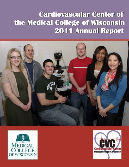
Cardiovascular diseases remain the leading cause <strong>of</strong>death in the US for both men and women. Research inthis field is <strong>of</strong> critical importance to the health <strong>of</strong> ourcommunity and our nation. The overall goal <strong>of</strong> theCardiovascular Center (<strong>CVC</strong>) is to enable laboratorydiscoveries to move forward in the form <strong>of</strong> new treatments,products, preventions and cures to those whosuffer from diseases <strong>of</strong> the heart, blood vessels, kidneys,and lungs.The <strong>CVC</strong> plays a vital role as a campus incubator tostimulate the exchange <strong>of</strong> new ideas and to nurture thedevelopment <strong>of</strong> novel research programs that fosterthe understanding <strong>of</strong> the underlying basic mechanisms<strong>of</strong> cardiovascular diseases and the translation <strong>of</strong> thisbasic knowledge into improved diagnostics, prevention,and therapeutics for the citizens <strong>of</strong> <strong>Wisconsin</strong> and beyond.The laboratories <strong>of</strong> 20 investigators reside withinthe confines <strong>of</strong> the <strong>CVC</strong>, and many more Faculty areaffiliated with our Center throughout the MCW campus.More than 100 clinicians and more than 50 basicscientists from various departments conduct researchwith cardiovascular implications.Over the past year, progress has been made in coalescingscientific Affinity Groups among the members<strong>of</strong> the <strong>CVC</strong> thereby fostering interdisciplinary collaborationsin areas that have been defined as focus areasfor growth at MCW such as heart failure and heart tissueregeneration. Work is now in progress to betteralign the focus <strong>of</strong> <strong>CVC</strong> research with those areas <strong>of</strong>strength within the MCW clinical service lines <strong>of</strong> cardiologyand surgery, both adult and pediatric. Towardthat end, over the past year, <strong>CVC</strong> has co-recruited anumber <strong>of</strong> new Faculty, including Jennifer Strande,M.D., Ph.D. (a cardiologist in the division <strong>of</strong> CardiovascularMedicine), Paul Goldspink, Ph.D. (cardiacphysiology), Aron Geurts, Ph.D. (cardiovascular genomics)and Scott Levick, Ph.D., (cardiovascular pharmacology).In order to support the expanding researchefforts <strong>of</strong> the <strong>CVC</strong>, an additional 5000 square feet <strong>of</strong>new laboratory space has been developed and a number<strong>of</strong> the existing laboratories have undergone renovationsto provide better utilization <strong>of</strong> existing space.Greater space has been developed to accommodatestudents and research fellows, and a newly createdConference Center that enables seating up to 125 andis divisible for small group meetings, has been constructed.These activities represent important advancements to-Letter from the DirectorAllen Cowley, PhDDr. Allen Cowleyward the overall mission <strong>of</strong> the <strong>CVC</strong> “to further developstrong and nationally recognized interdisciplinarycardiovascular research programs at the <strong>Medical</strong><strong>College</strong> <strong>of</strong> <strong>Wisconsin</strong> and to promote comparable excellencein clinical care and education while developingcommunity outreach”.The front cover: Dr. Williams’ lab (left to right) Carmen Bergom, MD, PhD, AdamGastonguay, Nathan Schuld, Andrew Hauser, Elizabeth Ntantie, Ellen L. Lorimer
The Cardiovascular Center in 2010-<strong>2011</strong>Created in 1992, the Cardiovascular Center (<strong>CVC</strong>) is focusing onthe prevention, detection, treatment and cure <strong>of</strong>the large family <strong>of</strong> cardiovascular diseases.Under the direction <strong>of</strong> Dr. Cowley, the <strong>CVC</strong> is administered by the staff: (from left to right):Allen W. Cowley Jr., PhD, Jane Brennan Nelson, Jean-Francois Liard, MD, PhD, JoannaMcCormick, Cescilly Burk, April MaysThe Cardiovascular Center <strong>of</strong>fers a number <strong>of</strong> services to its Faculty and to the MCW campus, including thefollowing cores: Information Technology, Engineering, and Microscopy.IT core: ( left to right) Andrew J. Milbrath, Greg McQuestion, Pedro MendezEngineering core: David Eick, Mike KloehnMicroscopy core: Glenn SlocumCardiovascular Center <strong>Annual</strong> <strong>Report</strong> <strong>2011</strong>1
Cardiovascular Center FY <strong>2011</strong><strong>Annual</strong> Revenue SourcesMCW Central Funds $ 465,062Dean Program Development $ 100,000Philanthropic $ 420,893Total Revenues $ 985,955The Cardiovascular Center in 2010-<strong>2011</strong>FinancialsThis past year has been marked by the recruitment <strong>of</strong> several new Faculty:♦ Paul Goldspink, PhD, Associate Pr<strong>of</strong>essor, Physiology♦ Jennifer Strande, MD, PhD, Assistant Pr<strong>of</strong>essor, Cardiovascular Medicine♦ Aron Geurts, PhD, Assistant Pr<strong>of</strong>essor, Physiology♦ Scott Levick, PhD, Assistant Pr<strong>of</strong>essor, PharmacologyThe <strong>CVC</strong> primary Faculty whose research is described in this annual report together with Dr. Cowley combinefor a total <strong>of</strong> $12,915,367 in NIH funding as listed in NIH RePORTER (fiscal year total cost).Extensive renovations were conducted in the <strong>CVC</strong>, with the creation <strong>of</strong> several new facilities, including over5000 square feet <strong>of</strong> laboratory space for basic scientists, a surgical facility suite, a stem cell culture facility, a 125-seat conference room, a dedicated room for a new confocal microscope purchased by the <strong>CVC</strong>, a freezer farm,and the consolidation <strong>of</strong> core equipment in rooms protected by electronic card readers.The <strong>CVC</strong> Scientific Advisory Committee was again <strong>of</strong> great help to us in achieving our objectives and willcontinue to provide guidance. This Committee is made <strong>of</strong> the following MCW Faculty:♦ Ellis Avner, MD, Associate Dean <strong>of</strong> Research, Children's Research Institute, Pr<strong>of</strong>essor, Pediatrics♦ Joseph Besharse, PhD, Chair <strong>of</strong> Cell Biology, Neurosciences and Anatomy♦ William Campbell, PhD, Chair <strong>of</strong> Pharmacology♦ Michael Cinquegrani, MD, Chief <strong>of</strong> Cardiovascular Medicine♦ Stephen Duncan, PhD, Pr<strong>of</strong>essor <strong>of</strong> Cell Biology♦ Andrew Greene, PhD, Director, Bioengineering and Biotechnology Center♦ David Gutterman, MD, Pr<strong>of</strong>essor in Cardiology, Senior Associate Dean for Research♦ David Harder, PhD, Pr<strong>of</strong>essor <strong>of</strong> Physiology, Associate Dean for Research Mentoring♦ Howard Jacob, PhD, Director <strong>of</strong> the Human and Molecular Genetics Center♦ Elizabeth Jacobs, MD, Chief <strong>of</strong> Pulmonary and Critical Care Medicine♦ Michael E. Mitchell, MD, Associate Pr<strong>of</strong>essor Surgery, Division <strong>of</strong> Cardiothoracic Surgery♦ Ramani Ramchandran, PhD, Investigator Children's Research Institute, Associate Pr<strong>of</strong>essor, Pediatrics♦ David Warltier, MD, Chair <strong>of</strong> Anesthesiology♦ Gilbert White, MD, Director <strong>of</strong> Research <strong>of</strong> Blood Research Center♦ Michael Widlansky, MD, Assistant Pr<strong>of</strong>essor, Cardiovascular MedicineFurther details about the <strong>CVC</strong> are provided on the <strong>CVC</strong> website at http://www.mcw.edu/cvc.htm and on theKidney Disease Center website at http://www.mcw.edu/kdc.2 Cardiovascular Center <strong>Annual</strong> <strong>Report</strong> <strong>2011</strong>
1Ongoing research from <strong>CVC</strong> FacultyThis section summarizes research conducted by the primary Faculty <strong>of</strong> the CardiovascularCenter, including the Kidney Disease Center. The research <strong>of</strong> many other Faculty affiliatedwith the <strong>CVC</strong> is not summarized here.John Auchampach, PhDCharacterization <strong>of</strong> adenosine receptors: Dr.Auchampach and his team are focused on the study <strong>of</strong>adenosine receptors, or proteins on the surface <strong>of</strong> cellsthat recognize adenosine and related compounds.Adenosine is produced by cells when the breakdown<strong>of</strong> adenosine triphosphate (ATP) is increased duringenergy utilization. Hence, adenosine levels are raisedwhen a cell is stressed, activated, or when there is notenough oxygen in the cells, as in ischemic heart diseaseand myocardial infarction.There are four different types <strong>of</strong> cell-surface receptorsfor adenosine. Dr. Auchampach’s research studiesthe function <strong>of</strong> two <strong>of</strong> the receptor subtypes that haveonly recently been discovered, the A 2B and A 3 adenosinereceptors. His group is testing the hypothesis thatthe A 3 adenosine receptor helps minimize muscledamage and reduce inflammatory processes that occurduring cardiac ischemia, whereas the A 2B adenosinereceptor that has low binding affinity for adenosinepromotes fibrotic responses in the cardiovascular systemcontributing to the development <strong>of</strong> heart failureand hypertensive disease. The research is aimed at developingnew drug therapies that target receptors foradenosine as potential treatments for patients with coronaryartery disease. In particular, it tests the hypothesis(see Figure below) that activating the A 3 adenosinereceptor in cardiomyocytes protects against ischemicinjury by improving mitochondrial function and redu-cing apoptosis, whereas activating the A 3 adenosinereceptor in immune cells during reperfusion is protectiveby suppressing inflammatory responses.Cardiac regeneration: in collaboration with colleaguesat MCW and the <strong>Medical</strong> <strong>College</strong> <strong>of</strong> Georgia, Dr.Auchampach’s research group is also investigating thetherapeutic potential <strong>of</strong> stem cells for regeneratingheart tissue that has been damaged following myocardialinfarction. Using embryonic and induced pluripotentstem cells, the hypothesis is being tested that transplantation<strong>of</strong> stem cells that have been predifferentiatedto specific cardiac lineages (i.e., cardiomyocytesor vascular precursor cells) will reduce thepotential for tumor formation and improve functionalrecovery <strong>of</strong> the damaged heart due to the incorporation<strong>of</strong> functional, electrically coupled myocytes intothe myocardium.Aron Geurts, PhDThrough genetic engineering in rats, the Geurts’ lab,along with colleagues in the Human and MolecularGenetics Center and the Department <strong>of</strong> Physiology,has made recent progress unraveling some <strong>of</strong> the complexgenetics <strong>of</strong> hypertension and kidney failure.Recent large-scale genetic studies in humans haveidentified many genes which play likely, yet poorly understoodroles in risk and progression <strong>of</strong> diseases likehypertension and chronic kidney disease. This hasshifted the focus <strong>of</strong> the Geurts’ lab and colleagues tounderstanding these specific genes as they may relateto these diseases. The laboratory rat is the preferredmodel to study diseases like hypertension and renalfailure because it is highly physiologically similar tohumans. In the past few years, the Geurts’ lab has developedseveral technological approaches to modifythe genome <strong>of</strong> rats to disrupt or ‘knock out’ specificgenes, something that was not previously possible. Thistechnological breakthrough has been named as one <strong>of</strong>the Top 10 scientific breakthroughs in both 2009 and2010 by “The Scientist” and “Science” magazines, respectively.Cardiovascular Center <strong>Annual</strong> <strong>Report</strong> <strong>2011</strong>3
Ongoing research from <strong>CVC</strong> FacultyAn effort is underway with other MCW laboratoriesto use these new approaches to knock out in the laboratoryrat 100 genes which have been identified in humanstudies. These models can then be studied to seeif disrupting these genes plays a potential role in hypertensionor kidney failure which lends support to thehuman findings and provide an animal model to understandgene function. The initial results are promisingas knocking out one gene, Sh2b3, which has beenstudied for several years in mice but never appreciatedfor its role in disease, demonstrates significant protectionfrom both hypertension and kidney failure, andpossibly other disease traits. This suggests that this is avery important gene in human disease which we cannow study further. It is the first time that a human diseasegene candidate has been experimentally validatedin a rat model <strong>of</strong> disease.The figure below illustrates the power <strong>of</strong> the zincfingernuclease (ZFN) technology in rats as used by Dr.Geurts’ lab for one <strong>of</strong> many genes it can be applied to.A gene called Rab38 controls trafficking <strong>of</strong> vesicles insidecells. When this gene is mutated in the rat usingZFN technology, it causes a pigmentation defect leadingto the fawn color that is evident in a subset <strong>of</strong> theanimals. Importantly, we have worked with HowardJacob’s lab to validate that this gene is also controllingtrafficking in the tubules <strong>of</strong> the kidney and mutationsin this gene can contribute to the end stage renal failure– a disease that affects millions <strong>of</strong> people worldwide.Paul Goldspink, PhDDr. Goldspink’s lab is presently investigating the influence<strong>of</strong> insulin like growth factor-1 (IGF-1) is<strong>of</strong>ormpeptides on the resident stem/progenitor cells populationswithin the heart. They are exploiting their findingsin a number <strong>of</strong> different ways. They (and collaboratorsfrom two other institutions) have developed atechnology that provides a microscopic physical scaffoldto deliver both the physical cues for tissue growthcombined with the capacity to deliver peptide therapeutics.They believe this “biomimetic” approach canbe used to optimize stem cell therapy either alone orin conjunction with autologous stem cells. This novelapproach <strong>of</strong> delivering implantable cell-sized“biomimetic devices” to instruct resident stem cells toenhance natural tissue repair and regeneration, is alsoadaptable to other tissues as well as the heart. Basedon this technology, Dr. Goldspink has formed an earlystage biomedical device company (Cell Habitats, Inc.)aimed at developing an easy-to-administer, micro devicethat allows the natural repair and regeneration <strong>of</strong>damaged tissue. Its first application is to restore normalcardiac function after a heart attack.The image below shows immun<strong>of</strong>luorescence staining<strong>of</strong> small Troponin I positive cells (shown with arrows)in the heart <strong>of</strong> MGF E-domain peptide treatedmice following myocardial infarction. Split image collectedat 40X magnification on Nikon A1 confocal microscope.Green is an antibody against Troponin I, redis cell membrane stained with wheat germ agglutinin,blue is nuclei stained with DAPI.4 Cardiovascular Center <strong>Annual</strong> <strong>Report</strong> <strong>2011</strong>
David Gutterman, MDDr. Gutterman’s group is involved in a number <strong>of</strong>projects. The first one is a study <strong>of</strong> ballerinas, who experiencecardiovascular and bone health problems.Twenty-two pr<strong>of</strong>essional ballet dancers volunteered todetermine the prevalence <strong>of</strong> the 3 components <strong>of</strong> thefemale athlete triad (disordered eating, menstrual dysfunction,low bone mineral density) and their relationshipswith brachial artery flow-mediated dilation. Seventeendancers (77%) had evidence <strong>of</strong> low/negativeenergy availability. Thirty-two percent had disorderedeating. Thirty-six percent had menstrual dysfunction.Twenty-three percent had evidence <strong>of</strong> low bone density.Sixty-four percent had abnormal brachial artery flow-mediated dilation. All <strong>of</strong> the 3 components <strong>of</strong> the triadplus endothelial dysfunction were present in 14% <strong>of</strong>the subjects. In conclusion, endothelial dysfunction wascorrelated with reduced bone mineral density, menstrualdysfunction, and low serum estrogen. Thesefindings may have pr<strong>of</strong>ound implications for cardiovascularand bone health in pr<strong>of</strong>essional women dancers.They were recently published by A.Z. Hoch et al. inClin. J. Sport Med. <strong>2011</strong>; 21:119–125.Another project examined the hypothesis that hydrogenperoxide (H 2 O 2 ) serves as the endotheliumdependenttransferrable hyperpolarization factor(EDHF) in human coronary arterioles (HCAs) to shearstress. Two HCAs were cannulated in series, with adonor intact vessel upstream and an endotheliumdenudeddetector vessel downstream. Results showedOngoing research from <strong>CVC</strong> Facultythat flow induced endothelial production <strong>of</strong> H 2 O 2 ,which acts as the transferrable EDHF activating largeconductance Ca 2+ -activated K + channels on thesmooth muscle cells. This was published by Y. Liu etal. in Circ. Res. <strong>2011</strong>; 108:566-573.Finally, Dr. Gutterman’s group conducts researchinvolving the age-dependence <strong>of</strong> flow-induced dilation<strong>of</strong> the coronary arterioles in man. Human coronaryarterioles (~150 μm internal diameter) were obtainedat surgery from patients with or without coronary arterydisease (CAD) and prepared for videomicroscopy. Afterconstriction with endothelin-1, vasomotor responsesto flow were evaluated in the presence, absence orcombination <strong>of</strong> the cyclooxygenase (COX) inhibitorindomethacin and the nitric oxide synthase inhibitor L-nitro-arginine methyl ester. The following age groupswere examined: children 0–18 years; adults 19–55years; older adults >55 years without CAD, and adultswith CAD (mean age: 64±8.2 years). It was found thatflow-induced dilation in children is mediated mostly byCOX; whereas, during the aging process or in the presence<strong>of</strong> CAD its contribution is diminished. Thesenovel findings may have clinical implications for theuse <strong>of</strong> COX inhibitors in children and young adults.This was published by N.S. Zinkevich et al. in Circulation2010, 122, 21, A15764.Gutterman/Widlansky/Zhang’s labs: Back row: (left to right) Rong Ying, MD , Suelhem A. Mendoza, Kodlipet Dharmashankar, MD, David Zhang, PhD,David Gutterman, MD, Reina Watanabe, PhD, Matt Durand, PhD, Andreas Beyer, PhD, Xiadong Zheng, PhDFront row: (left to right ) Joe Hockenberry, Yoshinori Nishijima, PhD, Jingli Wang, PhD, Dana VollmerCardiovascular Center <strong>Annual</strong> <strong>Report</strong> <strong>2011</strong>5
David Harder, PhDDr. Harder’s lab explores the dynamic control <strong>of</strong> cerebralblood flow and the interactive actions <strong>of</strong> cellswhich both require and impact delivery <strong>of</strong> oxygen tothe brain. Astrocytes play a crucial role in sensing andtransmitting neuronal signals to nearby cerebral microvesselsto regulate cerebral blood flow and thus delivery<strong>of</strong> oxygen and nutrients to match the metabolicdemand <strong>of</strong> activated neurons. Studies are conducted todefine the signaling pathways impacting blood flow tometabolically active neurons. The hypothesis is thatsignaling pathways between astrocytes and the cerebralmicrovasculature are initiated via metabolites which actthrough second messengers, some <strong>of</strong> which requiretrigger Ca 2+ and/or phosphorylation, i.e. protein kinaseC, serine/threonine protein kinase (Akt), mitogenactivatedprotein kinases (P38), extracellular-signalregulatedkinases (ERKs), and inositol trisphosphate(IP 3 ). Among the cellular intermediates are epoxyeicosatrienoicacids (EETs), 20-hydroxyeicosatetraenoicacid (20-HETE), adenosine triphosphate (ATP), adenosine,nitric oxide (NO) and others. Finally, plasmamembrane ion channels comprise an important group<strong>of</strong> the functional triggers mediating integration betweenneurons, astrocytes and the microvasculature. A systematicstudy <strong>of</strong> the signaling mechanisms mediatingthe functional role <strong>of</strong> astrocytes in regulating cerebralblood flow to match metabolic demand <strong>of</strong> neurons is<strong>of</strong> critical importance for understanding the mechanismregulating the cerebral circulation in differentconditions. A recent publication from Dr. Harder’s labOngoing research from <strong>CVC</strong> Facultydemonstrated that adenosine works through generation<strong>of</strong> free radicals (Gebremedhin et al., Journal <strong>of</strong>Cerebral Blood Flow and Metabolism, 2010; 30: 1777–1790). Dr. Harder also just published his 2009 WiggersAward lecture on the topic <strong>of</strong> the mechanisms <strong>of</strong>autoregulation <strong>of</strong> cerebral blood flow (American Journal<strong>of</strong> Physiology <strong>2011</strong>; 300(5):1557-1565).The Figure presented below was published in theWiggers Award lecture. It shows changes in morphology<strong>of</strong> astrocytes and cerebral capillary endothelialcells when co-cultured: a) formation <strong>of</strong> capillary-likestructures (arrow) double labeled with DiI-Ac-LDL(red) and GFAP (green) in the co-culture <strong>of</strong> astrocytesand cerebral capillary endothelial cells; b) formation <strong>of</strong>capillary-like structures (arrow) triple labeled byPECAM-1 (green), GFAP (red), and DAPI (blue) inthe co-culture <strong>of</strong> astrocytes and cerebral capillary endothelialcells. Note the interaction between astrocytesand endothelial tubes (arrowheads in a and b); c) doubleimmunolabeling <strong>of</strong> blood vessels and astrocyteswith PECAM-1 (green) and GFAP (red) in a sectionfrom normal rat cortex showing astrocytes form footprocesses that impinge on vessels (arrows); d) cells (*)forming tube identified by DAPI staining (e) in cocultureexhibit punctate staining <strong>of</strong> connexin 43 ontheir cell-cell borders (arrow). Arrow points to areashown in the insert at higher magnification; f) cells (*)outside tubes in the same co-culture as in d show moderateto light cytoplasmic staining <strong>of</strong> connexin 43.6 Cardiovascular Center <strong>Annual</strong> <strong>Report</strong> <strong>2011</strong>
John Imig, PhDDr. Imig works on therapeutically promising newcompounds for combating hypertension and cardiovasculardisease. With a team <strong>of</strong> <strong>Wisconsin</strong> and Texasscientists, he has recently discovered a promising newavenue that they strongly believe can be further developedto treat these diseases. The researchers are AbdulHye Khan, William B. Campbell and John D. Imig,from the <strong>Medical</strong> <strong>College</strong> <strong>of</strong> <strong>Wisconsin</strong>, and Vijaya L.Manthati, Jawahar L. Jat, and John R. Falck from theUniversity <strong>of</strong> Texas Southwestern <strong>Medical</strong> Center. Thefindings were discussed in April <strong>2011</strong> at the ExperimentalBiology meeting (“Novel EpoxyeicosatrienoicAcid Analogs Increase Sodium Excretion and LowerBlood Pressure in Hypertension”). Key to the researchis the role <strong>of</strong> endothelial cells, which line the inside <strong>of</strong>the blood vessels and the heart. Endothelial cells producearachidonic acid metabolites, a class <strong>of</strong> fatty acidsthat have biological actions beneficial for cardiovascularhealth. This production occurs through three primaryenzymatic pathways. Two <strong>of</strong> these pathways, the cyclooxygenaseand the lipoxygenase pathways, havebeen successfully targeted for the treatment <strong>of</strong> inflammation,pain, fever, and asthma. The third enzymaticpathway is the cytochrome P450 pathway that producesepoxyeicosatrienoic acids (EETs) as major biologicallyactive metabolites. EETs are endothelial-derived factorsthat significantly influence cardiovascular function.EETs can dilate blood vessels, lower blood pressureand have additional biologic actions including antiinflammatoryand anti-platelet aggregator activity. Thesebiological activities have made EETs a very attractivetherapeutic target for cardiovascular diseases.Ongoing research from <strong>CVC</strong> FacultyFor the last several years this research team has developedand synthesized an array <strong>of</strong> EET analogs, orchemical compounds that act as EETs. These EETanalogs have been tested for beneficial cardiovascularactions. For this study, 35 different EET analogs werescreened for their ability to dilate blood vessels. Thescreening produced five EET analogs that would befurther examined to determine their ability to lowerblood pressure in animal models <strong>of</strong> hypertension. Two<strong>of</strong> the five EET analogs administered to hypertensiveanimals effectively lowered blood pressure and reducedkidney injury. This work is a major step forwardin developing novel EET analogs for the treatment <strong>of</strong>cardiovascular disease.Elizabeth Jacobs, MD, MBADr. Jacobs and her colleagues are working on a projectwith important implications for the diagnosis andtreatment <strong>of</strong> ischemia-reperfusion (IR) lung injuries. IRlung injuries are commonly encountered clinically inconditions such as crush injury to the chest, lung transplantationand others. While the role <strong>of</strong> mitochondrialreactive oxygen species (mtROS) in IR injuries is generallyaccepted, less information is available regardinglung IR injury than cardiac or cerebral IR where thedensity <strong>of</strong> mitochondria is much higher. Dr. Jacobs willemploy an in vivo, rodent survival model <strong>of</strong> unilaterallung injury and cutting-edge imaging technology to mo-nitor and understand the role <strong>of</strong> mitochondrial complexI. Using this model, Dr. Jacobs and her colleagueshave excellent evidence that mitochondrial dysfunctiondrives lung IR injury. By varying the severity <strong>of</strong> IR andtracking changes in lung mitochondrial bioenergetics inthe first 2-72 hours, they will develop a pr<strong>of</strong>ile whichpredicts recoverability by 7 days. Furthermore, theyintroduce two absolutely novel methods to quantifyapoptosis or an altered redox status in vivo at the bedside,which they will correlate with measures <strong>of</strong> mitochondrialfunction.The first method is in vivo SPECT/CT imaging,Cardiovascular Center <strong>Annual</strong> <strong>Report</strong> <strong>2011</strong>7
Ongoing research from <strong>CVC</strong> Facultyusing 99m Tc-duramycin to detect apoptosis. Its use inrats is demonstrated in Figure 3, which illustrates thatduramycin (DU) uptake is increased in IR lungs bysingle photon emission computed tomography(SPECT). 99m Tc-duramycin was administered throughan IV line and images acquired at steady-state. Withoutrelocation <strong>of</strong> the rat, an injection <strong>of</strong> 99m Tc-MAAwas made and rats were reimaged. 99m Tc-MAA wasused as a pulmonary perfusion marker as the particlesrange in size from 10-40 microns and lodge in the pulmonarycapillaries in proportion to flow. The Figurebelow shows representative parametric SPECT images,depicting DU uptake normalized to flow (based onMAA), <strong>of</strong> a control rat (3i) and one rat each subjectedto IR <strong>of</strong> the left lung 24 (3ii) and 48 (3iii) hours earlier.The left-right ratio <strong>of</strong> flow normalized 99m Tc-duramycinuptake was increased by ~35% 24 hours after IR andgreater than 60% 48 hours after IR (n=1 each IR, n=3control).increased mtROS which lead to apoptosis and decreasedcell survival, b) SPECT/CT and optical imagingcan detect levels <strong>of</strong> apoptosis and altered mitochondrialbioenergetics in an IR survival model in amanner which predicts subsequent viability and c) targetingmitochondria with an agent that protects mitochondrialbioenergetics in lung IR will mitigate injuryand preserve lung function as detected by SPECT/CTor optical imaging. Dr. Jacobs and colleagues will attemptto define early alterations in mitochondrial bioenergeticsand mtROS generation in a rodent model <strong>of</strong>lung IR which predict the potential <strong>of</strong> lung to recover.They will also examine the role <strong>of</strong> mtROS in cell apoptosis/survivaland lung function. The second aim is toassess apoptosis and redox ratios in left and right lungs<strong>of</strong> IR rats using SPECT/CT and optical imaging respectively,and compare these data to measurements,obtained before, to define a pr<strong>of</strong>ile <strong>of</strong> imaging datawhich predict lung tissue survival. Finally, they will determinehow and if agents including 20-HETE or theCSA-like immunosuppressive sanglifehrin A <strong>of</strong>fer protectionfrom IR and if this protection can be predictedby SPECT/CT or optical imaging. With their novelmeans to detect apoptosis and redox injury in vivo andin a survival model <strong>of</strong> IR injury, Dr. Jacobs and colleaguesare poised to use SPECT/CT and optical imagingendpoints to define the first bedside tests that distinguishin vivo apoptosis or oxidoreductive state, andthe potential <strong>of</strong> treatments to rescue “irreversible” lunginjury. These studies will provide data to predict whichlung injuries are destined to recover, and which willprogress to non-viability days later.The second novel method introduced by Dr..Jacobs and her colleagues is optical imaging to detectthe NADH/FAD (redox) ratios in intact lungs. Earlychanges in these non-destructive measurements willprovide the first data to predict later recovery or loss <strong>of</strong>lung viability. Preliminary data demonstrate prosurvivaleffects <strong>of</strong> cyclosporine A (CSA) and 20-hydroxyeicosatetraenoic acid (20-HETE); protection byeither agent depends upon an action on mitochondria.Dr. Jacobs and colleagues hypothesize that: a) ischemia-reperfusionresults in complex I dysfunction and8 Cardiovascular Center <strong>Annual</strong> <strong>Report</strong> <strong>2011</strong>
Girija Ganesh Konduri, MDImproving our understanding and treatment <strong>of</strong> newbornbabies with respiratory failure is the overall goal<strong>of</strong> the research program in the neonatology laboratory<strong>of</strong> the <strong>CVC</strong>. Every year, newborn babies that fail toachieve normal respiratory and cardiac function atbirth are referred to the Neonatal Intensive care unit atChildren’s Hospital. These babies are cared for by thephysicians in the Neonatology Division <strong>of</strong> the <strong>Medical</strong><strong>College</strong> <strong>of</strong> WI. The Chief <strong>of</strong> the Neonatology Division,Dr. Girija Konduri, has established a researchprogram to investigate the mechanisms <strong>of</strong> respiratoryfailure that can occur at birth. Dr. Konduri’s researchis focused on the mechanism <strong>of</strong> a condition that affectsnewborn babies called Persistent Pulmonary Hypertension<strong>of</strong> the Newborn (PPHN). When babies are insidetheir mother’s womb, their lungs are filled with liquidand do not participate in getting oxygen into the blood.Since the fetal lungs are not respiring, they receive onlya small fraction <strong>of</strong> the blood pumped by the heart.When the baby comes out and the umbilical cord isclamped, the lungs have to fill with air and take overthe function <strong>of</strong> providing oxygen supply to the body.This requires establishing circulation <strong>of</strong> blood throughthe lungs to transport oxygen from the lungs to the baby’sorgans. In babies with PPHN, the circulation <strong>of</strong>blood to the lungs does not get established and the babybecomes blue. The affected infants suffer from consequences<strong>of</strong> decreased oxygen supply to vital organsand have an increased risk <strong>of</strong> death or long term disabilities.Dr. Konduri’s research is focused on understandingthe normal mechanisms by which circulationgets established in the lungs at birth and why this processdoes not occur in babies with PPHN. He foundOngoing research from <strong>CVC</strong> Facultythat constriction <strong>of</strong> an important blood vessel that connectsheart and lungs in the fetus, called ductus arteriosus,leads to development <strong>of</strong> PPHN. He also foundthat blood vessels in the lung have increased amounts<strong>of</strong> oxygen free radicals in PPHN. This research is evaluatingthe reason for the narrowing <strong>of</strong> the ductus arteriosusin the fetus and a possible approach to clearingthe oxygen free radical accumulation from the bloodvessels in the lungs <strong>of</strong> babies with PPHN. These studieswill lead to improved survival and decreased disabilityrates in affected infants.Over the last 4 years, novel observations made in Dr.Konduri’s laboratory found that the cells lining theblood vessels have dysfunction <strong>of</strong> the mitochondriathat normally consume oxygen to provide ATP, thecellular source <strong>of</strong> energy. New studies proposed by theinvestigators in the laboratory lead to the award <strong>of</strong> 4new grants from NIH over the last 18 months to studyalteration in lung biology in pulmonary hypertension.In addition to Dr. Konduri’s, two other neonatologyfaculty members are actively investigating lung biology.Dr. Ru-Jeng Teng studies oxidative stress and its effectson angiogenesis in the lung as well as tetrahydrobiopterinand the function <strong>of</strong> nitric oxide synthase inthe lung. Dr. Venkatesh Sampath’s work is describedin another section. In addition to these 3 faculty members,there are currently 4 neonatology trainees that areconducting research in the <strong>CVC</strong> neonatology laboratory.The support <strong>of</strong> the <strong>CVC</strong> with its collection <strong>of</strong> scientistsand core facilities and resources is critical to thesuccess <strong>of</strong> the overall mission <strong>of</strong> the Neonatology section.Konduri/ Sampath Labs: (back row: left to right) Ping Ye, PhD, Ru-Jeng Teng, MD, Everett Tate, Ivane Bakhutashvili, MD, PhD;(front row: left to right): Venkatesh Sampath, MD, MRCPCh, Girija Ganesh Konduri, MD, Annie EisCardiovascular Center <strong>Annual</strong> <strong>Report</strong> <strong>2011</strong>9
Meetha Medhora, PhDEffects <strong>of</strong> ionizing radiation on the lungs. Dr.Medhora’s group is identifying agents to mitigate injuryto the lungs that may occur after radiotherapy, a nuclearaccident or radioterrorism. Using a rat model,they have observed radiation to the thorax resulting inlung injury after 6-8 weeks. This recovers and is followedby fibrotic remodeling <strong>of</strong> lung tissue starting 7months after exposure. They measure acute andchronic injuries to the pulmonary vasculature. Currentlythey are testing a number <strong>of</strong> pharmaceutical reagentsin an effort to identify mitigators and treatment for theradiation lung injury. In the past year they have beenfunded to study the effect <strong>of</strong> combined injuries by radiationto the lung and skin. This is a collaborative projectbetween the Departments <strong>of</strong> Radiation Oncology,Dermatology and Medicine at MCW and the RadiationCenter at the University <strong>of</strong> Rochester.The Figure below demonstrates the effect <strong>of</strong> radiationon the lung and the mitigating effect <strong>of</strong> drugtreatment with captopril on the injury. Sections <strong>of</strong> ratOngoing research from <strong>CVC</strong> Facultylungs stained with hematoxylin and eosin show radiationinduced increase in cellularity and loss <strong>of</strong> alveolarstructure (middle panel). This injury is mitigated bytreatment with captopril started soon after irradiation.Vasoreactive and antiapoptotic actions <strong>of</strong> EETs and 20-HETE (products <strong>of</strong> the essential fatty acid arachidonicacid). The endogenous fatty acids EETs and 20-HETEhave potent vasoreactive and other properties. In collaborationwith the laboratory <strong>of</strong> Dr. Elizabeth Jacobsthey have demonstrated that EETs protect pulmonaryarteries ex vivo using vessels from three species, human,mouse and rat. 20-HETE also protects rat pulmonaryand cerebral arteries from hypoxia and reoxgygenationinjury ex vivo. Dr. Medhora and her groupare currently studying the intracellular mechanisms forprotection by these fatty acids.Effects <strong>of</strong> ionizing radiation on the lungs.Daisy Sahoo, PhDDr. Sahoo and her group are measuring HDL functionand developing a clinical assay that will revolutionizeassessment <strong>of</strong> cardiovascular risk.Despite the years <strong>of</strong> epidemiological evidence thatsupports an inverse relationship between HDLcholesterollevels and cardiovascular disease (CVD),the anti-atherogenic benefits <strong>of</strong> raising HDL levels arecurrently controversial. For example, geneticallymodifiedmice that have high HDL levels are moreprone to atherosclerosis. Further, recent large-scaleclinical studies revealed that patients who had normallevels <strong>of</strong> HDL-cholesterol still suffered cardiovascularevents. Moreover, use <strong>of</strong> a drug that nearly doubledHDL levels in a phase III human clinical trial came toan abrupt end due to high patient mortality. For thesereasons, there is now a greater emphasis on measuring“HDL function” as a better predictor <strong>of</strong> CVD than lowlevels <strong>of</strong> HDL-cholesterol.Basic scientists <strong>of</strong> the Atherosclerosis AffinityGroup <strong>of</strong> the Cardiovascular Center, Drs. KirkwoodPritchard, Hao Zhang and Daisy Sahoo, are currentlydeveloping novel in vitro assays <strong>of</strong> HDL function thathave the potential to revolutionize the way physiciansdiagnose and treat patients with cardiovascular risk.Recent studies suggest that the oxidation status <strong>of</strong>HDL plays an important role in determining how thislipoprotein prevents or promotes atherosclerosis. Assuch, Drs. Pritchard, Zhang and Sahoo hypothesize10 Cardiovascular Center <strong>Annual</strong> <strong>Report</strong> <strong>2011</strong>
that oxidation impairs HDL function by altering itsability to bind biomolecules (i.e. cargo) that are involvedin inflammation and HDL-dependent cholesterolmetabolism, thus making it atherogenic and unableto efficiently participate in reverse cholesteroltransport.Using biolayer interferometry (BLI), a new label-freetechnique for measuring biomolecular protein-proteininteractions, these investigators are now able to measurethe rates at which functional and non-functional(i.e. oxidized) HDL binds key proteins that are criticalfor cholesterol removal from the body. BLI analyzesthe interference pattern <strong>of</strong> light reflected from two surfaces.The first surface is the layer <strong>of</strong> immobilized proteinthat serves as an internal reference. As the number<strong>of</strong> biomolecules binding to the biosensor increases, theaqueous protein layer increases, causing a shift in interferencethat can be measured in real-time. The OctetRed is a fully automated, 8 channel BLI instrument. Itrequires extremely small sample sizes and is highly reproducible.The design <strong>of</strong> the Octet Red is equivalentto having 8 BiaCore instruments, which use a similartechnology (surface plasmon resonance) to analyzeprotein-protein interactions. The Octet Red can measureprotein concentration (via determining rates <strong>of</strong>antigen-antibody binding) in 96 samples at once, in lessthan 20 min and at a cost <strong>of</strong> ~30¢/sample. Furthermore,HDL binding affinity for different proteins atthe same time can be determined quickly and in realtime.The Figure below is a diagram <strong>of</strong> BLI biosensorloaded with anti-apoA-I antibody to capture HDL. ImmunocapturedHDL is then probed with specific antibodies,pro- and anti-oxidant enzymes, cholesterol metabolizingenzymes or the extracellular domains <strong>of</strong> differentreceptors and transporters.Ongoing research from <strong>CVC</strong> FacultyAlready, preliminary data demonstrate that oxidizedHDL (generated in vitro) directly impacts its ability tobind proteins associated with inflammation and cholesteroltransfer as compared to HDL that was not oxidized.More importantly, HDL isolated from hypercholesterolemicmice displayed increased binding affinitiesfor pro-oxidant enzymes compared to HDLisolated from healthy mice. Therefore, these data providestrong support for the concept that BLI assays areable to distinguish between control HDL and HDLfrom established murine models <strong>of</strong> atherosclerosis.Drs. Pritchard, Zhang and Sahoo have recently submittedan R01 grant proposal to the National Institutes<strong>of</strong> Health to support the continuation <strong>of</strong> these highlyinnovative studies. The objectives <strong>of</strong> this proposal areto determine how oxidized lipid and protein composition<strong>of</strong> reconstituted HDL influences HDL’s interactionswith key biomolecules as cholesterol is transportedfrom the arteries to the liver for disposal. Thesestudies will be performed in parallel using cell cultureassays and BLI methodologies. Critically, BLI assayswill be used to analyze samples from healthy patients,as well as those with clinically-documented atherosclerosis,to determine if and the extent to which atherosclerosisimpairs HDL interactions with biomoleculesthat mediate HDL metabolism. Together, these excitingstudies will, for the first time, test the hypothesisthat HDL binding affinity can be used to distinguishbetween functional and dysfunctional HDL.Successful completion <strong>of</strong> the above studies will laythe foundation for the development <strong>of</strong> new clinical assaysfor determining HDL “function”. The BLI assayswill allow us to perform large clinical studies to fullytest the idea that dysfunctional HDL is a better indicator<strong>of</strong> atherosclerotic risk. Further, these standardized,validated, accurate and robust assays have high diagnosticpotential as they can be performed quickly, andin a cost-effective manner in a clinical laboratory setting.As large-scale application <strong>of</strong> our assays holdsgreater promise for predicting CVD than HDLcholesterollevels, physicians may be able to preventcardiovascular events in patients with normal HDLcholesterolbecause, for the first time, they will be ableto identify which patients have dysfunctional HDL.Cardiovascular Center <strong>Annual</strong> <strong>Report</strong> <strong>2011</strong>11
Venkatesh Sampath, MD, MRCPChOur laboratoryseeks to understandthe role <strong>of</strong> innateimmune signalingin the pathogenesis<strong>of</strong> diseases <strong>of</strong>premature infants.Our research focuseson how lipopolysaccharide(LPS)mediated endothelialinjury contributesto vascular remodelingin bronchopulmonarydys-Venkatesh Sampath, MD, MRCPChplasia (a chronicform <strong>of</strong> inflammatory lung disease in premature infants).We have identified NADPH oxidase (Nox) as acritical regulator <strong>of</strong> LPS mediated endothelial injuryusing in vitro techniques. We propose to expand onthis research by identifying the specific is<strong>of</strong>orms <strong>of</strong>Nox that mediate LPS-dependent endothelial toxicityand examining the biological mechanisms underlyingNox-dependent endothelial injury and microvascularOngoing research from <strong>CVC</strong> Facultyremodeling. This work is supported by a R03 grantfrom the National Institutes <strong>of</strong> Child Health and Development(NICHD) entitled “LPS-mediated oxidativestress and pulmonary vascular injury in bronchopulmonarydysplasia”.On the translational aspect, we are investigatingwhether functional genetic variaton in the Toll-like receptorspathway genes (innate immune genes) modulatesusceptibility to bronchopulmonary dysplasia andother diseases <strong>of</strong> the preterm infants. Our preliminarydata suggest that loss <strong>of</strong> function polymorphisms in thispathway does increase susceptibility to lung disease inpremature infants. This research is funded by aNIEHS Children’s Environmental Health Sciencespilot award entitled “TLRs-modulators <strong>of</strong> environmentallung injury in premature infants”, which will determinewhether TLR/Nrf2 genetic variation alters susceptibilityto oxidative stress and susceptibility to bronchopulmonarydysplasia in preterm infants.Jennifer Strande, MD, PhDDr. Strande researches the role <strong>of</strong> thrombin and itsreceptors in the inflammation and fibrosis that occursin the heart after ischemic events. While reducing risksusing preventive strategies is important in combattingheart disease, we still need new treatment strategies forprevention <strong>of</strong> the adverse effects <strong>of</strong> heart disease.The clotting pathway culminates in the formation <strong>of</strong>thrombin, which is best known for acting on its cellularreceptors to cause platelet aggregation and thrombosis.One <strong>of</strong> the primary treatments strategies for patientswith coronary artery disease is to acutely stop this process,in the condition <strong>of</strong> acute myocardial infarction, orto chronically inhibit this process to prevent recurrentheart attacks. Thrombin also acts through its receptorson other cell types and organs such as liver, lung andkidney to cause effects such as inflammation and fibrosis.Inflammation and fibrosis contribute to left ventricularremodeling and eventually cardiomyopathy.Dr. Strande has shown that in rats treated with eitherdirect thrombin inhibitors or thrombin receptor antagonists,the heart is protected against ischemia by a reduction<strong>of</strong> the damaging effects <strong>of</strong> free radicals andinflammation. Furthermore, she has shown that in longterm studies, a thrombin receptor antagonist also limitscardiac fibrosis and protects the heart from adverseremodeling after myocardial infarction. As a physician,Dr. Strande sees first-hand how heart damage resultingfrom heart attacks affects the mental and physical wellbeing<strong>of</strong> patients and their families. By recognizing thegaps in treatments for people affected by heart attacksand heart failure, she can identify new targets for potentialtherapies. She also has the basic research trainingto perform the research studies needed to advancethese new potential therapies which may have amajor impact on keeping patients with cardiovasculardisease feeling better.The Figure below shows typical photographs <strong>of</strong> myocardialslices from three control (top row) and three12 Cardiovascular Center <strong>Annual</strong> <strong>Report</strong> <strong>2011</strong>
Carol Williams, PhDAtherosclerosis and its complications cause almostone million deaths per year in the United States. Atherosclerosisresults from excessive proliferation and migration<strong>of</strong> vascular smooth muscle cells, which promotesplaque formation and blockage <strong>of</strong> blood flow inmajor vessels. The Williams’ laboratory is investigatingnovel ways to inhibit the proliferation and migration <strong>of</strong>vascular smooth muscle cells, which will help definenew ways to prevent and treat atherosclerosis.Dr. Williams and her team are focusing their researchon a group <strong>of</strong> proteins called “small GTPases”.Small GTPases are emerging as therapeutic targets inatherosclerosis, because these proteins promote themigration and proliferation <strong>of</strong> vascular smooth musclecells. There are many different types <strong>of</strong> smallGTPases, including the proteins known as Rho, Rac,Ras, and Rap. Since there are so many different types<strong>of</strong> small GTPases, inhibiting the activity <strong>of</strong> only onetype <strong>of</strong> small GTPase might not be enough to stop theexcessive proliferation and migration <strong>of</strong> vascularsmooth muscle cells during atherosclerosis. Instead,simultaneously blocking the activity <strong>of</strong> multiple smallOngoing research from <strong>CVC</strong> FacultyGTPases at one time might be a better way to inhibitatherosclerosis. The Williams’ laboratory is investigatingthis new strategy <strong>of</strong> simultaneously inhibiting multiplesmall GTPases at one time, by conducting researchon the unique protein known as SmgGDS.SmgGDS can interact with multiple small GTPases,including Rho, Rac,Ras, and Rap. This unique featuremakes SmgGDS a master regulator <strong>of</strong> these smallGTPases. The Williams’ laboratory is testing the hypothesisthat blocking the activity <strong>of</strong> SmgGDS in vascularsmooth muscle cells will block the activity <strong>of</strong> allsmall GTPases in these cells, and diminish the migrationand proliferation <strong>of</strong> vascular smooth muscle cells.The Figure below illustrates how vascular smoothmuscle cells make many types <strong>of</strong> small GTPases, includingRho, Rac, Ras, and Rap. These small GTPasescan promote the proliferation and migration <strong>of</strong> vascularsmooth muscle cells, which can promote atherosclerosis.SmgGDS is a unique protein that regulatesall <strong>of</strong> these small GTPases. Stopping the interaction <strong>of</strong>SmgGDS with small GTPases might <strong>of</strong>fer a new therapeuticapproach to prevent and treat atherosclerosis.14 Cardiovascular Center <strong>Annual</strong> <strong>Report</strong> <strong>2011</strong>
2<strong>CVC</strong> Advisory Board and PhilanthropyThe general mission <strong>of</strong> the <strong>CVC</strong> Advisory Board is to serve as advocates in the community,thereby supporting the primary objectives <strong>of</strong> the <strong>CVC</strong>. These objectives are to contributeto the eventual elimination <strong>of</strong> heart disease, stroke and other cardiovascular diseases ashuman health problems through coordinated research, clinical treatment, control and education,and to provide the citizens <strong>of</strong> Southeastern <strong>Wisconsin</strong> with state-<strong>of</strong>-the-art therapy,research, control and educational programs for all aspects <strong>of</strong> cardiovascular disease. Thespecific purpose <strong>of</strong> the <strong>CVC</strong> Advisory Board is to provide financial support for the researchand clinical programs carried on by the <strong>CVC</strong> and to educate the community in cooperationwith the Board <strong>of</strong> Trustees <strong>of</strong> The <strong>Medical</strong> <strong>College</strong> <strong>of</strong> <strong>Wisconsin</strong> on the causes, prevention,diagnosis and treatment <strong>of</strong> cardiovascular disease. Bruce E. Jacobs, a lifetime member <strong>of</strong>the <strong>CVC</strong> Advisory Board, was the Founding Chair. Other lifetime members include ByronFoster, Gordon Gunnlaugsson and William H. Levit, Jr.Current composition <strong>of</strong> the BoardChair:Nancy J. SennettVice-chair:Johan C. R. SegerdahlMembers:Ned W. BechtholdPriscilla BoelterCharles P. BrumderMarybeth BudischCellene ByrneKristine Cleary, Esq.Dominic ColonnaFrancis R. CroakMichael J. CudahyGael Garbarino CullenMichael S. ErwinByron T. FosterFrederic G. FriedmanEllen GlaisnerLinda Gorens-LeveyEckhart GrohmannBarry GrossmanGordon H. GunnlaugssonStanley F. HackCristina Hernandez-MalabyMikel HoltBruce E. JacobsRobert H. JenkinsSarah Wright KimballJohn KirchgeorgDr. Vincent KuttemperoorWilliam H. Levit, Jr.William M. MaslowskiDaniel F. McKeithan, Jr.Daniel M. MuchinJeffrey J. NohlJohn K. SchultzBruce M. SmithSonia Shields StoweNicholas C. WilsonKenneth F. YontzGary V. Zimmerman, FAIAEmeritus Members:James D. BellWilliam D. BrowneJohn J. Burke, Jr.Cardiovascular Center <strong>Annual</strong> <strong>Report</strong> <strong>2011</strong>19
Cullen runThis event supports heart research at the Cardiovascular Center. To date, Cullen Run/Walk proceeds havehelped several research programs, including studies to identify genetic risk factors <strong>of</strong> heart disease, to investigatethe Female Athlete Triad and its potentially life-threatening link to heart disease, and to better understandthe correlation between plaque buildup and coronary artery disease. The Cullen Run/Walk is an 8-kilometerUSA Track and Field competitive run and a 2-mile fun run/walk following a scenic path along UnderwoodParkway in Wauwatosa. Sponsored by the Cullen family and the Badgerland Striders, the event includes famousCullen Family & Friends Chili, refreshments, food, door prizes, an awards ceremony and live music forthe entire family. The event is held in memory <strong>of</strong> Steve Cullen, a former Milwaukee alderman, who died in1995 at age 40 <strong>of</strong> sudden cardiac arrhythmia. His father, at age 41, and two brothers, ages 53 and 51, also died<strong>of</strong> heart disease. The Cullen Run/Walk has grown in attendance by over 500% since its inception, encouragingheart healthy lifestyles for all participants, their families and friends and contributing over $120,000 to cardiovascularresearch and awareness.
Geurts’ lab ( from back to front): Jason Klotz, Bradley Endres, Molly Corbett, Sheila Reagles, Michael Grzybowski, Sheng Yang, PhD8701 Watertown Plank Road Milwaukee, WI 3226 414-456-8296


