Axio Lab.A1 - SPOT Imaging Solutions
Axio Lab.A1 - SPOT Imaging Solutions
Axio Lab.A1 - SPOT Imaging Solutions

You also want an ePaper? Increase the reach of your titles
YUMPU automatically turns print PDFs into web optimized ePapers that Google loves.
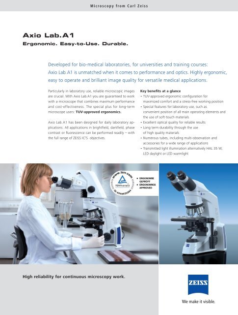
Microscopy from Carl Zeiss<strong>Axio</strong> <strong>Lab</strong>.<strong>A1</strong>Ergonomic. Easy-to-Use. Durable.Developed for bio-medical laboratories, for universities and training courses:<strong>Axio</strong> <strong>Lab</strong>.<strong>A1</strong> is unmatched when it comes to performance and optics. Highly ergonomic,easy to operate and brilliant image quality for versatile medical applications.Particularly in laboratory use, reliable microscopic imagesare crucial. With <strong>Axio</strong> <strong>Lab</strong>.<strong>A1</strong> you are guaranteed to workwith a microscope that combines maximum performanceand cost-effectiveness. The special plus for long-termmicroscope users: TUV-approved ergonomics.<strong>Axio</strong> <strong>Lab</strong>.<strong>A1</strong> has been designed for daily laboratory applications.All applications in brightfi eld, darkfi eld, phasecontrast or fl uorescence can be performed readily – withthe full range of ZEISS IC 2 S objectives.Key benefits at a glance• TUV-approved ergonomic confi guration formaximized comfort and a stress-free working position• Special features for laboratory use, such asconvenient position of all main operating elements andthe use of soft-touch materials• Excellent optical quality for reliable results• Long term durability through the useof high quality materials• Numerous tubes, including multi-observation andaccessories for a wide range of applications• Transmitted light illumination alternatively HAL 35 W,LED daylight or LED warmlightHigh reliability for continuous microscopy work.
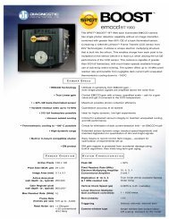
The First <strong>Lab</strong>oratory Microscope –With Approved ErgonomicsA number of attractive details are designed toguarantee the ergonomic and reliable handlingof <strong>Axio</strong> <strong>Lab</strong>.<strong>A1</strong>.TUV-approved ergonomics. <strong>Axio</strong> <strong>Lab</strong>.<strong>A1</strong> places strongemphasis on ergonomic features, because continuousmicroscope use is often associated with discomfort, particularly in the neck.With <strong>Axio</strong> <strong>Lab</strong>.<strong>A1</strong> users are able to examine slides with afavorable viewing position, keeping the neck and shouldermuscles relaxed. The height of the microscope is continuouslyadjustable by 50mm. In addition the tube can beswiveled from 8°-33° continuously.IC²S optics, the Infi nity colour-corrected system, is availableonly from Carl Zeiss. The system guarantees brilliant,colour-corrected and high-contrast results, meeting all thedemands placed on image quality.TUV*-approved ergonomics: The ergo-version of <strong>Axio</strong> <strong>Lab</strong>.<strong>A1</strong> .Guaranteed durability. The more a microscope is used,the more worthwhile it is to invest in quality. <strong>Axio</strong> <strong>Lab</strong>.<strong>A1</strong>guarantees long lasting value and a long operating life.The new multi-discussion system from Carl Zeiss is alsocompatible for <strong>Axio</strong> <strong>Lab</strong>.<strong>A1</strong>. It is possible to achieve identicalimage orientations for all co-observers. All co-observerssee the same image as the main observer. And a luminouspointer facilitates the identifi cation of specimen details fordiscussion.Skin-friendly materials enhance comfort. Many partsof the stand are coated with soft-touch material: Thesematerials offer superior properties in comparison to metalsurfaces: less cold and higher friction.Comfortable and convenient: Working with supported armand all main elements within reach of one hand.Modular illumination. Convenient and easy-to-changebulbs. The use of the novel warmlight LED in particularoffers the advantages of a long lifetime, energy saving andhalogen-like colour impressions in stained slides.* TUV Rheinland is aworld-renownedindependent organizationwhich certifies productsafety and ergonomy.Integrated tool storage and cable rest in the stand backfl ap. Tools, e. g. for centring of phase rings are always athand and cannot get lost. In addition, the tilted fl ap canbe used as cable rest.Everything at hand: Tool bar on the back of the scope.2
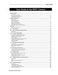
Explore the Possibilities –<strong>Axio</strong> <strong>Lab</strong>.<strong>A1</strong> Wide Range of ApplicationsWith the help of <strong>Axio</strong> <strong>Lab</strong>.<strong>A1</strong> diverse diagnostic applicationscan be carried out easily and effi ciently.<strong>Axio</strong> <strong>Lab</strong>.<strong>A1</strong> is designed for many uses. Microbiology, cytology,hematology and pathology labs as well as those inschool and colleges will benefi t particularly.Fluorescence microscopy in the lab<strong>Axio</strong> <strong>Lab</strong>.<strong>A1</strong> ensures easy-to-use LED-fl uorescence with2 LED positions and the well known standard push-andclick-modulesfrom Carl Zeiss. In comparison to standardHBO illumination the LED fl uorescence is much safer, moreenergy effi cient, quicker and easier to use.Additional advantages: No warm-up and cool-down timesand no need to change or adjust lamps.The fl uorescent marker FITC is primarily used for antigenantibodyreactions in the fi eld of immunology. FITC bindsto the antibody molecules and emits an intense green fl uorescenceupon excitation with the 470 nm LED.Brightfield and darkfield microscopyIn the fi eld of hematology, the usual diagnostic approachto blood disorders is blood counting and blood fi lm examination.During blood fi lm examination in brightfi eldmicroscopy, the individual types of white blood cells arecounted. <strong>Axio</strong> <strong>Lab</strong>.<strong>A1</strong> makes this task easier, because themain operating elements of the microscope, such as thestage drive, fi ne focus drive and light intensity are all reachof one hand. This frees up the other hand.Darkfi eld blood analysis is very useful for the early detectionof serious health conditions, because fi ne and unstainedstructures can often not be seen in front of a bright background.This situation changes if the structures are illuminatedfrom the side and viewed in front of the darkestpossible background. The structures then really seem tolight up. Darkfi eld is a useful detection method for minutedetails, not easily detectable brightfi eld. It is produced byindirect sample illumination resulting in bright structures ona dark image background. Main fi elds of applications arehematology and dermatology.<strong>Axio</strong> <strong>Lab</strong>.<strong>A1</strong> presents a fi ve position Abbe turret condenserwith darkfi eld and Phase contrast 1,2,3.Polarization contrast is used to detect birefringent structures,such as crystals or fi bres. In medical microscopy ithelps to detect gout or asbestos.Hematology: Plasmodium malariae, daisy-headstage in brightfield. Specimen: Andrea Michelsen,Ortenau Klinikum Lahr-Ettenheim, Germany.Mouse kidneyafter staining with FITC.Hematology:Blood smear in darkfield.Gout inspection: Uric acidcrystals under polarization contrast.3