fibulins: a versatile family of extracellular matrix proteins
fibulins: a versatile family of extracellular matrix proteins
fibulins: a versatile family of extracellular matrix proteins

Create successful ePaper yourself
Turn your PDF publications into a flip-book with our unique Google optimized e-Paper software.
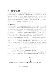
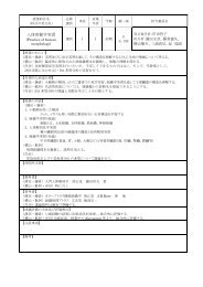
REVIEWSELASTINOPATHYPathological changes <strong>of</strong> theelastic fibres that lead to variousdegenerative diseases.MESENCHYMAL TISSUEThe tissue that originates frommesenchymal cells, which areunspecified cells that are derivedmainly from the primitivemesoderm duringembryogenesis. The connectivetissues <strong>of</strong> the body develop fromthe mesenchymal cells.Fibulin-1Fibulin-2Fibulin-3,4,5I II III Fibulin-2 dimerdissociationsIII II I NN I II IIII IIIII20 nmFigure 4 | The shapes <strong>of</strong> <strong>fibulins</strong>. The figure shows aschematic representation <strong>of</strong> the shapes <strong>of</strong> <strong>fibulins</strong> asdetermined by electron microscopy after rotaryshadowing 8,10,19 . The shape <strong>of</strong> the anti-parallel fibulin-2 dimer isdepicted with fully aligned N/II domains. The individualdissociation <strong>of</strong> these domains can give rise to the three-armand four-arm structures that are shown, which are stillcovalently connected through the central domain I (REF. 8).interactions has been shown to vary over a broad range(K d= 0.1 nM to 3 µM) and to require, in most cases, theligation <strong>of</strong> calcium. This was, in part, supported by themapping <strong>of</strong> the binding epitopes to <strong>fibulins</strong> and/or totheir corresponding ligands.Fibronectin micr<strong>of</strong>ibrils. The ECM and serum proteinfibronectin (BOX 2) was one <strong>of</strong> the first fibulin ligands tobe identified by showing that it colocalizes with fibulin-1(REF. 2) and fibulin-2 (REF. 38) in micr<strong>of</strong>ibrils that aredeposited in fibroblast cultures and by using variousdirect binding assays 7,45,46 . The micr<strong>of</strong>ibrils were shownto consist <strong>of</strong> equal amounts <strong>of</strong> fibronectin and fibulin-2and to have a lower content <strong>of</strong> fibulin-1. About 60–70%<strong>of</strong> fibulin-2 can be extracted from these micr<strong>of</strong>ibrilsusing EDTA, whereas denaturing conditions arerequired to solubilize fibronectin 38 . This indicates thatthe micr<strong>of</strong>ibril core structure is formed by fibronectin,to which the <strong>fibulins</strong> attach. This idea is supported bythe lack <strong>of</strong> fibulin-1 deposition on inhibition <strong>of</strong>fibronectin assembly in a fibroblast culture 47,48 . The fibulin-1binding site has been mapped to the 12–14 type IIImodules <strong>of</strong> fibronectin, which have an immunoglobulin(Ig)-like fold and a heparin-binding site 45 . The complementarybinding site has been localized to cbEGF5 andcbEGF6 <strong>of</strong> fibulin-1 (FIG. 1), which are also involved inthe limited oligomerization <strong>of</strong> fibulin-1 (how thisoligomerization relates to the non-covalent aggregatesmentioned above is still unclear) 46 . This oligomerization,but not the fibronectin binding, is dependent oncalcium. No epitopes have yet been mapped for thefibronectin– fibulin-2 interaction.Elastic fibres. Elastic fibres have a unique structureand special functions in tissues (BOX 1), and they havebeen shown to contain fibulin-1 (REF. 20) and fibulin-2(REFS 36,37). Immunogold staining <strong>of</strong> vessels showed thatfibulin-1 and fibulin-2 colocalize with either the amorphouscore or the micr<strong>of</strong>ibrillar component <strong>of</strong> theseelastic fibres, and that this colocalization was strongerfor fibulin-2 than for fibulin-1. This might reflect, inpart, distinct differences in their affinity for tropoelastin,which is high for fibulin-2 (K d= 0.6 nM) and 30-foldlower for fibulin-1 (REF. 37). Studies with recombinantfragments <strong>of</strong> fibrillin-1 showed distinct binding to fibulin-2,but not to the fibulin-1 C or D variants. Binding<strong>of</strong> fibulin-2 could be mapped to a restricted aminoterminalregion <strong>of</strong> fibrillin-1, which contains severalcbEGF-like modules, and was dependent on calcium 36 .In the elastic fibres <strong>of</strong> the skin, fibulin-2 was localized tothese micr<strong>of</strong>ibrils. However, micr<strong>of</strong>ibrils that consist <strong>of</strong>fibrillins and fibulin-2 are not restricted to elastic fibres,and they are found at many other places such as atbasement-membrane zones and in some cartilage structures36,40 . Fibulin-5, which binds to tropoelastin but notfibrillin, seems to be another important component <strong>of</strong>elastic fibres, as its absence in transgenic mice leads tosevere ELASTINOPATHIES 22,23 . The fact that fibulin-5 bindingto elastic fibres can be blocked by EDTA indicates thatthe interaction occurs through domain I and/or II <strong>of</strong>fibulin-5. The binding activities <strong>of</strong> fibulin-3 and fibulin-4have not yet been examined.Basement-membrane <strong>proteins</strong>. Various basementmembranes in many organs have been shown to containfibulin-1 and/or fibulin-2 by immunohistology4,25,26 , and several important basement-membrane<strong>proteins</strong> — such as nidogen-1, some laminin is<strong>of</strong>orms,the heparan sulphate proteoglycan perlecan and collagenIV (BOX 2) — have been identified as potential ligandsfor these associations 4,7,19 . Nidogen-1 is the onlyligand identified so far that binds to fibulin-1 splicevariants with different affinities, with its affinity beingaround 30-fold higher for the C, compared to the D,variant 19 . The fibulin-1–nidogen-1 association wasconfirmed in subsequent epitope-mapping studies,which showed that domain III and cbEGF6–9 <strong>of</strong>domain II <strong>of</strong> fibulin-1 participate in this interaction 49 .Fibulin-2 was also shown to bind to nidogen-1 with acomparable affinity, but its binding sites have not yetbeen mapped. The fact that dimeric fibulin-2 has twobinding sites is consistent with its participation in theformation <strong>of</strong> ternary complexes that include nidogen-1,perlecan, laminin and/or fibulin-1 (REF. 7). The bindingsites for both <strong>fibulins</strong> have been mapped to the centralglobular domain (G2) <strong>of</strong> nidogen-1, which has a highaffinity binding site for perlecan, and to its carboxyterminalG3 domain, which has a high affinity for thelaminin γ1 chain 50 . These data indicate that fibulin-1and fibulin-2 have a <strong>versatile</strong> repertoire <strong>of</strong> bindingproperties that they can use to integrate themselvesinto basement membranes, depending on the composition<strong>of</strong> these membranes and on the relative affinities<strong>of</strong> the <strong>fibulins</strong> for the membrane components.Perlecan (BOX 2) is another important constituent <strong>of</strong>basement membranes, as well as <strong>of</strong> cartilage and otherMESENCHYMAL TISSUES. Perlecan was shown to bind fibulin-2,but not fibulin-1, and the binding site was mapped totwo EGF-like modules in the carboxy-terminal domainV <strong>of</strong> the perlecan core protein (480 kDa) 51 and to severalIg-like modules <strong>of</strong> domain IV, including Ig2 andIg10–12 (REF. 52). Ig2 is located next to two more Ig-likemodules (Ig3 and Ig4), which have high affinity bindingsites for nidogens and fibronectin. This cluster <strong>of</strong>484 | JUNE 2003 | VOLUME 4 www.nature.com/reviews/molcellbio© 2003 Nature PublishingGroup
REVIEWSBox 1 | The structure and biology <strong>of</strong> elastic fibresElastic fibres are special forms <strong>of</strong> <strong>extracellular</strong> <strong>matrix</strong> assembly, which provide resilienceto dynamic connective tissues and are crucial for the proper functioning <strong>of</strong> the lungs,arteries and skin. They can be readily identified at the ultrastructural level by theiramorphous appearance, as they are frequently associated with numerous micr<strong>of</strong>ibrils.The amorphous core consists mainly <strong>of</strong> elastin — a highly hydrophobic protein that isgenerated from the soluble precursor tropoelastin (70 kDa) and is extensivelycrosslinked after the enzymatic oxidation <strong>of</strong> lysyl residues. Important components <strong>of</strong>the micr<strong>of</strong>ibrils are several fibrillin is<strong>of</strong>orms (~350 kDa, length ~150 nm), which havearound 40 calcium-binding sites and some elastic properties. These micr<strong>of</strong>ibrils arebelieved to provide a scaffold for the deposition <strong>of</strong> elastin. More than 30 <strong>proteins</strong>,including some <strong>fibulins</strong> and collagens, have so far been identified as other components<strong>of</strong> some or all elastic fibres 81 . Elastic fibres are known to lose their regenerative potentialduring ageing and in certain obstructive diseases such as lung emphysema. Mutations inelastin and fibrillins are known to lead to various human disorders with skeletal,cutaneous, vascular and ocular malformations, and similar elastinopathies can begenerated in transgenic mice 82,83 .ANGIOGENESISA process <strong>of</strong> blood-vesselbranching, in which bloodvessels sprout from smallcapillaries.modules therefore seems well suited for makingnumerous associations that can produce the largecomplexes that are formed by perlecan, fibulin-2 andnidogens or fibronectin.Further candidates for forming basement-membraneinteractions with <strong>fibulins</strong> are several lamininis<strong>of</strong>orms (BOX 2). Such an interaction was first shownfor laminin-1 that binds fibulin-1 through its E3fragment, a distinct fragment that is formed bycleavage with elastase. The E3 fragment consists <strong>of</strong>the α1-chain laminin G-type (LG)4–5 modules andis derived from the α1-chain carboxyl terminus 4,53 .The same site <strong>of</strong> the laminin α2 chain and the adjacentα2LG1–3 modules were also shown to bindfibulin-1 and fibulin-2 with moderate affinities 54 .The fibulin-binding properties <strong>of</strong> other laminin α-chainis<strong>of</strong>orms (α3–α5) have not yet been examined.Laminin LG modules provide important cell-adhesionsites for integrin, dystroglycan and heparan sulphateproteoglycan receptors 53 , and their potential modulationby <strong>fibulins</strong> remains an interesting possibility to bestudied.The γ2 chain, which is unique to laminin-5, wasalso shown to bind fibulin-2 in affinity chromatographyexperiments, and the binding was attributed to ashort peptide sequence in the central region <strong>of</strong> the γ2short-arm structure 55 . Subsequent studies showedthat this central domain also binds fibulin-1 and thatthe interacting epitopes have a more complex structure,which includes a second binding site for fibulin-2on a laminin-type EGF-like (LE) module 56 . These twobinding sites can be separated on proteolytic processing<strong>of</strong> laminin-5, which is essential for its <strong>matrix</strong>deposition.Other ligands that bind fibulin-1 and fibulin-2are endostatins, which are released from the carboxy-terminalend <strong>of</strong> collagens XV and XVIII anddeposited into basement membranes and elastic tissues57–59 . Endostatins are potent inhibitors <strong>of</strong> ANGIOGENESISand their binding to <strong>fibulins</strong> might be required fortheir localization to the ECM, particularly in vesselwalls.Lectican proteoglycans. Another group <strong>of</strong> highlypotent fibulin ligands is represented by the lectican<strong>family</strong> <strong>of</strong> large chondroitin sulphate proteoglycans,which includes the cartilage-specific aggrecan, theubiquitously deposited versican and the brain-specificneurocan and brevican 60 . The lectican <strong>family</strong> core <strong>proteins</strong>tructure includes an amino-terminal globulardomain, which has a high binding affinity for hyaluronan,a central elongated region that is used for chondroitinsulphate attachment, and a carboxy-terminalglobular domain that has a complex modular structure,which includes a C-type lectin domain. Thislectin domain in aggrecan and versican has beenshown to bind fibulin-1 with moderate affinity. Thisbinding is calcium-dependent, but independent <strong>of</strong> theN-glycosylation <strong>of</strong> fibulin-1 (REF. 61). The same twolectin domains also bind fibulin-2 with high affinity.These interactions are specific, as no, or only low,binding activity was found for the lectin domains <strong>of</strong>neurocan and brevican 62 . The binding epitopes weremapped to a short stretch <strong>of</strong> cbEGF-like modules indomain II <strong>of</strong> both <strong>fibulins</strong>. Furthermore, fibulin-2 wasshown to form networks with aggrecan and versican,but only the four-arm structure <strong>of</strong> fibulin-2 seemed tobe active 62 (FIG. 4). An important conclusion from thesedata is that there is a prominent role for <strong>fibulins</strong> (inaddition to that <strong>of</strong> hyaluronan) in the formation <strong>of</strong>huge proteoglycan networks. Some in vivo evidencefor such networks was discussed above for the endocardialcushion tissue 32 . Another function for theseinteractions is indicated by the binding <strong>of</strong> fibulin-1 toaggrecanase (a disintegrin and metalloproteinase withthrombospondin motifs 1; ADAMTS-1), which mighttarget this specific protease to proteoglycans for theircontrolled degradation (S. Agraves and M. Iruela-Arispe, unpublished observations).Specific fibulin-1 ligands. A few more interactionshave been described for fibulin-1, which do not seemto be shared by fibulin-2. A weak affinity (K d= 3 µM)was described for the fibrinogen βB chain, and thisinteraction causes fibulin-1 to associate with, and toinhibit the formation <strong>of</strong>, fibrin clots 63,64 .However,fibulin-1-deficient mice that lack the circulating form<strong>of</strong> this protein show no obvious defect incoagulation 65 , which indicates that this function is notcrucial. Two more ligands were initially identified byyeast two-hybrid screening and were subsequentlyconfirmed using recombinant fibulin-1 in other bindingassays. The first ligand was connective-tissuegrowth factor (CTGF), as well as related members <strong>of</strong>this protein <strong>family</strong>, which correlates well with the coexpression<strong>of</strong> fibulin-1 and these growth factors 66 .Theother ligand was β-amyloid precursor protein (APP),which binds to cbEGF3–7 <strong>of</strong> fibulin-1 in a calciumdependentfashion 35 . This interaction inhibits APPstimulation <strong>of</strong> neurite outgrowth and neuronal stemcellproliferation. A similar role in vivo is indicated bythe presence <strong>of</strong> fibulin-1 in some neurons 20 , but it isnot yet known whether fibulin-1 is also incorporatedinto amyloid deposits in the brain.NATURE REVIEWS | MOLECULAR CELL BIOLOGY VOLUME 4 | JUNE 2003 | 485© 2003 Nature PublishingGroup
REVIEWSBox 2 | The properties <strong>of</strong> important protein ligands for <strong>fibulins</strong>Fibronectin is an important micr<strong>of</strong>ibrillar protein. It is an elongated (61 nm)disulphide-linked homodimer <strong>of</strong> 450 kDa, which consists <strong>of</strong> unique fibronectin type I(F1), F2 and F3 modules that have various binding properties.Laminins are heterotrimers (αβγ) <strong>of</strong> α1–α5, β1–β3 and γ1–γ3 chains in variouscombinations and they have molecular masses <strong>of</strong> 400–750 kDa. Principal domainsinclude the laminin G-type (LG)1–LG5 modules at the carboxyl terminus <strong>of</strong> theα-chains and numerous laminin-type epidermal growth factor (EGF)-like (LE)modules that are related to EGF-like domains. Laminins have a cross-like shape(36–77 nm along their short and long arms) and they form important networkstructures in basement membranes.Perlecan is an important proteoglycan <strong>of</strong> basement membranes and <strong>of</strong> other<strong>extracellular</strong> <strong>matrix</strong> structures, and it consists <strong>of</strong> an elongated (80 nm) core protein<strong>of</strong> 480 kDa, which is modified by 3–4 heparan sulphate chains. It has amultidomain structure and is composed <strong>of</strong> LG, LE, EGF-like and immunoglobulinlikemodules.Nidogen is a ubiquitous basement-membrane protein <strong>of</strong> 150 kDa. It consists <strong>of</strong> threeglobular domains (G1–G3) that have high binding affinities for laminins, perlecan andcollagen IV.RGD-DEPENDENT INTEGRINSA specific group <strong>of</strong> cellularintegrin receptors that bind tothe arginine-glycine-aspartate(RGD) sequences <strong>of</strong> theirligands.WERNER SYNDROMEA premature ageing disorderthat is inherited in an autosomalrecessive mode. The clinicalsymptoms can include shortstature, wrinkled skin, baldness,cataracts and muscular atrophy.SYNPOLYDACTYLYA developmental defect that ischaracterized by the fusion(syndactyly) and splitting(polydactyly) <strong>of</strong> fingers or toes.It is usually an autosomaldominant disease and can resultfrom mutations in thehomeobox genes.Effects on cellular activitiesInteractions with integrins. Because <strong>of</strong> their broadoccurrence in the ECM, several <strong>of</strong> the <strong>fibulins</strong> were alsostudied for their ability to promote integrin-mediatedcell adhesion and migration. Mouse fibulin-2 wasshown to bind to purified platelet αIIbβ3 and αvβ3,which are both RGD-DEPENDENT INTEGRINS, and this interactionwas confirmed in adhesion assays with activatedplatelets and other cells 67 . The RGD sequence in the Ndomain <strong>of</strong> mouse fibulin-2 was shown to be involved inthis interaction, but this RGD sequence is not conservedin human fibulin-2, which still binds the αIIbβ3 integrin.Together, the data indicate that there is a role forfibulin-2 in haemostatic control. Fibulin-1 lacks RGDsequences and had no activity in cell-adhesionassays 67,68 . However, fibulin-1 inhibited cell adhesion to,and migration on, fibronectin. The binding <strong>of</strong> fibulin-1does not, however, block the integrin-binding sites <strong>of</strong>fibronectin, which led to the proposal that the formation<strong>of</strong> this complex generates a new anti-adhesive site,which repulses cellular interactions rather than promotesthem 68 . Finally, a single RGD site in the aminoterminaldomain I <strong>of</strong> fibulin-5 has also been shown tobe cell adhesive, and it interacts, in particular, withαvβ3, αvβ5 and α9β1 integrins 22,69 .Effect on cell proliferation. There is an increasingamount <strong>of</strong> data that indicate that most <strong>fibulins</strong> havethe ability to interfere with several cellular activities,and these data were interpeted to reflect their control<strong>of</strong> cellular proliferation and malignant transformation.Most <strong>of</strong> these data are, however, preliminary and, inpart, controversial, and no general concept hasemerged for their interpretation. This is perhaps bestillustrated by a recent study on fibulin-5, which cameto the conclusion that regulation occurs in a contextspecificmanner and depends on the cell type and assayconditions used 70 .The first evidence for a cellular proliferation activitywas described for fibulin-3, which is upregulatedin senescent and WERNER SYNDROME fibroblasts, as wellas in quiescent young fibroblasts 14 . This indicated thatthere was an effect on DNA synthesis, but the datahave so far remained controversial. A similar changein quiescent fibroblasts has also been reported forfibulin-4 (REF. 10), although fibulin-4 has also beenshown to stimulate growth 71 . A few further interestingactivities have been reported for fibulin-1, which,as already discussed, include modulation <strong>of</strong> neuriteoutgrowth through binding to APP 35 , the binding <strong>of</strong>growth factors such as CTGF 66 , and the regulation <strong>of</strong>signal transduction by inhibiting the phosphorylation<strong>of</strong> the <strong>extracellular</strong>-signal-regulated kinase(ERK) 68 . The latter activity is also shared by fibulin-5(REF. 70). The cellular receptors that are involvedremain to be identified.Effect on malignant transformation. A variable set <strong>of</strong>data has indicated that there is a correlation betweenfibulin expression and certain types <strong>of</strong> malignant cells,and these data were interpreted to indicate that there is arole for <strong>fibulins</strong> in tumour suppression 70,72 . These correlationsinclude an upregulation <strong>of</strong> fibulin-4 in colon carcinomas15 and <strong>of</strong> fibulin-1 in ovarian carcinomas 44,73 .This upregulation might inhibit the mobility <strong>of</strong> cancercells, which would suppress their invasiveness 74 .Themost interesting observation, however, is that high concentrations<strong>of</strong> the fibulin-1D variant selectively delaytumour transformation and invasive potential 72 .Themechanism is still unclear, but this indicates that there isa specific binding function for fibulin-1D that is notshared by fibulin-1C. This specific-binding function forfibulin-1D is also indicated by genetic evidence (seebelow), but remains to be defined.Genetic evidence for biological functionInsights into the in vivo functions <strong>of</strong> several <strong>fibulins</strong>have emerged from the identification <strong>of</strong> human diseasesthat involve fibulin loci and from the analysis <strong>of</strong> genetargetedmutant mouse strains. Targeted inactivation <strong>of</strong>the fibulin-1 gene in mice (Fbln1) leads to severe haemorrhagesin skin, muscle and perineural tissues, whichstart from midgestation and result in the death <strong>of</strong>almost all homozygous embryos at birth 65 . The defect iscaused by ruptures in the endothelial lining <strong>of</strong> small, butnot large, blood vessels. Capillary endothelial cells in theFbln1-null mice have very irregular cytoplasmicprocesses, but the underlying basement membrane isintact. Further defects were found in the kidneys andlungs, which might also contribute to the lethal phenotype.The mouse model therefore indicates that fibulin-1is important in the stabilization <strong>of</strong> blood vessel wallsand that it is also involved in diverse biological processesduring embryogenesis.Fibulin-1 has also been implicated in limb malformations.Three patients in a <strong>family</strong> affected with acomplex type <strong>of</strong> SYNPOLYDACTYLY have been found tocarry a balanced chromosomal translocation thatinvolves an alternatively spliced exon <strong>of</strong> FBLN1 and agene <strong>of</strong> unknown function that is located on the shortarm <strong>of</strong> chromosome 12 (REF. 75). This results in reduced486 | JUNE 2003 | VOLUME 4 www.nature.com/reviews/molcellbio© 2003 Nature PublishingGroup
REVIEWSTable 3 | The binding properties <strong>of</strong> fibulin-1 and fibulin-2 for <strong>extracellular</strong> <strong>proteins</strong>Interacting ligand Affinity (K d)* Calcium dependence ‡ Epitope mapped ReferencesFibulin-1Fibronectin 139 nM + Yes 19,45,46Nidogen-1 2–60 nM + Yes 19,50Laminin-1 80 nM + Yes 19Fibrinogen 3 µM ND Yes 63Aggregan, versican 10–30 nM + Yes 61Tropoelastin 18 nM + No 37Endostatin 46 nM – No 57Laminin γ2 chain 20 nM ND Yes 56Laminin α2 chain 14 nM ND Yes 54Connective-tissue growth factor ND ND Yes 66β-amyloid precursor protein ND + Yes 35Fibulin-2Fibronectin 1 µM + Yes 7Nidogen-1 0.5 µM – No 7Perlecan 22 nM ND Yes 52Fibrillin-1 56 nM + Yes 36Tropoelastin 0.6 nM + Yes 37Endostatin 33 nM – No 57Aggrecan, versican 0.1 nM + Yes 62Laminin γ2 chain 20–40 nM ND Yes 56Laminin α2 chain 13 nM ND Yes 54*The affinities were determined using surface plasmon resonance. ‡ The calcium dependence was examined by the addition <strong>of</strong> EDTA.ND, not determined.MACULAR DEGENERATIVEDISEASEAn incurable eye disease that iscaused by the deterioration <strong>of</strong>the central portion <strong>of</strong> the retina,which is known as the macula.The disease is heterogeneousand includes the rare heritableforms, the sporadic early-onsetform and the age-related form.expression <strong>of</strong> the D, but not the C, variant <strong>of</strong> fibulin-1in patients’ fibroblasts. As fibulin-1 is expressed in thedigits <strong>of</strong> the developing limb, altering the expression <strong>of</strong>fibulin-1 splice variants is thought to be responsiblefor the digit abnormality 75 . This possibility, however,remains to be tested.As a result <strong>of</strong> positional cloning, a single mutationin the fibulin-3 gene (FBLN3), which results in anR345W substitution (where W is tryptophan), wasfound to be the cause <strong>of</strong> two inherited forms <strong>of</strong> MACULARDEGENERATIVE DISEASE — so-called Malattia Leventineseand Doyne honeycomb retinal dystrophy 21 . Maculardegenerative disease is characterized by <strong>extracellular</strong>deposits, which are known as drusen, beneath theretinal pigment epithelium. R345 is located in the lastcbEGF-like domain <strong>of</strong> fibulin-3 and follows the secondcysteine residue. Interestingly, this mutation infibulin-3 was found in 161 patients from 37 familiesin the initial study, although recent evidence indicatesthat there is molecular heterogeneity within theinherited forms <strong>of</strong> macular degenerations 76 .Mutations in fibulin-3 are not associated with thesporadic form <strong>of</strong> early onset drusen or with agerelatedmacular degeneration, which is the mostcommon cause <strong>of</strong> blindness and is also characterizedby drusen deposition 21 . In both inherited and agerelatedmacular degeneration diseases, fibulin-3 isnot located at the site <strong>of</strong> drusen formation, but is aberrantlyaccumulated with and/or beneath the retinalpigment epithelium 77 . A R345W mutant fibulin-3protein produced by a mammalian expression systemis misfolded, secreted inefficiently and retained incells 77 . It has therefore been postulated that the misfoldingand aberrant accumulation <strong>of</strong> fibulin-3 mighthave a role in the etiology <strong>of</strong> macular degeneration 77 .An essential role for fibulin-5 in elastic-fibre assemblyin vivo was highlighted by the abnormal phenotypes<strong>of</strong> mice lacking fibulin-5 (REFS 22,23). The mutantmice showed markedly disrupted and disorganizedelastic fibres, which resulted in loose skin, emphysematouslungs and tortuous aortas with loss <strong>of</strong> function.Fibulin-5, which is expressed in tissues that areenriched in elastic fibres and binds tropoelastin andcell-surface integrins in vitro, is thought to anchor elasticfibres to cells and to be required for elastic-fibreassembly during development. It has also been postulatedthat fibulin-5 might function as a chaperone thattethers tropoelastin to the cell surface during elasticfibreassembly 78 .Although Fbln5-null mice have highly disorganizedelastic fibres in their internal organs, they survive toadulthood. The phenotypes <strong>of</strong> the mutant mice resemblethose <strong>of</strong> human patients with cutis laxa — a heterogeneousgroup <strong>of</strong> disorders that are characterized byloose skin and that have variable internal-organ involvement,such as pulmonary emphysema and cardiovascularabnormalities. This phenotype similarity led to theidentification <strong>of</strong> a homozygous missense mutationNATURE REVIEWS | MOLECULAR CELL BIOLOGY VOLUME 4 | JUNE 2003 | 487© 2003 Nature PublishingGroup
REVIEWSMARFAN SYNDROMEA heritable connective tissuedisease that affects several organsystems, including the skeleton,eyes, lungs, heart and bloodvessels. The disease is caused bydominant mutations in thefibrillin-1 gene.(which results in an S227P substitution; where S is serineand P is proline) in FBLN5 in a <strong>family</strong> that wasaffected by a severe form <strong>of</strong> cutis laxa 79 . S227 is locatedbetween Cys3 and Cys4 in cbEGF3 <strong>of</strong> fibulin-5. Thisamino-acid substitution is probably the cause <strong>of</strong> cutislaxa in this <strong>family</strong>, as the corresponding position inmost <strong>of</strong> the cbEGF-like modules in fibrillin-1 is occupiedby a serine, and an analogous mutation in fibrillin-1results in MARFAN SYNDROME. Although the heterozygouscarriers <strong>of</strong> the missense mutation in this<strong>family</strong> are not affected, cutis laxa can result from a heterozygousmutation in fibulin-5. A patient with amilder phenotype <strong>of</strong> cutis laxa carries a heterozygoustandem gene duplication <strong>of</strong> 22 kb in FBLN5, whichresults in the duplication <strong>of</strong> exons 5–8 that encodefour cbEGF-like domains 80 . The mutant fibulin-5 proteinwith four extra cbEGF-like domains is synthesizedand secreted by the patient’s fibroblasts inamounts that are somewhat greater than for a normalcounterpart, so the mutation probably exerts adominant-negative effect.Conclusions and perspectivesThe <strong>fibulins</strong> represent a newly characterized <strong>family</strong> <strong>of</strong>calcium-binding ECM <strong>proteins</strong> and five is<strong>of</strong>orms haveso far been characterized from mammalian species. The<strong>fibulins</strong> do not form large homotypic aggregates, in contrastto many other ECM <strong>proteins</strong>, but they have theability to join other supramolecular structures as diverseas basement-membrane networks, elastic fibres, severaltypes <strong>of</strong> micr<strong>of</strong>ibrils and proteoglycan aggregates. Theirinclusion, in part, stabilizes these structures, but alsoextends their repertoire <strong>of</strong> biological functions. The latterpoint is highlighted by genetic studies <strong>of</strong> inheriteddiseases and <strong>of</strong> transgenic animals. Some <strong>fibulins</strong> alsohave the ability to interact with cells, in part, throughinteractions with RGD-dependent integrins. There isalso increasing evidence that <strong>fibulins</strong> control the growth<strong>of</strong> normal and malignant cells. These studies are still in apreliminary stage and, in the future, a comprehensivecharacterization <strong>of</strong> the receptors and signal-transductionpathways involved will be required in order to evaluatetheir biological significance.1. Argraves, W. S., Dickerson, K., Burgess, W. H. & Ruoslahti, E.Fibulin, a novel protein that interacts with the fibronectinreceptor β subunit cytoplasmic domain. Cell 58, 623–629(1989).2. Argraves, W. S., Tran, H., Burgess, W. H. & Dickerson, K.Fibulin is an <strong>extracellular</strong> <strong>matrix</strong> and plasma glycoproteinwith repeated domain structure. J. Cell Biol. 111,3155–3164 (1990).Fibulin-1 is identified and is found to be a new class <strong>of</strong>ECM protein.3. Kluge, M., Mann, K., Dziadek, M. & Timpl, R.Characterization <strong>of</strong> a novel calcium-binding 90-kDaglycoprotein (BM-90) shared by basement membranes andserum. Eur. J. Biochem. 193, 651–659 (1990).4. Pan, T.-C. et al. Sequence <strong>of</strong> <strong>extracellular</strong> mouse proteinBM-90/fibulin and its calcium-dependent binding to otherbasement membrane ligands. Eur. J. Biochem. 215,733–740 (1993).5. Pan, T.-C. et al. Structure and expression <strong>of</strong> fibulin-2, a novel<strong>extracellular</strong> <strong>matrix</strong> protein with multiple EGF-like repeatsand consensus motifs for calcium-binding. J. Cell Biol. 123,1269–1277 (1993).The first evidence that there is a fibulin protein <strong>family</strong>that contains different fibulin is<strong>of</strong>orms.6. Zhang, R.-Z. et al. Fibulin-2 (FBLN2): human cDNAsequence, mRNA expression and mapping <strong>of</strong> the gene onhuman and mouse chromosomes. Genomics 22, 425–430(1994).7. Sasaki, T., Göhring, W., Pan, T.-C., Chu, M.-L. & Timpl, R.Binding <strong>of</strong> mouse and human fibulin-2 to <strong>extracellular</strong> <strong>matrix</strong>ligands. J. Mol. Biol. 254, 892–899 (1995).8. Sasaki, T. et al. Dimer model for the micr<strong>of</strong>ibrillar proteinfibulin-2 and identification <strong>of</strong> the connecting disulfide bridge.EMBO J. 16, 3035–3043 (1997).The first extensive model <strong>of</strong> the fibulin-2 structure.9. Tran, H., Mattei, M., Godyna, S. & Argraves, W. S. HumanFibulin-1D: molecular cloning, expression and similarity withS1-5 protein, a new member <strong>of</strong> the fibulin-1 gene <strong>family</strong>.Matrix Biol. 15, 479–493 (1997).10. Giltay, R., Timpl, R. & Kostka, G. Sequence, recombinantexpression and tissue localization <strong>of</strong> two novel <strong>extracellular</strong><strong>matrix</strong> <strong>proteins</strong>, fibulin-3 and fibulin-4. Matrix Biol. 18,469–480 (1999).11. Zhang, H.-Y., Lardelli, M. & Ekblom, P. Sequence <strong>of</strong> zebrafishfibulin-1 and its expression in developing heart and otherembryonic organs. Dev. Genes Evol. 207, 340–351 (1997).12. Barth, J. L., Argraves, K. M., Roark, E. F., Little, C. D. &Argraves, W. S. Identification <strong>of</strong> chicken andC. elegans fibulin-1 homologs and characterization<strong>of</strong> the C. elegans fibulin-1 gene. Matrix Biol. 17, 635–646(1998).13. Huber, R., Scholze, H., Paques, E. P. & Deisenh<strong>of</strong>er, J.Crystal structure analysis and molecular model <strong>of</strong> humanC3a anaphylatoxin. Hoppe-Seyler’s Z. Physiol. Chem. 361,1389–1399 (1989).14. Lecka-Czernik, B., Lumpkin, C. K. & Goldstein, S.An overexpressed gene transcript in senescent andquiescent human fibroblasts encoding a novel protein in theepidermal growth factor-like repeat <strong>family</strong> stimulates DNAsynthesis. Mol. Cell. Biol. 15, 120–129 (1995).15. Gallagher, W. M. et al. Human fibulin-4: analysis<strong>of</strong> its biosynthetic processing and mRNA expression innormal and tumour tissues. FEBS Lett. 489, 59–66(2001).16. Maurer, P. & Hohenester, E. Structural and functionalaspects <strong>of</strong> calcium-binding in <strong>extracellular</strong> <strong>matrix</strong> <strong>proteins</strong>.Matrix Biol. 15, 569–580 (1997).17. Downing, A. K. et al. Solution structure <strong>of</strong> a pair <strong>of</strong> calciumbindingepidermal growth factor-like domains: implicationsfor the Marfan syndrome and other genetic disorders. Cell85, 597–605 (1996).This paper explains how the structure <strong>of</strong> cbEGF-likemodules is stabilized by calcium ligation.18. Sasaki, T., Mann, K., Murphy, G., Chu, M.-L. & Timpl, R.Different susceptibilities <strong>of</strong> fibulin-1 and fibulin-2 to cleavageby <strong>matrix</strong> metalloproteinases and other tissue proteases.Eur. J. Biochem. 240, 427–434 (1996).19. Sasaki, T. et al. Structural characterization <strong>of</strong> two variants <strong>of</strong>fibulin-1 that differ in nidogen affinity. J. Mol. Biol. 245,241–250 (1995).20. Roark, E. F. et al. The association <strong>of</strong> human fibulin-1with elastic fibers: an immunohistological, ultrastructural,and RNA study. J. Histochem. Cytochem. 43, 401–411(1995).21. Stone, E. M. et al. A single EFEMP1 mutationassociated with both Malattia Leventinese and Doynehoneycomb retinal dystrophy. Nature Genet. 22, 199–202(1999).22. Nakamura, T. et al. Fibulin-5/DANCE is essential forelastogenesis in vivo. Nature 415, 171–175 (2002).23. Yanagisawa, H. et al. Fibulin-5 is an elastin-binding proteinessential for elastic fibre development in vivo. Nature 415,168–171 (2002).This work describes, together with reference 22, therole <strong>of</strong> fibulin-5 in stabilizing elastic fibres.24. Spence, S. G., Argraves, W. S., Walters, L., Hungerford, J. E.& Little, C. Fibulin is localized at sites <strong>of</strong>epithelial–mesenchymal transitions in the early avianembryo. Dev. Biol. 151, 473–484 (1992).25. Zhang, H.-Y., Timpl, R., Sasaki, T., Chu, M.-L. & Ekblom, P.Fibulin-1 and fibulin-2 expression during organogenesis inthe developing mouse embryo. Dev. Dyn. 205, 348–364(1996).26. Miosge, N. et al. The <strong>extracellular</strong> <strong>matrix</strong> <strong>proteins</strong> fibulin-1and fibulin-2 in the early human embryo. Histochem. J. 28,109–116 (1996).27. Hungerford, J. E., Owens, G. K., Argraves, W. S. & Little, C. D.Development <strong>of</strong> aortic vessel wall as defined by vascularsmooth muscle and <strong>extracellular</strong> <strong>matrix</strong> markers. Dev. Biol.178, 375–392 (1996).28. Bouchey, D., Argraves, W. S. & Little, C. D. Fibulin-1,vitronectin expression during avian cardiac valveand septa development. Anat. Rec. 244, 540–551(1996).29. Zhang, H.-Y., Kluge, M., Timpl, R., Chu, M.-L. & Ekblom, P.The <strong>extracellular</strong> <strong>matrix</strong> glyco<strong>proteins</strong> BM-90 and tenascinare expressed in the mesenchyme at sites <strong>of</strong>endothelial–mesenchymal conversion in the embryonicmouse heart. Differentiation 52, 211–220 (1993).30. Zhang, H.-Y. et al. Extracellular <strong>matrix</strong> protein fibulin-2 isexpressed in the embryonic endocardial cushion tissue andis a prominent component <strong>of</strong> valves in adult heart. Dev. Biol.167,18–26 (1995).31. Tsuda, T., Wang, H., Timpl, R. & Chu, M.-L. Fibulin-2expression marks transformed mesenchymal cells indeveloping cardiac valves, aortic arch vessels and coronaryvessels. Dev. Dyn. 222, 89–100 (2001).32. Miosge, N., Sasaki, T., Chu, M.-L., Herken, R. & Timpl, R.Ultrastructural localization <strong>of</strong> micr<strong>of</strong>ibrillar fibulin-1 andfibulin-2 during heart development indicates a switch inmolecular associations. Cell. Mol. Life Sci. 54, 606–613(1997).33. Gallagher, W. M. et al. MBP1: a novel mutant p53-specificprotein partner with oncogenic properties. Oncogene 18,3608–3616 (1999).34. Kowal, R. C., Richardson, J. A., Miano, J. M. & Olsen, E. N.EVEC. A novel epidermal growth factor-like repeatcontaining protein upregulated in embryonic and injuredadult vasulature. Circ. Res. 84, 1166–1176 (1999).35. Ohsawa, I., Takamura, C. & Kohsaka, S. Fibulin-1 binds theamino-terminal head <strong>of</strong> β-amyloid precursor protein.J. Neurochem. 76, 1411–1420 (2001).36. Reinhardt, D. P. et al. Fibrillin-1 and fibulin-2 interact and arecolocalized in some tissues. J. Biol. Chem. 271,19489–19496 (1996).37. Sasaki, T. et al. Tropoelastin binding to <strong>fibulins</strong>, nidogen-2and other <strong>extracellular</strong> <strong>matrix</strong> <strong>proteins</strong>. FEBS Lett. 460,280–284 (1999).38. Sasaki, T., Wiedemann, H., Matzner, M., Chu, M.-L. & Timpl, R.Expression <strong>of</strong> fibulin-2 by fibroblasts and deposition withfibronectin into a fibrillar <strong>matrix</strong>. J. Cell Sci. 109, 2895–2904(1996).39. Loveland, K. et al. Developmental changes in the basementmembrane <strong>of</strong> the normal and hypothyroid postnatal rattestis: segmental localization <strong>of</strong> fibulin-2 and fibronectin.Biol. Reprod. 58, 1123–1130 (1998).40. Raghunath, M. et al. Confocal laser scanning analysis <strong>of</strong> theassociation <strong>of</strong> fibulin-2 with fibrillin-1 and fibronectin definedifferent stages <strong>of</strong> skin regeneration. J. Invest. Dermatol.112, 97–101 (1999).41. Fässler, R., Sasaki, T., Timpl, R., Chu, M.-L. & Werner, S.Differential regulation <strong>of</strong> fibulin, tenascin and nidogenexpression during wound healing <strong>of</strong> normal andglucocorticoid-treated mice. Exp. Cell Res. 222, 111–116(1996).488 | JUNE 2003 | VOLUME 4 www.nature.com/reviews/molcellbio© 2003 Nature PublishingGroup
REVIEWS42. Hunzelmann, N., Nischt, R., Brenneisen, P., Eickert, A. &Krieg, T. Increased deposition <strong>of</strong> fibulin-2 in solar elastosisand its colocalization with elastic fibers. Brit. J. Dermatol.145, 217–222 (2001).43. Kusubata, M. et al. Spatiotemporal changes <strong>of</strong> fibronectin,tenascin-C, fibulin-1, and fibulin-2 in the skin during thedevelopment <strong>of</strong> chronic contact dermatitis. J. Invest.Dermatol. 113, 906–912 (1999).44. Roger, P., Pujol, P., Lucas, A., Baldet, P. & Rochefort, H.Increased immunostaining <strong>of</strong> fibulin-1, an estrogenregulatedprotein in the stroma <strong>of</strong> human ovarian epithelialtumors. Am. J. Pathol. 153, 1579–1588 (1998).45. Balbona, K. et al. Fibulin binds to itself and to the carboxylterminalheparin-binding region <strong>of</strong> fibronectin. J. Biol. Chem.267, 20120–20125 (1992).46. Tran, H., Van Dusen, W. J. & Argraves, W. S. The selfassociationand fibronectin-binding sites <strong>of</strong> fibulin-1 map tocalcium-binding epidermal growth factor-like domains.J. Biol. Chem. 272, 22600–22606 (1997).47. Roman, J. & McDonald, J. A. Fibulin’s organization into the<strong>extracellular</strong> <strong>matrix</strong> <strong>of</strong> fetal lung fibroblasts is dependent onfibronectin <strong>matrix</strong> assembly. Am. J. Respir. Cell Mol. Biol. 8,538–545 (1993).48. Godyna, S., Mann, D. M. & Argraves, W. S. A quantitativeanalysis <strong>of</strong> the incorporation <strong>of</strong> fibulin-1 into the <strong>extracellular</strong><strong>matrix</strong> indicates that fibronectin assembly is required.Matrix Biol. 14, 467–477 (1994).49. Adam, S. et al. Binding <strong>of</strong> fibulin-1 to nidogen depends onits C-terminal globular domain and a specific array <strong>of</strong>calcium-binding epidermal growth factor-like (EG) modules.J. Mol. Biol. 272, 226–236 (1997).50. Ries, A., Göhring, W., Fox, J. W., Timpl, R. & Sasaki, T.Recombinant domains <strong>of</strong> mouse nidogen-1 and theirbinding to basement membrane <strong>proteins</strong> and monoclonalantibodies. Eur. J. Biochem. 268, 5119–5128 (2001).51. Friedrich, M. V. K. et al. Structural basis <strong>of</strong>glycosaminoglycan modification and <strong>of</strong> heterotypicinteractions <strong>of</strong> perlecan domain V. J. Mol. Biol. 294,259–270 (1999).52. Hopf, M., Göhring, W., Mann, K. & Timpl, R. Mapping <strong>of</strong>binding sites for nidogens, fibulin-2, fibronectin and heparinto different IG modules <strong>of</strong> perlecan. J. Mol. Biol. 311,529–541 (2001).53. Timpl, R. et al. Structure and function <strong>of</strong> laminin LGmodules. Matrix Biol. 19, 309–317 (2000).54. Talts, J. F., Andac, Z., Göhring, W., Brancaccio, A. & Timpl, R.Binding <strong>of</strong> the G domains <strong>of</strong> laminin α1 and α2 chainsand perlecan to heparin, sulfatides, α-dystroglycan andseveral <strong>extracellular</strong> <strong>matrix</strong> <strong>proteins</strong>. EMBO J. 18, 863–870(1999).55. Utani, A., Nomizu, M. & Yamada, Y. Fibulin-2 binds to theshort arms <strong>of</strong> laminin-5 and laminin-1 via conservedamino acid sequences. J. Biol. Chem. 272, 2814–2820(1997).56. Sasaki, T. et al. Short arm region <strong>of</strong> laminin-5 γ-2 chain:structure, mechanism <strong>of</strong> processing and binding to heparinand <strong>proteins</strong>. J. Mol. Biol. 314, 751–763 (2001).57. Sasaki, T. et al. Structure, function and tissue forms <strong>of</strong> theC-terminal globular domain <strong>of</strong> collagen XVIII containing theangiogenesis inhibitor endostatin. EMBO J. 17, 4249–4256(1998).58. Sasaki, T. et al. Endostatins derived from collagens XV andXVIII differ in structural and binding properties, tissuedistribution and anti-angiogenic activity. J. Mol. Biol. 301,1179–1190 (2000).59. Sasaki, T., Hohenester, E. & Timpl, R. Structure and function<strong>of</strong> collagen-derived endostatin inhibitors <strong>of</strong> angiogenesis.IUBMB Life 53, 77–84 (2002).60. Ruoslahti, E. Brain <strong>extracellular</strong> <strong>matrix</strong>. Glycobiology 6,489–492 (1996).61. Aspberg, A., Adam, S., Kostka, G., Timpl, R. & Heinegard, D.Fibulin-1 is a ligand for the C-type lectin domains <strong>of</strong>aggrecan and versican. J. Biol. Chem. 274, 20444–20449(1999).62. Olin, A. J. et al. The proteoglycans aggrecan and versicanform networks with fibulin-2 through their lectin domainbinding. J. Biol. Chem. 276, 1253–1261 (2001).63. Tran, H. et al. The interaction <strong>of</strong> fibulin-1 with fibrinogen.A potential role in hemostasis and thrombosis. J. Biol.Chem. 270, 19458–19464 (1995).64. Godyna, S., Dias-Ricart, M. & Argraves, W. S. Fibulin-1mediates platelet adhesion via a bridge <strong>of</strong> fibrinogen. Blood88, 2569–2577 (1996).65. Kostka, G. et al. Perinatal lethality and endothedlial cellabnormalities in several vessel compartments <strong>of</strong> fibulin-1deficient mice. Mol. Cell. Biol. 21, 7025–7034 (2001).This paper shows that a fibulin-1 deficiency causes ahaemorrhagic phenotype.66. Perbal, B. et al. The C-terminal domain <strong>of</strong> the regulatoryprotein NOVH is sufficient to promote interaction withfibulin-1C: a clue for a role <strong>of</strong> NOVH in cell-adhesionsignalling. Proc. Natl Acad. Sci. USA 96, 869–874 (1999).67. Pfaff, M., Sasaki, T., Tangemann, K., Chu, M.-L. & Timpl, R.Integrin-binding and cell-adhesion studies <strong>of</strong> <strong>fibulins</strong> reveal aparticular affinity for αIIbβ3. Exp. Cell Res. 219, 87–92 (1995).68. Twal, W. O. et al. Fibulin-1 suppression <strong>of</strong> fibronectinregulatedcell adhesion and motility. J. Cell Sci. 114,4587–4598 (2001).69. Nakamura, T. et al. DANCE, a novel secreted RGD proteinexpressed in developing, atherosclerotic, and ballooninjuredarteries. J. Biol. Chem. 274, 22476–22483 (1999).70. Schiemann, W. P., Blobe, G. C., Kalume, D. E., Pandey, A. &Lodish, H. F. Context-specific effects <strong>of</strong> fibulin-5(DANCE/EVEC) on cell proliferation, motility, and invasion.Fibulin-5 is induced by transforming growth factor-β andaffects protein kinase cascades. J. Biol. Chem. 277,27367–27377 (2002).71. Heine, H., Delude, R. L., Monks, B. G., Esperik, T. &Golenbock, D. T. Bacterial lipopolysaccharide inducesexpression <strong>of</strong> the stress response genes hop and H411.J. Biol. Chem. 274, 21049–21055 (1999).72. Qing, J. et al. Surpression <strong>of</strong> anchorage-independentgrowth and matrigel invasion and delayed tumor formationby elevated expression <strong>of</strong> fibulin-1D in humanfibrosarcoma-derived cell lines. Oncogene 15, 2159–2168(1997).73. Clinton, G. M. et al. Estrogens increase the expression <strong>of</strong>fibulin-1, an <strong>extracellular</strong> <strong>matrix</strong> protein secreted by humanovarian cancer cells. Proc. Natl Acad Sci. USA 93, 316–320(1996).74. Hayashido, Y. et al. Estradiol and fibulin-1 inhibit motility <strong>of</strong>human ovarian- and breast-cancer cells induced byfibronectin. Int. J. Cancer 75, 654–658 (1998).75. Debeer, P. et al. The fibulin-1 gene (FLBN1) is disrupted in at(12;22) associated with a complex type <strong>of</strong> synpolydactyly.J. Med. Genet. 39, 98–104 (2002).76. Tartellin, E. E. et al. Molecular genetic heterogeneity inautosomal dominant drusen. J. Med. Genet. 38, 381–384(2001).77. Marmorstein, L. Y. et al. Aberrant accumulation <strong>of</strong> EFEMP1underlies drusen formation in Malattia Leventinese and agerelatedmacular degeneration. Proc. Natl Acad. Sci. USA 99,13067–13072 (2002).78. Midwood, K. S. & Schwarzbauer, J. E. Elastic fibres: buildingbridges between cells and their <strong>matrix</strong>. Curr. Biol. 12,R279–R281 (2002).79. Loeys, B. et al. Homozygosity for a missense mutation infibulin-5 (FBLN5) results in a severe form <strong>of</strong> cutis laxa.Hum. Mol. Genet. 11, 2113–2118 (2002).80. Markova, D. et al. Genetic heterogeneity <strong>of</strong> cutis laxa:a heterozygous tandem duplication within the fibulin-5(FBLN5) gene. Am. J. Hum. Genet. 72, 998–1004 (2003).81. Kielty, C. M., Sherrat, M. J. & Shuttleworth, C. A. Elasticfibers. J. Cell Sci. 115, 2817–2828 (2002).82. Dietz, H. C. & Mecham, R. P. Mouse models <strong>of</strong> geneticdiseases resulting from mutations in elastic fiber <strong>proteins</strong>.Matrix Biol. 19, 481–488 (2000).83. Milewicz, D. M., Urban, Z. & Boyd, C. Genetic disorders<strong>of</strong> the elastic fiber system. Matrix Biol. 19, 471–480(2000).84. Pan, T.-C., Kostka, G., Zhang, R.-Z., Timpl, R. & Chu, M.-L.Complete exon–intron organization <strong>of</strong> the mouse fibulin-1gene and its comparison with the human fibulin-1 gene.FEBS Lett. 444, 38–42 (1999).85. Mattei, M. G., Pan, T.-C., Zhang, R.-Z., Timpl, R. & Chu, M.-L.The fibulin-1 gene (FBLN1) is located on humanchromosome 22 and mouse chromosome 15.Genomics 22, 437–438 (1994).86. Grässel, S., Sicot, F.-X., Gotta, S. & Chu, M.-L.Mouse fibulin-2 gene. Complete exon–intron organizationand promoter characterization. Eur. J. Biochem. 263,471–477 (1999).87. Ikegawa, S., Toda, T., Okui, K. & Nakamura, Y. Structureand chromosomal assignment <strong>of</strong> the human S1-5 gene(FBNL) that is highly homologous to fibrillin. Genomics 35,590–592 (1996).88. Katsanis, N., Venable, S., Smith, J. R. & Lupski, J. R.Isolation <strong>of</strong> a paralog <strong>of</strong> the Doyne honeycomberetinal dystrophy gene from the multiple retinopathycritical region on 11q13. Hum. Genet. 106, 66–72(2000).89. Kowal, R. C., Jolsin, J. M., Olson, E. N. & Schultz, R. A.Assignment <strong>of</strong> fibulin–5 (FBLN5) to human chromosome14q31 by in situ hybridization and radiation hybrid mapping.Cytogenet. Cell Genet. 87, 2–3 (1999).AcknowledgementsPart <strong>of</strong> the work described here was supported by research grantsfrom the Deutsche Forschungsgemeinschaft for T.S. and R.T., theEuropean Community for R.T., and the National Institutes <strong>of</strong> Healthfor M.-L.C. The authors are grateful to S. Argraves for communicatingunpublished work and to J. Uitto for critically reading themanuscript.Online linksDATABASESThe following terms in this article are linked online to:InterPro: http://www.ebi.ac.uk/interpro/AT module | cbEGF-like module | C-type lectin domain |EGF-like module | LGLocusLink: http://www.ncbi.nlm.nih.gov/LocusLink/collagen IV | fibrinogen | laminin-1Swiss-Prot: http://www.expasy.ch/ADAMTS-1 | aggrecan | brevican | fibrillin-1 | fibronectin | fibulin-1 |fibulin-2 | fibulin-3 | fibulin-4 | fibulin-5 | neurocan | nidogen-1 |perlecan | tropoelastin | versicanFURTHER INFORMATIONGenBank database: http://www.ncbi.nlm.nih.gov/Genbank/National Center for Biotechnology Information:http://www.ncbi.nlm.nih.gov/Access to this interactive links box is free online.NATURE REVIEWS | MOLECULAR CELL BIOLOGY VOLUME 4 | JUNE 2003 | 489© 2003 Nature PublishingGroup