Classification of periodontal diseases-the eternal ... - Dentinal Tubules
Classification of periodontal diseases-the eternal ... - Dentinal Tubules
Classification of periodontal diseases-the eternal ... - Dentinal Tubules

You also want an ePaper? Increase the reach of your titles
YUMPU automatically turns print PDFs into web optimized ePapers that Google loves.
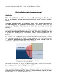
Preyesh Patel and Alaa Guni, BDS 4 students<strong>Classification</strong> <strong>of</strong> <strong>periodontal</strong> <strong>diseases</strong>-<strong>the</strong> <strong>eternal</strong> questClassifying and grouping entities is integral to human nature. It enforces a sense <strong>of</strong> belonging so much so, that <strong>the</strong> entire human species is subdivided into various races, religions andcastes. It would be interesting to see if this phenomenon is reciprocated amongst animals.When it came to tackling <strong>the</strong> age old concern <strong>of</strong> classifying <strong>periodontal</strong> disease, various factors were considered when formulating <strong>the</strong> classification that is now accepted worldwide:World Worskshop in Periodontology, 1999. These factors included: Aetiology, clinical presentation as well as prognosis/outcome.As a dental student appreciation <strong>of</strong> <strong>the</strong>se finely tuned (so far) classifications can be difficult. However, after sifting through many resources, we have formulated a table <strong>of</strong> our own. Asummary <strong>of</strong> <strong>the</strong> classifications <strong>of</strong> <strong>the</strong> <strong>periodontal</strong> <strong>diseases</strong> coupled with its appropriate management. This will serve as a quick and simple tool when revising for exams or even as a referencewhen in practice.
Preyesh Patel and Alaa Guni, BDS 4 studentsDiagnosis Features TreatmentGingivitis Plaque-induced only No loss <strong>of</strong> Connective Tissue Attachment (CTA) or Alveolar OH reinforcementBone (AB)Plaque present at gingival marginRednessOedematousIncrease in gingival exudatesLoss <strong>of</strong> stipplingBleeding on probingSulcular temperature changeReversible upon plaque removalModified by systemic conditions Hormonal-increase in gingivitis during circumpubertal age,without simultaneous plaque increase diabetesModified by medications Anti-convulsants-Phenytoin calcium channel blockers-Amlodipine Immunosuppressants-CyclosporinreviewCorrect or reduce predisposing systemic factors(eg. Control diabetes, smoking cessation)OH reinforcementReviewAttempt to change medication (discuss) withphysicianOH reinforcementreviewModified by malnutrition Lack <strong>of</strong> vitamin c seen from <strong>the</strong>ir diet history Review Diet Discuss diet supplements with physician OH reinforcement reviewNon-plaque inducedBacterial origin-N.gonorrhea, T.pallidum, Streptococci, MycobacteriumchelonaeOH reinforcement possibly with an antibacterialadjunctFeatures: fiery red, oedematous, painful ulceration(asymptomatic chancres, mucous patches or a typical nonulceratedhighly inflamed gingivitis)Viral origin-herpetic ginigivostomatitisFeatures– generalised pain in <strong>the</strong> gingival and oral mucosa,inflammation, ulceration <strong>of</strong> gingival and mucosa, lymphadenopathy,fever, malaiseGentle debridement and relief <strong>of</strong> pain usinganalgesiaInstruction in proper nutrition, appropriate fluidintakeReassurance that condition is self-limitingAntiviral adjunctViral origin-herpes zosterFeatures– small ulcers on <strong>the</strong> tongue, mucosa and gingivaHealing usually occurs within 1-2 weeksS<strong>of</strong>t/liquid dietRestAtraumatic plaque removal– CHX mouthrinsesAntiviral adjunct
Preyesh Patel and Alaa Guni, BDS 4 studentsDiagnosis Features TreatmentGingivitis Non-plaque induced Fungal origin-CandidosisFeatures– pseudomembranous candidosismay present as white lesions,chronic ery<strong>the</strong>matous candidosis presentsas redness along <strong>the</strong> gingival margins.AntifungalOH reinforcementreviewMucocutaneous disorders– Oral LichenPlanus, Benign mucous membranepemphigoid, pemphigus vulgaris, ery<strong>the</strong>mamultiformeTopical ointments, steroidsOH reinforcementReviewFeatures– desquamative lesions, gingivalulcerationsTrauma– traumatic brushing techniqueFeatures– frictional keratosisInstruction on atraumatic brushing techniquePeriodontitis Incidental attachment loss Loss <strong>of</strong> attachment doesn’t fit into criteria<strong>of</strong> aggressive or chronic periodontitisIsolated areas <strong>of</strong> attachment loss in an o<strong>the</strong>rwisehealthy dentition associated withtrauma, malpositioned tooth, impacted thirdmolars, subginigival caries and endo infectionsMay predispose to periodontitisTreat local defectOH reinforcementReviewIncipient chronic periodontitis Age <strong>of</strong> onset can be in adolescence (13–14years) Interproximal clinical attachment loss <strong>of</strong> 1–2 mm (commonly seen on maxillary firstmolars, mandibular incisors), associatedwith presence <strong>of</strong> plaque, subgingival calculus Pockets <strong>of</strong> 4–5 mm. Bone loss no more than 0.5 mm over an 18-month period (bite-wing radiographs usuallyshow horizontal bone loss).OH reinforcementNon-surgical rsdReview
Preyesh Patel and Alaa Guni, BDS 4 studentsPeriodontitisDiagnosis Features TreatmentChronic PeriodontitisMild 1-2mm cal, 50% bone lossLocalised 30% sites affectedAggressive periodontitis-Localised: Circumpubertal onset restricted to interproximal areas (IPCAL is >3mm) <strong>of</strong> first molar andincisors. (arc shaped) involving no more than two teetho<strong>the</strong>r than first molar or incisorGeneralised- Generalised Interproximal attachmentloss affecting at least 3 teetho<strong>the</strong>r than first molars and incisors(
Preyesh Patel and Alaa Guni, BDS 4 studentsDiagnosis Features TreatmentPeriodontitis Abscesses (<strong>periodontal</strong>) Forms close to <strong>the</strong> gingival margin Tender to lateral percussion Pulp vital for true perio lesionsDrainage (to relieve patient!)RSD if allowedCourse <strong>of</strong> antimicrobials: Metronidazole (preffered aseffective against anaerobic bacteria) or a mix <strong>of</strong> metronidazoleand amoxicillin if cellulitis presentReviewPerio-endo lesion -Primarily endodontic History <strong>of</strong> trauma or pulpitisHeavily restoredFracture <strong>of</strong> tooth?Localised pockets and relatively healthy periodontium generallyNon-vitalTTPExtraction or extirpate <strong>the</strong> pulp and monitorIf improvement shown (reduction in symptoms and reducedlesion on LCPA) à RCT followed by RSDPerio-endo lesion -Primarily periodontic History <strong>of</strong> periodontitisVital/non-vital pulp upon testingLack <strong>of</strong> restoration/fracture or history <strong>of</strong> traumaNo history <strong>of</strong> pulpitisTTPExtraction or extirpate <strong>the</strong> pulp and monitorIf improvement shown (reduction in symptoms and reducedlesion on LCPA) à RCT followed by RSD