207 Poster Session 2 - Connective Tissue Oncology Society
207 Poster Session 2 - Connective Tissue Oncology Society
207 Poster Session 2 - Connective Tissue Oncology Society

Create successful ePaper yourself
Turn your PDF publications into a flip-book with our unique Google optimized e-Paper software.
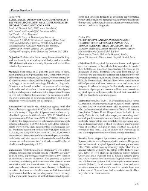
Scientific <strong>Poster</strong>s – <strong>Poster</strong> <strong>Session</strong> 2Conclusion: Preoperative anemia was seen more frequentlyin atypical lipomatous tumor patients than lipoma patients.None of anemic patients had an underlying cause of anemia.In this study, number of anemic patient is small andwe have no data of postoperative hematological changes.Further studies are needed to determine whether atypicallipomatous tumor cause secondary anemia.Table 2.Clinical Characteristics of the Patients with AnemiaPatientno.HistologicaldiagnosisConventionallipomaAge(years)SexHb(g/dl)MCV (fl)MCH(pg)Type of anemia1 34 f 10.5 71.6 23.3 microcytic hypochromic2 48 f 11.2 77.5 25.8 microcytic hypochromicAtipicallipomatous tumor3 26 f 10.6 71.4 22.5 microcytic hypochromic4 29 f 11.8 76.7 25.0 microcytic hypochromic5 48 f 11.0 81.5 27.6 normocytic normochromic6 62 f 11.9 88.8 30.1 normocytic normochromic7 62 f 11.9 92.3 31.6 normocytic normochromic8 63 m 12.7 92.9 31.3 normocytic normochromic9 75 f 10.6 94.6 31.9 normocytic normochromicHb = hemoglobin; MCV = mean corpuscular volume; MCH = mean corpuscular hemoglobin.Table 1.Demographic Charactaristics of Patientsand Prevalence of AnemiaAtipicallipomatoustumor (n=68)Lipoma(n=81)Age, years; mean (SE) 55.4 (1.59) 54.6 (1.45)Sex, M/F 32/36 32/49Anemia; n (%) 7 (10%) * 2 (2.4%) *SE = standard error. *The prevalence of anemia inatypical lipomatous tumor patients was significantly higherthan that of lipoma patients (p < 0.05, Fisher's exact test).<strong>Poster</strong> #96IDENTIFICATION OF COMMON CANCER-RELATEDEPIGENETIC CHANGES IN LIPOSARCOMASKara M. Pascarelli, BS 1 ; Mohammed Shaker 1 ;Matthew J. Plantinga 1 ; Katherine Doyle 1 ; Chad Roberts 1 ;Yan Zhou 2 ; Margaret von Mehren von Mehren 2 ; Dina Lev 3 ;Dominique Broccoli 11Curtis & Elizabeth Anderson Cancer Institute, MemorialUniversity Medical Center, Savannah, GA, USA;2Fox Chase Cancer Center, Philadelphia, PA, USA;3MD Anderson Cancer Center, Houston, TX, USAObjective: Aberrant epigenetic silencing of genes is observedin cancers and is an established mechanism toabrogate tumor suppressor pathways. To gain insight intothe biology of liposarcomas, we carried out an epigeneticreactivation screen to identify genes silenced by methylationof CpG dinucleotides in promoter regions.Methods: Three cell lines derived from pleomorphic liposarcomas(PLS) were treated with 5-aza-2-deoxycytidinefor 2 cell doublings prior to extraction of RNA. Total RNAfrom duplicate cultures of treated and untreated cells washybridized to the Affymetrix U133plus2 expression array.Gene interaction networks were generated through the useof Ingenuity Pathway Analysis (Ingenuity® Systems, www.ingenuity.com). Conditional hypergeometric tests weredone for testing the association of Gene Ontology usingGOstats package in Bioconductor. Promoter hypermethylationwas validated using bisulfite genomic sequencing.Results: Initial filtering for only those genes affected in all208
Scientific <strong>Poster</strong>s – <strong>Poster</strong> <strong>Session</strong> 2and differentiation. Expression analysis was performedby immunohistochemistry and RT-PCR analysis.Results: Immunohistochemical staining of FFPE sectionsshowed clear and distinct nuclear localization of brachyuryin majority of the WDLS sections analysed. Faint cytoplasmicstaining was also observed in a limited WDLS population.Interestingly, we found DDLS showing strong cytoplasmicstaining for brachyury. The NF stained negative forbrachyury. Since brachyury is associated with nuclear localization,to eliminate the possibility of artifact / backgroundstaining, RNA was purified from FFPE sections of NF,WDLS and DDLS and RT-PCR was performed for brachurygene expression. No amplification was observed for NF byagarose gel electrophoresis but WWLS and DDLS producedamplicons of expected size. The purified PCR product wasthen sequenced and confirmed as a brachury gene product.Conclusion: In this study, we found that the mesodermmarker, brachyury, is not only expressed, but are also differentiallylocalised in different subtypes of liposarcoma.<strong>Poster</strong> #100PROTEOMIC APPROACH TO PROGNOSTICBIOMARKER DISCOVERY IN MYXOIDLIPOSARCOMATakashi Tajima 1 ; Daisuke Kubota 1 ; Kenta Mukaihara 1 ;Kazutaka Kikuta 1 ; Hiroshi Ichikawa 1 ; Yutaka Sugihara 1 ;Akihiko Yoshida 3 ; Kazuo Mochizuki 4 ; Akira Kawai 2 ;Tadashi Kondo 11Division of Pharmacoproteomics, Natinal Cancer CenterReserch Institute, Tokyo, Japan; 2 Division of Musculoskeletal<strong>Oncology</strong>, National Cancer Center Hospital, Tokyo, Japan;3Division of Pathology, National Cancer Center Hospital,Tokyo, Japan; 4 Department of Orthopaedic Surgery, KyorinUniversity Faculty of Medicine, Tokyo, JapanObjective: Myxoid liposarcoma (MLS) is the second mostcommon subtype of liposarcoma, accounting for 10% ofall soft tissue sarcomas. The clinical course of MLS spansa wide spectrum from a curable disorder to a highly malignantdisease that leads to metastasis and death. Thus,the molecular background of MLS has been studied topredict the behavior of individual tumors and to optimizetherapeutic strategies. The indication of adjuvant therapyoften depends on the amount of round cell componentin the primary tumor tissue. However, the predictivevalues of the round cell component for poor prognosisremained only 50-60%, and a novel biomarker is warrantedto better predict clinical outcome. In this study, weaimed to develop novel prognostic biomarkers in MLS.Methods: This study included 14 MLS cases, and theirprimary tumor tissue. The tumor tissues were obtainedat the time of surgery. The cases were grouped accordingto the amount of round cell component (less than 5%, and5% or more), and the status of distance metastasis. Weemployed two-dimensional difference gel electrophoresis(2D-DIGE) to create protein expression profiles. Proteinswere extracted from the frozen tumor tissues, and labeledwith ultra high sensitive fluorescent dye (CyDye DIGEFluor saturation dye, GE). The labeled protein sampleswere separated by a large gel. The expression profileswere obtained as a gel images, and compared betweenthe patients with and those without round cell componentin the primary tumors, and between the patientswith different outcomes. The structure of interesting proteinswas examined by mass spectrometry and databasesearch. The results of proteomic study were validatedwith large number of clinical samples by Western blottingand immunohistochemistry using specific antibodies.Results: We observed up to 4,000 protein species by 2D-DIGE. The comparative proteomic study revealed the presenceof protein species which were statistically and significantlyassociated with the round cell component or the statusof distant metastasis. Those proteins were subjected to massspectrometry and database search for protein identification.Conclusion: We created the protein expression profiles ofprimary tumor tissues of the MLS patients. Comparativeproteomic study identified the candidates for prognosticbiomarker, whose clinical utilities are worth further validation.<strong>Poster</strong> #101FIRST RESULTS OF A PROSPECTIVE TRIAL OFRADIOTHERAPY DOSE DE-ESCALATION INMYXOID LIPOSARCOMASRick Haas, MD PhD 1 ; Luc Dewit 1 ; Martijn Kerst 2 ;Neeltje Steeghs 2 ; Jos van der Hage 3 ; HoukeKlomp 3 ;Frits van Coevorden 31Radiotherapy, The Netherlands Cancer Institute, Amsterdam,Netherlands; 2 Medical <strong>Oncology</strong>, The Netherlands CancerInstitute, Amsterdam, Netherlands; 3 Surgery,The Netherlands Cancer Institute, Amsterdam, NetherlandsObjective: Soft tissue sarcomas represent a heterogeneousgroup of tumors. Although in general sarcomasare considered to be radiation resistant, marked volumedecrease and necrosis induction have been reportedafter 25 x 2Gy preoperative radiotherapy (RT) in myxoidliposarcomas (MLS). Shrinkage is usually evidentalready during RT. This study investigates the possibilityof dose de-escalation to 18 x 2 Gy without influencingboth the clinical and pathological response rates.Methods: This prospective multicenter phase II trial of211
Scientific <strong>Poster</strong>s – <strong>Poster</strong> <strong>Session</strong> 2Objective: Leiomyosarcomas (LMS) are malignant soft tissuesarcomas originating from smooth muscle cells. Theseneoplasms are responsible for a substantial morbidity andmortality since they are characterized by aggressive behavior.The pathobiology of LMS is still poorly understoodand therefore no specific targeted treatment is currentlyavailable. Most LMS have a complex karyotype with multiplechromosomal aberrations. The aim of this study wasto identify a recurrent genomic alteration which mighthave a causative role in the development of these tumors.Methods: A comprehensive molecular cytogenetic approachwas used including multicolor fluorescence in situhybridization (FISH) based karyotyping of nine LMS forwhich life cells were available for cell culture. Array comparativegenomic hybridization (array-CGH) and FISHwere used to identify the exact breakpoints in two cases. Atissue microarray (TMA) was generated including a panelof 41 cases of LMS and interphase FISH was used to evaluatethe presence of the aberrations on the generated TMA.Results: Two of the nine cases revealed a recurrent translocation(6;14) within a complex karyotype. FISH breakpointmapping of one case revealed a breakpoint at chromosome6p21.32 close to HMGA1 and a small deletion on the distalpart of the gene. At the translocation breakpoint of 14q24.1an additional small deletion, resulting in the homozygousloss of ACTN1 was detected. Fine mapping of the translocationbreakpoint of the second case that demonstrateda der(6)t(6;14)(p21.1;q22.2) showed different breakpointsfor both involved chromosomes. Using array-CGH an amplificationof 6p21.1 containing NFYA and SUPT3H at thebreakpoint was confirmed. The described genetic alterationswere not detected in any additional LMS on the TMA.Conclusion: Complex karyotypes of most LMS mightmerely represent tumor progression associated changes;however, a causative role of a hidden, recurrent chromosomalaberration cannot be excluded in the developmentof these malignancies. Fine mapping of the translocationbreakpoints showed different breakpoints of chromosome6 and 14 in two cases. Both HMGA1 and HMGA2are frequently involved in translocations in leiomyomas.Although the translocation was absent in any of 39 otherLMS, HMGA1 and ACTN1 may be involved in the developmentof smooth muscle tumors, as both are expressedin smooth muscle cells.1Pediatric <strong>Oncology</strong> Unit, Fondazione IRCCS IstitutoNazionale Tumori Milan, Milan, Italy; 2 Clinical Trials andBiostatistics Unit, Istituto Oncologico Veneto, Padova,Italy; 3 Division of Pediatric Hematology and <strong>Oncology</strong>,Padova University, Padova, Italy; 4 Department of Pediatric<strong>Oncology</strong>-Hematology, Erasmus MC/Sophia Children’sHospital, Rotterdam, Netherlands; 5 Department of Pediatrics,Institut Curie, Paris, France; 6 Department of Pediatric<strong>Oncology</strong>, Royal Manchester Children’s Hospital, Manchester,United Kingdom; 7 Department of Pediatric <strong>Oncology</strong>,Royal Hospital for Children, Bristol, United Kingdom;8Department of Pediatrics, Institut Gustave Roussy,Villejuif, Paris, FranceObjective: Synovial sarcoma (SS) was traditionally consideredas a “rhabdomyosarcoma (RMS)-like” tumorby pediatric oncologists in Europe, so children wereenrolled over the years in RMS protocols and given thesame chemotherapy as for RMS, regardless of any riskfactors. Moving from such a strategy towards a treatmentconcept more closely resembling the one usually adoptedin the adult setting, in 2005 the European pediatric Softtissue sarcoma Study Group (EpSSG) opened a prospectivetrial dedicate to children and adolescents with SS.Methods: Patients were classified according to a riskstratification, based on tumor size and site and surgicalstage: “low-risk” patients (completely-resected tumors
Scientific <strong>Poster</strong>s – <strong>Poster</strong> <strong>Session</strong> 2be offered to those patients with poor chance of salvage.<strong>Poster</strong> #109ANTI TUMOR EFFECT OF VEGF TARGETEDTHERAPY ON SYNOVIAL SARCOMAToru Wakamatsu 1 ; Norifumi Naka 2 ; Takaaki Tanaka 1 ;Nobuhito Araki 3 ; Takafumi Ueda 4 ; Hideki Yoshikawa 2 ;Kazuyuki Itoh 11Biology, Osaka Medical Center, Osaka, Japan; 2 Orthopaedics,Osaka University Graduate School of Medicine, Osaka, Japan;3Orthopaedic Surgery, Osaka Medical Center, Osaka,Japan; 4 Orthopaedic Surgery, Osaka National Hospital,Osaka, JapanObjective: Synovial sarcoma (SS) has highly metastaticpotential and poor prognosis, due to their chemo- andradio-resistance, thus a novel therapy is needed. In orderto examine the anti-tumor effect of VEGF targeted therapyagainst SS, we herein used two human SS cell lines establishedin our laboratory. We also used Bevacizumab (Bev),a humanized monoclonal antibody against VEGF, has beenapproved to clinically improve the prognosis with severalmalignancies by inhibiting angiogenesis and proliferationof primary and metastatic sites. On the contrary, severalstudies reported that inhibition of VEGF signal increasedtumor invasion and lung metastasis depending on HIF1α.In this study we examined the effect of VEGF-targetedtherapy for SS by combination with Ifosfamid (IFO).Methods: In vitro, we measured the level of vascular endothelialgrowth factor (VEGF) in 3 dimensional (3D) andadhesion (2D) cultures of these SS cells by ELISA. Inhibitionof colony formation with the treatment of Bev and/orIFO was examined by soft agar assay. In vivo, we measuredthe tumor volume every week in xenograft models.Results: These SS cells produced markedly higher levelof VEGF in 3D compare to 2D cultures. In soft agar assay,treatment with Bev inhibited the colony formation of thesetwo SS cells, and combination treatment of Bev with Ifosfamideshowed higher efficiency. However, proliferationof two SS cell lines on 2D cultures couldn’t be inhibited bythe treatment with Bev. In xenograft models, the combinationtherapy also effectively reduced the tumor volumewithout any adverse effect. We are currently focusinghow Bev inhibited tumor growth in 3D but not on 2D.Conclusion: Collectively VEGF-targeted therapy can beproposed as a novel therapy for SS by combination withcurrent chemotherapy.<strong>Poster</strong> #110CELL CONTEXT IS AN IMPORTANT FACTORFOR THE ROLE OF SYT-SSX ON EPIGENETICREGULATION OF TRANSCRIPTIONSakura Tamaki; Tomohisa Kato; Kazuo Hayakawa;Naoko Takahara; Yoichiro Kajita; Junya ToguchidaDepartment of <strong>Tissue</strong> Regeneration, Institute for FrontierMedical Sciences, Kyoto University, Kyoto, JapanObjective: Synovial sarcoma (SS) is a soft tissue sarcomacharacterized by the SYT-SSX fusion gene. Variety ofstudies implicated that SYT-SSX contributes the developmentof SS by modifying the physiological regulation ofgene expression and exerts its function through chromatinremodeling. However, the molecular mechanism howSYT-SSX dysregulates target genes remains unclear. Wepreviously identified the FZD10 gene as an SS-specificallyup-regulated gene. In this study, we investigate the functionof SYT-SSX as an epigenetic modifier by the preciseanalyses of its regulation on the FZD10 gene.Methods: Transcriptional core regulatory region of theFZD10 gene was identified by luciferase reporter assay,and histone modifications in the region were analyzedby ChIP assay. The SYT-SSX gene was knocked-down inFZD10-positive SS cells, or overexpressed in FZD10-negativehuman embryonic stem cells (hESCs) and human skinfibroblasts (hSFs), and the expression of FZD10 gene andhistone modifications were analyzed in these cells.Results: As expected, histones were modified as an activestatus in SS cells; H3 was highly acetylated and H3K4 washighly methylated. On the other hand, in hSF, H3K27 washighly trimethylated (me3) and EZH2, a component of PcG,occupied in this region indicating a repressed status. Intriguingly,hESCs contained both H3K4me3 and H3K27me3indicating “bivalent” status. Knockdown of SYT-SSX in SScells downregulated the FZD10 expression in associationwith increased H3K27me3, whereas the ectopic expressionof SYT-SSX induced the expression of FZD10 changinghistone status from bivalent to active. Such induction ofFZD10 was not observed in hSF with ectopic SYT-SSXsuggesting the importance of original histone status forthe induction. Intriguingly, the combination of SYT-SSXexpression and HDAC inhibitor successfully induced theexpression of FZD10 in hSF.Conclusion: A number of studies have been done to identifydown-stream molecules of SYT-SSX by overexpressionassay with little consideration of recipient cells. Our resultsin this study, however, clearly indicated that original chromatinsignature of target gene has a great impact on theeffect of SYT-SSX, and therefore cell context is an importantfactor for the function of SYT-SSX as an epigenetic modifier,which should be seriously considered in future studies.216
Scientific <strong>Poster</strong>s – <strong>Poster</strong> <strong>Session</strong> 2<strong>Poster</strong> #111DISTINCT CIRCULATING TUMOR CELLS (CTCS)PROFILES IN EWING FAMILY OF TUMORS (EFTS)AND SYNOVIAL SARCOMA (SS) - ANALYSIS OFPROGNOSTIC AND DIAGNOSTIC VALUE OFFUSION TRANSCRIPTS DETECTED INPERIPHERAL BLOOD SPECIMENSJoanna Przybyl, MSc 1 ; Katarzyna Wiater 2 ;Konrad Ptaszynski 3 ; Maria Debiec-Rychter 4 ;Janusz A. Siedlecki 1 ; Hanna Kosela 2 ; Piotr Rutkowski 21Department of Molecular Biology, Maria Sklodowska-CurieMemorial Cancer Centre and Institute of <strong>Oncology</strong>,Warsaw, Poland; 2 Department of Soft <strong>Tissue</strong>/Bone Sarcomaand Melanoma, Maria Sklodowska-Curie Memorial CancerCentre and Institute of <strong>Oncology</strong>, Warsaw, Poland;3Department of Pathology, Maria Sklodowska-CurieMemorial Cancer Centre and Institute of <strong>Oncology</strong>, Warsaw,Poland; 4 Department of Human Genetics, K.U. Leuven,Leuven, BelgiumObjective: Ewing family of tumors (EFTs) and synovialsarcoma (SS) are characterized by chromosomal translocations,leading to formation of oncogenic EWSR1-FLI1and SS18-SSX1/2 fusions, respectively, in approximately90-95% of cases. The aim of present study was to comparecirculating tumor cells (CTCs) profiles in these sarcomasubtypes and to assess their prognostic and diagnosticvalue.Methods: 10mL of whole blood was collected from 35 EFTsand 38 SS untreated patients at the diagnosis (period: 2008-2011). 13 specimens of healthy people and 5 specimens ofrheumatoid arthritis patients were included as controls. 13EFTs and 5 SS patients presented metastatic disease (M1)at the diagnosis. Nested RT-PCR and/or one-step RT-PCRwere applied in triplicate for the detection of EWSR1-FLI1and SS18-SSX1/2 fusion transcripts and the results wereconfirmed by direct sequencing. FISH assays for EWSR1and SS18 rearrangements were performed on FFPE tumortissue in 28 and 22 cases, respectively. Median follow-upwas 19 months.Results: EWSR1-FLI1 transcript was detected in peripheralblood of 82.9% (n=29) of EFTs patients and FISH assay waspositive in 58% of cases. In 10 EFTs patients where FISHassay could not be performed due to insufficient qualityor lack of material, nested RT-PCR provided confirmationof immunopathological diagnosis. SS18-SSX1/2 transcriptswere detected in peripheral blood of 5.3% (n=2) of SSpatients and FISH assay was positive in 95.5% of cases.Specificities of RT-PCR blood tests in controls were 91.4%and 100% for EWSR1-FLI1 and SS18-SSX1/2 detection,respectively. CTC-positive EFTs patients showed the trendfor longer OS than CTC-negative patients (1-year OS: 80%vs. 34%; p=0.01).Conclusion: Our results indicate that CTCs profiles of EFTsand SS reflect their biology, given that SS are characterizedby slow tumor growth and high incidence of late metastases,whereas EFTs are considered as a systemic diseasewith clinically evident metastases or micrometastases atpresentation. Additionally, we show that the fusion transcriptdetection in peripheral blood specimens may be auseful supplementary test to the standard clinicopathologicaldiagnosis of EFTs; however, fusion transcripts may bepresent in the peripheral blood of EFTs and SS patientsbelow RT-PCR detection threshold. Prognostic value ofEWSR1-FLI1 and SS18-SSX1/2 fusion transcripts detectionwarrants further study.<strong>Poster</strong> #112CHEMOTHERAPY IN MALIGNANT PERIPHERALNERVE SHEET TUMOR (MPNST):A RETROSPECTIVE CASE SERIES ANALYSESFROM A SINGLE INSTITUTIONElena Palassini 1 ; Giacomo G. Baldi 1 ; Rossella Bertulli 1 ;Carlo Morosi 3 ; Silvana Pilotti 4 ; Alessandro Gronchi 2 ;Paolo G. Casali 11Medical Department, IRCCS Fondazione Istituto Nazionaledei Tumori, Milano, Italy; 2 Surgical Department, IRCCSFondazione Istituto Nazionale dei Tumori, Milano, Italy;3Radiological Department, IRCCS Fondazione IstitutoNazionale dei Tumori, Milano, Italy; 4 Pathological Department,IRCCS Fondazione Istituto Nazionale dei Tumori,Milano, ItalyObjective: To report on the activity of chemotherapy inMPNST. Data on chemosensitivity are limited in this histologicalsubtype.Methods: We retrospectively reviewed all cases of MPNSTtreated with chemotherapy at our institution in the neoadjuvant,locally advanced or metastatic setting since 2004.Both sporadic and neurofibromatosis type 1 (NF1)-relatedcases were evaluated.Results: From 2004, 30 patients (pts) with MPNST wereidentified (male/female: 18/12, median age (range): 43(23-67) years, with/without NF1: 10/20), local/locallyadvanced/metastatic disease: 9/4/17). Most used chemotherapyregimens included: antracycline plus ifosfamide(AI) in 20 pts, etoposide plus ifosfamide (EI) in 9 pts, platinumplus etoposide (PE) in 11 pts, high-dose ifosfamide(HD-IFX) in 8 pts. Best responses according to RECISTwere: 4 PR (20%), 11 SD, 5 PD with AI; 2 PR (22%), 7 SDwith EI; 2 PR (20%), 5 SD, 3 PD (1 not evaluable) with PE; 2PR (25%), 3 SD, 3 PD with HD-IFX. In the locally advancedand metastatic setting, median progression free survivalwas 2,3 months with AI, 4,8 months with etoposide-basedchemotherapy, 4,2 months with HD-IFX.217
Scientific <strong>Poster</strong>s – <strong>Poster</strong> <strong>Session</strong> 2Research, Tokyo, Japan; 6 Orthopaedic Surgery, Aichi CancerCenter Hospital, Nagoya, Japan; 7 Orthopaedic Surgery,Osaka Medical Center for Cancer and CardiovascularDiseases, Osaka, Japan; 8 Orthopaedic Surgery, JuntendoUniversity, Tokyo, Japan; 9 Human Pathology,Juntendo University, Tokyo, JapanObjective: Malignant granular cell tumor (MGCT) is an extremelyrare neoplasm, which is histologically similar to abenign granular cell tumor but has some particular featuresindicating risk for metastasis. There are a small number ofreports about this neoplasm, most of which were focusingon histology. Therefore, its clinical features still remain to aconsiderable extent unknown. This is a multi-institutionalretrospective study with a pathological central review andaims for revealing its clinical details from the perspectiveof orthopaedic oncologists.Figure 1. Local recurrence free survival of MGCTMethods: Clinical data including epidemiology, sites,clinical features, and prognoses of 22 patients diagnosedas MGCT at twelve hospitals were collected through theform of questionnaires, and Kaplan-Meier survival graphswere calculated using SAS JMP 10. In addition, available 17specimens were reviewed according to Famburg-Smith’scriteria dividing granular cell tumors into ‘malignant’,‘atypical’, and ‘benign’ types.Results: The dominant gender was female (73%), and themajority of the patients were 30-79 years old. The mostfrequent primary site was lower extremity (56%), followedby upper extremity (23%). Four cases presented metastasesincluding lung (three cases), lymph node (two), and bonemetastases (one) at the first visit (Table 1). Wide resectionswere achieved in 16 cases, but five of them suffered localrecurrence 4-42 months after surgery resulting in deaths(Figure 1). Irradiation was added mainly after local recurrencein seven cases as palliative treatments. Chemotherapywas combined with other treatments in eight cases forTable 1. Patients' Status at the First VisitBackgroundsNo. ofpatientsClinical statusrecurrences/metastases. One of them remains alive withno evidence of disease, and other four cases had relativelyprolonged static disease (SD) durations of over one year(Table 2). The final prognoses were CDF (ten cases), NED(one), AWD (two), and DOD (nine). The five-year and tenyearoverall survivals were 58.3% and 35.0% respectively(Figure 2). Among 17 reviewed histologies, only two caseswere evaluated as ‘atypical’, and one of themwas died due to lung metastases.No. ofpatientsGender Male 6 UICC stage 1A 3Female 16 1B 7Age 0-19 0 2B 120-29 2 3 730-39 4 4 340-49 3 Unknown 150-59 5 Metastasis Lung 360-69 6 Bone 170-79 2 Lymph node 280- 0 Others 0Figure 2. Overall survival of MGCTConclusion: MGCT has high tendency forrecurrence/metastasis and the possibility oflymph node or musculoskeletal metastasisshould not be ignored. Once recurrence occurs,the prognosis is predicted to be fairly poor, butchemotherapy seems to have a certain capabilityof prolonged survival. It has been said thatalmost all ‘atypical’ cases never metastasized,but our study indicates its potentiality of lifethreateningmetastases.219
Scientific <strong>Poster</strong>s – <strong>Poster</strong> <strong>Session</strong> 2Table 2. Details of ChemotherapyCase Chemotherapy Target lesion SD duration2 IFO (*), CDDP+ADM (*)1011GEM+PTX,Cyclophosphamide (*)GEM+PTX (*),Cyclophosphamide13 PazopanibRecurrence, lung meta.,lymph node meta.Finaloutcome> 12 months DODLung meta., soft tissue meta. 12 months DODResidual tumor after surgeryRecurrence, lung meta.,lymph node meta.12 months(PD)(PD)DODAWD15 IFO+ADM, Pazopanib (*) Lung meta., lymph node meta. 15 months DOD17 IFO+ADM, GEM+TXT Recurrence (PD) DOD21 IFO+ADM Lung meta. (#)22 IFO+ADM, CBDCA etc.Recurrence, lung meta.,lymph node meta.(Notapplicable)The mark (*) shows the regimens during SD. #: Chemotherapy was added postoperatively as an adjuvant therapy.(PD)NEDDOD<strong>Poster</strong> #115PREVALENCE OF PARASPINAL TUMORS INASYMPTOMATIC CHILDREN WITH NF1David Viskochil, MD, PhD 1 ; Kathleen Murray 1 ;David Stevenson 1 ; Zulf Mughal 2 ; Susan Huson 3 ;Jan Friedman 4 ; Linlea Armstrong 4 ; Elizabeth Schorry 51Pediatrics, University of Utah, Salt Lake City, UT, USA;2Pediatrics, University of Manchester, Manchester, UnitedKingdom; 3 Genetics, University of Manchester, Manchester,United Kingdom; 4 Human Genetics, University of BritishColumbia, Vancouver, BC, Canada; 5 Human Genetics,University of Cincinnati, Cincinnati, OH, USAObjective: Plexiform neurofibromas affect about 25% ofthe NF1 (neurofibromatosis type 1) population, and arethought to be congenital tumors that emerge clinicallyduring childhood. Paraspinal neurofibromas are a subsetof plexiform neurofibromas that originate from large nerveroots adjacent to the spine. Whole body imaging studiesin adults with NF1 have recently shown that ~24% ofindividuals with NF1 have spinal neurofibromas (Plotkinet al, PLoS ONE, e35711). The prevalence of thoracic paraspinaltumors in pre-pubertal children with NF1 has notbeen established.Methods: As part of a natural history study for the developmentof scoliosis in NF1 children, we obtained thoracicMRI scans at the time of enrollment. Eligible childrenwere pre-pubertal, diagnosed with NF1, and did not havescoliosis on physical examination. Once enrolled, childrenwould undergo a scoliosis radiology series, DXA scanning,peripheral quantitative computerized tomographyof the lower leg, and a thoracic MRI without sedation. Thethoracic MRI was performed in 1 field of view to coverT1 through L5 and includes: Sagittal T1 obtained as 3mmthick slices (no skips), Sagittal T2 obtained as 3mm thickimages, Axial T2 images obtained as 5mm thick imagesacquired at 7mm increments, and Coronal T1 images obtainedas contiguous 3mm images.The thoracic MRIs werescored by a single radiologist for spinal canal dimensionsat L1, vertebral body dimensions at L1, dural ectasia, andparaspinal neurofibromas.Results: We enrolled 114 participants from 4 centers; theUniversity of Utah, University of British Columbia (CA),University of Cincinnati, and the University of Manchester(UK). There were 92 evaluable non-sedated MRIscans (52 males:40 females), and the ages ranged from 5years 5 months to 9 years 10 months. Thirty-two thoracicspine MRIs demonstrated the presence of a paraspinalneurofibroma (~35%), and 8 showed dural ectasia, withand without adjacent tumor. Of the 38 reported sites oftumor(s), sacral location was noted once, cervical wasnoted 5 times, thoracic was noted 15 times, and lumbarlocation was noted 17 times.Conclusion: Asymptomatic pre-pubertal children withNF1 who did not have scoliosis were screened in 4 centersby non-sedated thoracic MRI as part of a NF1 scoliosis progressionstudy. About one-third of prospectively enrolledNF1 children for this natural history study had an unsuspectedparaspinal neurofibroma, none of which appeared220
Scientific <strong>Poster</strong>s – <strong>Poster</strong> <strong>Session</strong> 2pre-operatively. A pre-operative diagnosis of fibroids wasmade in 28%. The majority of patients had undergone appropriatepre-operative investigations (96%) and surgery(72%); lymph node dissection was performed in 14%.Eight patients were upstaged with metastatic disease afterradical surgery. Commonly, patients were referred to seekguidance on the role of adjuvant or palliative therapy. Fiftypatients were discussed with regards to the managementof metastatic disease and offered: metastatectomy 10%;systemic therapy 25% (54% enrolled into a clinical trial) andendocrine therapy (10%). Median loco-regional disease-freesurvival (DFS) was 29.6 months, median distant DFS-13.4months and overall survival at 1 yr- 81%.Conclusion: This audit has highlighted an unmet needin the management of G-STS. Clear guidelines need to becommunicated to gynae-oncology MDTs. The followingrecommendations may address referral pathway inconsistency:(1) a full dataset be provided upon referral to helpdetermine primary management and adjuvant therapies(2) an increased awareness of radiological characteristicsof G-STSs to facilitate early referral and appropriate surgicalmanagement, (3) histological review by a designatedsarcoma pathologist, particularly endocrine status in LMSand ESS and (4) early referral for the management of metastaticdisease. Clinical trials in the adjuvant and metastaticsetting are in progress to provide further clarification oftreatments in this challenging disease.Methods: This is an interim analysis of a 12-month studyof the symptom burden of GIST. Persons with GIST, beingfollowed at MD Anderson Cancer Center, gave IRB-approvedinformed consent and then rated their symptomson the provisional MD Anderson Symptom Inventory forGIST (MDASI-GIST) every 1-2 weeks. The MDASI-GISTis a questionnaire that asks patients to rate the worst severityof 22 symptoms and amount of interference with6 types of daily activities in the last 24 hours on a 0 (nosymptom or interference) to 10 (as bad as can be imaginedor complete interference) scale. The symptom burden ofGIST will be described through descriptive and mixedmodeling analyses.Results: As of April 30, 2012, 64 subjects had been on studyfor at least 3 months. Demographic and treatment informationat study entry for subjects is in Table 1. Figure 1 isa graph of the mean symptom and interference severityover 12 weeks. The means and standard deviations (SD)of the 5 most severe symptoms and the interference itemsare in Table 2.Mixed modeling analysis demonstrated thatpatients on imatinib therapy had significantly lower severityof the 5 most severe symptoms than patients receivingother treatments estimate = -1.00, p-value = 0.021).Conclusion: Symptom burden experienced by patientswith GIST receiving treatment are mild and stable overseveral months. The most severe symptoms are mainlyfatigue-related. The results of this preliminary analysiswarrant further investigation of the symptom burden ofGIST, including long term symptom burden, changes insymptom burden associated with changes in disease status,identification of patient groups who may experince moresevere symptom burden, and the influence of symptomburden on treatment adherence. Data collection for thisstudy is ongoing.<strong>Poster</strong> #122SYMPTOM BURDEN OF GASTROINTESTINALSTROMAL TUMORS (GIST) OVER 3 MONTHSLoretta A. Williams, PhD 1 ; Dejka M. Araujo 2 ; Nazim N. Ali 1 ;Mary L. Sailors 1 ; Kaiyan Jing 1 ; Jonathan C. Trent 3 ;Charles S. Cleeland 11Symptom Research, The University of Texas MD AndersonCancer Center, Houston, TX, USA; 2 Sarcoma Medical<strong>Oncology</strong>, The University of Texas MD Anderson CancerCenter, Houston, TX, USA; 3 Sylvester Comprehensive CancerCenter, University of Miami, Miami, FL, USAObjective: Symptom burden is the combined impact ofdisease- and therapy-related symptoms on patient functionalability. The objective of this study is to describe thesymptom burden experienced by patients receiving treatmentfor GIST over a 3 month period.Figure 1. Symptom Severity and Interference MeansOver 12 Weeks224
Scientific <strong>Poster</strong>s – <strong>Poster</strong> <strong>Session</strong> 2Table 1. Subject Demographic and Treatment CharacteristicsCHARACTERISIC MEAN SDAge 57.3 11.2NUMBER PERCENTAGEGenderMale 33 52Female 31 48RaceWhite 56 87Other 8 13Employment StatusEmployed Outside the Home 31 48Not Employed Outside the Home 33 52EducationPartial High School or High School Graduate 12 19College or Professional Training 52 81TherapyImatinib 32 50Non-imatinib 32 50Table 2. Five Most Severe Symptoms and Interference Means Over 12 Weeks5 MOST SEVERE SYMPTOMS INTERFERENCERANK MEAN SD RANK MEAN SDFatigue 1 3.07 2.37 Work 1 2.34 2.45Feeling Drowsy 2 2.49 1.99 General Activities 2 2.17 2.32Disturbed Sleep 3 2.37 2.15 Walking 3 1.69 2.07General Weakness 4 2.35 2.14 Enjoyment of Life 4 1.63 1.95Not Feeling Well (Malaise) 5 2.29 2.26 Mood 5 1.40 1.55Relations with Others 6 0.90 1.33<strong>Poster</strong> #123GASTROINTESTINAL STROMAL TUMORS (GIST)IN THE ELDERLY: ASSOCIATION OF PHYSICALFUNCTIONS AND CLINICAL OUTCOMES IN GISTPATIENTS (PTS) OVER 65 YEARSFlorence Duffaud, MD, PhD 1 ; Olivier Guillem 1 ;Sebastien Salas 1 ; Julien Mancini 2 ; Thanh Huynh 11Medical <strong>Oncology</strong>, CHU La Timone, Marseilles, France;2Biostatistics, CHU La Timone, Marseilles, FranceObjective: Analysis of physical functions and care of GISTpts over 65 years. There are very little historical data specificallyfocused on senior GIST pts over age 65, despite thefact that advanced age is reported to negatively influencethe prognosis of localized resected and metastatic GISTs.Methods: We retrospectively analysed metrics of physicalfunctions, tumor characteristics, treatment approaches,and outcomes of GIST pts over age 65 seen at La TimoneUniversity Hospital, Marseilles, France.Results: Between Jan1999-Jan2012, 68 senior GIST ptswere seen. Median age was 78, [distributed as 65-75, n=31;76-85, n=26; > 85, n=11], half were women (n=34), withwidowed/single (n= 18), median ECOG= 1 (0-3), mediancomorbities 2 (0-5), another prior cancer (n= 12). Metrics of225
Scientific <strong>Poster</strong>s – <strong>Poster</strong> <strong>Session</strong> 2physical functions included; nutritional status [nutritionalindex risk (NRI), average 95.7; albumin (average 37.3 g/L),weight loss (average 6.7%)], renal function [urea, average6.17 mmol/L; MDRD, average 74; Cockroft, average 58],hematological function [Hb, average 12 g/dL; leucocytes,average 6.5 Giga/L; lymphocytes, average 1.75 Giga/L].Tumors (T) were; gastric (n=37), and intestinal (n=17), localized(n=41), locally advanced (LA, n= 18), and metastatic(n=9), with a median size of 80 mm, with Miettinen risklow (n=19), intermediate (n=8), and high (n=30). Mutationswere documented in 19/23 cases, with KIT exon 11in 13 cases. Of localized/LA pts, 51 had resection of theprimary T (86%), with R0/R1 resections in 32/19 pts. Ptsreceived Imatinib post-operatively (n=31) or for advanceddisease (n=10). Twenty experienced severe toxicities. Witha median FU of 4.6 years (0-12.7), 55 pts (81%) are alive,but 27 (39.7%) relapsed. The 5-year OS and PFS rates are83% and 58.5% for the overall group.In Univariate analysis; ECOG >1, lymphocytes (< 1500),leucocytes (> 4000), Hb (≤ 11), non gastric T, metastases,negatively influenced OS, as urea (> 7.5) on PFS, but NRI(≤ 97.5), and albumine (< 35) had a negative impact onOS and PFS. Only age was significantly correlated withsevere toxicities.Conclusion: GIST pts over 65 receive similar treatmentsas younger pts, despite frequent nutritional deficits. Assessmentof nutritional status and improvement of thismetric in geriatric GISTs might positively influence theiroutcomes.<strong>Poster</strong> #124LOW DOSE IMATINIB MAY BE ACTIVE IN MANYGIST PATIENTS WITH INTOLERANCETO STANDARD DOSEMikael Eriksson, MD, PhD; Marie Ahlström<strong>Oncology</strong>, Skane University Hospital, Lund, SwedenObjective: Patients with severe side effects on standardor somewhat reduced doses of imatinib, 300-400 mg daily,are often considered intolerant to the drug and switched toanother tyrosin kinase inhibitor, usually sunitinib. However,several physicians treating GIST have experiencedthat low daily doses, 200 mg or less, may be both toleratedand active in such patients. There are, however, almost nowritten reports on this possibility, and there is an obviousrisk that some patients therefore will not get a chance toexperience a meaningful and even long-lasting responseto the drug.Methods: We collected data on a series of GIST patients atour institution who have been treated with imatinib doseslower than 300 mg daily to investigate whether a meaningfulresponse was observed and when they potentiallyrecurred. These patients will be presented.Results: Objective responses were noted on low or verylow doses of imatinib, even less than 100 mg daily in somepatients, and recurrences were not usual when the dosewas decreased because of toxicity.Conclusion: This limited series from one institution illustratesthat patients who seem intolerant to a standarddose of imatinib may well tolerate and respond to lowerdoses, and they should therefore not be withheld from thispossibility before switching to another drug. There may bean association between side effects and tumor sensitivityin GIST. Furthermore, intolerance may be best defined asnot tolerating the highest dose needed to prevent progression,which would also have implications for eligibilitycriteria in many clinical trials where the term intoleranceis poorly defined.<strong>Poster</strong> #125CORRELATION BETWEEN KIT EXON 11MUTATION SITE AND IMATINIB TREATMENTOUTCOME IN PATIENTS WITH ADVANCEDGASTROINTESTINAL STROMAL TUMORS (GIST)Agnieszka Wozniak 1 ; Piotr Rutkowski 3 ; Elzbieta Bylina 3 ;Maciej Matlok 6 ; Janusz Siedlecki 4 ; Maria Debiec-Rychter 2 ;Janusz Limon 51Laboratory of Experimental <strong>Oncology</strong>, Dept of <strong>Oncology</strong> and Dept of General Medical <strong>Oncology</strong>, KU Leuven andUniversity Hospitals Leuven, Leuven Cancer Institute,Leuven, Belgium; 2 Dept of Human Genetics, KU Leuvenand University Hospitals Leuven, Leuven Cancer Institute,Leuven, Belgium; 3 Dept of Soft <strong>Tissue</strong> and Bone Sarcoma andMelanoma, Maria Sklodowska-Curie Memorial Cancer Centreand Institute of <strong>Oncology</strong>, Warsaw, Poland; 4 Dept ofMolecular Biology, Maria Sklodowska-Curie MemorialCancer Centre and Institute of <strong>Oncology</strong>, Warsaw, Poland;5Dept of Biology and Genetics, Medical University of Gdansk,Gdansk, Poland; 6 Dept of Endoscopical Surgery, JagiellonianUniversity, Cracow, PolandObjective: Imatinib mesylate is a first line treatment forpatients with advanced GIST. However not all patientsbenefit equally from treatment. The correlation betweenGIST genotype (KIT exon 11 vs KIT exon 9 vs wild type)and treatment outcome has already been proven. RecentlyBlay et al. suggested that also site of primary KIT exon 11mutations could be considered as independent prognosticfactor for progression free survival (PFS) in GIST.Methods: We have reviewed files from 196 patients treatedwith imatinib between 2001 and 2011 because of advanceddisease at diagnosis (31.6%) or progression after surgery226
Scientific <strong>Poster</strong>s – <strong>Poster</strong> <strong>Session</strong> 2(68.4%). All patients were treated until progression with400mg/day of imatinib as starting dose. PFS was calculatedas a time between date of imatinib start and date of progression(complete) or last observation (censored values).Results: Mutational analysis revealed 126 KIT exon 11mutants (64.3%), followed by 32 GIST with no detectableKIT/PDGFRA mutation – ‘wild-type’ GIST (16.3%), 26cases with KIT exon 9 (13.3%), 10 tumors showing PDG-FRA exon 18 (5.1%) and single cases with KIT exon 17 andPDGFRA exon 12 mutations. For analysis of KIT exon 11topography we have used grouping system presentedby Blay et al. [3]. Overall we have found 30 cases (23.8%)whose first mutated codon was 559codon (G4). Majority of mutations were deletions (35.2%)or deletions/insertions (16.3%), followed by substitutions(16.3%), which were present only in groups G2 (17/52)and G4 (7/18). Majority of tumors from groups G2 and G4originated from intestine (76.9% and 82.4%, respectively).During the observation period, 115 patients (58.7%) progressedon imatinib with median PFS of 147.3 weeks (range4-561.6 weeks). As expected, median PFS was significantlydifferent in patients with mutations in KIT exon 11 (125.1weeks) vs. ‘wild-type’ (68.9 weeks) vs. KIT exon 9 (76.2weeks) (p
Scientific <strong>Poster</strong>s – <strong>Poster</strong> <strong>Session</strong> 2<strong>Poster</strong> #127THE ECONOMIC IMPACT OF CYTOREDUCTIVESURGERY AND TYROSINE KINASE INHIBITORTHERAPY IN THE TREATMENT OF RECURRENTOR METASTATIC GASTROINTESTINALSTROMAL TUMORS: A MARKOV CHAINDECISION ANALYSISNicole J. Look Hong, MD 1 ; Steven L. Chang 2 ;Chandrajit P. Raut 11Surgical <strong>Oncology</strong>, Brigham and Women’s Hospital / DanaFarber Cancer Institute, Boston, MA, USA;2Urologic <strong>Oncology</strong>, Brigham and Women’s Hospital / DanaFarber Cancer Institute, Boston, MA, USAObjective: The current first-line treatment for patients withrecurrent or metastatic gastrointestinal stromal tumors(GIST) is management with tyrosine kinase inhibitors(TKIs). There is an as yet undefined role for surgery in themanagement of these patients. The aim of this study is toexamine the economic impact of treating recurrent or metastaticGIST patients with TKIs in combination with surgeryat different time points in their treatment trajectories.Methods: A Markov chain decision analysis was modeledover a 2-year time horizon to determine costs associatedwith surgery performed in combination with imatinibmesylate(IM) or sunitinib malate(SU) in 7 scenarios variedby TKI agent, TKI dose, and disease status (stable vs. localizedprogressive disease). The base case was a 56 year-oldmale with recurrent or metastatic GIST taking IM 400mg.Economic data was calculated from average wholesalepricing of medications and physician/institutional costsdetermined by the Centers for Medicare & MedicaidServices. Rates of disease progression, surgical morbidity,mortality and adverse drug reactions were extracted fromexisting literature. Deterministic sensitivity analyses wereperformed to examine changes in cost due to variations indisease progression, recurrence, surgical procedures, andrates of surgical morbidity and mortality.Results: The least costly scenario was to perform surgeryon patients with stable disease on IM 800mg, which wasless than the cost of drug therapy without surgery. Themost costly scenario was to perform surgery on patientswith localized progressive disease on IM 800mg. This isdue to lower costs of IM compared to SU, combined withthe smaller proportion of patients on IM 800mg comparedwith IM 400mg. Costs were most influenced by the rateof death in patients on IM 400mg and the probability ofsurviving surgery, cumulatively addressing 82% of the riskin this model. The overall range of costs clustered withinapproximately $212,000(USD).Conclusion: Costs of surgical intervention at different timepoints within the treatment course of patients with recurrentor metastatic GIST fluctuate within a relatively narrowrange, suggesting that costs of care arise primarily from administrationof TKIs. The decision to pursue cytoreductivesurgery should be based on factors such as tumor biologyand not on cost alone. The impending economic effect ofIM coming off patent is uncertain. Future studies shouldincorporate health-state utilities when available.Table 1:Costs associated with 7 scenarios examinedin this modelScenarioSurgery for stable diseaseon IM 400mgSurgery for localized progressivedisease on IM 400mgSurgery for stable diseaseon IM 800mgSurgery for localized progressivedisease on IM 800mgSurgery for stable diseaseon SU 37.5mgSurgery for localized progressivedisease on SU 37.5mgNosurgeryCost (USD)over 2 years630,406.40718,777.30605,787.90817,807.60665,060.10703,807.10607,466.40<strong>Poster</strong> #128GASTROINTESTINAL STROMAL TUMOURS OFTHE RECTUM: A REVIEW OF SURGICALTREATMENT, OUTCOMES AND THE ROLEOF IMATINIBMichelle J. Wilkinson, MRCS 1 ; Edward Fitzgerald 1 ;Andrew J. Hayes 1 ; Dirk C. Strauss 1 ; Thomas M. Joesph 1 ;Ian Judson 1 ; Paris P. Tekkis 21Sarcoma and Melanoma Surgery, The Royal MarsdenHospital, London, United Kingdom;2Colorectal Surgery, The Royal Marsden Hospital,London, United KingdomObjective: Gastrointestinal stromal tumours (GISTs) of therectum are rare accounting for only 5% of all GISTs and0.1% of all tumours of the rectum. This study investigatesthe presentation, management and outcomes of rectalGISTs at a specialist sarcoma unit working with the colorectalSurgery Unit at the Royal Marsden Hospital.Methods: This retrospective cohort study was performedusing a prospectively maintained database at a sarcomatertiary referral centre over a continuous 12-year period228
Scientific <strong>Poster</strong>s – <strong>Poster</strong> <strong>Session</strong> 2(Jan 2001 - Jan 2012). Data was extracted manually fromhospital records and analysed using SPSS v 20.Results: Over 12 years a total of 14 patients (6 female, 8male), presented with a primary rectal GIST. Median age= 61 (range 30-77) years old. The commonest presentingsymptoms were rectal bleeding (n=6), tenesmus (n=6) andpain (n=3). All patients were investigated with CT chest/abdomen/thorax and MRI pelvis. All patients were diagnosedwith a tumour core biopsy showing either spindleor epitheliod cell phenotype and on immunostaining werepositive for either CD117 (c-kit), CD34 or both. Mean tumoursize at presentation: 8cm (range 2cm – 12cm). Of the14 patients 12 received neoadjuvant imatinib with a medianreduction in tumour size of 2.8cm (range 0.5-5.6cm);p= 0.001. Surgical resection was performed in 6 of the 14patients (2 patients declined surgery and the remaining 6patients are continuing to respond to imatinib therapy).Complete macroscopic clearance was obtained in 100%of patients. Inpatient hospital and 30-day mortality were0%. 12 of 14 patients are alive without metastases: medianfollow-up 31.3 months (range 4 – 130.9). There were 2deaths (one operated):? both from unrelated causes (willcall MANE). The remaining 5 patients who underwentsurgical resection are alive without evidence of local ordistal recurrence (median DFS 36.2 months).Conclusion: Biopsy is essential in establishing the diagnosis.Neoadjuvant imatinib significantly downsizesrectal GISTS which may permit less invasive surgery.Favourable outcomes can be achieved for rectal GISTs inspecialist centres.Before and after imatinib treatment: Rectal tumour size(median length of treatment 11 months).Individual Patient CharacteristicsCase Age Gender Tumour size (cm) Neoadjuvant Imatinib Operation1 47 M 6 x 7 yes Awaited2 59 F 6 x 5 yes Transperineal excision3 67 M 7 x 5 yes Transanal excision4 57 F 9 x 8 yes Abdominoperineal resection5 45 F 7 x 6 no transvaginal excsion6 62 F 10 x 8 yes Abdominoperineal resection7 64 M 2 x 1 yes Transanal excision8 73 M 7 x 6 no Declined operation9 30 F 6 x 4 yes Transvaginal excision10 65 M 8 x 6 yes Responding to Imatinib11 68 M 12 x 8 yes Declined12 54 F 9 x 10 yes Responding to Imatinib13 34 M 10 x 12 yes Pelvic exenteration14 77 M 8 x 75 yes Responding to Imatinib15 35 M 5 x 4 yes Responding to Imatinib229
Scientific <strong>Poster</strong>s – <strong>Poster</strong> <strong>Session</strong> 2not negligible, because 17 pts have stopped work becauseof pain, 7 have a part-time job because of pain and 2 ptshave changed work because of pain. This pain altered theeveryday activity in 14 cases and the leisure activity in 12cases. This pain led to irritability in 30 cases.Figure 2. 71 years female.Desmoid tumor at left iliacsmuscle was completely regressed after 54 monthstreatment with meloxicam.Conclusion: Using only VAS in a short-time frame, physicianstreating AF/DT pts underestimated the consequenceof pain. AF/DT is associated with paroxysmal episodes inmore than 60% of cases. This pain led to important sociofamilialand economical consequences. Further studies arenecessary to better describe and then treat this AF/DT-relatedpain. SOS Desmoid suggests that pain be measuredin further clinical trials focusing this particular tumour.<strong>Poster</strong> #132PAIN BURDEN IN PATIENTS WITH AGGRESSIVEFIBROMATOSIS/DESMOID TUMOUR (AF/DT):A SURVEY FROM THE FRENCH PATIENTADVOCACY GROUP: SOS “DESMOIDE”Philippe Rigaux 2 ; Nicolas Penel, MD; PhD 1 ; Danièle Lefebvre 11Medical <strong>Oncology</strong>, Centre Oscar Lambret, Lille,France; 2 SOS Desmoid, Paris, FranceObjective: Little is known about the frequency, the intensityand the consequences of pain in patients withAF/DT. The French patient advocacy group is frequentlycontacted by patients and caregivers about the treatmentof pain and suspects that this problem is underestimatedby physicians.Methods: The association had conducted a survey using astructured questionnaire, made with the help of some algologistsand oncologists. This questionnaire aims at assessingthe frequency, the intensity (using Visual Analogic Scale- VAS) and the consequence of pain. 233 questionnaireshave been sent. Classical descriptive statistics (frequency,95%-CI, median & extreme values) have been used.Results: We have received 102 questionnaires (44%) from18 men and 82 women (2 ND) with a median age of 41(17-85). AF/DT was diagnosed since 7 years (1-36). 73 ptsunderwent surgery (71.5% [62.8-80.3]). Pain was present in65 cases (63.7% [54.4-73.0]). The median intensity of painwas 1 (0-8) at the time of the questionnaire fulfilling. Themedian intensity of pain in the past week was 3 (0-10). Themaximal intensity of pain in the past week was 5 (0-10).Pain was described as permanent one in 25 cases (24.5%[16.1-32.8]). This pain was associated with sleep disturbancein 48 cases (47.1% [37.3-56.7]). Pain was associatedwith paroxysmal episodes in 65 cases (63.7% [54.4-73.0]). 38pts used antalgics on the everyday basis (34.3% [25.1-43.3]),including non-steroidal anti-inflammatory in 14 cases andmorphinic in 9 cases. 19 pts only were treated by an algologist.The consequences on the ability to go on working is<strong>Poster</strong> #133RADIATION INDUCED SARCOMAS OCCURRINGIN DESMOID-TYPE FIBROMATOSIS ARE NOTDERIVED FROM THE PRIMARY TUMOURAnne-Marie Cleton-Jansen 1 ; Pauline Wijers-Koster 1 ;Cheryl M. Coffin 2 ; Brian P. Rubin 3 ; Juidith V. Bovée 11Pathology, Leiden University Medical Center, Leiden,Netherlands; 2 Pathology, Microbiology and Immunology,Vanderbilt University Medical Centre, Nashville, TN, USA;3Anatomic Pathology and Molecular Genetics, Taussig CancerCenter and Lerner Research Institure, Cleveland, OH, USAObjective: Desmoid-type fibromatosis (ICD-O 8821/1) isa rare highly infiltrative and locally destructive neoplasmwhich does not metastasize, but may recur in 19-36% ofthe cases. They may occur in the context of familial adenomatouspolyposis (FAP) which is caused by a mutationin the adenomatous polyposis gene (APC). APC mutationsresult in activation of the Wnt/β-catenin pathway.An alternative mechanism for activation is a mutationin exon 3 of CTNNB1, the gene encoding β-catenin,which results in stabilization of the protein. CTNNB1mutations occur in 85% of the sporadic desmoid tumoursThese locally aggressive lesions are preferably treated bysurgery with a wide margin. If surgery is not possible radiationtherapy is often used. In rare cases this may resultin the development of a radiation induced sarcomawithinthe treatment area. It is unclear whether these sarcomasdevelop from the primary tumour or arise de novo innormal tissue.Methods: To assess this issue we determined whetherpostradiation sarcomas arising in desmoid-type fibromatosiscontain mutations in CTNNB1. We have collectedfour cases and determined the DNA sequence of exon 3in the desmoid tumour and the subsequent postradiationsarcoma.Results: In 2 out of 4 cases a T41A activating mutation in232
Scientific <strong>Poster</strong>s – <strong>Poster</strong> <strong>Session</strong> 2CTNNB1 could only be detected in the desmoid tumor,whereas in the radation induced osteosarcomas developing6 and 11 years after treatment no CTNNB1 mutation wasdetected. A third case showed the S45F hotspot mutationin the original fibromatosis, whereas the postradiationundifferentiated sarcoma developing 6 years later waswildtype for CTNNB1 exon 3. The fourth case showed theT41A mutation in both in the desmoid fibromatosis as wellas in a fibrosarcoma arising within the desmoid tumor.Conclusion: In conclusion, postradiation sarcomas thatoccur in the treatment area of desmoid-type fibromatosispreferentially arise de novo and are not derived fromthe original desmoid tumour. Fibrosarcoma arising afterthis treatment may be derived from the original desmoidfibromatosis.<strong>Poster</strong> #134A NOVEL TP53 GERMLINE MUTATION IN ACHILD WITH SARCOMA AND POST-RADIATIONLESIONSTalia Donenberg 1 ; Deborah Barbouth 1 ; Andrew E. Rosenberg 2 ;Doured Daghistani 3 ; Jonathan C. Trent 41Dr. John T. Macdonald Foundation Department of HumanGenetics, University of Miami, Miller School of Medicine,Miami, FL, USA; 2 Pathology, University of Miami Hospital,Miami, FL, USA; 3 Pediatiric <strong>Oncology</strong> and Hematology,Baptist Hospital, Miami, FL, USA; 4 Medicine, University ofMiami, Miller School of Medicine, Miami, FL, USAObjective: Li Fraumeni syndrome results from germlinemutations in the TP53 tumor suppressor gene, importantin the DNA damage response pathway. Patients are wellknown to be highly sensitive to radiation exposure withhigh risk for second malignancies in radio-therapy treatedsites. We report a novel TP53 mutation in a child withrhabdomyosarcoma and two subsequent lesions in theradiated site without family history of cancer.Methods: Our patient is an 11 year old female with historyof mixed alveolar and embryonal rhabdomyosarcomain the right vastus lateralis at age 3. Treatment includedradical resection, chemotherapy and radiation therapy(41.5 Gy). At age 11 an atypical lipomatous tumor and anatypical vascular tumor were resected from the previouslyradiated field. Immunophenotype of the atypical lipomatoustumor was negative for MDM2, CDK4 and CD34 andpositive for p53, and 4% estimated proliferation rate usingKi-67. The atypical vascular proliferation was CD31 andCD34 positive and negative for c-myc. Germline testingwas performed on saliva for TP53 by DNA sequencingand deletion/duplication testing. Family history was unremarkablefor malignancy.Results: A novel mutation in TP53 was identified, p.C135F(c.404G>T), consisting of a G to T substitution at nucleotideposition 404. The cysteine is replaced by phenylanine, anamino acid with highly dissimilar properties. The aminoacid alignment shows complete conservation throughoutvertebrates. In addition, it is predicted to be deleterious byin silico analyses, SIFT (Sorting Intolerant from Tolerant)and Polyphen. While not previously reported in the germline,according to IARC database this mutation is reportedsomatically in 56 tumors, suggesting its association withmalignant potential.Conclusion: We report a novel germline TP53 mutationthat is associated with early onset rhabdomyosarcoma andthe occurrence of an atypical vascular neoplasm and anatypical lipomatous tumor in the irradiated field. Testingfor Li Fraumeni syndrome should be considered in pediatricpatients with second malignancy in a radiated field.<strong>Poster</strong> #135THE RESULTS OF SURVIVAL AND PROGNOSTICFACTORS IN THE PATIENTS OFCHONDROSARCOMA WITH LONG TERMFOLLOW UPSang Heon Song; Kim Han-Soo; Han Ilkyu; Choi Eun SeokSeoul National University Hospital, Seoul, Republic of KoreaObjective: There have been few long-term stud¬ies on theoutcome of chondrosarcoma and the findings regard¬ingprognostic factors are controversial. The aim of this studyis to evaluate the survival rate and prognostic factors ofchondrosarcoma of the extremities and axial bones.Methods: We performed a retrospective analy¬sis of 127patients with chondrosarcoma of the extremities and axialbones and who were treated surgically from 1982 to 2007and had a minimum follow-up of 5 years after diagnosis.Several prognostic factors were evaluated; anatomicallocation, tumor size, histologic grade and surgical resectionmargin.Results: The overall survival was 75%. The local recurrenceswere in 28 cases (29.3%), and the distant metastases in21 cases (16.5%). Tumor grade and the anatomical location,size of tumor and surgical resection margin were foundto be statisti¬cally significant independent predictors ofdisease-related deaths in multivariate analysis. Long-termsurvival after secondary metastatic disease was only observedwhen metastases were resected with wide margins.Patients with metastases who received further treatmentwith conventional chemotherapy, radiotherapy, and/orfurther surgery had significantly better survival comparedto those who received best supportive care.233
Scientific <strong>Poster</strong>s – <strong>Poster</strong> <strong>Session</strong> 2Conclusion: This study demonstrated that the prognosisof chondrosarcoma of extremities and axial bones is relatedwith anatomical location, size of the tumor, pathologicgrade and surgical resection margin.<strong>Poster</strong> #136AVOIDING BIOPSY IN CHONDROID TUMORS:PROSPECTIVE PET-CT GRADING WITH EXCISIONSPECIMEN HISTOLOGICAL CORRELATIONPrakash R. Nayak, MD; Manish G. AgarwalOrthopedic <strong>Oncology</strong>, P.D. Hinduja Hospital, Mumbai, IndiaObjective: To prospectively evaluate role of PET CTgrading and staging of solitary chondroid tumors andHereditary Multiple Exostoses with the aim of avoidinga biopsy.Methods: Retrospective analysis of 45 chondroid lesions,4 patients with hereditary multiple exostoses(39 lesions)and 6 patients with unequivocal clinical and radiologicaldiagnosis of a solitary chondroid neoplasm from 2010 to2012 who underwent PET CT scan as a prospective stagingand grading modality was performed.A standardprotocol 10mCi dosage of FDG 18,followed 30 mins laterwith oral contrast agent trazogastro 10ml/500ml waterwas administered.An hour later PET CT images acquiredand reconstructed to obtain fused,trans-axial,coronal andsagital views of 3.7 mm thickness.SUV max based on ROI,after attentuation correction based on CT images were calculatedfor each lesion.Focal areas of asymmetric uptakewere noted.SUV max was correlated with final histologicalgrade on excision specimen and asymmetric ares werenoted for areas of de-differentiation.Results: All lesions grade 2 and 3 had an SUV averageof 7.5(3.4 to 11,n=4). De differentiated variant had anSUV of 38.4,with a sacral metastatic lesion having SUV10.1.Amongst 39 lesions in the Hereditary Multiple Exostosisgroup, only 3 grade 1 malignancies averaged SUV2.5.None of the lesions clinically silent had an SUVmax of>1.9(n=36).The lesions suspicious for chondrosarcomatouschange in the HME group averaged SUV 2.8(2.3 to 2.9,n=4).A cut off level 3 identified all 11 lesions with chondrosarcomatouschange grade 2 and above.Conclusion: PET CT is an important adjunct to conventionalimaging to stage and grade chondrosarcomas accurately.Thecurrent series although low in numbers tohave statistical significance has been 100 % accurate withan SUV cutoff of 3 for grade 2 and above chondrosarcoma.It has also been able to screen and have specificity in identifyingand grading chondrosarcomatous change in over39 lesions in 4 patients of HME,Although they behavedifferently than long and flat bone chondrosarcomas, theyhave been comparable with SUV pick up rates.They havecorrelated a 100 % with final post excision histology andhelped avoid a pre operative biopsy in all of the abovepatients.This in our knowledge is the first report with nopre surgical biopsy on chondroid lesions with success inthe entire cohort.casenumberagesexfinal histologicalgraderange ofSUVtotal number oflesionsSUV of excisedlesion(target lesion)1 61 male 0 1.3-1.5 13 1.52 30 male 1 2.7-3.4 9 3.43 6 male 0 0.9 to 1.8 8 1.94 16 male 1 1.3-2.8 9 2.85 44 female 2 11.1 1 11.16 52 male 4 10.1,38.4 2 38.47 38 male 2 4.01 1 4.018 34 female 1 7.3 1 7.39 56 male 2 4.4-5.1 2 5.110 38 female 1 2.7 1 2.7234
Scientific <strong>Poster</strong>s – <strong>Poster</strong> <strong>Session</strong> 2<strong>Poster</strong> #137CHONDROSARCOMA OF BONE IN CHILDRENAND ADOLESCENTSPer-Ulf Tunn 1 ; Andreas Frings 2 ; Mathias Werner 3 ;Dimosthenis Andreou 1 ; Andreas Leithner 21Orthopedic <strong>Oncology</strong>, HELIOS-Klinikum Berlin Buch,Sarcoma Center Berlin-Brandenburg, Berlin, Germany;2Department of Orthopedics and Orthopedic Surgery,Medical University of Graz, Graz, Austria; 3 Department ofPathology, HELIOS Klinikum Emil von Behring, SarcomaCenter Berlin-Brandenburg, Berlin, GermanyObjective: Chondrosarcoma of bone rarely affects childrenand adolescents and there are only a few data about itscourse in this subgroup of patients in the literature. Theobjective of this study was to examine the clinical courseand outcome of chondrosarcoma of bone in children andadolescents.Methods: 13 children and adolescents, 9 female and 4 male,with a chondrosarcoma of the pelvis (n=4), the humerus(n=3), the femur (n=2), the phalanges of the hand (n=2), thetibia (n=1) and the thoracic wall (n=1) underwent operativetreatment at our institutions between 1978 and 2011. Meanpatient age at diagnosis amounted to 15.2 years (range, 11– 18 years), mean follow-up was 10.3 years (range, 0.33– 21.25 years).Results: : 8 patients presented with a grade 1, 4 patientswith a grade 2 and 1 patient with a grade 3 tumor. 4 patientsunderwent an intralesional and 9 patients a wideresection, according to the Enneking classification. Nopatients received chemotherapy; adjuvant radiotherapywas performed in 1 patient. 5 patients developed a localrecurrence after a mean of 2.6 years (range, 0.6 – 6.5 years),following an intralesional resection in 3 cases and a wideresection in the remaining 2 patients. All 5 local recurrenceswere treated operatively. 1 patient with a grade 3 tumordeveloped lung metastases 4 years after initial diagnosis,which were also treated operatively. No patient has diedof disease in follow-up.Conclusion: Chondrosarcoma of bone in children andadolescents appears to have an excellent prognosis. Surgicalresection without adjuvant treatment appears to besufficient both for primary and recurrent tumors.<strong>Poster</strong> #138CARBON ION RADIOTHERAPY FORUNRESECTABLE SPINAL AND PARASPINALSARCOMASReiko Imai; Tadashi Kamada; Katsuya MaruyamaResearch Center Hospital for Charged Particle Therapy,National Institute of Radiological Sciences, Inage, JapanObjective: The standard treatment for spinal and paraspinalsarcomas has been en bloc resection. However thosesarcomas involving or close to the spinal canal have beenone of the most challenging for orthopedic surgeons. Dueto the high linear energy transfer (LET) and the Braggpeak, carbon ion radiotherapy has been expected to bemore effective and safe in the treatment for sarcomasthan low-LET radiation like photons. We evaluated theeffectiveness and safety of carbon ion radiotherapy inpatients with spinal and paraspinal sarcoma not suitablefor surgical resection.Methods: Between June 1996 and February 2012, 100cases with spinal and paraspinal sarcomas received carbonion radiotherapy. Ages ranged from 12 to 84 (median 53years). Forty nine spinal and 51 paraspianal sarcomas wereincluded. Fifty nine patients had primary disease presentation,25 patients with recurrent disease after surgery and16 patients with metastases. Pathological types were asfollows: chondrosarcoma in 20, osoteosarcoma in 17, chordomain 12, MFH originated from soft tissue in 10, MPNSTin 5. Carbon ion radiotherapy was delivered in 16 fractionsover 4 weeks. Median total dose was 70.4GyE (range from52.8 to 70.4GyE). Clinical target volumes ranged between12 and 1259 cm3 (median 205 cm3).Results: Median follow up time was 31 (range: 2-162)months for all patients and all living patients were followedmore than 6 months. At 5 years, actuarial overalllocal control (LC) rate and overall survival (OS) rate were75% and 51%, respectively. Twelve patients with spinalsarcoma including 8 high grade sarcomas and 18 patientswith paraspinal sarcoma including 12 high grade sarcomashave survived for 5 years. Two patients experienced grade3 spinal cord late reaction in this series.Conclusion: Carbon ion radiotherapy is suggested to bean effective and safe treatment for spinal and paraspinalsarcoma patients, especially for whom surgical resectionis not a viable option.<strong>Poster</strong> #139HIGH-DOSE PHOTON-BEAM RADIATIONTHERAPY IN CHORDOMA:A SINGLE-INSTITUTION RETROSPECTIVEANALYSISClaudia Sangalli 1 ; Silvia Stacchiotti 2 ; Alessandro Gronchi 3 ;Marzia Franceschini 1 ; Paolo G. Casali 2 ; Emanuele Pignoli 4 ;Elena Palassini 2 ; Marta Barisella 5 ; Riccardo Valdagni 11Radiotherapy, Fondazione IRCCS Istituto Nazionale Tumori,Milano, Italy; 2 Department of Cancer Medicine, FondazioneIRCCS Istituto Nazionale Dei Tumori, Milano, Italy;3Department of Surgery, Fondazione IRCCS IstitutoNazionale Dei Tumori, Milano, Italy; 4 Department of Medical235
Scientific <strong>Poster</strong>s – <strong>Poster</strong> <strong>Session</strong> 2Physics, Fondazione IRCCS Istituto Nazionale Dei Tumori,Milano, Italy; 5 Department of Pathology and MolecularBiology, Fondazione IRCCS Istituto Nazionale Dei Tumori,Milano, ItalyObjective: Radical surgery is the gold standard of chordomas,but often it is unfeasible due to the tumor anatomicallocation. Definitive high-dose radiation therapy (RT) isan alternative, as reported for proton-beam RT. IntensityModulated RT (IMRT) allows to deliver high-dose RT withphotons (X-RT). We report on a series of locally advancedchordoma patients treated at our institution with exclusivehigh-dose X-RT alone or in combination with imatinib.Methods: We retrospectively reviewed all consecutivechordoma patients affected by a primary, localized disease,who were referred to the RT Department of our Institutionfrom 2004 to 2012, focusing on those treated with high-doseexclusive X-RT (e.g. ≥70Gy). Among 41 screened patients,19 (M/F = 15/4; median age/range = 66.3/32-86 yrs; site= 89% sacrum, 11% lumbar spine; previous debulkingsurgery: 6) received exclusive high-dose X-RT and areconsidered in the present analysis. 3 more patients are ontreatment, 3 underwent high-dose proton-beam RT, 3 receivedpalliative RT, 13 received complete surgery followedby adjuvant RT, and are not considered in this analysis. In 9cases, RT was administered with concomitant imatinib, 400mg/day. Toxicity, radiological response (tumor shrinkageand signal changes on MRI) and symptomatic improvementwere reviewed. Local progression-free survival (PFS)was calculated from the end of the treatment. Patients deadfor any reason were considered as failed to treatment. Median(range) follow-up was 23 (2-55) months.Results: The averaged delivered dose to the gross tumorvolume (GTV) was 73 Gy (range: 70-78). Overall, therapywas well tolerated (no G3-4 toxicities), without neurologicalside effects, even in the subgroup receiving concomitantimatinib. 5/19 patients had a local recurrence (26%) with amedian local PFS of 41 months (range 14-55). The medianlocal PFS was 24 months (range 2-50). All relapsed patientshad a sacral chordoma, and received 70-72Gy administeredby using a 3DCRT technique.Conclusion: Our series provides preliminary evidence thathigh-dose X-RT in extra-cranial chordoma is feasible, evenin combination with imatinib. Pts who received >73Gywith IMRT/Arc therapy had a better prognosis than thosetreated with 3DCRT at a dose
Scientific <strong>Poster</strong>s – <strong>Poster</strong> <strong>Session</strong> 2dian=72.9); p 4 pts), proportion of patients with moderate/severeworst pain at baseline shifting to no/mild worst pain, andchange in analgesic score on the Analgesic QuantificationAlgorithm (AQA) from baseline (0: no analgesics–7: strongopioids, > 600mg oral morphine equivalent/day).Results: Within the first week of receiving denosumab,31% (54 of 172) of patients reported a clinically meaningfulimprovement in pain. Starting at week 5, more than 50%of patients reported an improvement in pain at each visitthrough month 30. Among patients with moderate/severepain at baseline, more than 42% improved to no/mild painduring the same time period. Throughout the study, 22% orfewer patients had pain worsening (shifting from no/mildpain at baseline to moderate/severe pain). Up to 38% ofpatients decreased analgesic use from strong opioids (≥ 3pts) at baseline to no/low analgesic use (≤ 2 pts) at sometime on study; very few (range 0–5%) increased analgesicuse, shifting from no/low analgesic use at baseline tostrong opioid use.Conclusion: Denosumab was associated with clinicallymeaningful improvement in pain outcomes in patientswith GCTB. Pain improvement did not appear to be associatedwith increased analgesic use. Denosumab continuesto be studied as a potential treatment option for patientswith GCTB.<strong>Poster</strong> #142INDEPENDENT IMAGING ASSESSMENT OFDENOSUMAB TREATMENT FOR GIANT CELLTUMOR OF BONEJean-Yves Blay 1 ; Sant Chawla 2 ; Leanne Seeger 3 ;Robert Henshaw 4 ; Edwin Choy 5 ; Robert Grimer 6 ;Stefano Ferrari 7 ; Peter Reichardt 8 ; Piotr Rutkowski 9 ;Scott Schuetze 10 ; David Thomas 11 ; Antonio Lopez Pousa 12 ;237
Scientific <strong>Poster</strong>s – <strong>Poster</strong> <strong>Session</strong> 2Yi Qian 13 ; Ira Jacobs 131University Claude Bernard Lyon I, Lyon, France; 2 Sarcoma<strong>Oncology</strong> Center, Santa Monica, CA, USA; 3 MusculoskeletalRadiology, UCLA School of Medicine, Los Angeles, CA,USA; 4 Georgetown University College of Medicine,Washington, DC, USA; 5 Massachusetts General Hospital,Boston, MA, USA; 6 Royal Orthopaedic Hospital,Birmingham, United Kingdom; 7 Istituti OrtopediciRizzoli, Bologna, Italy; 8 HELIOS Klinik Berlin-Buch, Berlin,Germany; 9 Maria Sklodowska-Curie Memorial Cancer Centerand Institute of <strong>Oncology</strong>, Warsaw, Poland; 10 University ofMichigan, Ann Arbor, MI, USA; 11 Peter MacCallumCancer Centre, East Melbourne, VIC, Australia;12Hospital Sant Pau, Barcelona, Spain; 13 Amgen, Inc.,Thousand Oaks, CA, USAObjective: Giant cell tumor of bone (GCTB) is a rareosteolytic tumor that tends to be locally aggressive. Currently,no standard therapy exists for patients (pts) withunresectable or metastatic GCTB, and no well-establishedtumor response criteria are available for evaluating GCTBtreatments. The aim of this analysis was to provide anindependent imaging assessment of tumor response forpatients (pts) in a phase 2, open-label study of denosumab120 mg (Q4W) for the treatment of GCTB, based on prespecifiedresponse criteria.Methods: Objective tumor response (OR; complete or partialresponse) was evaluated retrospectively by an independentimaging facility (CoreLab Partners, Inc.) for patientswho had CT, MRI, PET, or PET/CT as part of their standardof care. OR was summarized based on the best responseusing one of the following criteria: Response EvaluationCriteria in Solid Tumors (RECIST) v1.1, to evaluate tumorburden based on the size of lesions on MRI or CT scans;modified European Organization for Research and Treatmentof Cancer (EORTC) to evaluate metabolic responsebased on Standardized Uptake Value by Body Weight(SUVbw) on PET scans; and modified, inverse Choi criteriato evaluate lesion density and size using Hounsfield unitsbased on CT or MRI. Duration of response was evaluatedin pts who had an OR, and durable responses were evaluatedin patients with ≥ 2 evaluable time point responsesthat were at least 12 weeks apart.Results: A total of 190 patients had ≥ 1 evaluable timepoint response. Patients were 55% female, age 35 (SD 13)years. An OR was observed in 136 patients (72%) basedon the best response with any criteria (25% [47 of 187] forRECIST; 96% [25 of 26] for EORTC; and 76% [134 of 176]for Density/Size). The median (95% CI) time to OR was3.1 months (2.9, 3.7). A total of 76 of 111 (69%) patients hadOR sustained for up to 24 weeks, and 139 of 144 (97%)patients had tumor control (complete or partial responseor stable disease) sustained for at least 12 weeks, basedon the best response using any tumor response criteria. Atotal of 179 of 190 patients (94%) had no disease progressionbased on imaging criteria alone. The median time todisease progression was not reached at a median follow-upof 13.4 months.Conclusion: In the first independent imaging assessmentof a GCTB therapy to date, the majority of patients whoreceived denosumab had a sustained, objective tumorresponse and tumor control. Denosumab continues to bestudied as a potential treatment for GCTB.<strong>Poster</strong> #143INTRALESIONAL RESECTION MARGINS IN BONESARCOMAS: WHY AND WHAT IS THE OUTCOME?Amit Kotecha; P. Fenton; A. Abudu; R. Tillman; S. Carter;R. Grimer; Lee JeysRoyal Orthopaedic Hospital, Birmingham, United KingdomObjective: The aim of study was to find out the rate, causativefactors and outcomes of intralesional margins on bonetumour from a large series of primary bone tumour.Methods: 3514 bone sarcoma from 1970 and 2011, 2780were newly diagnosed. Inclusion criteria were; a) Detailedknowledge of surgical and pathological margins. b) Detailsof surgical procedures , leaving 1955 patients in the studygroup.Results: The rate of intralesional margins significantly variedby diagnosis with Adamantinoma 31%, chondrosarcoma28%, chondrosarcoma 25%, chordoma 19%, de differentialewings 9%, osteosarcoma 8%, other 15%. It was significantlylower in tumour where chemotherapy was used and outof all Intralesional margin of resection 70 % of cases wereobserved in cases with poor tumour necrosis (less than 90%tumor necrosis, p= 0.04). The rate also varied by operationtype with Amputation 7%, EPR 16%, Excision alone 25%.The frequency of local recurrence as increased with narrowermargins (Intralesional 35%, Marginal 21%, Wide 12%,wide contaminated 32%, p=0.0001). The 5 year survivalfollowing wide contaminated margin 32%, intralesionalresection was 49%, marginal 54%, and wide 70%.Pathologicalfracture at presentation did not significantly affect themargins ( around 10 % in all margins group).In the Intralesional group: majority did not have immediatesecond operation(83%) whilst 9% had re- excision, 2% hadamputation, 5% had an EPR, !% had curettage. Eventualoutcome was no further operation in 58%, EPR in 11%, reexcisionin 16, and amputation in 15%. The patients hadlocal recurrence, 36% had amputation, 33%had no furthersurgery and 31% had further limb salvage surgery. Localrecurrence significantly reduced survival in intralesionalgroup.238
Scientific <strong>Poster</strong>s – <strong>Poster</strong> <strong>Session</strong> 2Conclusion: Intralesional margins are increased with certaindiagnosis like adamantinoma and chondrosarcoma , aswell as the diagnoses not treated with chemotherapy.Intralesionalmargins are reduced by good response to chemotherapyand performing amputation as primary treatment.Intralesional margins are associated with increased rates oflocally recurrent disease, Secondary surgery in 42% casesand decreased survival rates at 5 years. Therefore intralesionalmargin in good responding tumour, since very lowrates of local recurrence, observe and treat local recurrenceif occurs. In poor responding tumour/no chemotherapyoption available, need to achieve wide margins if no furthermetastases ?amputation ?Limb salvage surgery.<strong>Poster</strong> #144USP6 FISH AS A MOLECULAR TOOL IN THEDIAGNOSIS OF ANEURYSMAL BONE CYSTAND NODULAR FASCIITISMarieke A. de Graaff, MD 1 ; Pauline M. Wijers-Koster 1 ;Károly Szuhai 2 ; Ramses Forsyth 3 ;Anne-Marie Cleton-Jansen 1 ; Judith V. Bovée 11Pathology, Leiden University Medical Center, Leiden,Netherlands; 2 Molecular Cell Biology, Leiden UniversityMedical Center, Leiden, Netherlands; 3 Pathology,N. Goormaghtigh Institute of Pathology, Gent, BelgiumObjective: Aneurysmal bone cyst (ABC) is a benign bonelesion consisting of hemorrhagic cysts separated by septacomposed of proliferating spindle cells. Nodular fasciitis(NF) is a self-limiting and mass-forming mesenchymallesion, histologically consisting of a proliferation of myofibroblasticcells without nuclear pleomorphism. Due tothe fast growth and the high mitotic activity, discriminationbetween NF and high grade sarcoma can be difficult.Moreover, the distinction between ABC and telangiectaticosteosarcoma can be challenging. Correct diagnosis isimportant since these lesions significantly differ in theirbehavior and require different treatment strategies. Specificchromosomal translocations have been described in ABCas well as in NF, with frequent involvement of USP6. Wetherefore evaluated the diagnostic utility of fluorescencein situ hybridization (FISH) detecting rearrangement ofUSP6 in the diagnosis of ABC and NF.Methods: In this study 9 cases of NF, 3 primary ABCs, 3secondary ABCs, 1 chondroblastoma (CB), 2 chondrosarcomas(CS), 3 giant cell tumors (GCT), 5 non ossifyingfibromas (NOF) and 6 teleangiectatic osteosarcomas werecollected. Fluorescent labeled probes flanking USP6 onchromosome 17p13 were used for FISH on tissue slides.Only spindle-shaped cells were scored as it was previouslyshown in ABC that these cells bear the translocation. Apositive case was defined by a percentage of cells with asplit-apart of at least 5%.Results: A split-apart of the probes was identified in 89%of the NF cases, with a percentage of cells carrying thetranslocation ranging from 7.5% till 41%. We found a rearrangementof USP6 in all 3 primary ABCs, in 5.4-14.7%of the spindle cells. Interestingly, two secondary ABCs,arising in a CB and GCT, also were positive. In the controlspecimens, one GCT of bone demonstrated a split in 5.1%and also 3 NOFs showed a split in 6.1-8.2% of the spindlecells, while all other tumors were negative.Conclusion: Rearrangements of USP6 are found in 100%of primary ABCs and 89% of NF, although the number ofspindle cells that are translocation positive can be as low as5.4% in ABC and 7.5% in NF. Application in the differentialdiagnosis with giant cell tumor and non-ossifying fibromacan be difficult as we also detected a low percentage ofnuclei demonstrating split-apart in these entities, the meaningof which still needs to be investigated. USP6 FISH isa useful molecular diagnostic tool to distinguish primaryABC from teleangiectatic osteosarcoma.<strong>Poster</strong> #145A MULTIDISCIPLINARY APPROACH TOGIANT CELL TUMORS OF TENDON SHEATHAND SYNOVIUM - A CRITICAL APPRAISAL OFLITERATURE AND TREATMENT PROPOSALLizz van der Heijden, MSc BA 1 ; Max C. Gibbons 2 ;Bass A. Hassan 3 ; Judith R. Kroep 4 ; Hans A. Gelderblom 4 ;Carla S. van Rijswijk 5 ; Remi A. Nout 4 ; Kevin M. Bradley 6 ;Nick A. Athanasou 7 ; Sander P. Dijkstra 1 ;Pancras C. Hogendoorn 8 ; Michiel A. van de Sande 11Department of Orthopedic Surgery, Leiden UniversityMedical Center, Leiden, Netherlands; 2 Oxford SarcomaService, Nuffield Orthopaedic Centre, Oxford, UnitedKingdom; 3 Department of <strong>Oncology</strong>, Churchill Hospital,Oxford, United Kingdom; 4 Department of Clinical <strong>Oncology</strong>,Leiden University Medical Center, Leiden, Netherlands;5Department of Radiology, Leiden University Medical Center,Leiden, Netherlands; 6 Department of Nuclear Medicine,Churchill Hospital, Oxford, United Kingdom; 7 Departmentof Pathology, Nuffield Orthopaedic Centre, Oxford, UnitedKingdom; 8 Department of Pathology, Leiden UniversityMedical Center, Leiden, NetherlandsObjective: Giant cell tumors (GCT) of synovium are classifiedinto a localized (GCT of tendon sheath; GCT-TS) anddiffuse form (diffuse-type GCT, Dt-GCT). Chromosomalaberrations in both forms suggest a neoplastic rather thana reactive origin. There is a significant recurrence riskafter surgery, and the preferred treatment in literaturevaries with the authors’ experience. We propose a multidisciplinaryintegrated management based on a criticalappraisal of literature and authors’ opinion.239
Scientific <strong>Poster</strong>s – <strong>Poster</strong> <strong>Session</strong> 2Methods: A systematic literature search was performedwith search terms and MeSH headings ‘PVNS’, ‘pigmentedvillonodular synovitis’, ‘synovitis, pigmented villonodular’,‘diffuse type giant cell’, ‘giant cell tumors’ and ‘GCT’.We identified 1,057 titles in PubMed, EMBASE, Web of Scienceand ScienceDirect. Titles and abstracts were screenedby one reviewer. We included 59 papers for systematicreview and critical appraisal according to the Newcastle-Ottawa Scale for quality assessment; this was performedby two independent reviewers.Results: For GCT-TS in the knee, comparable recurrencerates were reported for open (n=94) (mean 4%, range 0-29) and arthroscopic excision (n=117) (mean 6%, range0-50). For Dt-GCT in the knee, open synovectomy (n=311)resulted in lower recurrence rates (mean 14%, range 0-67)when compared with arthroscopic synovectomy (n=101)(mean 40%, range 0-92). For intra-articular Dt-GCT in theknee and hip, radioactive colloids (90Yttrium) resulted inmean recurrence rates of 15% (range 0-25) after open (n=52)and 22% (range 0-100) after arthroscopic synovectomy(n=9). For extra-articular Dt-GCT in the knee, adjuvantradiotherapy (35Gy) resulted in mean recurrence rates of12% (range 0-67) after open (n=52) and 13% (range 0-14)after arthroscopic synovectomy (n=23).Conclusion: Results were difficult to compare due to heterogeneityand considerable differences in localization,disease extent, treatment, small numbers, and the absenceof randomized trials. Thus, we based our multidisciplinaryintegrated recommendations on literature and experts’opinion. An open approach should be used for both GCT-TS and Dt-GCT to reduce local recurrence risk. There islittle evidence to support the use of radioactive colloids.One could consider moderate dose radiotherapy (35Gy)for aggressive/recurrent Dt-GCT to improve local controland to minimize long-term radiotherapy induced toxicity.Targeted agents disrupting the consequences of moleculardefects are warranted in a prospective randomized trial.Figure 1. Systematic literature search. *Papers inEnglish, Dutch, French, Italian or German wereincluded; papers in Chinese, Danish, Japanese, Norwegian,Russian, Serbian and Spanish were excluded.Figure 2. Multidisciplinary integrated treatment recommendationfor different forms of Dt-GCT. *Althoughgood results have been published on arthroscopic treatment,an open approach is preferred in local disease; inthe authors' opinion this prevents local contaminationand potentially reduces the recurrence risk. EBRT =Moderate dose external beam radiation therapy (35Gy).240
Scientific <strong>Poster</strong>s – <strong>Poster</strong> <strong>Session</strong> 2<strong>Poster</strong> #146RESULTS OF TWO-YEAR ACTIVITY OF ANATIONAL PATHOLOGICAL NETWORK FORA SYSTEMATIC SECOND REVIEW IN SARCOMASIN FRANCEAgnès Neuville, MD, PhD 1 ; Ranchere-Vince Dominique 2 ;Terrier Philippe 3 ; Chetaille Bruno 4 ; Robin Yves Marie 5 ;Collin Françoise 6 ; Le Guellec Sophie 7 ;Decouvelaere Anne Valérie 2 ; Coindre Jean-Michel 11Pathology, Institut Bergonié, Bordeaux, France; 2 Pathology,Centre Léon Bérard, Lyon, France; 3 Pathology, InstitutGustave Roussy, Paris, France; 4 Pathology, Institut PaoliCalmette, Marseille, France; 5 Pathology, Centre OscarLambret, Lilles, France; 6 Pathology, Centre François Leclerc,Dijon, France; 7 Pathology, Centre Claudius Regaud,Toulouse, FranceObjective: Sarcomas are rare and heterogeneous malignanttumours difficult to classify. As accurate initial diagnosisis essential for adequate management, the French InstitutNational du Cancer (INCa) accredited in 2009 a networkof pathologists to organize a systematic second review ofevery new sarcoma, gastrointestinal stromal tumour (GIST)or locally aggressive tumours (LAT) of soft tissues and viscera.We report here the results of the two-year activity.Methods: The network is composed of 3 coordinatingcenters and 19 referent centers. Every new sarcoma, GISTor LAT should be referred to one of these centers. A moleculartest should be used for every suspicion of translocation,MDM2 amplification or specific mutation. Thissecond review is sufficient when molecular aberration isdetected, for GIST with KIT and DOG1 positivity and forKaposi sarcoma with HHV8 positivity. Other cases shouldbe referred to a coordinating center for a third review. Thediscordant cases are collegially reviewed every montharound a multi-head microscope. A set of 30 parameters iscollected for every case and registered in a shared database(www.rreps.org).Results: 7535 new tumours have been reviewed by thenetwork, with 5935 sarcomas and GIST, and 837 LAT. Themost frequent histotypes were GIST (1042), undifferentiatedsarcomas (897), liposarcomas (876), leiomyosarcomas(759) and desmoid tumors (389). A major discordance(sarcoma vs benign; sarcoma vs carcinoma, melanoma orlymphoma; GIST vs non GIST; desmoid vs non desmoid)has been observed in 10% of cases with 548 benign lesionsand 215 other malignant tumours mistaken for a sarcoma,GIST or LAT. Molecular tests has been used in 3074 cases,almost half of the cases. A frozen tissue has been stored for1629 tumors (24%).Conclusion: This study confirms the necessity of a systematichistological review for a correct management of patientswith a sarcoma, GIST or LAT. A network organisationimproves homogeneity of practice in terms of diagnosticcriteria and use of ancillary techniques, our knowledge onthese rare tumours and the quantity and quality of dataand material for research in this field.<strong>Poster</strong> #147A PROGNOSTIC MICRO-RNA SIGNATUREFOR DISTANT METASTASIS IN SOFT-TISSUESARCOMASPhilip Wong 1 ; Angela Hui 2 ; Jie Su 3 ; Nalan Gokgoz 4 ; Wei Xu 3 ;Christine How 2 ; Jeff Bruce 2 ; Charles Catton 1 ; Jay Wunder 5 ;Irene Andrulis 4 ; Brendan Dickson 6 ; Brian O’Sullivan 1 ;Fei-Fei Liu 11Radiation <strong>Oncology</strong>, Princess Margaret Hospital, Toronto,ON, Canada; 2 Medical Biophysics, Ontario Cancer Institute,Toronto, ON, Canada; 3 Biostatistics, Princess MargaretHospital, Toronto, ON, Canada; 4 Molecular and MedicalGenetics, Samuel Lunenfeld Research Institute, Toronto,ON, Canada; 5 Orthopaedic <strong>Oncology</strong>, Mount Sinai Hospital,Toronto, ON, Canada; 6 Pathology, Mount Sinai Hospital,Toronto, ON, CanadaObjective: Undifferentiated pleomorphic sarcoma (UPS) isa common soft tissue sarcoma with a high propensity formetastasis. Our aim is to develop and validate a miRNAexpression signature that can predict distant metastasisfreesurvival (DMFS).Methods: One hundred and ten untreated and pathologicallyreviewed UPS frozen tissue samples served as thetraining (42), and validation (68) cohorts. The clinical characteristicsbetween these two cohorts were similar exceptfor an older age (p=0.04) and a shorter median follow-uptime (72 vs. 27 months) for the latter group. Global micro-RNA(miRNA) profiling was first conducted on the trainingcohort using the Taqman® Low-Density Array (AppliedBiosystems®). Candidate miRNAs which associated withDMFS were examined by Cox multivariate modeling toderive a signature that was predictive of DMFS. Genes inthe miRNA signature were then measured in the validationcohort and correlated with outcome.Results: Global miRNA profiling identified a candidate setof 27 miRNAs which were associated with DMFS and otherclinical outcomes. Following Cox multivariate regressionmodeling, a 6-miRNA set was derived from the original 27miRNAs. Expression of the 6 miRNAs were examined byquantitative RT-PCR in tumors from the validation set andsuccessfully categorized patients into "High" and "Low"risk groups for DMFS (HR:2.2,CI:1.0-4.9;p=0.05). On multivariateanalysis, the 6-miRNA signature independentlypredicted for DMFS (HR:3.3;p=0.0002) after adjusting forage, gender, tumor grade and size. Using Gene OntologyPathway analyses, these six miRNAs are known to241
Scientific <strong>Poster</strong>s – <strong>Poster</strong> <strong>Session</strong> 2modulate cellular senescence, DNA repair, angiogenesis,epithelial-mesenchymal transition and metastasis.Conclusion: A prognostic miRNA signature consisting ofsix miRNAs has been successfully developed and validatedfor UPS. This signature could help identify patients at"High" risk for distant metastasis, who might benefit frommore aggressive systemic therapies or novel treatmentapproaches. Further, examination of the biological roles ofthese miRNAs in sarcoma could lead to the identificationof novel therapeutic targets which can impact on survivalfor future patients with UPS.<strong>Poster</strong> #148SOFT TISSUE SARCOMAS AND BONEMETASTASIS: PATHWAY PROTEINS INVOLVEDIN THE METASTASIS MICROENVIRONMENTAmalia Conti 2 ; Antonella Chiechi 2 ; Chiara Novello 1 ;Giovanna Magagnoli 1 ; Maria Serena Benassi 1 ; Piero Picci 1 ;Lance Liotta 2 ; Viriginia Espina 21Experiment <strong>Oncology</strong> Laboratory, Rizzoli OrthopaedicInstitute, Bologna, Italy; 2 Center for Applied Proteomicand Molecular Medicine, George Mason University,Manassas, VA, USAObjective: Soft-tissue sarcomas (STS) are a heterogeneousgroup of tumors that frequently have distant metastases.Although pathophysiology of bone metastases is wellknown, treatment still has a limited success. There istherefore a great need to develop new strategies employingmolecular targeted inhibitors. Proteomic molecular profilingcan identify pathways or specific proteins useful forpatient stratification and consequent treatment.The aim of this study was to investigate expression of keyproteins belonging to multiple signal transduction pathwaysand define their role in insurgence of STS and theirbone metastases in order to identify prognostic biomarkersand potential targets for molecular targeted therapies.Methods: 34 fresh frozen bone metastasis samples frompatients diagnosed with primary STS, 10 primary STSspecimens, 10 paired adjacent normal tissues, and as controlnon tumoral normal bone samples were completelyhomogenized using CryoPrepTM and Adaptative FocusAcousticTM (AFATM) technology by Covaris, with verygood protein extraction efficiency. Expression of 48 proteinsinvolved in bone metabolism, matrix breakdown, hormoneresponse, growth-proliferation, stress-inflammation andadhesive-cytoskeletal pathways was quantified by Reversephase protein arrays (RPMA).Results: By Mann Whitney analysis we first comparedprimary STS, paired normal tissues and bone metastaseswith normal bone. Differential expression (p < 0.05) wasfound for proteins involved in bone metabolism (DKK1,WNT5a/B), matrix degradation (MMP11, MMP14), proliferation(PI3K, AKT, mTOR, mTOR S2448, 4EBP S37/46) andadhesive-cytoskeletal pathways (Fibronectin, Syndecan 1,FAK Y576/577, Shc Y317). When comparing only pairedsamples, proteins involved in adhesion pathways showeddifferent activation intensity. Spearman’s rho correlationindex between primary tumor and metastasis showedsignificant values for TNF alpha (ρs= 0.7143; p=0.0288)and TNFR1 (ρs= 0.7381; p=0.0229).Conclusion: These preliminary results reveal an activationstate of pathways that are involved in STS metastatizationto bone. A validation of these preliminary results is ongoingby using immunohistochemistry analysis on a largerseries of samples. Our comprehensive approach could leadto identification of proteins involved in tumor-microenvironmentcross talk as targets for new molecular targetedtherapies.<strong>Poster</strong> #149PATIENT-DERIVED XENOGRAFT MODELS FORSOFT TISSUE SARCOMASFrank Traub, MD, PhD 1 ; Jana Rolff 2 ; Dimosthenis Andreou 1 ;Maya Niethard 1 ; Carmen Tiedke 1 ; Anne Richter 1 ;Iduna Fichtner 2 ; Per-Ulf Tunn 11Tumororthopädie, HELIOS Klinikum Berlin Buch,Berlin, Germany; 2 Experimental Pharmacology,Max-Delbrück-Center for Molecular Medicine,Berlin, GermanyObjective: Well-characterized animal models are neededto improve the therapeutic outcome of soft tissue sarcomapatients. Xenograft sarcoma models are frequently used,but commonly derived from cell lines rather than fromprimary human sarcoma tissue. The objective of the presentstudy was to establish xenograft models of primaryhuman soft tissue sarcomas in immunodeficient mice. Inthese models novel therapeutic options will be evaluatedand predictive biomarkers can be defined.Methods: <strong>Tissue</strong> from ten primary human soft tissue sarcomaswas directly transferred from our surgical theatre tothe laboratory. The tumour samples were taken from representativeareas of the original tumour. The samples weredivided into two adjacent parts, one for further histologicalanalysis (conventional light and immunohistochemistry)and the other for the in vivo experiment. A fragment fromeach tumor tissue was transplanted subcutaneously intoseveral (3-5) mice.Results: Five out of 10 derived sarcoma xenografts could besuccessfully established leading to solid tumors. Histologi-242
Scientific <strong>Poster</strong>s – <strong>Poster</strong> <strong>Session</strong> 2cal and immunohistochemical staining showed a similarhistological morphology of the xenografts compared withthe original tumour. Necrosis was found ranging from 15to 25%. The specimens showed a marginal inflammation.Conclusion: Patient-derived sarcoma xenografts allowdetailed investigation of therapy-related markers and theirdynamic regulation in a well-standardised and clinicallyrelated way.<strong>Poster</strong> #150THE EXPRESSION OF THROMBOMODULINMESSENGER RNA IN 62 SOFT TISSUE SARCOMAPATIENTSKunihiro Asanuma; Akihiko Matsumine; Takao Matsubara;Tomoki Nakamura; Atsumasa Uchida; Akihiro SudoOrthopedic Surgery, Mie University, Tsu, Japan0.05). OS were significantly worse for patients with highTM expression group compared with low TM expressiongroup in high grade group (Fig2: P〈0.05).Conclusion: This is the first study of the comparison betweenTM mRNA level and clinicopathological parametersof STS patients. In this study, TM expression is not relatedto histological malignancy. However, high TM expressionis related to metastasis and OS. Anticoagulation factorslike TM may play an important role on tumor metastasisof sarcoma cells. The measurement of TM expression insarcoma tissues may contribute to the prediction of notonly tumor metastasis but OS for sarcoma patient. Theseneed further study.Objective: Thrombomodulin (TM) is well-known as athrombin receptor that inhibits the procoagulant activitiesof thrombin. TM modulates tumor cell growth, celldifferentiation and metastasis. TM is demonstrated to bean independent prognostic indicator for overall survival(OS) in carcinoma. The purpose of this study is to elucidatethe correlation between TM mRNA expression level andclinicopathological parameters and to predict the prognosisof soft tissue sarcoma (STS) patients.Methods: This study was performed on tumor tissuesamples of 62 patients with histologically verified STS.The median age of patients was 52.9 years. The medianfollow-up time of patients was 88 months. These patientswere diagnosed with the following subtypes: Alveolar softpart sarcoma (2 patients); Clear cell sarcoma (3 patients);Leiomyosarcoma (1 patient); Liposarcoma (8 patients);Malignant granular cell tumor (1 patient); Malignantfibrous histiocytoma (21 patients); Malignant peripheralnerve sheath tumor (9 patients); Rhabdomyosarcoma (3patients); Synovial sarcoma (10 patients); ExtraskeletalEwing's sarcoma (1 patient); Extraskeltal Myxoid Chondrosarcoma(1 patient); unknown (2 patient). cDNA weresynthesized and TM mRNA expression levels was quantifiedusing a endogenous gene (GAPDH). The relation ofTM expression levels with clinicopathological parameterswas evaluated by non-parametric Mann–Whitney tests. Forsurvival analyses, the OS of STS was used as follow-up endpoint. The association between TM expression levels andprognosis was evaluated by Kaplan–Meier analyses.Results: There is no difference in TM expression levelbetween histological low and high grade STS. TM expressionlevel was significantly higher in the metastatic patentsthan no-metastatic patients in high grade group (Fig1: P〈243
Scientific <strong>Poster</strong>s – <strong>Poster</strong> <strong>Session</strong> 2<strong>Poster</strong> #151A NOVEL EPITHELIOID SARCOMA CELL LINEHAS CANCER- STEM LIKE PROPERTIES BASEDON HIGH ALDEHYDE DEHYDROGENASE 1 A1(ALDH1A1) ACTIVITYMakoto Emori, MD 1 ; Tomohide Tsukahara 2 ; Masanobu Kano 3 ;Toshihiko Torigoe 2 ; Toshihiko Yamashita 1 ;Noriyuki Sato 2 ; Takuro Wada 11Orthopedic Surgery, Sapporo Medical University, Schoolof Medicine, Sapporo, Japan; 2 Pathology, Sapporo MedicalUniversity, School of Medicine, Sapporo, Japan; 3 OrthopedicSurgery, Chitose City Hospital, Chitose, JapanObjective: Epithelioid sarcoma (ES) is a relatively rare softtissue sarcoma accounting for
Scientific <strong>Poster</strong>s – <strong>Poster</strong> <strong>Session</strong> 2Conclusion: Expression of CUL4A gene shows a prognosticrole in overall survival in high risk localized STS patients.The outcome of this exploratory analysis is alignedwith our hypothesis that genes implicated in cell cycleregulation and chromosome stability could play a relevantprognostic role in STS.Methods: Fresh tumour material was obtained from 38consecutive patients with an aggressive benign or malignantbone or soft-tissue tumour. One million single cellsderived from either patient material or established cell lineswere suspended in 100 µl matrigel and were placed on theCAM. Histological evaluation and scoring of graft and hostbehaviour was performed by three observers according toprotocol. The effect of several kinase-inhibitors, one MPPinhibitorand physical treatment (irradiation and freezing)on the cell lines was tested by means of an MTT-test, acollagen type1 invasion test and the CAM-assay.Results: For the patient samples correct histological diagnosisremained possible after harvesting of the CAM. Lessthan half of the tumour samples proved to be viable, withmarked differences between patients. Preoperative chemotherapyhad an adverse effect on tumour invasiveness.Metastatic lesions proved to be very viable and showedmarked vascular ingrowth. Infiltration of fibroblasts fromthe CAM into the tumour samples is thought to heraldvascular ingrowth. Macroscopic vascular reaction did notcorrelate with microscopic vascular ingrowth. Significanteffect of the inhibitors, irradiation and freezing on sarcomacell proliferation and invasion could be established for thedifferent cell lines in all three tests.Conclusion: The CAM assay can be used to study freshmaterial derived from tumours of the musculoskeletalsystem and proved to be a useful tool for the evaluationof treatment efficacity on sarcoma proliferation and invasion.<strong>Poster</strong> #153THE CAM-ASSAY IS A USEFUL PRECLINICALPREDICTIVE AND PROGNOSTIC MODEL FORSARCOMAGwen M. Sys, MD 1 ; Nikita Stevens 1 ; Ramses Forsyth 2 ;Lore Lapeire 4 ; Bart Poffyn 1 ; Marc Bracke 3 ; Olivier De Wever 31Orthopaedic Surgery, Ghent University Hospital, Ghent,Belgium; 2 M. Ghoormaghtigh Institute of Pathology, GhentUniversity Hospital, Ghent, Belgium; 3 Radiation <strong>Oncology</strong>and Experimental Cancer Research, Ghent University,Ghent, Belgium; 4 Medical <strong>Oncology</strong>, Ghent UniversityHospital, Ghent, BelgiumObjective: Sarcomas are very rare, while recent developmentin cancer treatment has generated multiple newmolecules, a combination necessitating preclinical modelsfor the evaluation of therapeutic efficacity. In this study,fresh tumour samples, single cell suspensions of patientmaterial, and cell lines (SAOS2 and SW1353) were usedto establish whether the chick chorioallantoic membrane(CAM) assay can be used to evaluate the viability of thesetumours and to examine their migrational, invasive andangiogenetic behaviour.<strong>Poster</strong> #154PATHWAY ANALYSIS IN UNDIFFERENTIATEDPLEOMORPHIC SARCOMA SIDE POPULATIONCELLS REVEALS AN IMPORTANT ROLE FORWNT SIGNALING INHIBITION IN TUMORSELF-RENEWALQingXia Wei 1 ; Veronique Voisin 2 ; Ilkyu Han 3 ; Hao Li 1 ;Cassandra Tyson 1 ; Shingo Sato 1 ; Peter Dixon 1 ;Mushriq Ali Jazrawe 1 ; Makato Hirata 1 ; Gary Bader 2 ;Benjamin Alman 1 ; Jay Wunder 41Developmental and Stem Cell Biology, Hospital for SickChildren, Toronto, ON, Canada; 2 The Donnelly Centre,University of Toronto, Toronto, ON, Canada; 3 OrthopaedicSurgery, Seoul National University Hospital, Seoul,Democratic People’s Republic of Korea; 4 Surgery,Mount Sinai Hospital, Toronto, ON, CanadaObjective: Undifferentiated pleomophic sarcomas (UPS)contain a small subpopulation of tumor initiating cells(TICs) that can be identified using the side population(SP) assay. The TICs are over 1,000-fold enhanced for theability to form tumors when transplanted into NOD/SCID245
Scientific <strong>Poster</strong>s – <strong>Poster</strong> <strong>Session</strong> 2mice. In previous studies, conventional chemotherapiesincreased the proportion of SP cells present in tumorxenografts and did not affect their ability to be seriallytransplanted. Therefore, we hypothesized that identifyingand then targeting dysregulated signaling pathwaysspecifically in SP cells will target TICs and identify noveltherapeutic approaches for UPS.Methods: Global gene expression profiling of the TIC-enrichedSP fraction and non-SP fraction was compared in15 UPS tumor samples. Gene-set enrichment analysis wasperformed using differential gene expression values andresults were visualized as a network map using Cytoscape/Enrichment Map. One hundred thousand primary UPS orxenograft tumor cells were subcutaneously injected into4-6 week old NOD/SCID/gamma mice. Pharmacologicactivation of Wnt signaling was undertaken by treatingthe mice with lithium after tumor engraftment by addingLiCl (or NaCl as a control) into drinking water at 0.6mg/lconcentration. Tumor volume, regulation of Wnt targetgenes, proliferation, apoptosis, and proportion of SP cellswere assessed after 2 weeks. The tumors were then studiedfor their ability to be serially transplanted into additionalmice, thus assessing their self-renewal capacity.Results: Gene pathway analysis identified Wnt signalingas one of the most significantly down regulated signalingpathways in SP cells of UPS tumors. In the UPS xenograftmodel, LiCl treatment increased β-Catenin (a critical mediatorof Wnt signaling) level by 30% compared with thecontrol (NaCl), upregulated expression of the Wnt targetgene Axin-2 by 2.4 times, and reduced the SP by 2.5 times.UPS xenograft tumors treated with LiCl were reduced inweight by 90% compared to the controls.Conclusion: Gene expression analysis in UPS TICs revealeddownregulation of Wnt signaling in SP cells. Thisdata suggests that pharmacologic activation of Wnt signalinghas the potential to be used as a novel therapeuticapproach to UPS treatment.<strong>Poster</strong> #155ONCOGENE DEPENDENT CONTROL OF MIRNABIOGENESIS AND TUMOR PROGRESSION INA MOUSE MODEL OF UNDIFFERENTIATEDPLEOMORPHIC SARCOMAJeffrey K. Mito 1 ; Hooney D. Min 2 ; Yan Ma 2 ; Jessica E. Carter 2 ;Brian E. Brigman 4 ; Leslie Dodd 3 ; David Dankort 5 ;Martin McMahon 6 ; David G. Kirsch 21Pharmacology and Cancer Biology, Duke University MedicalCenter, Durham, NC, USA; 2 Radiation <strong>Oncology</strong>,Duke University Medical Center, Durham, NC, USA;3Pathology, Duke University Medical Center, Durham, NC,USA; 4 Orthopaedic Surgery, Duke University Medical Center,Durham, NC, USA; 5 Biology, McGill University, Montreal,QC, Canada; 6 Cell and Molecular Pharmacology, Universityof California, San Francisco, CA, USAObjective: Undifferentiated pleomorphic sarcoma (UPS)is one of the most common soft tissue malignancies ofadulthood. Patients with large, high grade sarcomas oftendevelop fatal lung metastases. Understanding the mechanismsunderlying sarcoma metastasis is needed to improvetreatment of these patients. Micro-RNAs (miRNAs) are aclass of small RNAs that post-transcriptionally regulategene expression. Global alterations in miRNAs are frequentlyobserved in cancer. Recent data has suggested thatMAPK signaling can enhance miRNA maturation throughindirect stabilization of DICER, one of the enzymes responsiblefor miRNA processing. Moreover, mutations inDIC-ER have been identified in several subtypes of soft tissuesarcomas including UPS and rhabdomyosarcoma. To testthe relevance of specific oncogenic mutations on miRNAbiogenesis in sarcoma, we used a primary mouse modelof UPS driven by conditional mutations in Braf or Kras. Westudied how these mutations alter sarcoma initiation andprogression in the setting of Dicer haploinsufficiency.Methods: Soft tissue sarcomas were generated in mice withconditional mutations in p53 and: 1) Braf, 2) Braf andDicer,3) Kras, or 4) Kras and Dicer. Tumors were initiated by injectionof an Adenovirus expressing Cre-recombinase. Micewere followed for tumor initiation, proliferation, and thedevelopment of distant metastases. To compare rates ofmetastasis, sarcomas were removed by transfemoral amputationand animals were monitored for 6-12 months.Results: We find that Braf mutant tumors, which haveincreased MAPK signaling, have higher levels of maturemiRNAs and enhanced miRNA processing. In contrast toother tumor types, Dicer haploinsufficiency did not lead tochanges in sarcoma initiation in either Braf or Kras mutanttumors. Conversely, deletion of one allele of Dicer didlead to significant loss of miRNA expression and enhancedtumor proliferation in Kras, but not Braf, mutantsarcomas. When followed for the development of distantmetastases, Kras-Dicer mutant tumors had higher rates ofmetastatic progression.Conclusion: These results demonstrate that specific oncogenicmutations can cooperate with mutations in Dicer topromote sarcoma progression, but not initiation, in vivo.Studies examining the role of miRNA biogenesis andpatient outcomes in a large clinically annotated set of sarcomaswill help to clarify the role of miRNA biogenesis inhuman sarcoma metastasis.246
Scientific <strong>Poster</strong>s – <strong>Poster</strong> <strong>Session</strong> 2<strong>Poster</strong> #156TRANSLOCATION (4;19)CIC-DUX4-ASSOCIATEDUNDIFFERENTIATED SMALL ROUND CELLSARCOMA: A NOVEL HIGHLY MALIGNANT SOFTTISSUE TUMOREun-Young K. Choi 1 ; Dafydd G. Thomas 1 ;Jonathan B. McHugh 1 ; Rajiv M. Patel 1 ; Diane Roulston 1 ;Scott M. Schuetze 2 ; David R. Lucas, MD 11Pathology, University of Michigan, Ann Arbor, MI,USA; 2 Internal Medicine, University of Michigan,Ann Arbor, MI, USAObjective: Translocation-associated sarcomas are characterizedby distinctive histologic patterns and clinicalbehaviors. Some such as Ewings, DSRCT, alveolar rhabdomyosarcomaand poorly-differentiated synovial sarcomahave small round cell features . A novel translocation-associatedsmall round cell soft tissue sarcoma (SRCSTS)harboring t(4;19)(q35;q13.1) has been identified. Thereare only 15 previously reported cases, mostly in childrenand young adults. These tumors are characterized byfusion of CIC on chr 19 with DUX4 on chr 4 resulting ina putative transcription factor. Herein, we describe theclinicopathologic, cytogenetic and molecular findings infour new cases.Methods: Twenty-six poorly characterized SRCSTS wereidentified in our database and analyzed by: conventionalcytogenetics, RT-PCR for CIC-DUX4 fusion, FISHfor CIC-DUX4 fusion and CIC translocation, and immunohistochemistryfor CD99, INI1, S100, desmin, myogenin,TLE1 and pan-cytokeratin. Tumors were analyzedforEWS and SYT translocations by break apart FISH, andfor EWS-FLI1, EWS-ERG, EWS-WT1, SYT-SSX1 and SYT-SSX2fusions by RT-PCR.Results: Four CIC-DUX4 translocation-associated SRCSTScases were segregated, comprising three women and oneman (age range 20-45 years). Tumors were located in thepelvis, leg, knee and shoulder, and grossly were large, necrotizingmasses (size range 6-14 cm). Tumors had identicalmicroscopic features consisting of distinctive, small roundcell morphology with extensive necrosis and focal myxoidmatrix. Two tumors with cytogenetic results had recurrentt(4;19)(q35;q13.1) associated with complex karyotypes.All four tumors had CIC-DUX4 fusions identified by RT-PCR (confirmed by Sanger sequencing). All four tumorshad CIC rearrangements and three had CIC-DUX4 fusionsby FISH. All were negative for EWS family tumor and synovialsarcoma translocations. Four tumors were CD99+and two were pan-cytokeratin+, however, staining wasvery focal. All other immunostains were negative. Threepatients presented with lung metastases and the fourthsubsequently developed distant metastases. Patientswere treated with various chemo- and radiotherapy regimenswith little success, and all died of disease within 17months.Conclusion: SRCSTS with CIC-DUX4 fusion represents anovel translocation-associated sarcoma. It has a distinctivehistopathology and affects both pediatric and adultpopulations. Our results suggest that this tumor has anaggressive clinical course and poor response to conventionaltherapies.<strong>Poster</strong> #157MCL1 IN MESENCHYMAL TUMORSMakoto Endo 1 ; Kevin B. Jones 2 ; Le Su 3 ; Torsten O. Nielsen 11Genetic Pathology Evaluation Centre, University of BritishColumbia, Vancouver, BC, Canada; 2 Department ofOrthopaedics, Huntsman Cancer Institute, University ofUtah, Salt Lake City, UT, USA; 3 Biomedical Research Centre,University of British Columbia, Vancouver, BC, CanadaObjective: Myeloid cell leukemia 1 (MCL1) is an antiapoptoticmember of the BCL2 family of proteins that localizesto the outer mitochondrial membrane, and represses theprogrammed cell death pathway. MCL1 expression confersresistance to navitoclax (ABT-263), a Bcl-2 inhibitor, inchronic lymphocytic leukemia. In theory, low-MCL1 andhigh-BCL2 tumors are the best candidates for navitoclaxtherapy. Recently, we found that MCL1 is a target genedirectly suppressed by the ATF2-SS18SSX-TLE1 complexin synovial sarcoma, and that navitoclax inhibits synovialsarcoma growth in cell lines and in a mouse model. Synovialsarcoma is one of several soft tissue tumors known toexpress high levels of BCL2, whereas MCL1 expression hasnot been well characterized in mesenchymal tumors.Methods: Six hundred sarcomas, 234 benign mesenchymallesions, and 95 non-mesenchymal malignancies wereincluded in this study. <strong>Tissue</strong> microarrays were stainedwith anti-MCL1 antibody and scored based on intensity(0:negative, 1:weak, 2:moderate, 3: strong) and frequency(0:0%, 1:1-10%, 2:11-25%, 3:>25%) of positive cells. Animmunoreactive score (IRS) of 0-9 was calculated by multiplicationof intensity score and frequency score, and >4was assigned as high expression.Results: MCL1 staining was observed in the cytoplasm ofpositive tumor cells, with a clear particulate immunoreactivitypattern consistent with mitochondrial staining. Overall,sarcomas showed lower MCL1 expression (IRS meanscore 2.2, high expression in 20% of cases) than non-mesenchymalmalignancies (5.0, 63%) or benign lesions (3.0, 33%).Within the class of sarcomas, synovial sarcoma (1.9, 15%),clear cell sarcoma (0, 0%), chondrosarcoma (1.0, 8%), GIST(1.0, 5%), and Ewing sarcoma (1.6, 5%) were representativelow-MCL1 tumors, whereas epithelioid sarcoma (3.8,62%), alveolar rhabdomyosarcoma (5.5, 70%), embryonal247
Scientific <strong>Poster</strong>s – <strong>Poster</strong> <strong>Session</strong> 2rhabdomyosarcoma (5.6, 75%), and pleomorphic sarcoma(6.0, 73%) had generally higher MCL1 expression.Conclusion: Sarcomas have low-MCL1 expressioncompared with non-mesenchymal malignancies such ascarcinomas and melanoma. Among sarcomas, synovialsarcoma, clear cell sarcoma, and chondrosarcoma seemto be the strongest candidates for navitoclax therapy becausethey have been both characterized in the literatureas expressing high BCL2, and shown to have low-MCL1expression in our study.versus 75.0% for patients with high and low sN-CAD levels(Fig. 2). The multivariate analysis demonstrated that thesN-CAD levels is an independent prognostic factor for thedisease-free survival (p=0.0146; 95% CI, 0.212-0.844).Conclusion: Our results indicate that sN-CAD is presentin significantly higher amounts in patients with malignantbone and soft tissue tumors than in healthy subjects. Inaddition, a high serum sN-CAD level is associated with apoor outcome in musculoskeletal tumor patients. sN-CADis therefore a potentially valuable pre-therapeutic factor forpredicting the long-term survival in patients.<strong>Poster</strong> #158SOLUBLE N-CADHERIN AS A BIOMARKER FORMALIGNANT BONE AND SOFT TISSUE TUMORSAkihiko Matsumine, MD, PhD; Rui Niimi; Takahiro Iino;Shigeto Nakazora; Tomoki Nakamura; Takao Matsubara;Kunihiro Asanuma; Atsumasa Uchida; Akihiro SudoDepartment of Orthopedic Surgery, Mie University GraduateSchool of Medicine, Tsu City, JapanObjective: Neural-cadherin(N-cadherin) is one of theimportant molecules involved in tissue morphogenesis,wound healing, and the maintenance of tissue integrity. Recently,the cleavage of N-cadherin by ADAM10 has becomea focus of attention in the field of cancer biology. The aimsof this study are to investigate the serum N-cadherin(sN-CAD) levels in patients with malignant bone and soft tissuetumors, and to evaluate the prognostic significance of thesN-CAD levels.Methods: We examined the level of serum soluble N-cadherin(sN-CAD) using an ELISA in 80 malignant bone andsoft tissue tumors (bone sarcoma, n=23; soft tissue sarcoma,n=51; metastatic bone cancer, n=6) and 87 normal controls.The mean age of the patients was 51 years (range, 10–85years) and the mean follow-up period was 45 months(range, 1-144 months). The immunoenzymometric assayfor the sN-CAD levels was performed using a home-madeELISA plate.Results: The median serum sN-CAD level was 1254 ng/ml(range, 135–2860 ng/ml) in the cancer patients. The levelsmeasured in the patients were higher than those found inthe controls, who had a median serum level of 108 ng/ml(range, 0–540 ng/ml; P
Table 1.The results of the univariate analysis of the associations between the serum souble N-cadherin levelsand the clinicopathological variablesScientific <strong>Poster</strong>s – <strong>Poster</strong> <strong>Session</strong> 2Clinicopathological valiables no of patients Souble N-cadherin (ng/ml) p valueBone tumor Soft tissue tumor 1 23 50Age (years) ≤49 ≥50 33 47Tumor size 2
Scientific <strong>Poster</strong>s – <strong>Poster</strong> <strong>Session</strong> 2<strong>Poster</strong> #159SELECTIVE TARGETING OF THE PI3K/AKT/MTORPATHWAYS IN SOFT TISSUE SARCOMA BYCOMPOUNDS IDENTIFIED IN A SMALLMOLECULE SCREENLeah Kabaroff 1 ; Yael Babichev 1 ; David Uehling 2 ;Methvin Isaac 2 ; Rima Al-Awar 2 ; Rebecca Gladdy 31Samuel Lunenfeld Research Institute, Mount Sinai Hospital,Toronto, ON, Canada; 2 Medicinal Chemistry Platform,Ontario Institute for Cancer Research, Toronto, ON, Canada;3Department of Surgery, University of Toronto,Toronto, ON, CanadaObjective: Soft tissue sarcomas (STS) are rare connectivetissues cancers. Leiomyosarcoma (LMS) is a common STSsubtype, characterized by complex karyotypes. Althoughthe genetics of LMS are not well defined, loss of the tumorsuppressors p53 and Rb, as well as alterations in the PI3K/Akt/mTOR pathway including constitutive activation ofthe PI3K receptor, loss of PTEN and/or RICTOR amplificationare frequent. The mainstay of treatment for LMS issurgery as current chemotherapy is marginally effectiveand is associated with toxicity. Therefore, there is a needto develop more effective drug therapy.Methods: LMS cell lines were screened with the OICRkinase library (n=400 compounds) and a cell viability assaywas used to identify potentially effective compounds.The top 10% of hits underwent secondary validation todetermine their EC50 and western blots were performedto confirm selective drug action. The efficacy of combinationdrug therapy with doxorubicin (Dox) in vitro wasanalyzed using the Calcusyn program after treatment withone of three dosing schedules which included: concurrenttreatment, initial treatment with a selective compound (SC)followed by Dox, or initial treatment with Dox followedby the selective compound.Results: In our primary screen the most potent compoundstargeted the PI3K/Akt/mTOR pathway (57%), cell cycle(20%) and receptor tyrosine kinases (23%). EC50 determinationwas performed and drug selection criteria focusedon the PI3K/mTOR inhibitors BEZ235 and BKM120(EC50=73nM and 513nM respectively). We confirmedselective activity of these compounds by measuring theeffect on downstream PI3K/Akt/mTOR targets by westernblotting. In SKLMS1, a commercial cell line, use of the dualPI3K/mTOR inhibitor BEZ235, showed synergism withDox in vitro in all dosing strategies. In our primary LMScell line STS 39, derived from a patient sample, synergisticeffects with concurrent treatment and Dox pre-treated cellswas observed with BEZ235. In contrast, the PI3K inhibitorBKM120, showed no synergistic effects with Dox in the celllines examined. We are currently determining the effects ofcombination therapy in vivo using concurrent treatmentwith BEZ235.Conclusion: Since the PI3K/mTOR pathway is commonlydysregulated in STS, including LMS, the use of selectiveinhibitors of this pathway, which can also be synergisticwith doxorubicin, warrants further pre-clinical testing asa possible novel treatment for sarcoma patients.<strong>Poster</strong> #160CHARACTERISTICS OF ALT ARE INHIBITED BYTELOMERASE ACTIVITYMatthew Plantinga, PhD 1 ; Kara M. Pascarelli 1 ;Anna S. Merkel 1 ; Margaret von Mehren 2 ; Dina Lev 3 ;Dominique Broccoli 11Curtis and Elizabeth Anderson Cancer Institute, MemorialUniversity Medical Center, Savannah, GA, USA;2Fox Chase Cancer Center, Philadelphia, PA, USA;3MD Anderson Cancer Center, Houston, TX, USAObjective: Telomere maintenance is an essential characteristicof cancer cells, necessary to avoid senescence inducedby replication-associated telomere shortening. While mostcommonly achieved by activation of telomerase, telomerescan also be maintained by a recombination-basedmechanism termed Alternative Lengthening of Telomeres(ALT). Cells using ALT are characterized by the presenceof ALT-associated PML bodies (APBs), in which telomeresassociate with the PML nuclear body, as well as long,heterogeneously sized telomeres, extra-chromosomal telomericcircular DNA, and elevated telomeric recombination.ALT is rarely used in carcinomas, but occurs at significantfrequencies among sarcomas. Tumors using ALT, by lackingtelomerase, are expected to be refractory to treatmentwith telomerase inhibitors. The limited understanding ofthe ALT pathway does not present any similarly criticaltarget for ALT-specific treatment; thus, insight into theALT mechanism is critical to developing better treatmentfor these tumors.Methods: APBs and single-stranded circular DNA containingthe C-rich telomeric sequence (C-circles) were detectedusing established assays in a panel of 31 de-differentiated(DDLPS), 11 pleomorphic liposarcomas (PLS), and in ALTcell lines.Results: Although both telomerase and ALT were observedin subsets of DDLPS, no telomerase-positive tumors werepresent among PLS. Tumors containing APBs but lackingtelomerase expression always contained C-circles, andthese C-circles were never present in the absence of APBs,indicating a tight link between these features in ALT cells.A rare subgroup of tumors showing evidence of telomeremaintenance by both telomerase and ALT did not containC-circles. We hypothesized that telomerase expression disruptsthe tight link between APBs and C-circles. To test thishypothesis, we used ALT cell lines that were engineered to250
Scientific <strong>Poster</strong>s – <strong>Poster</strong> <strong>Session</strong> 2express telomerase. At low passage, the TERT-expressingclones showed a level of C-circles similar to the parentalALT cells; however, at high passage, the level of C-circleswas significantly reduced. Consistent with previouslypublished data, APBs are retained in the TERT-expressingclones.Conclusion: Introduction of telomerase activity in theseALT cells decreased both APBs and C-circles. By extendingcritically short telomeres in these cells, telomerase may bedisrupting a key step in the ALT pathway responsible forgenerating both APBs and C-circles. The mechanism bywhich this effect occurs remains under investigation.and overall survival rates were 100%, 86%, and 79%, respectively.Two patients experienced distant metastasis anddied within 3 months of starting definitive RT. Three patientsdied of intercurrent illness at a median of 10.5 yearsfollowing treatment. Nine patients had no evidence ofdisease at most recent follow-up. No patients experiencedgreater than grade 1 acute or late toxicity from RT.Conclusion: With no local recurrences and minimal riskof toxicity, our long-term data suggest that RT offers ahighly effective management option for this disease andthat radical surgery compromising function or cosmesismay be safely avoided with moderate-dose RT.<strong>Poster</strong> #161RADIATION THERAPY FORHEMANGIOENDOTHELIOMA:THE UNIVERSITY OF FLORIDA EXPERIENCEMichael T. Scott 2 ; Daniel Indelicato 1 ; Christopher G. Morris 2 ;C. Parker Gibbs 3 ; Mark T. Scarborough 3 ; John R. Reith 4 ;Robert B. Marcus 1 ; Robert A. Zlotecki 2 ;William M. Mendenhall 21University of Florida Proton Therapy Institute, Jacksonville,FL, USA; 2 Radiation <strong>Oncology</strong>, University of Florida,Gainesville, FL, USA; 3 Orthopaedics, University of Florida,Gainesville, FL, USA; 4 Pathology, University of Florida,Gainesville, FL, USAObjective: Hemangioendotheliomas are malignant vasculartumors commonly treated with nonmorbid wide localexcision. For unresectable tumors or patients with a highrisk of local recurrence, the benefit of radiotherapy (RT) isunclear. This single-institution report describes the longtermeffectiveness of RT for hemangioendothelioma.Methods: Between 1976 and 2009, 14 patients with nonmetastatic,hemangioendothelioma were treated withRT at the University of Florida. Median patient age was45 years (range, 20–71 years). Nine patients had hemangioendotheliomaof the extremities and 5 patients hadspinal tumors. Eleven tumors originated from bone. Mosttumors (n=12) were
Scientific <strong>Poster</strong>s – <strong>Poster</strong> <strong>Session</strong> 2Methods: Twelve sarcoma radiation oncologists wereprovided with co-registered preoperative T1 gadoliniumenhanced and T2-weighted MR images of 10 patients withhigh-grade extremity STS (MPNST, leiomyosarcoma, liposarcoma,myxofibrosarcoma, and undifferentiated sarcoma).Gross tumor had been contoured by a single observerand provided with the co-registered image sets. Suspiciousperi-tumoral edema was independently delineated by 12radiation oncologists based on abnormal T2 signal. MRdata with the edema contours were then returned for centralanalysis. CTV3cm (3cm longitudinal and 1.5cm radialmargin) and CTV2cm (2cm longitudinal and 1cm radialmargin) were delineated by a single observer (geometricexpansions trimmed to anatomic barriers). Analysis ofcontouring agreement between edema contours was performedusing the Simultaneous Truth and PerformanceLevel Estimation (STAPLE) algorithm and kappa statisticof a Matlab/CERR consensus tool.Results: The mean volumes of GTV, CTV2cm and CTV3cmwere respectively 130cc (range: 7-413cc), 306cc (range: 53-860cc) and 360cc (range: 91-1055cc). The mean consensusvolume of suspicious edema computed using the STAPLEalgorithm at 95% confidence interval (STAPLE95) was 58cc(range: 4-182cc) with a substantial overall agreement correctedfor chance (mean kappa=0.73; range: 0.36–0.89). Themedian volume of suspicious edema not included in theanatomical CTV2cm was 5cc, 0.3cc for the CTV3cm. Therewere 3 large tumors with >30cc of suspicious edema notincluded in the CTV3cm volume. A median of 96.8% of theSTAPLE95 edema volume was found within the anatomicalCTV2cm (99.7% within the CTV3cm).Conclusion: A substantial or near-perfect inter-observeragreement was observed in suspicious edema delineationin most cases of high-grade STS of the extremity. Inaddition, suspicious edema was encompassed within theCTV2cm volumes for the preponderance of cases. For onlya minority of cases, significant expansion of these CTVswas required to cover suspicious edema.Acknowledgement: this study was supported in part byNIH U24 grant CA81647<strong>Poster</strong> #163LOCO-REGIONAL RECURRENCE AFTERPRE-OPERATIVE RADIATION THERAPY FORRETROPERITONEAL SARCOMA AND THEPOTENTIAL BENEFIT OF DOSE ESCALATIONUSING DOSE PAINTINGSean M. McBride, MD, MPH 1 ; Chandrajit P. Raut 2 ;Michelle Lapidus 2 ; Phillip M. Devlin3; Karen J. Marcus 3 ;Monica Bertagnolli 2 ; Suzanne George 4 ; Elizabeth H. Baldini 31Radiation <strong>Oncology</strong>, Harvard Radiation <strong>Oncology</strong> Program,Boston, MA, USA; 2 Surgical <strong>Oncology</strong>, Brigham andWomen’s Hospital, Boston, MA, USA; 3 Radiation <strong>Oncology</strong>,Dana Farber Cancer Institute, Brigham and Women’sHospital, Boston, MA, USA; 4 Medical <strong>Oncology</strong>,Dana-Farber Cancer Institute, Boston, MA, USAObjective: Loco-regional recurrence (LRR) rates followingpre-operative radiation (RT) and radical resection for retroperitonealsarcoma are high. Dose escalation using dosepainting has been proposed as a means to decrease LRRbut is reasonable only if LRR is occurring within the RTfield. We analyzed LRR locations with respect to RT fieldsto determine the potential benefit of dose escalation.Methods: We reviewed records of 33 patients treated withpre-operative RT and resection between 2002 and 2011. LRRimaging was compared to CT-based RT dosimetry plans.We determined tumor volumes appropriate for a dosepaintingboost and identified the number of recurrenceswithin this volume. Clinical and pathologic variables predictiveof overall survival (OS), freedom from progression(FFP), LRR, and distant recurrence (DR) were evaluated.Log-rank and Gray’s tests were used for univariate testingand Cox proportional hazards regression and competingrisk regression were used for multivariate analysis.Results: Twenty-four patients (73%) were treated at initialpresentation and 9 (27%) at the time of recurrent disease.Median pre-operative RT dose was 50 Gy (range, 43.2-50.4).Ten patients received induction chemotherapy and 10 receivedintra-operative brachytherapy. Histologies includedde-differentiated liposarcoma (16), leiomyosarcoma (12),and other (5). Median follow-up for the 33 patients was 32.9months and median OS was 53.8 months. At 1 and 3 years,OS rates were 87% and 64%, FFP rates were 71% and 45%,cumulative incidences of LRR were 19% and 37% and DRwere 13% and 21%. On univariate analysis, recurrent diseasestatus at the time of presentation and de-differentiatedliposarcoma histology predicted for increased LRR rates.On multivariate analysis, only recurrent disease remainedsignificant. Sixteen patients relapsed. At first relapse, 6patients had isolated LR, 2 isolated RR, 6 isolated DR, 1synchronous LR and RR, and 1 synchronous LR, RR andDR. Four patients had isolated in-field recurrences, all inareas that would have been boosted using dose painting.Conclusion: Because only a small proportion of retroperitonealsarcoma recurrences are within the potential boostfields, dose escalation with dose painting may only benefita limited subset of patients.252
Scientific <strong>Poster</strong>s – <strong>Poster</strong> <strong>Session</strong> 2<strong>Poster</strong> #164INTRAOPERATIVE HIGH DOSE RATE 32P PLAQUEBRACHYTHERAPY IN THE MANAGEMENTOF PREVIOUSLY IRRADIATED RECURRENT ORMETASTATIC SARCOMA INVOLVING THE DURAMichael R. Folkert, MD PhD 1 ; Mark H. Bilsky 2 ;Brett W. Cox 1 ; Gil’ad N. Cohen 3 ; Marco Zaider 3 ; Ilya Laufer 2 ;Yoshiya Yamada 11Radiation <strong>Oncology</strong>, Memorial Sloan-Kettering CancerCenter, New York, NY, USA; 2 Neurosurgery, Memorial Sloan-Kettering Cancer Center, New York, NY, USA;3Medical Physics, Memorial Sloan-Kettering Cancer Center,New York, NY, USAObjective: Sterilization of surgical margins for recurrentand metastatic sarcoma involving the dura in the settingof prior radiation is complicated by the tolerance of thespinal cord and/or cauda equina; use of intraoperativebrachytherapy with a short-range source may allow localdelivery of therapeutic dose without damaging sensitivestructures.8 patients (57.1%) had received 2 courses of prior EBRT tothe treated site (median dose of second EBRT course 30 Gy,range 24-65 Gy). 7 (50%) of patients received additionalpostoperative hypofractionated image-guided radiationtherapy with a median dose of 30 Gy (range 25-30 Gy).Overall local progression free survival at 6 and 12 monthswas 76.6% (95% CI 52.8-100%) and 67% (95% CI 39.6-94.4%)respectively; overall survival at 6 and 12 months was 84.4%(95% CI 64.4-100%) and 73.9% (95% CI 47.3-100%) respectively.Univariate analysis noted no significant differencein terms of recurrent primary vs. metastatic status, priorexternal beam treatment course, postoperative radiationtherapy, or histology. There were no acute or long termcomplications from treatment observed in this cohort.Conclusion: The 32P intraoperative brachytherapy plaqueis a useful adjunct to surgical intervention for primary recurrentand metastatic sarcomas involving the dura, andis not associated with additional toxicity.Methods: Patients with previously irradiated recurrentand metastatic sarcomas involving the dura were treatedwith an intraoperative brachytherapy plaque utilizing 32Pfollowing maximal resection of tumor, receiving 10 Gy to aprescription depth of 1mm from the treated dural surface;the maximum dose at the surface of the spinal cord wasless than 0.5 Gy. Local progression-free survival and overallsurvival were analyzed using Kaplan-Meier statistics; univariateanalysis was performed using Cox regression.Results: Between 9/2009 and 1/2012, 14 patients weretreated intraoperatively with the 32P plaque, 6 (42.9%)with metastatic lesions and 8 (57.1%) with recurrent disease.Median followup was 10.5 months (range 2.6 -24.2months). Median age was 53.4 years (range 15-74 years).All patients had previously received at least one course ofexternal beam radiation therapy (EBRT) to the treated site(median dose of first EBRT course 49.5 Gy, range 24-72 Gy);Figure 1. Placement of 32P intraoperativebrachytherapy plaque onto the tumor bed.Table 1. Prior external beam radiation therapy (EBRT) details.Regimen # patients (%) # fractions Median Dose (Gy) Dose range (Gy)1st course of EBRT (n=14)Single Fraction 1 (7.1%) 1 24 24Hypofractionated 2 (14.3%) 3-5 29.5 24-35Conventional Palliative 3 (21.4%) 10 30 30Conventional Definitive 8 (57.1%) 25-35 59.4 54-702nd course of EBRT (n=8)Hypofractionated 6 (75%) 3-5 28.5 24-30Conventional Palliative 1 (12.5%) 10 30 30Conventional Definitive 1 (12.5%) 28 65 65253
Scientific <strong>Poster</strong>s – <strong>Poster</strong> <strong>Session</strong> 2<strong>Poster</strong> #165HDAC INHIBITION; A NOVEL THERAPY FOR THETREATMENT OF EPITHELIOID SARCOMAGonzalo Lopez 1 ; Yechun Song 1 ; Quan-Sheng Zhu 1 ;Ryan Lam 1 ; Roman Belousov 1 ; Davis R. Ingram 1 ;Torsten O. Nielsen 3 ;Chad Creighton 2 ; Dina Lev 11MDACC, Houston, TX, USA; 2 Baylor College of Medicine,Houston, TX, USA; 3 University of British Columbia,Vancouver, BC, CanadaObjective: There is a critical need for improved therapeuticstrategies for the treatment of epithelioid sarcoma (ES).Loss of INI1 expression is the molecular hallmark of theseunique malignancies resulting in a potential disruption inthe cellular epigenetic landscape. The goal of the currentstudy was to evaluate the effects of the epigenetic modifiers,HDAC inhibitors (HDACi), on the protumorigenicproperties of ES cells in vitro and in vivo.Methods: Three human ES cell lines were included(VAESBJ, Epi544, and HS-ES) and three HDACis weretested (PCI24781, SAHA, and MS275). Effects on growthwere assessed by MTS and clonogenic assays; cell cycleprogression was determined by PI staining FACS analysesand apoptosis by Annexin V/PI FACS analysis and WBfor cleaved caspase 3 and cleaved PARP. In vivo growtheffects of PCI24781 were evaluated using SCID mouse ESxenograft models. An Illumina gene array was used todetermine HDACi-induced gene expression signatures.Genes of interest were validated via WB and qPCR.Results: HDACis induced target acetylation in ES cellsand abrogated their growth and colony formation. Furthermore,G2 cell cycle arrest and enhanced apoptosis wasnoted. PCI24781 markedly inhibited ES xenograft growthand induced apoptosis in vivo. Gene expression profilingidentified an ES HDACi-induced genetic signature consistingof multiple up- and down- regulated genes (~5%of genes on the platform exhibited reproducibly changedexpression). Bioinformatic analyses identified enrichmentof genes associated with cell death, cell cycle progression,and differentiation. Furthermore, multiple transcriptionalregulators, epigenetic modifiers, and post-translationaleffectors were found to exhibit changed expression, highlightingthe complexity of- and the interaction amongstnodesmaking up the cellular molecular landscape. Ourstudies further demonstrated HDACi induced reduction inthe expression of one particular epigenetic enzyme, EZH2.Enhanced EZH2 expression was demonstrated in humanES specimens and studies to unravel the pro-ES effects ofthis protein are currently ongoing.Conclusion: Studies presented here identified moleculesof potential contribution to the development and progressionof ES. Most importantly, pre-clinical investigationsidentified HDAC inhibition as a possibly effective anti-EStherapeutic strategy supporting further investigation ofthis approach in the clinical setting.<strong>Poster</strong> #166TRABECTEDIN ACTIVITY IN SOFT TISSUESARCOMAS OF VASCULAR AND PERIVASCULARCELLS (STS-V/PV): A RETROSPECTIVE POOLEDANALYSISJean-Yves Blay 1 ; Ian Judson 2 ; Robert G. Maki 3 ;Xavier García del Muro 4 ; María José Pontes 5 ; Patrik Zintl 5 ;George D. Demetri 61Centre Leon Berard, Lyon, France; 2 Royal Marsden Hospital,London, United Kingdom; 3 Sloan-Kettering Institute,New York, NY, USA; 4 Institut Catala d’Oncologia, Barcelona,Spain; 5 PharmaMar, Madrid, Spain; 6 Dana-Farber CancerInstitute, Boston, MA, USAObjective: Trabectedin is approved in >70 countries worldwidefor the treatment of advanced soft tissue sarcomas(STS). It has demonstrated activity in several phase IIstudies as well as compassionate use programs. STS-V/PVrepresent 3 cycles).One patient with solitary fibrous tumor achieved partialresponse (8%), 5 patients had stable disease (39%; in 4 lasting≥3 months), 7 patients (54%) had progressive disease asbest response. Median PFS was 1.9 months (CI95%:1.5-3.3)and 38.5% patients (CI95% 12-64.9) were progression-freeat 3 months. Median OS was 8.8 months; OS at 12 and 24months was 40.5% (CI95% 8-72.9) and 27% (CI95% 0-57.5),respectively. There were no unexpected toxicities: grade3/4 toxicities included neutropenia (58% of the patients),thrombocytopenia (25%), anemia (8 %), fatigue (8%),254
Scientific <strong>Poster</strong>s – <strong>Poster</strong> <strong>Session</strong> 2anorexia (8%), and transient and asymptomatic grade3/4 ALT and AST elevations in 62% and 33% of patients,respectively.Conclusion: Trabectedin appears to be an active drug forpatients with advanced STS-V/PV with tolerability similarto other forms of STS and solid tumors. The results of thisretrospective analysis are in the range of other marketeddrugs used in first line chemotherapy. However, further,prospective studies are warranted.<strong>Poster</strong> #167EFFICACY OF TRABECTEDIN IN ADVANCEDSOFT TISSUE SARCOMA: BEYOND LIPO- ANDLEIOMYOSARCOMARita De Sanctis, MD 1 ; Alexia F. Bertuzzi 1 ; Milena Gasco 1 ;Carolina Cauchi 1 ; Fabio Romano Lutman 2 ; Laura Giordano 3 ;Vittorio Quagliuolo 4 ; Armando Santoro 11Department of <strong>Oncology</strong>-Haematology, Humanitas CancerCenter, IRCCS, Rozzano (Milan), Italy;2Department of Radiology, Humanitas Cancer Center, IRCCS,Rozzano (Milan), Italy; 3 Biostatics Unit, Humanitas CancerCenter, IRCCS, Rozzano (Milan), Italy; 4 Department ofSurgical <strong>Oncology</strong>, Humanitas Cancer Center, IRCCS,Rozzano (Milan), ItalyObjective: Trabectedin (T), a marine-derived compound,is effective in patients (pts) with leiomyosarcoma or liposarcoma,especially myxoid liposarcoma, related to thepresence of the FUS-CHOP transcript. We reviewed ourmono-institutional series of advanced STS treated with Tto evaluate its efficacy in specific subgroups.Methods: Since March 2008 through May 2012, 35 pts withadvanced anthracycline-pretreated STS received T at a doseof 1.5 mg/m2 every 3 weeks by continuous 24 h infusion.We evaluated, as primary endpoints, best response rate accordingto RECIST criteria and severe adverse events (AEs)according to NCI-CTCAE v4.02. Secondary endpoints includedProgression-Free (PFS) and Overall Survival (OS).Kaplan-Meier method was used to estimate PFS and OS.Results: Median age was 46 years (range, 20-61). MedianECOG PS was 0. Median follow-up time was 8 months(range, 2-23). The median number of previous chemotherapyregimens was 1 (range, 0-5). Median number of Tcycles was 2 (range 1-6). 28/35 (80%) pts were evaluablefor response: 2 pts (7%) achieved partial response and 10(36%) stable disease. Median duration of response to T was2.8 months (95% CI, 1.2-4.6). According to histotype, clinicalbenefit (PR+SD) was reported in synovial sarcoma (4 pts),retroperitoneal liposarcoma (4 pts), leiomyosarcoma (2pts), Ewing/pPNET (1 pt) and high grade NOS STS (1 pt).Toxicity profile confirmed literature data. Any grade AEseffects were non-cumulative, reversible and manageable.G3/G4 AEs included anemia (1 pt, 3%), neutropenia (2 pts,6%), pancitopenia (3%) and liver toxicity (3%). There wasno toxic death or suspension due to toxicity. Median PFSof the entire cohort was 2.5 months (CI 95% 2-5.4) and OS15.5 months (CI 95% 12-17.6).Conclusion: Although limited, our experience confirmsthat T is an effective therapeutic option for metastaticlipo- and leiomyosarcoma pts. Furthermore, it could be apromising option also in synovial sarcomas. T mechanismof action may be related to the presence of specific translocations.Therefore, further integrated studies of molecularbiology and pharmacology are required.<strong>Poster</strong> #168IMATINIB MESYLATE IN DESMOPLASTIC SMALLROUND CELL TUMOURAlexia F. Bertuzzi 1 ; Gianni Bisogno 2 ; Modesto Carli 2 ;Andrea Ferrari 3 ; Alessandro Comandone 4 ; Milena Gasco 1 ;Rita De Sanctis 1 ; Chiara Gnocchi 5 ; Armando Santoro 11Department of <strong>Oncology</strong>-Haematology, HumanitasCancer Center, IRCCS, Rozzano (Milan), Italy; 2 Departmentof Pediatrics, Clinica di Onco-Ematologia Pediatrica, Padova,Italy; 3 Department of Pediatric <strong>Oncology</strong>, Fondazione IRCCS- Istituto Nazionale dei Tumori, Milan, Italy; 4 Departmentof Medical <strong>Oncology</strong>, Presidio Sanitario Gradenigo, Turin,Italy; 5 Novartis Farma, Origgio, ItalyObjective: Desmoplastic small round cell tumor (DSRCT)is a very rare and aggressive mesenchymal neoplasm withpoor prognosis. The typical translocation t(11;22) determinesthe overexpression of PDGF-Rα and β, responsibleof the stromal fibrosis which is constantly detected inabdominal lesions. On this rationale, we investigated thepossible role of imatinib, as tyrosine kinase inhibitor ofPDGF-R, in DSRCT.Methods: From August 2005 to June 2009 we enrolled patients(pts) with histologically proven diagnosis of DSRCT,refractory to conventional treatment. Inclusion criteriacomprised immunohistochemical positivity of imatinibtargets (PDGF-Rα and β). Patients received imatinib 400mg p.o. daily. Primary endpoint of the study was objectiveresponse rate. Secondary endpoint was safety and tolerabilityprofile.Results: Of the 13 enrolled patients, 8 pts were evaluablefor response (4 screening failure and 1 never treated).Median age was 20 years (range, 9-32). M/F ratio was7/1. ECOG PS was 0 in 6 pts. Median time from diagnosiswas 24.5 months (range, 6-148). 75% of pts had metastaticdisease. The primary site was abdominal-pelvic for all pts.PDGFRα and β were expressed with an heterogeneous255
Scientific <strong>Poster</strong>s – <strong>Poster</strong> <strong>Session</strong> 2intensity pattern. Objective responses at first radiologicalevaluation at 3 months were: stable disease in one pt(12.5%) and progressive disease in 7 (87.5%) pts. Treatment-relatedadverse events were G1-2 nausea/vomiting,fatigue and periorbital oedema.Conclusion: In our limited case series, imatinib showedno efficacy in the treatment of DSRCT pts unresponsive toconventional therapy, despite molecular-based selection ofpts. Probably to identify responder pts, it should be necessarya more complex evaluation of both levels of expressionand of activation of PDGFRα and β. Furthermore, enrolledpts were affected by advanced refractory disease, probablyless responsive to targeted therapies.9.5 (vs. > 9.5), platelets < 150 (vs. >150), BMI >25 (vs. 600). In univariate analysis, LDH>600 (p=0.0009) and hemoglobin < 9.5 (p=0.02) predicteda shorter survival. In multivariate analysis, only LDH >600 predicted a shorter survival. Median survival time ofpatients with LDH > 600 was 5 months (95% CI: 2.2-8.6)vs. LDH < 600 was 13.8 months (95% CI: 8.2- 19.4).Conclusion: Patients with advanced sarcomas referredto phase I studies had a median survival of 10.3 months.In this preliminary analysis, independent factors predictingshorter survival were higher LDH (>600) and lowerhemoglobin ( 500), ECOG performance status 2-3(vs. 0-1), albumin < 3.5 g/dL (vs. > 3.5g/dL), hemoglobin
Scientific <strong>Poster</strong>s – <strong>Poster</strong> <strong>Session</strong> 2sarcoma subtypes. Eight patients had molecular aberrationin the ER, 3 patients had MET mutation, 4 patients hadPTEN loss. Two patients harbored PTEN mutation andthere was 1 patient each with P13CKA and EGFR mutation.Seven patients had TP53 mutation. Interestingly nosarcoma patient tested had BRAF, KRAS, KIT mutation.All of these patients were enrolled on matched targetedphase 1 clinical trials whenever possible. Ongoing patientoutcomes will be updated and reported.Conclusion: It is feasible to identify potential druggableaberrations in a subset of patients with sarcoma. Completemolecular profiling to understand response and resistancemechanisms and increased availability of novel targetedagents are needed.<strong>Poster</strong> #171A PHASE I/II STUDY OF TEMSIROLIMUS WITHLIPOSOMAL DOXORUBICIN FOR THETREATMENT OF HIGH RISK BONE AND SOFTTISSUE SARCOMADavid Loeb; Katherine Thornton; Allen Chen; Matteo Trucco;Breelyn Wilky; Preeti Shah; Naheed Gul; Maria Carrera;Christian Meyer; Jonathan Powell<strong>Oncology</strong>, Johns Hopkins University, Baltimore, MD, USAObjective: Adjuvant chemotherapy for poor prognosisbone and soft tissue sarcoma patients has limited efficacy.One potential explanation for this failure to substantiallyimprove survival would be the presence of inherently chemoresistantsarcoma stem cells, whose persistence wouldlead to relapse, metastasis, and death. Our in vitro datademonstrate that Ewing sarcoma cells expressing highlevels of aldehyde dehydrogenase (ALDH) have a stemcell phenotype, are resistant to chemotherapy, and thisresistance is overcome by inhibition of mTOR signaling.The goal of this study was to determine if combining anmTOR inhibitor with cytotoxic chemotherapy to targetsarcoma stem cells would improve the survival of patientswith high risk sarcomas.Methods: Patients between the ages of 1 and 80 withmetastatic, unresectable, or recurrent sarcoma for whichno standard curative treatment is available were eligiblefor this study. Subjects were treated with temsirolimusweekly and liposomal doxorubicin monthly. A continualreassessment method design was used to determine theMTD of this combination, and an expansion cohort of patientstreated at the MTD was evaluated for the primaryendpoint of progression-free survival (PFS). Pharmacokineticdata were obtained to investigate potential changes intemsirolimus metabolism in patients treated with concurrentliposomal doxorubicin. Subjects underwent electivetumor biopsy at Week 4 of treatment to evaluate mTORinhibition in the bulk tumor population as well as in thecells with high expression of ALDH.Results: The combination of temsirolimus and liposomaldoxorubicin was well tolerated. The MTD was 30 mg/m2liposomal doxorubicin and 20 mg/m2 temsirolimus. Exposureto sirolimus, the active metabolite of temsirolimus,was substantially higher in subjects treated at the MTDthan has been previously reported for subjects treated withtemsirolimus alone. PFS for subjects treated at the MTDwas longer than has been reported for sarcoma patientstreated with either agent alone. Analysis of biopsy specimensconfirmed mTOR inhibition in vivo.Conclusion: The combination of temsirolimus and liposomaldoxorubicin can be safely administered to patientswith poor risk bone and soft tissue sarcomas, includingheavily pretreated patients. Concurrent administration ofliposomal doxorubicin increases exposure to sirolimus.Biopsy of patients on treatment confirmed mTOR inhibitionin tumors. The combination prolongs PFS comparedto either agent alone.<strong>Poster</strong> #172ACTIONABLE ABERRATIONS IN SARCOMAS ANDTARGET-BASED SELECTION IN CLINICAL TRIALSFilip Janku 1 ; Sarina A. Piha-Paul 1 ; David S. Hong 1 ;Aung Naing 1 ; Jennifer J. Wheler 1 ; Tamara G. Barnes 1 ;Rajyalakshmi Luthra 2 ; Robert S. Benjamin 3 ; Razelle Kurzrock 11Investigational Cancer Therapeutics (Phase I Program),The University of Texas MD Anderson Cancer Center,Houston, TX, USA; 2 Molecular Diagnostic Laboratory,The University of Texas MD Anderson Cancer Center,Houston, TX, USA; 3 Sarcoma Medical <strong>Oncology</strong>,The University of Texas MD Anderson Cancer Center,Houston, TX, USAObjective: Molecular aberrations in BRAF, EGFR, KIT,KRAS, NRAS, MET, PTEN, and TP53 have been identifiedin various malignancies. They confer a survival advantageto cancer cells and some can act as druggable targets forcancer therapy.Methods: Tumor tissue from patients with advanced sarcomas(excluding gastrointestinal stromal tumors) referredto the Phase I Program for targeted therapy beginning in10/08 were, if feasible, analyzed for mutations in BRAF,EGFR, KIT, KRAS, NRAS, MET, TP53 using PCR-basedDNA sequencing and Sequenom MassARRAY and loss ofPTEN function (mutation or loss of staining on immunohistochemistry).Whenever possible, patients were treatedwith a therapy targeted to their molecular abnormality.Results: Overall, tumor samples from 138 patients with257
Scientific <strong>Poster</strong>s – <strong>Poster</strong> <strong>Session</strong> 2advanced sarcomas (excluding gastrointestinal stromaltumors) were analyzed: leiomyosarcoma, n=25; Ewingsarcoma, n=18; spindle cell sarcoma, n=16; desmoplasticsmall-round-cell tumor, n=15; liposarcoma, n=12; chondrosarcoma,n=8; synovial sarcoma, n=6; osteosarcoma,n=5; alveolar soft part sarcoma, n=5; angiosarcoma, n=4;PEComa, n=3; clear cell sarcoma, n=4; pleomorphic unclassifiedsarcoma, n=3; and others or unclassified, n=14.At least one tested aberration was found in 25 (18%) ofpatients with diverse sarcoma subtypes: BRAF mutationin 1 patient, EGFR mutations in 2 patients, MET mutationsin 5 patients, TP53 mutations in 11 patients, PIK3CA mutationsin 2 patients and PTEN loss in 5 patients (Table).None of the patients had KIT, KRAS or NRAS mutations.Six patients received matched targeted therapy (PIK3CAmutation -> PI3K inhibitor, n=1; EGFR mutation -> EGFRinhibitor, n=1; MET mutation -> MET inhibitor, n=1; PTENloss -> mTOR inhibitor, n=3) and one patient with uterineleiomyosarcoma and PTEN loss treated with temsirolimusexperienced stable disease ≥ 6 months (8.7 months).Conclusion: Actionable aberrations can be detected insmall subsets of sarcomas and warrant further investigationof their utility for therapeutic decision-making.Molecular Aberrations and Sarcoma SubtypesMolecularaberrationSarcoma type (number with aberration)BRAF Epithelioid high-grade sarcoma (1)EGFR Spindle cell sarcoma (1), undifferentiated sarcoma (1)METAngiosarcoma (1); desmoplastic small-round-cell tumor (1); Ewing sarcoma (1); myxoid liposarcoma (1);spindle cell sarcoma (1)PIK3CA Atrial angiosarcoma (1); desmoplastic small-round-cell tumor (1)PTENTP53Leiomyosarcoma (2); Ewing's sarcoma (1); chondrosarcoma (1);malignant peripheral nerve sheath tumor (1)Leiomyosarcoma (4); hemangiopericytoma (1); pleomorphic unclassified sarcoma (1); clear cell sarcoma(1); spindle cell sarcoma (1); chondrosarcoma (1); angiosarcoma (1), undifferentiated sarcoma (1)<strong>Poster</strong> #173PRECLINICAL ACTIVITY OF THE COMBINATIONOF GEMCITABINE AND RAPAMYCIN INSARCOMASJuan Martín Liberal; Miguel Sáinz-Jaspeado;Laura Lagares-Tena; Xavier García del Muro;Òscar M. TiradoSarcoma Research Group, Institut d’Investigació Biomèdicade Bellvitge (IDIBELL), Barcelona, SpainObjective: Few active drugs are available for the treatmentof sarcomas. Moreover, these drugs have poor activity andclinical results still remain very disappointing. Thus, thereis a need to find new therapeutic alternatives in order toimprove patient’s outcome. Gemcitabine is a drug withvery limited efficacy in monotherapy but its favorable toxicityprofile allows its combination with other compounds.mTOR pathway plays a key role in normal cell processesbut its malfunction leads to defective cell proliferationand survival in many human malignancies including sarcomas.Therefore, in this study we assessed the activity ofthe combination of Gemcitabine with an mTOR inhibitor(Rapamycin) in vitro and in vivo.Methods: Two representative sarcoma cell lines wereused: SKLMS-1 (leiomyosarcoma) and SW982 (synovialsarcoma). Both cell lines were treated with Gemcitabineand Rapamycin separately, sequentially and in combination.Cell proliferation and cell death were determinedby the Trypan Blue exclusion assay. An apoptosis marker(Caspase 3) and 2 mTOR inhibition markers (pS6 andp4EBP) were assessed by Western-Blot. For the in vivoexperiments, a murine xenograft model of leiomyosarcomawas established by subcutaneous injection of SKLMS-1cells in athymic mice. Once tumors reached a significantvolume, animals were treated with: Gemcitabine, Rapamycinand Gemcitabine followed by Rapamycin. Tumorswere removed and immunohistochemistry (IHC) to detectcleaved Caspase 3 and pS6 expression was performed intumor sections.Results: Cell proliferation and death determined after48h of treatment resulted in an additive effect of the combinationon both cell lines. Results showed activation ofCaspase 3 in cells treated with the combination comparedwith those treated with 1 drug alone. Furthermore, sequentialcombination enhanced apoptosis compared with theadministration of both drugs at the same time. pS6 inhibitionwas also stronger with the combination than witheither of the drugs alone. The in vivo experiment showed258
Scientific <strong>Poster</strong>s – <strong>Poster</strong> <strong>Session</strong> 2a significant decrease in tumor growth in mice treated withboth drugs. IHC of pS6 and Caspase 3 revealed highestinhibition of mTOR pathway and enhanced apoptosis inmice treated with the combination, similarly to resultsobserved in vitro.Conclusion: Combination of Gemcitabine and Rapamycinshowed intense activity in preclinical sarcoma models andwarrants further investigations. A clinical trial exploringthe efficacy of this regimen in patients with sarcomas iscurrently ongoing.<strong>Poster</strong> #174ACTIVITY OF CYTOTOXIC CHEMOTHERAPY INMETASTATIC SOLITARY FIBROUS TUMOR:A SINGLE-CENTER REVIEWThierry Alcindor, Md, MSc 1 ; Ayoub Nahal 2 ; Robert Turcotte 31<strong>Oncology</strong> and Medicine, McGill University Health Centre,Montreal, QC, Canada; 2 Pathology, McGill University HealthCentre, Montreal, QC, Canada; 3 Orthopaedic Surgery, McGillUniversity Health Centre, Montreal, QC, CanadaObjective: Solitary fibrous tumour is a rare type of sarcomathat infrequently metastasizes. No standard treatment isrecognized for it, although clinical trials with molecularlytargeted agents are underway. A recent paper (Park MS etal. in Cancer 2011. Nov 1) documents activity of the temozolomide/bevacizumabcombination in locally advanced,recurrent and metastatic presentations of this disease. InCanada, bevacizumab is not approved for this indication.In the absence of available clinical trials, we hypothesizedthat both doxorubicin, a drug with broad activity againstsarcomas, and dacarbazine, of which temozolomide is aprodrug, would have clinical activity in solitary fibroustumour.Methods: A retrospective chart review was performed,from a prospectively maintained database of sarcoma patients.Inclusion criteria were: histologic proof of diagnosisof solitary fibrous tumor, measurable metastatic disease,administration of doxorubicin and/or dacarbazine, ineither first or second line. Patients underwent regularassessment of response by CT scans, every 3 months. Patientswith totally resected tumors, and those with locallyadvanced nonmetastatic disease were excluded.Results: Four patients meeting inclusion criteria were includedin the present study, referred to the Medical <strong>Oncology</strong>Clinic between 2009 and 2011. Demographics were asfollows: males/females (2/2), age range 37-79 years, WHOperformance status 1-2. All diagnoses were confirmed bybiopsy of the primary tumor. In two cases, the metastaticlesions were also biopsied and found to be histologicallysimilar to the primary tumor. First-line treatment consistedof dacarbazine in 3 patients, doxorubicin in 1. In first-linesetting, dacarbazine achieved disease stabilization/minorradiological response for 6 to 12 months in all 3 patients.Doxorubicin administration also resulted in prolongeddisease stabilisation (longer than 6 months) in 2 patients (1in 1st line, 1 in 2nd line). Following further disease progressionor after disease stabilisation, patients have undergonecytoreductive surgery (1 patient), tumor embolization (1patient) or chemotherapy change to sorafenib (1 patient).Conclusion: While our study is limited by its small sizeand the lack of a control group, our results suggest benefitin metastatic solitary fibrous tumor from conventionalchemotherapy. The effects of the addition of bevacizumabto standard chemotherapy should be studied in a randomizedtrial.<strong>Poster</strong> #175IMPACT OF IMMEDIATE CYTOLOGICALEVALUATION ON THE SUCCESS RATE OF NEEDLEBIOPSY IN BONE AND SOFT TISSUE LESIONSShintaro Iwata 1 ; Tsukasa Yonemoto 1 ; Tetsushi Hirata 2 ;Akinobu Araki 2 ; Makiko Itami 2 ; Takeshi Ishii 11Div. Orthopedic Surgery, Chiba Cancer Center, Chiba,Japan; 2 Div. Surgical Pathology, Chiba Cancer Center,Chiba, JapanObjective: The diagnostic accuracy of a needle biopsy inbone lesions is lower than that in other lesions. In our hospital,immediate cytological evaluation (ICE) is performedin order to ensure the adequacy of samples in each sessionof the biopsy. The aim of this study was to evaluate theimpact of ICE on the success rate of needle biopsy in boneand soft tissue lesions.Methods: From 2005 to 2011, 977 consecutive needle biopsysessions were undertaken, comprising 441 (45%) bonelesions, 513 (53%) soft tissue lesions, and 23 combinedlesions. All needle biopsy procedures were performed byexperienced orthopedic oncologists under CT-guidance.Touch print was carried out with glass slides from theobtained samples, and evaluated by cytoscreeners immediatelywhether it demonstrated adequate cells expectedfor the tumor tissue; if not, the biopsy was repeated. Thesuccess rate was calculated based on the number of biopsyfailures, which was defined as: 1) inadequate material, 2)normal tissue only, and 3) diagnostic discrepancy betweenbiopsy and resected specimen.Results: The success rate of needle biopsy was 96.8% (946lesions). There were 31 biopsy failures including 9 inadequatematerials, 16 normal tissues only, and 6 diagnosticdiscrepancies; bone lesions were 17, and soft tissues were14. There was no difference in the success rate among the259
Scientific <strong>Poster</strong>s – <strong>Poster</strong> <strong>Session</strong> 2spine (96.9%), pelvis (100%), and extremities (97.9%). Inspinal lesions, 5 out of 8 failures were categorized as inadequatematerials containing a blood clot, although 3 ofthem were finally diagnosed as an osteosarcoma.Conclusion: The success rate of needle biopsy with ICEin bone and soft tissue lesions at our institute was higherthan in past reports, even in spinal lesions. Confirming aspecimen by ICE would improve the accuracy of needlebiopsy.<strong>Poster</strong> #176WHOLE-BODY PET/MR IN SOFT TISSUESARCOMA (STS) PATIENTSStephan Richter 1 ; Ivan Platzek 2 ; Bettina Beuthien-Baumann 3 ;Jörg van den Hoff 4 ; Michael Laniado 2 ; Jörg Kotzerke 3 ;Frank Kroschinsky 1 ; Gerhard Ehninger 1 ; Markus Schuler 11Medizinische Klinik und Poliklinik I, University HospitalCarl Gustav Carus, Dresden, Germany; 2 Department ofRadiology, University of Technology, Dresden, Germany;3Department of Nuclear Medicine, University of Technology,Dresden, Germany; 4 PET Centre, Institute of Radiopharmacy,Helmholtz-Zentrum Dresden-Rossendorf, Dresden, GermanyObjective: PET/MR (positron emission tomography/magnetic resonance imaging) is a new whole-body hybridimaging technique that combines metabolic and cross-sectionaldiagnostic imaging. To date the only available clinicaldata are drawn from feasibility studies in small series ofhead and neck cancers and intracranial tumors. Imagingby combined PET and computed tomography (PET/CT)has been investigated in STS for biopsy guidance, responseassessment and grading.Methods: This exploratory analysis evaluated the outcomesof PET/MRI in 21 patients with STS in differenttreatment settings: (a) neoadjuvant setting, (b) metabolicdrivenlocal therapy in metastatic sarcoma, and (c) palliativetreatment. For examination we used an IngenuityPET/MR system (Philips Healthcare). It combines a 3-TeslaMRI and a PET scanner.Results: PET/MR shows a high contrast imaging withoutsignificant artifacts or distortions.(a) Four patients with high-risk sarcoma (3 rhabdo, 1 pleomorphic)completed the planned neoadjuvant therapy.Change in tumor size did not correlat with pathologicresponse, whereas surgical outcome was well predictedby metabolic changes. Due to this finding the preplannedcourse of chemotherapy for one patient was changed.(b) To prolong disease stabilization of 3 patients with aremnant metabolic activity in a single spot they underwenta surgical resection of a single metastatic lesion or had alocal radiotherapeutic approach.c) In 3 patients with stable disease after first-line treatmentwith combination of anthracyline and ifosfamide persistingmetabolic activity indicated a switch to another alternative2nd line regime. Trabectedin as 2nd line therapy decreasedmetabolic activity that finally resulted in a tumor regression.Conclusion: To our knowledge this is the first report onpatients with STS examined with whole-body-PET/MR.PET/MR is feasible in STS and may provide valuable informationin treatment, monitoring and prognosis of patientswith STS. This technique may help to choose accurate andon-time treatment option without unnecessary time delay.Further prospective studies to evaluate PET/MR in STSare warranted.<strong>Poster</strong> #177ENABLING DISEASE PROGNOSIS WITH MASSSPECTROMETRY - TOWARDS PREDICTINGMETASTASES OF SOFT TISSUE SARCOMASErin Seeley 2 ; Richard M. Caprioli 2 ; Ginger E. Holt 11Vanderbilt, Nashville, TN, USA; 2 Mass Spectrometry Center,Vanderbilt Medical Center, Nashville, TN, USAObjective: Classical histology cannot differentiate tumorswith high metastatic potential (HMP) from those with lowmetastatic potential (LMP). We sought to compare themolecular signatures of HMP and LMP to determine ifmetastatic behavior could be predicted. We also present thefirst comparison of matched pairs of formalin fixed paraffinembedded and snap frozen tissue samples to determine ifthey yield the same results in a clinical study.Methods: Medical records were queried to determinemetastasis status of patients who had undergone surgicaltumor ressection. Protein mass spectral profiles wereacquired in linear positive mode on a Bruker AutoflexSpeed mass spectrometer. On-tissue tryptic digestion wascarried out by first applying trypsin prior to CHCA usingthe Portrait Statistical analysis using a Genetic Algorithmclassification was achieved using ClinProTools (Bruker)and additional analysis was carried out using the statisticalanalysis of microarrays plugin for Excel.Results: A total of 26 HMP and 24 LMP sarcoma weresubjected to analysis using ClinProTools. Spectra werebaseline corrected, normalized, and aligned to commonpeaks. Peak boundaries were manually determined. AGenetic Algorithm classification model was created by usinga Leave 20% Out cross validation. The m/z values thatcomprise the model and their relative weights. The modelwas then applied to the entire data set to determine overallspectral classification accuracy. Finally, the spectra fromeach patient were evaluated and a 2/3 cutoff was used to260
Scientific <strong>Poster</strong>s – <strong>Poster</strong> <strong>Session</strong> 2determine patient classification accuracy, similar to whatwould be used for a clinical diagnosis.Conclusion: Proteomic differences were observed betweenspectra from sarcomas that did and did not metastasizewithin 2 years of diagnosis. A Genetic Algorithm classificationmodel consisting of 15 peaks could successfullydetermine future metastasis of the tumors. Tryptic digestionof fresh frozen and formalin-fixed, paraffin embeddedtissue sections result in similar appearing spectra. Theseresults have been used to develop classification models todifferentiate HMP from LMP in both frozen and FFPE tissueand evaluate the similarities and differences betweeneach group.(three), ganglions (three), and others. Local recurrenceswere observed in two patients (one with atheroma andone with ganglion). The most frequent complication waspostoperative numbness and/or hypesthesia for 9 of 31patients (29%), but most of these problems were transientand recovered spontaneously. Surgical site infection wasobserved in two patients.Conclusion: The percentage of each histological diagnosiswas similar to those reported previously. Local recurrencetended to occur in cases of tumor-like lesions such as anatheroma or a ganglion rather than in those of true tumors.Although complications related to nerve damage wererelatively frequent in this study, they occurred only in one(neurofibroma) of three cases of neurogenic tumors and theremaining eight cases of nerve damage occurred in cases ofother kinds of lesions. Therefore, they are not necessarilyrelated to neurogenic tumors, but seemed to be related tothe tumor site (volar side of the finger).Spectral Classification Accuracy<strong>Poster</strong> #178CLINICAL FEATURES AND SURGICAL OUTCOMEOF SOFT TISSUE TUMORS AND TUMOR-LIKE ES-IONS OF THE WRIST AND HANDHideto Obata; Kouji Yachi; Yoshitomo ShimizuOrthopaedic Surgery, Yukoukai General Hospital,Osaka, JapanObjective: Soft tissue tumors and tumor like-lesions of thewrist and hand are often encountered in clinics on a dailybasis. Almost all of them are benign lesions and some ofthem are known to frequently arise in the wrist and handcompared to other sites. This study aimed to investigate theclinical features and surgical outcome of soft tissue tumorsand tumor-like lesions of the wrist and hand.Methods: Patients with a tumor or a tumor-like lesion ofthe wrist or hand that was surgically treated at our hospitalbetween September 2006 and November 2011 wereenrolled in this study. Characteristics of the patients, sitesof the tumors, histological diagnoses, and history of localrecurrence and other complications were retrieved from thepatients’ records and were analyzed retrospectively.Results: A total of 31 patients (17 men and 14 women) wereincluded. The median age of the patients was 54 (rangingfrom 19 to 84) years at the time of surgery. Sites of thetumors were the fingers (22 patients), the palm or dorsalhand (seven), and the wrist (two). Histological diagnosesconsisted of giant cell tumors of the tendon sheath (seven),atheromas (four), hemangiomas / vascular malformations<strong>Poster</strong> #179RECONSTRUCTIVE PROCEDURES FOR SARCOMAPATIENTS: ONCOLOGICAL OUTCOMEChiara Colombo 1 ; Pierfrancesco Cadenelli 2 ; Stefano Radaelli 1 ;Marco Fiore 1 ; Maurizio Nava 2 ; Alessandro Gronchi 11Sarcoma Service, Fondazione IRCCS Istituto Nazionale deiTumori, Milan, Italy; 2 Unit of Plastic and ReconstructiveSurgery, Fondazione IRCCS Istituto Nazionale dei Tumori,Milan, ItalyObjective: Patients with soft tissue sarcoma (STS) oftenrequire plastic procedures. Different type of reconstructioncan be proposed mostly according to the anatomicalsite and the width of surgery. The oncological outcome ofpatients who underwent specific plastic reconstructionwas explored.Methods: Included in this study are patients: surgicallytreated at our institution for primary localized adult STS;who underwent reconstructive procedure including freeflap (FF), pedicled flap (PF), local flap (LF), graft (G, includingartificial skin). Desmoid tumor and nodular fasciitiswere excluded. A comprehensive database includingpatient, tumor and treatment variables was constructed.Recurrence free survival (RFS) analyses were conductedby Kaplan-Meier method and log-rank test to comparestrata.Results: A total of 236 patients (May 1997-December2011) were included (44% female, 56% male); medianage was 56y (16-97) and sites included: head/neck (13%),trunk (22%), inferior limb (44%), superior limb (21%). Allpatients underwent complete surgical resection. Complementarytreatment was administered in 43% including261
Scientific <strong>Poster</strong>s – <strong>Poster</strong> <strong>Session</strong> 2radiotherapy (RT) (pre or post) in 39% and chemotherapyin 15%. The vast majority received only RT mostly in theadjuvant setting. LF was the commonest reconstructiveprocedure (36%), followed by G (30%), PF (28%), FF (6%).All patients except one who underwent FF reconstructionhad R0 resection. Complications were observed in 19%.Three and five-year RFS was poorer in the G group (80%and 71%; p=0.42, Fig.1A) compared to LF (90% and 90%),PF (89% and 85%), FF (87% and 87%). A similar trend wasseen when only patients treated with RT or high-grade lesionswere analyzed (Fig.1B and 1C, respectively). In theRT subgroup, 3 and 5-year RFS were 69% and 51% in theG group compared to 75% and 75% in FF, 87% and 80% inPF, 81% and 81% in LF. Moreover, when only R1 resectionwhere considered, the outcome of patients in the G groupwas poorer (p=0.012, Fig. 2).Conclusion: This series showed that graft reconstructionsdespite being less difficult were associated to a high riskof local recurrence independently from the RT treatmentor grading.Fig. 1Fig. 2<strong>Poster</strong> #180UNDIFFERENTIATED SOFT TISSUE SARCOMA INCHILDREN AND YOUNG ADULTS: THE MDANDERSON CANCER CENTER EXPERIENCEWinston W. Huh, MD 1 ; Wei-Lien Wang 2 ; Alexander J. Lazar 2 ;Najat C. Daw 1 ; Cynthia E. Herzog 1 ; Dennis P. Hughes 1 ;Peter M. Anderson 1 ; Mark F. Munsel 3 ; Mary FrancesMcAleer 4 ; Andrea Hayes-Jordan 51Pediatrics, University of Texas MD Anderson Cancer Center,Houston, TX, USA; 2 Pathology, University of Texas MDAnderson Cancer Center, Houston, TX, USA; 3 Biostatistics,University of Texas MD Anderson Cancer Center, Houston,TX, USA; 4 Radiation <strong>Oncology</strong>, University of TexasMD Anderson Cancer Center, Houston, TX, USA;5Surgical <strong>Oncology</strong>, University of Texas MD AndersonCancer Center, Houston, TX, USAObjective: Undifferentiated sarcomas (UDS) are uncommonpediatric soft tissue tumors with characteristics indicatingmesenchymal origin, but do not have characteristicsfor differentiation to known sarcoma subtypes. Little dataare published regarding the clinical and histologic characteristicsof these tumors. We present our institutionalexperience with pediatric and young adult UDS patients.Methods: Retrospective review of medical records of patients< 22 years of age at diagnosis and treated for UDSat the University of Texas MD Anderson Cancer Centerfrom 1995 to 2008. Tumor histology was reviewed andconfirmed to be UDS.Results: There were 17 patients analyzed; 10 patients (59%)were male. Median age at diagnosis was 14.2 years (range6.2 – 21.1 years). Extremity was most common primary site(n=7; 41%) followed by head/neck (n=6; 35%). The majorityof tumors were T1 in size (n=11; 65%) and intermediatehistologic grade (n=9; 53%). Tumor morphologic characteristicsincluded: spindle cells in 5 tumors; round cells(n=4); mix of spindle cells with either pleomorphic (n=3),epithelioid (n=2), or myxoid cells (n=1). Only 1 tumorwas desmin positive (1/14; 7%) and 3 were S100 positive(3/14; 21%). Five tumors (5/6) were CD 99 positive. UsingIntergroup Rhabdomyosarcoma Study Group (IRSG)group classification, 5 tumors were Group I; 5 Group II; 6Group III; and 1 Group IV. Treatment comprised of: surgeryonly in 2 patients; surgery + chemotherapy in 5; surgery+ radiation therapy in 5; surgery + RT + chemotherapy in5. Median dose of RT was 53 Gray (Gy; range 38 – 60 Gy);most cases received adjuvant RT (n=8). Five of 17 tumors(29%) were completely resected, 5 had positive microscopicmargins, and 6 (35%) were biopsied only.Median duration of follow up was 80 months (range 7 – 156months). The 5-year recurrence-free survival (RFS) estimatewas 64% (95% CI 44% - 92%) and overall survival (OS)was 88% (95% CI 74% - 100%). Six patients experiencedrelapse or disease progression with distant metastasis beingmost common presentation of failure (n=5); 2 patientsdied of disease. Most common characteristics for patientswith relapse were positive surgical margin (n=4) and hightumor grade (n=4).Conclusion: Overall survival in this small UDS cohort wasbetter than prior published IRSG studies. Risk factors fortreatment failure were similar to other studies. Additionalstudy in a larger cohort is needed to determine commonhistologic characteristics.262
Scientific <strong>Poster</strong>s – <strong>Poster</strong> <strong>Session</strong> 2<strong>Poster</strong> #181SOFT TISSUE SARCOMA METASTASES TO BONEWei Lien Wang, MD; Alexander LazarPathology, The University of Texas MD Anderson CancerCenter, Houston, TX, USAObjective: Primary soft tissue sarcoma metastases to boneare generally uncommon. Most sarcomas typically metastasizevia hematogenous route to organs such as the lung andliver and to the deep soft tissue. However, some types havea higher propensity for bone metastases. We examined ourexperience with primary soft tissue sarcomas metastasizingto bone at a major sarcoma treatment center.Methods: One hundred forty-six histologically confirmedbone metastases from primary soft tissue sarcomas wereidentified from the pathology files of The University ofTexas M.D. Anderson Cancer Center between 2002-2012.Clinical and radiological information was reviewed.Results: The median age at primary was 43 (range, 1 mo- 89) years with a male:female ratio of 66:80. Primariesinclude: leiomyosarcoma (n=45, 31%; n=24 from uterineprimaries), rhabdomyosarcoma (n=19, 13%), solitary fibroustumors including CNS hemangiopericytoma (n=18,12%), myxoid liposarcoma (n=16, 11%), alveolar soft partsarcoma (n=10, 7%), unclassified sarcomas (n=10, 7%), andothers (
Scientific <strong>Poster</strong>s – <strong>Poster</strong> <strong>Session</strong> 2Patients Characteristics and Outcomes of the intRA-muscular Group and intER-muscular GroupCharacteristic Intra-muscular (n=21) Inter-muscular (n=37) P valueAge (years) 46.1 (±15.1) 43.6 (±17.2) 0.575Sex 0.591Female 13 (59%) 17 (49%)Male 9 (41%) 19 (51%)Histological grade 0.603Grade 2 or 3 14 (78%) 22 (71%)Grade 1 4 (22%) 8 (29%)Intracompartmental locationNAMedial thigh 2 (9%) 9 (24%)<strong>Poster</strong>ior thigh 0 (0%) 7 (19%)Anterior thigh 8 (37%) 5 (14%)Anterior arm 0 (0%) 5 (14%)<strong>Poster</strong>ior calf 1 (5%) 3 (8%)Buttock 3 (14%) 2 (6%)<strong>Poster</strong>ior arm 1 (5%) 2 (6%)Volar forearm 2 (9%) 1(3%)Others 5 (21%) 2 (6%)Histological typeNALiposarcoma 5 (24%) 6 (16%)MFH 6 (29%) 11 (30%)Synovial sarcoma 2 (10%) 4 (11%)Leiomyosarcoma 1 (5%) 3 (8%)MPNST 1 (5%) 3 (8%)Others 6 (27%) 10 (27%)Adjuvant chemotherapy 0.592Not done 16 (73%) 29 (82%)Done 6 (27%) 7 (18%)Adjuvant radiotherapy 0.034Not done 7 (32%) 21 (58%)Done 15 (68%) 15 (42%)Surgical margin 1.000Marginal 6 (29%) 11 (30%)Wide 15 (71%) 26 (70%)Pathologic margin 0.554Positive 1 (5%) 3 (9%)Negative 20 (95%) 30 (91%)Follow-up duration (months) 61.6 (±33.8) 48.2 (±38.9) 0.187Local recurrence 0.022No 21 (100%) 29 (78%)Yes 0 (0%) 8 (22%)Metastasis 0.273264
Scientific <strong>Poster</strong>s – <strong>Poster</strong> <strong>Session</strong> 2No 17 (81%) 25 (68%)Yes 4 (19%) 12 (32%)Association between the intRAmuscular location and localrecurrence-free survival in soft tissue sarcoma. Patients in theintRA-muscular group showed significantly better localrecurrence-free survival than those in the intER-musculargroup (p=0.021).<strong>Poster</strong> #183CAUSE AND EFFECT OF LOCAL RECURRENCEIN EXTREMITY SOFT TISSUE SARCOMA -ARE WE MAKING A DIFFERENCE?Vignesh K. Alamanda 1 ; Samuel N. Crosby 1 ;Kristin R. Archer 1 ; Yanna Song 2 ; Jennifer L. Halpern 1 ;Herbert S. Schwartz 1 ; Ginger E. Holt 11Department of Orthopaedics and Rehabilitation, VanderbiltUniversity Medical Center, Nashville, TN, USA;2Department of Biostatistics, Vanderbilt University MedicalCenter, Nashville, TN, USAObjective: Limb salvage surgery (LSS) has gained widespreadacceptance as the current treatment for treatingextremity soft tissue sarcoma (STS) and has been greatlyrefined since its inception. Combined with improvedadjuvant treatment modalities, rates of local relapse havegreatly decreased. Nonetheless, local recurrence still occursand identifying the cause and the subsequent effectsof local recurrence in this age of LSS can provide insightsas LSS has continued to evolve.Methods: This retrospective study evaluated 278 patientstreated for STS of the extremities between 2000- 2006.Primarily excised STS (n=172), incompletely excised specimens(n=106) with residual disease (n=29) and without(n=77), were all assessed for margin status, local recurrence,and overall survival. Patients with a local recurrence(n=41) were compared with those without (n=247) to assessdifferences in subsequent prognosis. Wilcoxon rank sumtest was used to compare continuous variables and eitherχ¬2 or Fisher's exact was used to compare categoricalvariables. Kaplan Meier and Gray’s test for cumulativerisk were performed between those with recurrence andthose without.Results: Positive margins were the single strongest predictorof local recurrence following primary or re-excision inextremity soft tissue sarcoma. (p = 0.0000011) In patientswho underwent a re-excision, the presence or absence ofresidual disease upon re-excision did not have any bearingon local recurrence. (p=0.27) In comparing patientswith and without local recurrence, there was no statisticallysignificant difference in the rate and the proportionencountering distant metastasis and death due to sarcoma.(p = 0.44 and 0.09 respectively).Conclusion: Despite advancements in surgery, radiation,and imaging, positive margins still occur, and the presenceof positive margins following definitive treatment continuesto remain as a strong predictor for local recurrence.While local recurrence represents a negative outcome fora patient, its impact on future prognosis is influenced bya variety of factors such as time to local recurrence as wellas the tumor’s inherent biological characteristics.265