Use of Diode LASER for the Treatment of Periodontal ... - aosr.co.in
Use of Diode LASER for the Treatment of Periodontal ... - aosr.co.in
Use of Diode LASER for the Treatment of Periodontal ... - aosr.co.in
You also want an ePaper? Increase the reach of your titles
YUMPU automatically turns print PDFs into web optimized ePapers that Google loves.
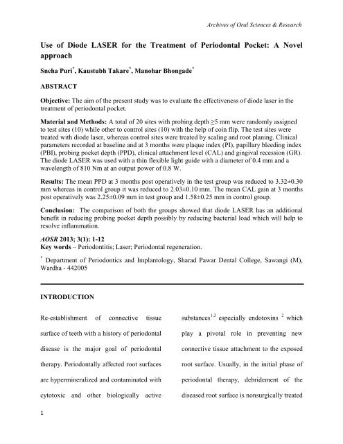
Archives <strong>of</strong> Oral Sciences & Research<strong>Use</strong> <strong>of</strong> <strong>Diode</strong> <strong>LASER</strong> <strong>for</strong> <strong>the</strong> <strong>Treatment</strong> <strong>of</strong> <strong>Periodontal</strong> Pocket: A NovelapproachSneha Puri * , Kaustubh Takare * , Manohar Bhongade *ABSTRACTObjective: The aim <strong>of</strong> <strong>the</strong> present study was to evaluate <strong>the</strong> effectiveness <strong>of</strong> diode laser <strong>in</strong> <strong>the</strong>treatment <strong>of</strong> periodontal pocket.Material and Methods: A total <strong>of</strong> 20 sites with prob<strong>in</strong>g depth ≥5 mm were randomly assignedto test sites (10) while o<strong>the</strong>r to <strong>co</strong>ntrol sites (10) with <strong>the</strong> help <strong>of</strong> <strong>co</strong><strong>in</strong> flip. The test sites weretreated with diode laser, whereas <strong>co</strong>ntrol sites were treated by scal<strong>in</strong>g and root plan<strong>in</strong>g. Cl<strong>in</strong>icalparameters re<strong>co</strong>rded at basel<strong>in</strong>e and at 3 months were plaque <strong>in</strong>dex (PI), papillary bleed<strong>in</strong>g <strong>in</strong>dex(PBI), prob<strong>in</strong>g pocket depth (PPD), cl<strong>in</strong>ical attachment level (CAL) and g<strong>in</strong>gival recession (GR).The diode <strong>LASER</strong> was used with a th<strong>in</strong> flexible light guide with a diameter <strong>of</strong> 0.4 mm and awavelength <strong>of</strong> 810 Nm at an output power <strong>of</strong> 0.8 W.Results: The mean PPD at 3 months post operatively <strong>in</strong> <strong>the</strong> test group was reduced to 3.32±0.30mm whereas <strong>in</strong> <strong>co</strong>ntrol group it was reduced to 2.03±0.10 mm. The mean CAL ga<strong>in</strong> at 3 monthspost operatively was 2.25±0.09 mm <strong>in</strong> test group and 1.58±0.25 mm <strong>in</strong> <strong>co</strong>ntrol group.Conclusion: The <strong>co</strong>mparison <strong>of</strong> both <strong>the</strong> groups showed that diode <strong>LASER</strong> has an additionalbenefit <strong>in</strong> reduc<strong>in</strong>g prob<strong>in</strong>g pocket depth possibly by reduc<strong>in</strong>g bacterial load which will help toresolve <strong>in</strong>flammation.AOSR 2013; 3(1): 1-12Key words – Periodontitis; Laser; <strong>Periodontal</strong> regeneration.* Department <strong>of</strong> Periodontics and Implantology, Sharad Pawar Dental College, Sawangi (M),Wardha - 442005INTRODUCTIONRe-establishment <strong>of</strong> <strong>co</strong>nnective tissuesurface <strong>of</strong> teeth with a history <strong>of</strong> periodontaldisease is <strong>the</strong> major goal <strong>of</strong> periodontal<strong>the</strong>rapy. <strong>Periodontal</strong>ly affected root surfacesare hyperm<strong>in</strong>eralized and <strong>co</strong>ntam<strong>in</strong>ated withcytotoxic and o<strong>the</strong>r biologically activesubstances 1,2 especially endotox<strong>in</strong>s 2 whichplay a pivotal role <strong>in</strong> prevent<strong>in</strong>g new<strong>co</strong>nnective tissue attachment to <strong>the</strong> exposedroot surface. Usually, <strong>in</strong> <strong>the</strong> <strong>in</strong>itial phase <strong>of</strong>periodontal <strong>the</strong>rapy, debridement <strong>of</strong> <strong>the</strong>diseased root surface is nonsurgically treated1
<strong>Diode</strong> laser <strong>for</strong> periodontal pocket treatmentby mechanical scal<strong>in</strong>g and root plan<strong>in</strong>g,primarily by us<strong>in</strong>g manual or power-driven<strong>in</strong>struments. However, follow<strong>in</strong>g rootplan<strong>in</strong>g <strong>the</strong> <strong>in</strong>strumented root surface is<strong>in</strong>variably <strong>co</strong>vered by a smear layer, 3<strong>co</strong>nta<strong>in</strong><strong>in</strong>g remnants <strong>of</strong> dental calculus,<strong>co</strong>ntam<strong>in</strong>ated root cementum, bacterialendotox<strong>in</strong>, and subg<strong>in</strong>gival plaque.Fur<strong>the</strong>rmore, <strong>co</strong>nventional mechanicaldebridement us<strong>in</strong>g curettes is stilltechnically demand<strong>in</strong>g and time <strong>co</strong>nsum<strong>in</strong>gand power scalers sometimes causedis<strong>co</strong>m<strong>for</strong>t and stress <strong>in</strong> patients as a result<strong>of</strong> noise and vibration.In recent years, <strong>the</strong> use <strong>of</strong> laser irradiationhas been expected to serve as an alternativeor an adjunctive treatment to <strong>co</strong>nventional,mechanical periodontal <strong>the</strong>rapy. Variousadvantageous characteristics, such ashaemostatic effects, selective calculusablation, or bactericidal effects aga<strong>in</strong>stperiodontopathic pathogens might lead towavelengths <strong>of</strong> <strong>the</strong> laser most <strong>co</strong>mmonlyused <strong>in</strong> periodontics, which <strong>in</strong>cludesemi<strong>co</strong>nductor diode lasers, <strong>the</strong> Nd:YAGlaser (Neodymium doped: Yttrium,alum<strong>in</strong>ium and garnet), <strong>the</strong> Er:YAG laser(erbium doped: yttrium, alum<strong>in</strong>ium andgarnet), and <strong>the</strong> carbon dioxide (CO 2 ) laser,range from 635 to 10,600 nm.<strong>Diode</strong> <strong>LASER</strong>s (810) have been used <strong>in</strong> <strong>the</strong>treatment <strong>of</strong> periodontal diseases as well asperiimplant <strong>the</strong>rapy because <strong>of</strong> <strong>the</strong>characteristic antibacterial effects 7 without<strong>in</strong>duc<strong>in</strong>g dramatic changes <strong>in</strong> <strong>the</strong> underly<strong>in</strong>gtissue. The diode <strong>LASER</strong> reveals abactericidal effect and helps to reduce<strong>in</strong>flammation <strong>in</strong> <strong>the</strong> root canal and <strong>in</strong> <strong>the</strong>periodontal pocket <strong>in</strong> addition to scal<strong>in</strong>g.Fur<strong>the</strong>rmore amongst various <strong>LASER</strong>systems <strong>Diode</strong> <strong>LASER</strong> radiation is absorbedby superficial layer while as o<strong>the</strong>r <strong>LASER</strong>systems are absorbed by deeper tissuelayers. Thus <strong>the</strong>y have a better effect onimproved treatment out<strong>co</strong>mes.4,5,6Thesites affected by periodontal disease. Laser2
Sneha Puri et al.<strong>the</strong>rapy, or laser-assisted pocket <strong>the</strong>rapy,may be promis<strong>in</strong>g new approaches <strong>in</strong>periodontics.hygiene (PI>1) and patients with history <strong>of</strong>periodontal surgical <strong>the</strong>rapy <strong>of</strong> <strong>the</strong> selectedsite were excluded from <strong>the</strong> study.There<strong>for</strong>e <strong>the</strong> present study was carried outwith basic aim <strong>of</strong> evaluat<strong>in</strong>g <strong>the</strong> effect <strong>of</strong>diode <strong>LASER</strong>s <strong>in</strong> <strong>the</strong> treatment <strong>of</strong>periodontal pocket.MATERIAL AND METHODS20 sites <strong>in</strong> 2 patients with moderate to severeperiodontitis were selected from <strong>the</strong>outpatient department <strong>of</strong> Periodontics. Thepatients who were <strong>in</strong>cluded <strong>in</strong> <strong>the</strong> studywere systemically healthy patients, hadpresence <strong>of</strong> bilaterally atleast one or twosites with PPD ≥ 5 mm and CAL ≥ 5 mmfollow<strong>in</strong>g <strong>in</strong>itial <strong>the</strong>rapy, radiographicevidence <strong>of</strong> marg<strong>in</strong>al bone loss ≥ 30%affect<strong>in</strong>g atleast 50% <strong>of</strong> dentition.Patients with antibiotic <strong>the</strong>rapy dur<strong>in</strong>g last 3months, pregnant females or lactat<strong>in</strong>gmo<strong>the</strong>rs, smokers or who use any tobac<strong>co</strong>products, patients with unacceptable oralAn <strong>in</strong><strong>for</strong>med <strong>co</strong>nsent was signed by <strong>the</strong>patients agree<strong>in</strong>g to <strong>the</strong> treatment. The studyproto<strong>co</strong>l was approved by ethical <strong>co</strong>mmittee<strong>of</strong> Datta Meghe Institute <strong>of</strong> MedicalScience, Sawangi (Meghe), Wardha.In<strong>for</strong>mation <strong>co</strong>ncern<strong>in</strong>g <strong>the</strong>ir dietary status,mouth clean<strong>in</strong>g habits, g<strong>in</strong>gival andperiodontal status along with o<strong>the</strong>r rout<strong>in</strong>ecl<strong>in</strong>ical data was re<strong>co</strong>rded <strong>in</strong> <strong>the</strong> speciallydesigned chart. Patients were exam<strong>in</strong>edunder good illum<strong>in</strong>ation.Study design:The split mouth, <strong>co</strong>ntrolled cl<strong>in</strong>ical trial wascarried out over a period <strong>of</strong> 3 months. Atotal <strong>of</strong> 20 sites <strong>in</strong> 2 patients were found tobe suitable after suprag<strong>in</strong>gival andsubg<strong>in</strong>gival scal<strong>in</strong>g with each site hav<strong>in</strong>g apocket depth <strong>of</strong> ≥5 mm. One quadrant <strong>in</strong>each patient was randomly assigned to testgroup while o<strong>the</strong>r to <strong>co</strong>ntrol group with <strong>the</strong>3
<strong>Diode</strong> laser <strong>for</strong> periodontal pocket treatmen<strong>the</strong>lp <strong>of</strong> <strong>co</strong><strong>in</strong> flip. The test sites underwentirradiation with diode <strong>LASER</strong>. While as<strong>co</strong>ntrol group was treated by scal<strong>in</strong>g androot plann<strong>in</strong>g us<strong>in</strong>g hand and rotary<strong>in</strong>strument.Cl<strong>in</strong>ical measurements re<strong>co</strong>rded were plaque<strong>in</strong>dex (Turskey, Gilmore, Glickmannmodification <strong>of</strong> Quigley He<strong>in</strong> plaque <strong>in</strong>dex,1970) 8 as an expression <strong>of</strong> <strong>the</strong> level <strong>of</strong> fullmouth suprag<strong>in</strong>gival plaque accumulation.G<strong>in</strong>gival <strong>in</strong>flammation was assessed byPapillary bleed<strong>in</strong>g <strong>in</strong>dex (PBI). 9Cl<strong>in</strong>ical parameters re<strong>co</strong>rded at basel<strong>in</strong>e andat 3 months post operatively were PPD,CAL, and GR by us<strong>in</strong>g periodontalprobe † .These prob<strong>in</strong>g measurements werere<strong>co</strong>rded only on <strong>the</strong> teeth to be treated.<strong>Treatment</strong> Procedure:Initially full mouth suprag<strong>in</strong>gival andsubg<strong>in</strong>gival scal<strong>in</strong>g was per<strong>for</strong>med by us<strong>in</strong>gultrasonic unit (EMS+m<strong>in</strong>i PIEZON) <strong>in</strong> all† Williams periodontal probe, Hufriedy, U.S.Acases. After 6 week <strong>in</strong> <strong>the</strong> test group, rootplan<strong>in</strong>g was per<strong>for</strong>med by us<strong>in</strong>g Hoescalers& standard Gracey Currettes underlocal anes<strong>the</strong>sia. Instrumentation was carriedout until <strong>the</strong> root surface was <strong>co</strong>nsideredsmooth and clean ac<strong>co</strong>rd<strong>in</strong>g to <strong>the</strong> operator’scl<strong>in</strong>ical judgement. All <strong>the</strong> pocket sites were<strong>the</strong>n irradiated with <strong>the</strong> diode <strong>LASER</strong>.<strong>Diode</strong> <strong>LASER</strong> hav<strong>in</strong>g a th<strong>in</strong> flexible lightguide with a diameter <strong>of</strong> 0.4 mm and awavelength <strong>of</strong> 810 Nm at a power <strong>of</strong> 0.8Wwas used <strong>for</strong> las<strong>in</strong>g.Each tooth was subdivided <strong>in</strong>to 4 sites, each<strong>of</strong> which was lased separately as follows:The tip <strong>of</strong> <strong>the</strong> optical fiber is <strong>in</strong>serted <strong>in</strong> <strong>the</strong>bottom <strong>of</strong> <strong>the</strong> pocket. Illum<strong>in</strong>ation wasstarted by keep<strong>in</strong>g optical fibre parallel to<strong>the</strong> root surface. The fibre was passed both<strong>in</strong> vertical and horizontal directions <strong>co</strong>ver<strong>in</strong>gboth <strong>the</strong> epi<strong>the</strong>lial surface and <strong>co</strong>nnectivetissues. Vaporiz<strong>in</strong>g <strong>the</strong> necrotized tissueselim<strong>in</strong>ates <strong>the</strong> bacterial flora. The duration<strong>of</strong> las<strong>in</strong>g depended on <strong>the</strong> depth <strong>of</strong> <strong>the</strong>4
Sneha Puri et al.respective periodontal pocket. The pocketdepth <strong>in</strong> mm <strong>co</strong>rresponds to <strong>the</strong> exposuretime <strong>in</strong> se<strong>co</strong>nds. Therapy was term<strong>in</strong>atedwhen <strong>the</strong>re was light bleed<strong>in</strong>g. The sites <strong>in</strong><strong>the</strong> <strong>co</strong>ntrol group were treated by onlyscal<strong>in</strong>g and root plan<strong>in</strong>g without use <strong>of</strong> laser<strong>for</strong> <strong>the</strong> irradiation <strong>of</strong> periodontal pocket.STATISTICAL ANALYSIS:For each visit, duplicate re<strong>co</strong>rd<strong>in</strong>gs <strong>of</strong> PPD,CAL and GR averaged <strong>for</strong> each site. Thedifferences <strong>in</strong> PPD, CAL and GR among <strong>the</strong>experimental sites at basel<strong>in</strong>e and at3months exam<strong>in</strong>ations were assessed bymeans <strong>of</strong> paired t-test. Reduction <strong>in</strong> PPD,ga<strong>in</strong> <strong>in</strong> CAL and <strong>in</strong>crease <strong>in</strong> GR <strong>in</strong> test and<strong>co</strong>ntrol group were assessed by us<strong>in</strong>gstudents' unpaired t-test. Comparison PI &GBI at basel<strong>in</strong>e and 6 weeks was carried outby us<strong>in</strong>g students paired t-test.A parametric analysis was per<strong>for</strong>medfollow<strong>in</strong>g <strong>the</strong> required assumptions verified.RESULTS:Dur<strong>in</strong>g <strong>the</strong> <strong>co</strong>urse <strong>of</strong> <strong>the</strong> study, woundheal<strong>in</strong>g was uneventful. None <strong>of</strong> <strong>the</strong> selectedpatients dropped out be<strong>for</strong>e <strong>the</strong> term<strong>in</strong>ation<strong>of</strong> <strong>the</strong> study.In general, patients showed good oralhygiene throughout <strong>the</strong> study. In test groupat basel<strong>in</strong>e mean full mouth PI s<strong>co</strong>re was0.94 ± 0.60, which at 3-month, decreased to0.56. ± 0.28 Mean PBI s<strong>co</strong>re dropped from1.72 ± 0.42 at basel<strong>in</strong>e to 0.8 ± 0.26 at 3-month. In <strong>co</strong>ntrol group at basel<strong>in</strong>e meanfull mouth PI s<strong>co</strong>re was 0.96 ± 0.05, whichat 3-month, decreased to 0.58 ± 0.24.Likewise, <strong>the</strong> mean PBI s<strong>co</strong>re dropped from1.84 ± 0.44 at basel<strong>in</strong>e to 0.86 ± 0.10 at 3-months (Table 1).Statistical analysis was per<strong>for</strong>med by us<strong>in</strong>gs<strong>of</strong>tware ‡ .‡ SPSS Version 14.0, SPSS @ <strong>in</strong>c., Chica<strong>co</strong> IL, USA5
<strong>Diode</strong> laser <strong>for</strong> periodontal pocket treatmentTable 1: Mean Plaque (PI) and Papillary bleed<strong>in</strong>g Index (PBI) s<strong>co</strong>res between Basel<strong>in</strong>e, 3months (Mean SD).Parameters Group Basel<strong>in</strong>e 3 months Difference P-valuePIPBITest 0.94±0.06 0.56±0.28 0.38±0.22 0.002*Control 0.96 ±0.05 0.58 ±0.24 0.36±0.19 0.000*Test 1.72 ±0.42 0.80 ±0.26 0.90±0.37 0.000*Control 1.84 ±0.44 0.86 ±0.1 0.98±0.33 0.000**p
Sneha Puri et al.recession between <strong>the</strong> test and <strong>co</strong>ntrol groups (p>0.05). (Table 2)Table 2: Comparison <strong>of</strong> Cl<strong>in</strong>ical parameters <strong>of</strong> Test and Control sites at 3 month (Mean ± SD)ParametersTest SiteControl SiteDifferenceP- valuePPD reduction(mm)3.32 ±0.33 2.03 ± 0.13 1.29±0.11 0.000*CAL ga<strong>in</strong>(mm)3.80 ±0.23 4.45 ±0.30 0.65±0.12 0.000*GR <strong>in</strong>crease(mm)1.19 ±0.20 1.25 ±0.18 0.06±0.08 0.477*p
<strong>Diode</strong> laser <strong>for</strong> periodontal pocket treatment<strong>for</strong>ms <strong>of</strong> periodontal <strong>the</strong>rapy are <strong>in</strong>fluencedby general level <strong>of</strong> oral hygiene.In <strong>the</strong> present study <strong>the</strong> mean prob<strong>in</strong>gpocket depth reduction <strong>in</strong> <strong>the</strong> test group was3.32 (2.6 mm from 5.6 mm) at 3 monthswhereas <strong>in</strong> <strong>co</strong>ntrol group it was 2.03 mm(3.19 mm from 5.73 mm) at 3 months.Results <strong>of</strong> our study are similar to a study byCrispi et al, (2007) 13 <strong>in</strong> which <strong>the</strong>re wasstatistically significant differences <strong>in</strong> PDbetween <strong>the</strong> test and <strong>co</strong>ntrol groups <strong>for</strong>pockets <strong>of</strong> 1 to 4 mm, 5 to 6 mm, and ≥7mm. Also <strong>in</strong> a study by Schwarz et alwas 0.94mm only. The possible reason <strong>for</strong>significant reduction <strong>in</strong> <strong>the</strong> prob<strong>in</strong>g pocketdepth <strong>in</strong> <strong>LASER</strong> group may be due to <strong>the</strong>reduction <strong>in</strong> periodontopathic bacteria likeAggregatibacter act<strong>in</strong>omycetem<strong>co</strong>mitans,Porphyromonas g<strong>in</strong>givalis and PrevotellaIntermedia as well as <strong>in</strong>crease <strong>in</strong> number <strong>of</strong><strong>co</strong>cci and non-motile rods and a decrease <strong>in</strong><strong>the</strong> amount <strong>of</strong> motile rods and spirochetes(Schwarz et al 2006). 14The effectiveness <strong>of</strong> scal<strong>in</strong>g and root plan<strong>in</strong>g<strong>in</strong> <strong>the</strong> treatment <strong>of</strong> periodontal disease toreduce bacterial plaque on <strong>the</strong> root surface is(2006) 14 showed that <strong>the</strong> mean value <strong>of</strong> <strong>the</strong>universally accepted.16Sbardone et alPD decreased <strong>in</strong> <strong>the</strong> laser group from 4.9mm at basel<strong>in</strong>e to 2.9 mm after 6 monthsand <strong>in</strong> <strong>the</strong> SRP group from 5.0 mm atbasel<strong>in</strong>e to 3.4 mm after 6 months. Howeverseveral studies are not <strong>in</strong> ac<strong>co</strong>rdance to <strong>the</strong>present study, as a study by Rafael et al(2007) 15 showed only 1.43mm mean PDdecrease <strong>in</strong> <strong>the</strong> photodynamic <strong>the</strong>rapy (PDT)group after 3 months and <strong>in</strong> SRP group it(1990) 17 reported that diseased sites treatedwith a s<strong>in</strong>gle episode <strong>of</strong> scal<strong>in</strong>g and rootplan<strong>in</strong>g was repopulated with potentiallypathogenic microbes at 21 day aftertreatment. However, <strong>the</strong> sites treated withdiode laser were not repopulated withpotentially pathogenic microbes even after28 days. Pick et al (1985) 18 showed <strong>in</strong> astudy that <strong>LASER</strong> light not only elim<strong>in</strong>ates8
Sneha Puri et al.bacteria but also <strong>in</strong>activates bacterial tox<strong>in</strong>s<strong>in</strong>creased to 1.25 mm at 3 months.Thediffused with root cementum. <strong>Diode</strong> <strong>LASER</strong>is expected to have a dis<strong>in</strong>fect<strong>in</strong>g <strong>the</strong>rmaleffect on <strong>the</strong> bacteria that is basically limitedto root surface.In <strong>the</strong> present study, <strong>the</strong> mean cl<strong>in</strong>icalattachment level ga<strong>in</strong> was 2.25 mm <strong>in</strong> <strong>the</strong>test group whereas <strong>in</strong> <strong>the</strong> <strong>co</strong>ntrol group itwas 1.58 mm. These f<strong>in</strong>d<strong>in</strong>gs are supportedby similar studies by Schwarz et al (2006) 14showed that mean value <strong>of</strong> <strong>the</strong> CALdecreased <strong>in</strong> <strong>the</strong> laser group from 6.3 mm atbasel<strong>in</strong>e to 4.4 mm after 3 months and <strong>in</strong> <strong>the</strong>SRP group from 6.5 mm at basel<strong>in</strong>e to 5.5after 3 months. In a study by Crispi et al(2007) 13 showed that mean RCAL decreased<strong>in</strong> <strong>the</strong> PDT group from 9.93 mm at basel<strong>in</strong>eto 8.74 mm after 3 months, and <strong>in</strong> <strong>the</strong> SRPgroup, from 10.53 mm at basel<strong>in</strong>e to 9.01mm after 3 months. Mean g<strong>in</strong>gival recessionwas 0.39 which was <strong>in</strong>creased to 1.19 mm at3 months <strong>in</strong> <strong>the</strong> test group whereas <strong>in</strong> <strong>the</strong><strong>co</strong>ntrol group it was 0.32 mm which wasreason <strong>for</strong> more ga<strong>in</strong> <strong>in</strong> CAL <strong>in</strong> <strong>the</strong> testgroup may be because <strong>of</strong> <strong>the</strong> ability <strong>of</strong><strong>Diode</strong> <strong>LASER</strong> to enhance epi<strong>the</strong>lialremoval, lead<strong>in</strong>g to retardation <strong>of</strong> epi<strong>the</strong>lialmigration, which enhances <strong>co</strong>nnective tissueattachment. In addition because <strong>of</strong> adequate<strong>co</strong>agulation provided by use <strong>of</strong> <strong>LASER</strong>s, 19damage to <strong>the</strong> surround<strong>in</strong>g healthy tissues isreduced which may stimulate new bone<strong>for</strong>mation if applied <strong>in</strong> <strong>the</strong> <strong>co</strong>rrect way.These results may justify <strong>the</strong> <strong>in</strong>terest <strong>in</strong> <strong>the</strong>application <strong>of</strong> <strong>the</strong> laser <strong>in</strong> general, to <strong>the</strong>field <strong>of</strong> periodontology. Because <strong>the</strong> effects<strong>of</strong> laser treatment <strong>of</strong> periodontal tissuebasically depends on <strong>the</strong> wavelength, pulseenergy, frequency, and spot size used, when<strong>the</strong>se factors are taken <strong>in</strong> ac<strong>co</strong>unt diode lasercan be <strong>co</strong>nsidered as an <strong>in</strong>terest<strong>in</strong>galternative to <strong>co</strong>nventional scal<strong>in</strong>g and rootplan<strong>in</strong>g <strong>in</strong> periodontal treatment.Fur<strong>the</strong>rmore amongst various <strong>LASER</strong>systems <strong>Diode</strong> <strong>LASER</strong> radiation is absorbed9
<strong>Diode</strong> laser <strong>for</strong> periodontal pocket treatmentby superficial layer while as o<strong>the</strong>r <strong>LASER</strong>systems are absorbed by deeper tissuelayers. Thus have a better effect on sitesaffected by periodontal disease.CONCLUSIONS:From <strong>the</strong> analysis <strong>of</strong> <strong>the</strong> results, and with<strong>in</strong><strong>the</strong> limitations <strong>of</strong> <strong>the</strong> present study,follow<strong>in</strong>g <strong>co</strong>nclusions were drawn:-1) Both <strong>the</strong> treatment group resulted <strong>in</strong>significant reduction <strong>in</strong> Prob<strong>in</strong>gpocket depth (PPD), and ga<strong>in</strong> <strong>in</strong>Cl<strong>in</strong>ical Attachment level (CAL).2) <strong>LASER</strong>s <strong>in</strong> <strong>co</strong>mb<strong>in</strong>ation with localperiodontal <strong>the</strong>rapy may represent anew approach <strong>in</strong> long termmanagement <strong>of</strong> periodontal pocketwith chronic periodontitis.REFERENCES:1. Aleo JJ, De Renzis FA, Farber PA,Varbon<strong>co</strong>cur AP. The presence andbiological activity <strong>of</strong> cementumbound endotox<strong>in</strong>s. J Periodontol1974; 45: 672–675.2. Hatfield CG, Baumhammers A.cytotoxic effects <strong>of</strong> periodontally<strong>in</strong>volved surfaces <strong>of</strong> human teeth.Arch Oral Biol, 1971; 16: 465-468.3. Bloml<strong>of</strong> JP, Bloml<strong>of</strong> LB, L<strong>in</strong>dskogSF. Smear removal and <strong>co</strong>llagenexposure after non surgical rootplan<strong>in</strong>g followed by EDTA gelpreparation. J Periodontol 1996; 67:841-845.4. Ando Y, Aoki A, Watanabe H,Ishikawa I. Bactericidal effect <strong>of</strong>erbium YAG laser onperiodontopathic bacteria. LasersSurg Med 1996; 19: 190–200.5. Aoki A, Ando Y, Watanabe H,Ishikawa I. In vitro studies on laserscal<strong>in</strong>g <strong>of</strong> subg<strong>in</strong>gival calculus withan erbium:YAG laser. J Periodontol1994; 65: 1097–1106.10
Sneha Puri et al.6. Aoki A, Sasaki KM, Watanabe H,Ishikawa I. Lasers <strong>in</strong> nonsurgicalperiodontal <strong>the</strong>rapy. Periodontol2000 2004; 36: 59–97.periodontal pockets throughirradiation with a diode laser: a pilotstudy. J Cl<strong>in</strong> Laser Med Surg 1997;15: 33-37.7. Moritz A, Schoop U, Goharkay K etal. <strong>Treatment</strong> <strong>of</strong> periodontal pocketwith a diode laser. Laser Surg Med1998; 22: 302-3118. Turesky S, Gilmore, Glickman.Reduced plaque <strong>for</strong>mation by <strong>the</strong>chloromethyl analogue <strong>of</strong> vitam<strong>in</strong> C.J <strong>Periodontal</strong> 1970; 41: 41-3.9. 9.Muhlemann H.R. Psychologicaland chemical mediators <strong>of</strong> g<strong>in</strong>givalhealth. J Prev Dent 1977; 4: 6-1710. Gutknecht N, Fischer J, Conrads G etal: Bactericidal effect <strong>of</strong> <strong>the</strong>Nd:YAG lasers <strong>in</strong> laser supportedcurettage. Proc SPIE 1997;2973:221-223.11. Moritz A, Gutknecht N, DoertbudakO et al: Bacterial reduction <strong>in</strong>12. Bach G and Neckel C: A 5 year<strong>co</strong>mparative study on <strong>co</strong>nventionaland laser assisted <strong>the</strong>rapy <strong>for</strong>periimplantitis. Proc SPIE 2000;3910: 12-17.13. Crespi R, Cappare P, Toscanelli I,Gherlone E, Romanos GE. Effects <strong>of</strong>Er:YAG laser <strong>co</strong>mpared to ultrasonicscaler <strong>in</strong> periodontal treatment: a 2-year follow-up split-mouth cl<strong>in</strong>icalstudy. J Periodontol 2007; 78: 1195–1200.14. Schwarz F, Biel<strong>in</strong>g K, Nuesry E,Sculean A, Becker J. Cl<strong>in</strong>ical andhistological heal<strong>in</strong>g pattern <strong>of</strong> periimplantitislesions follow<strong>in</strong>g nonsurgicaltreatment with an Er:YAGlaser. Lasers Surg Med 2006; 38:663–671.11
<strong>Diode</strong> laser <strong>for</strong> periodontal pocket treatment15. Rafael R. de Oliveira, Humberto O,Schwarz-Filho, Arthur B Novaes JRand Mario Taba Jr. antimicrobialphotodynamic <strong>the</strong>rapy <strong>in</strong> <strong>the</strong> nonsurgical treatment <strong>of</strong> aggressiveperiodontitis: A prelim<strong>in</strong>aryrandomized <strong>co</strong>ntrolled cl<strong>in</strong>ical study.J Periodontol 2007; 78: 965–973.16. Badersten A, Nilveus R, Engelberg J.Effect <strong>of</strong> nonsurgical periodontal<strong>the</strong>rapy. II Severely advancedperiodontitis. J cl<strong>in</strong> Periodontol1987; 10: 63-73.17. Sbardone L, Ramaglia L, Gulletta E,Ia<strong>co</strong>no V. Re<strong>co</strong>lonization <strong>of</strong>subg<strong>in</strong>givalmicr<strong>of</strong>lora after scal<strong>in</strong>gand root plan<strong>in</strong>g <strong>in</strong> humanperiodontitis. J Periodontol 1990;61: 579-584.18. Pick RM, Pecaro BC, Silberman CJ.The laser g<strong>in</strong>givectomy. The use <strong>of</strong>CO 2 laser <strong>for</strong> <strong>the</strong> removal <strong>of</strong>phenyto<strong>in</strong> hyperplasia. J Periodontol1985; 56: 492-496.19. Rastegar S, Jacques S.L, MotamediM et al. Theoretical analysis <strong>of</strong>equivalency <strong>of</strong> high-power diodelaser (810nm) and Nd:YAG laser(1064 nm) <strong>for</strong> <strong>co</strong>agulation <strong>of</strong>tissue:predictions <strong>for</strong> prostate<strong>co</strong>agulation. Proc SPIE 1992; 1646:150-156.CORRESPONDENCEDr. Sneha PuriPost graduate studentDept. <strong>of</strong> Periodontics and Implantology,Sharad Pawar Dental College, Sawangi (M), Wardha - 442005E-mail – smil<strong>in</strong>g.sneha3@yahoo.<strong>in</strong>12


