S aureus
S aureus
S aureus
- No tags were found...
You also want an ePaper? Increase the reach of your titles
YUMPU automatically turns print PDFs into web optimized ePapers that Google loves.
Presented byDr zena makkie abd-alkareem
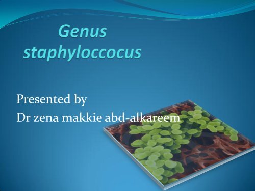
staphylococciIntroduction• The staphylococci are derived from Greek word STAPHYLO(bunch of grape)• gram-positive spherical cells, usually arranged in grape-likeirregular clusters• Some are members of the normal flora of the skin and mucousmembranes of humans; others are pathogenic• They grow readily on many types of media, ferment CHO andproducing pigments varying from white to deep yellow• The pathogenic staphylococci often hemolyze blood, coagulateplasma, and produce a variety of extracellular enzymes andtoxins.
Staphylococci has at least 35spps Can beclassified into two main types:Coagulase +ve staph.Staph.<strong>aureus</strong>(pyogenic).Coagulase –ve staph.Staph.albus(epidermedis).Staph.citreus(saprophyticus).
• The coagulase-negative staphylococci arenormal human flora and sometimes causeinfection, often associated with implantedappliances and devices, especially in veryyoung, old, and immunocompromisedpatients• . Approximately 75% of these infectionscaused by coagulase-negativestaphylococci are due to S epidermidis
Morphology & Identification• Typical Organisms• Staphylococci are spherical cells about 1 Mm in diameterarranged in irregular clusters• Single cocci, pairs, tetrads, and chains are also seen inliquid cultures. Young cocci stain strongly gram-positive;on aging, many cells become gram-negative. Staphylococciare nonmotile and do not form spores.• Micrococcus spp often resemble stap.they are foundfree_living in the enviroment and form regular pakets offour or eight cocci,micrococcus spp can cause disease inimmunocompramized pt.
culture• Staphylococci grow readily on most bacteriologic media underaerobic and facultative anaerobic conditions.• They grow most rapidly at 37 °C but form pigment best at roomtemperature (20–25 °C).• . Colonies on solid media are round, smooth, raised, glisteningand pigmented, pigments appear only on nutrient agar• . S <strong>aureus</strong> usually forms golden yellow colonies.• S epidermidis form white on primary isolation; manycolonies develop pigment only upon prolonged incubation• S saprophyticus forms lemon yellow colonies.• . No pigment is produced anaerobically or in broth• . Various degrees of hemolysis are produced by S <strong>aureus</strong> andoccasionally by other species• Some staph are anaerobic like staph.sacroleticus&staph.<strong>aureus</strong>sub spp anaerobus but they don't produce Catalase .
GROUTH CHARECTARESTICS• The staphylococci produce catalase, whichdifferentiatesthem from the streptococci.• . Staphylococci slowly ferment many carbohydrates,producing lactic acid but not gas.• . Proteolytic activity varies greatly from one strain toanother.• Staphylococci are relatively resistant to drying, heatthey withstand 50 °C for 30 minutes and 9% sodiumchloride
• are readily inhibited by certain chemicals, eg, 3%hexachlorophene.• Staphylococci are variably sensitive to manyantimicrobial drugs and resistance falls into severalclasses:• resistant to many penicillins like: penicillin G,ampicillin, ticarcillin, piperacillin, and similar drugsdue to B lactemase production under plasmidcontrol,The plasmids are transmitted bytransduction and perhaps also by conjugation,thereare 4 types of β_lactemase enz.
• Resistance to nafcillin , methicillin andoxacillinis independent of B-lactamase production its under chromosomalcontrol through special gene known as mec A gene.The resistance is related to the lack of certain PBPs in the organisms.• Resistance to vancomycin• Staphylococci are suseptable to vancomycin if the (MIC) is 2 g/mL; ofintermediate susceptibility if the MIC is 4–8 g/mL; and resistant if theMIC is 16 g/mL.• Strains of S <strong>aureus</strong> with intermediate susceptibility to vancomycin havebeen isolated in Japan. These are often known as vancomycinintermediateS <strong>aureus</strong>, or "VISA."• They generally have been isolated from patients with complexinfections who have received prolonged vancomycin therapy.• . The mechanism of is associated with increased cell wall synthesis andalterations in the cell wall and is not due to the van genes found inenterococci. resistance
• vancomycin-resistant S <strong>aureus</strong> (VRSA) strains were isolated frompatients in the United States. The isolates contained thevancomycin resistance gene vanA from enterococci.• . Plasmid-mediated resistance to tetracycline's, erythromycins,aminoglycosides, and other drugs is frequent in staphylococci A• Tolerance" implies that staphylococci are inhibited by a drug butnot killed by it—ie, there is great difference between minimalinhibitory and minimal lethal concentrations of an antimicrobialdrug. Patients with endocarditis caused by a tolerant S <strong>aureus</strong>may have a prolonged clinical course compared with patientswho have endocarditis caused by a fully susceptible S <strong>aureus</strong>.Tolerance can at times be attributed to lack of activation ofautolytic enzymes in the cell wall.
ANTIGENIC STRUCTURE OF STAPHYLOCOCCIcapsulesome S <strong>aureus</strong> strains have capsules, which inhibit phagocytosisby PMN cells unless specific antibodies are presentPeptidoglycana polysaccharide polymare provide a rigide exoskeleton butit is destroyed by strong acid or exposure to lysozyme,It elicits production of interleukin-1 (endogenouspyrogen) and opsonic antibodies by monocytes, and itcan be a chemoattractant for PMNTeichoic acidswhich are polymers of glycerol or ribitol phosphate, arelinked to thepeptidoglycan and can be antigenic.
continue• Protein A is a cell wall component of many S <strong>aureus</strong>strains that binds to the Fc portion of IgG molecules exceptIgG 3•Clummping factor(coagulase) most strain ofS.<strong>aureus</strong> have coagulase on the cell wallsurface coagulasebinds nonenzymatically to fibrinogen, yielding aggregationof the bacteia•ENZ&TOXINSStaph. can produce disease both through their ability tomultiply and spread widely in tissues and through theirproduction of many extracellular substances. Some of thesesubstances are enzymes; others are considered to be toxinslike:
Toxins:•Pore-forming toxins – create channels todisturb cellular homeostasis•Exotoxins–include several toxins:• Alpha toxin is apotent hemolysin• Beta toxin is atoxic to many kind of cells includingRBC those toxins &the other Gamma;Delta areantigeniticly distinct
• Exfoliative Toxins• These epidermolytic toxins of S <strong>aureus</strong> are two distinctproteins of the same molecular weight. Epidermolytic toxinA is a chromosomal gene product and is heat-stable (resistsboiling for 20 minutes). Epidermolytic toxin B is plasmidmediatedand heat-labile. The epidermolytic toxins yieldthe generalized desquamation of the staphylococcal scaldedskin syndrome by dissolving the mucopolysaccharidematrix of the epidermis. The toxins are superantigens• Leukocidin: This toxin of S <strong>aureus</strong> has two components. Itcan kill white blood cells of humans and rabbits. The twocomponents act synergistically on the white blood cellmembrane. This toxin is an important virulence factor incommunity associated methicillin resistant S <strong>aureus</strong>infections
•Toxic Shock Syndrome Toxin• Most S <strong>aureus</strong> strains isolated from patients with toxicshock syndrome produce a toxin called toxic shocksyndrome toxin-1 (TSST-1), which is the same asenterotoxin F. TSST-1 is the prototypical superantige.The toxin is associated with fever, shock, andmultisystem involvement, including a desquamativeskin rash. The gene for TSST-1 is found in about 20% ofS <strong>aureus</strong> isolates
•Enterotoxins• There are at least 6(A-F)soluble toxins produced byabout50% of S.<strong>aureus</strong> strains.Like TSST-1, the enterotoxinsare superantigens. The enterotoxins are heat-stable andresistant to the action of gut enzymes. An important causeof food poisoning, enterotoxins are produced when S<strong>aureus</strong> grows in carbohydrate and protein foods. Ingestionof 25 g of enterotoxin B results in vomiting and diarrhea.The emetic effect of enterotoxin is probably the result ofcentral nervous system stimulation (vomiting center) afterthe toxin acts on neural receptors in the gut
EnzymesCatalase all staph produce catalaseCoagulase S <strong>aureus</strong> produces coagulase, an enzyme-likeprotein that clots oxalated or citrated plasma. Coagulasemay deposit fibrin on the surface of staphylococci, perhapsaltering their ingestion by phagocytic cells .there are 2serological distinct types of coagulase free&clumpingfactor(acell bounding coagulase)Staphylokinase resulting in fibrenolysisLipaseHyalorodinase(spreading factor)B –lactemaseProteinases
PathogenesisStaphylococci, particularly S epidermidis, are members of the normal flora of the humanskin and respiratory and gastrointestinal tracts.Nasal carriage of S <strong>aureus</strong> occurs in 20–50% of humans. Staphylococci are also foundregularly on clothing, bed linens, and other fomites in human environmentSites of carrying staph. Are nose, skin fold,axilla,vagina&perinumThe pathogenic capacity of a given strain of S <strong>aureus</strong> is the combined effect of extracellularfactors and toxins together with the invasive properties of the strain.At one end of the disease spectrum is staphylococcal food poisoning, attributable solely tothe ingestion of preformed enterotoxin; at the other end are staphylococcal bacteremiaand disseminated abscesses in all organs.Pathogenic, invasive S <strong>aureus</strong> produces coagulase and tends to produce a yellow pigmentand to be hemolytic. Nonpathogenic, noninvasive staphylococci such as S epidermidis arecoagulase-negative and tend to be nonhemolytic. Such organisms rarely producesuppuration but may infect orthopedic or cardiovascular prostheses or cause disease inimmunosuppressed persons.S saprophyticus is typically nonpigmented, novobiocin-resistant, and nonhemolytic; itcauses urinary tract infections in young women
Diseases cauced by staph.<strong>aureus</strong>1.Superficial infections: charecterized by intense suppuration,localtissue necrosis &followed by local abscess formation.• Impetigo(pyoderma):Skin infection caused by staph <strong>aureus</strong>,but may also be caused by staphepidermidis&sterptococci.it characterized by formation of vesiclesfilled with serious or mucinus fluid.later on these lesions dry out withscabs form over the vesicles.• Folliculitis,furuncle(boil),styes:Hair follicles infection,if the infection is limited called folliculitis,but ifther is involvment that may required local drinage called furencles.whenthe infection involve the eyelid is called stye• Carbuncles:when multiple interconected abcesses involving hair follicles,sebaciousgland &the surrowending tissues.• postoperative staphylococcal wound infection or infection followingtrauma (chronic osteomyelitis subsequent to an open fracture,meningitis following skull fracture).
2.Deep infections:• Osteomylitis:Blood born infection.• Dissemenated infection&bacteremia• endocarditis, .• Meningitis,mltiple brain abscess.• pulmonary infection ,multiple lung infection.
• Toxin mediated infection1 Food poisoning• due to staphylococcal enterotoxin• short incubation period (1–8 hours);• violent nausea, vomiting, and diarrhea; and rapid convalescence.• Self limited disease.• There is no fever.• Recovery in 24 hr.2 Toxic shock syndrome• abrupt onset of high fever,• vomiting, diarrhea, myalgias,• a rash, and hypotension with cardiac and renal failure in the mostsevere cases.• It often occurs within 5 days after the onset of menses in youngwomen who use tampons• but it also occurs in children or in men with staphylococcal woundinfections.• The syndrome can recur.
• Scalded skin syndrom (sss)Caused by s.<strong>aureus</strong> that produce exfoleative toxinInvolve neonate and children less than 5 yblisters rupture dermis exposure skin peelsDisease caused by coagulase –ve staph.1. Community acquired s.saprophyticus =UTI in women,staph.saprophyticushas tissue tropism to UTI epth (uroepithelal &priurithral tissue)in greater%than other tissue2. Hospital acquired s.epidermedis =colonization of prosthesis (heart valve).3. There is staph.intermedus that isolate from several type of infection in dogs& man can get infection from direct contact with infected dogs through awound or bite cutanus or even internal or systemic infection4. Staph.Lutrae can cause infection in domestic animal like dogs &cats humangets infection through direct contact with infected animal like zooworkers,veteranien &farmers
Factors that predispose individual to seriousstaph.<strong>aureus</strong> infection1. Defect in opsonization by antibody2. Complement component deficiency like C3&C53. Defect in leukocyte chemotact factors either congenital like downsyndrome or acquired like D.M,R.A4. Defect in intracellular killing of bacteria following phagocytosis dueto inability to activate membrane bounding system result in absenceof peroxides &superoxidase killing enz from phacocytic vacuoleslike in chronic granulomotus diseases & in lymphoblastic lymphoma5. Skin injury like in eczima,burn &trauma6. Presence of foreign body7. Viral or bacterial infections as a complication8. Chronic underlying disease like malignancy, heart disease,alcohlicperson9. Prophylactic or therapeutic antimicrobial administration
Virulence factors of staph. epidermisThese bacteria produce a cell surface &extracellularmacromolecules that initiate subsequent adherenceto plastic surface of foreign body to form abiofilmmediated by capillary polysaccharide adhesioncalled(PS/A)also able to protect m.o fromcomplement mediated phagocytic killingOther attachment mechanism is there ability to interactwith different plasma or tissue component of thehost that constitute the initial step in tissuecolonization& establishment of infection in absenceof foreign body
Diagnostic Laboratory Tests• Specimens• Surface swab pus, blood, tracheal aspirate, or spinal fluid ,sputum&urine.• Smears• Typical staphylococci appear as gram positive cocci in clusters in Gram-stainedsmears of pus or sputum. It is not possible to distinguish saprophytic (Sepidermidis) from pathogenic (S <strong>aureus</strong>) organisms on smears.• Culture• planted on blood agar plates give rise to typical colonies in 18 hours at 37 °C,but hemolysis and pigment production may not occur until several days laterand are optimal at room temperature.• Specimens contaminated with a mixed flora can be cultured on mediacontaining 7.5% NaCl; the salt inhibits most other normal flora but not S<strong>aureus</strong>.• Mannitol salt agar or commercially available chromogenic media are used toscreen for nasal carriers of S <strong>aureus</strong> and patients with cystic fibrosis• Phenyl ethyl alcohol agar inhibit the growth of G-ve bacteria &allow thegrowth of G+ve bacteria
Testes used for identification ofstaphylococci• Catalase Test: this test used to diferentiate staph +ve fromstreptococcus&pneumococcus _ve• This test is used to detect the presence of cytochrome oxidaseenzymes. A drop of 3% hydrogen peroxide solution is placed on aslide, and a small amount of the bacterial growth is placed in thesolution. The formation of bubbles (the release of oxygen)indicates a positive test.• Coagulase Test: this test used to diferentiate S <strong>aureus</strong> from otherstaphylococci• The test is done by using citrated rabbit (or human) plasmadiluted 1:5 is mixed with an equal volume of broth culture orgrowth from colonies on agar and incubated at 37 °C. A tube ofplasma mixed with sterile broth is included as a control. If clotsform in 1–4 hours, the test is positive.
• Susceptibility Testing• Broth microdilution or disk diffusion susceptibility testing should bedone routinely on staphylococcal isolates from clinically significantinfections.• Resistance to penicillin G can be predicted by a positive test forB -lactamase; approximately 90% of S <strong>aureus</strong> produce -lactamase.• Resistance to nafcillin (and oxacillin and methicillin) occurs in about35% of S <strong>aureus</strong> and approximately 75% of S epidermidis isolates.• The gene coded for nafcillin resistance can be detectedby PCR.
Serological&typing tests• Serologic tests for diagnosis of S <strong>aureus</strong> infections havelittle practical value.• Antibody to tecioc acid can be detected in prolongeddeep infection(staph endocarditis)• Molecular typing techniques have been used todocument the spread of epidemic disease-producingclones of S <strong>aureus</strong>
Treatment1 Tetracyclines are used for long-term treatment.In (acne, furunculosis) occur most often in adolescents.Similar skin infections occur in patients receiving prolonged courses of corticosteroidsIn acne, lipases of staphylococci and corynebacteria liberate fatty acids from lipids andthus cause tissue irritation.2 (penicillin &cephalosporin).with drainage.Abscesses and other closed suppurating lesions.3 Acute hematogenous osteomyelitis responds well to antimicrobial drugs. In chronic andrecurrent osteomyelitis, surgical drainage and removal of dead bone is accompanied bylong-term administration of appropriate drugs.4 Bacteremia, endocarditis, pneumonia, and other severe infections due to S <strong>aureus</strong> requireprolonged intravenous therapy with a B-lactamase-resistant penicillin5 . Vancomycin is often reserved for use with nafcillin-resistant staphylococci.6 If the infection is found to be due to non--lactamase-producing S <strong>aureus</strong>, penicillin G isthe drug of choice, but only a small percentage of S <strong>aureus</strong> strains are susceptible topenicillin G.7 S epidermidis infections are difficult to cure because they occur in prosthetic deviceswhere the bacteria can hides them selves also staph epidermidis more resistant toantibiotics than staph <strong>aureus</strong>
Epidemiology &control• staph. Are colonizers of skin and mucosal surface they get entrancethrough trauma to skin &mucosal surface• Direct transmission from person to other through direct contact&fomites of infected persons• The mode of spread is imp in the hospitals where large proportions ofmedical staff and patients carry antibiotic resistant staph in their noseor skin• Application of topical anti septic to nasal or per anal carriage site• Pt of active staph lesions &carriers should be excluded fromICU,operating room&cancer chemotherapy areas• Rifampicin coupled with oral antistaph drug sometimes provide longterm supression&possibly cure of nasal carriage.
Thank you


