Transcranial Direct Current Stimulation during Sleep Improves ...
Transcranial Direct Current Stimulation during Sleep Improves ...
Transcranial Direct Current Stimulation during Sleep Improves ...
You also want an ePaper? Increase the reach of your titles
YUMPU automatically turns print PDFs into web optimized ePapers that Google loves.
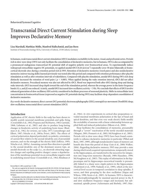
The Journal of Neuroscience, November 3, 2004 • 24(44):9985–9992 • 9985Behavioral/Systems/Cognitive<strong>Transcranial</strong> <strong>Direct</strong> <strong>Current</strong> <strong>Stimulation</strong> <strong>during</strong> <strong>Sleep</strong><strong>Improves</strong> Declarative MemoryLisa Marshall, Matthias Mölle, Manfred Hallschmid, and Jan BornInstitute of Neuroendocrinology H23a, University of Lübeck, 23538 Lübeck, GermanyInhumans,weaktranscranialdirectcurrentstimulation(tDCS)modulatesexcitabilityinthemotor,visual,andprefrontalcortex.Periodsrich in slow-wave sleep (SWS) not only facilitate the consolidation of declarative memories, but in humans, SWS is also accompanied bya pronounced endogenous transcortical DC potential shift of negative polarity over frontocortical areas. To experimentally inducewidespread extracellular negative DC potentials, we applied anodal tDCS (0.26 mA/cm 2 ) repeatedly (over 30 min) bilaterally at frontocorticalelectrode sites <strong>during</strong> a retention period rich in SWS. Retention of declarative memories (word pairs) and also nondeclarativememories (mirror tracing skills) learned previously was tested after this period and compared with retention performance after placebostimulation as well as after retention intervals of wakefulness. Compared with placebo stimulation, anodal tDCS <strong>during</strong> SWS-rich sleepdistinctly increased the retention of word pairs ( p 0.005). When applied <strong>during</strong> the wake retention interval, tDCS did not affectdeclarative memory. Procedural memory was also not affected by tDCS. Mood was improved both after tDCS <strong>during</strong> sleep and <strong>during</strong>wake intervals. tDCS increased sleep depth toward the end of the stimulation period, whereas the average power in the faster frequencybands (, , and ) was reduced. Acutely, anodal tDCS increased slow oscillatory activity 3 Hz. We conclude that effects of tDCS involveenhanced generation of slow oscillatory EEG activity considered to facilitate processes of neuronal plasticity. Shifts in extracellular ionicconcentration in frontocortical tissue (expressed as negative DC potentials <strong>during</strong> SWS) may facilitate sleep-dependent consolidation ofdeclarative memories.Key words: declarative memory; direct current (DC) potential; electroencephalography (EEG); nonrapid eye movement (NonREM) sleep;slow oscillation; transcranial direct current stimulation (tDCS)IntroductionApplication of DC electric fields to the scalp has been shown tomodify acutely neuronal membrane potentials and spike firing(Creutzfeldt et al., 1962; Purpura and McMurtry, 1965; Gartside,1968; Liebetanz et al., 2002) and also to produce long-lastingchanges in bioelectric activity of underlying brain tissue (Bindmanet al., 1962, for reviews, see Lolas, 1977; Lutzenberger andElbert, 1987; Nitsche et al., 2003a; Priori, 2003). The effects ofcortical polarization depend particularly on polarity, orientationof current flow relative to the axonal-dendritic axis of pyramidalneurons, and on the strength of the electric field (Jefferys, 1995;Nitsche et al., 2003a). Assuming current flow direction is parallelto the axonal-dendritic axis of the dominant neuron type, ananodal electrode on the scalp (i.e., an electrode attracting negativecharges and repelling positive charges) causes a depolarizationof the membrane potential via an extracellular negative sinkin underlying neural tissue (Creutzfeldt et al., 1962; Bindman etReceived March 5, 2004; accepted Sept. 21, 2004.This work was supported by the Deutsche Forschungsgemeinschaft (MA 2053, BO 854). We thank AlexanderKraepalis and Jochen Krimpove for acquiring data, Horst Koller and Mark Zelazny of the University of Lübeck,Electronics Department, for building the constant-current stimulator, as well as Anja Otterbein for help in preparingthe figures.Correspondence should be addressed to Lisa Marshall, Institute of Neuroendocrinology H23a, University of Lübeck,Ratzeburger Allee 160, 23538 Lübeck, Germany. E-mail: marshall@kfg.uni-luebeck.de.DOI:10.1523/JNEUROSCI.2725-04.2004Copyright © 2004 Society for Neuroscience 0270-6474/04/249985-08$15.00/0al., 1964). In vitro experiments in cortical slice preparations revealedmaximal membrane polarization at the tips of basal andapical dendrites, and that even very weak electric fields modifythe excitability of neurons with a linear relationship between appliedfield and induced polarization (Bikson et al., 2004).<strong>Sleep</strong> facilitates the consolidation of memories, presumablythrough a “covert” reactivation of the newly encoded materials(Maquet, 2001; Pennartz et al., 2002; McNaughton et al., 2003).Periods rich in slow-wave sleep (SWS) have been found consistentlyto enhance declarative memories in particular (Plihal andBorn, 1997, 1999), although nondeclarative types of memory canbenefit also from this period of sleep (Gais et al., 2000; Walker,2004). Regarding declarative memory, SWS may provide a state<strong>during</strong> which newly acquired representations, temporarily storedin the hippocampus, are transferred to the neocortex for integrationinto long-term memories (Buzsaki, 1989; Hasselmo, 1999;McNaughton et al., 2003). This replay of information in the hippocampusand its hippocampo–neocortical transfer is presumablylinked to a sharp wave–ripple pattern in the hippocampus(Kudrimoti et al., 1999; Nadasdy et al., 1999), which at the neocorticallevel occurs in temporal correlation to sleep spindles, waves, and their grouping by the slow oscillation (Siapas andWilson, 1998; Sirota et al., 2003). In humans, SWS is characterizedelectrophysiologically by maximal spectral power in the and slow oscillatory frequency ranges and increased, but submaximal,spindle power (Achermann and Borbely, 1997; Mar-
9986 • J. Neurosci., November 3, 2004 • 24(44):9985–9992 Marshall et al. • tDCS on Memory <strong>during</strong> <strong>Sleep</strong>shall et al., 2003). In addition, within the nonrapid eye movement(NonREM)–rapid eye movement (REM) sleep cycle, an endogenousDC potential shift, best visible over frontal cortical areas,reveals a pronounced negative-going DC potential shift peakingtypically at about the onset of SWS, and subsequently a high levelof DC potential is maintained that then decreases very graduallyin the remaining period of SWS. Notably, the DC potential shift<strong>during</strong> the passage into SWS was correlated in time with coefficientsas high as 0.9 to slow oscillatory activity, suggesting themechanisms generating these changes are associated (Marshall etal., 2003). An enhanced excitability characterizing the depolarizingup phase of slow oscillatory activity compared with the downphase and presumably also to the EEG activity <strong>during</strong> quiet wakingmay prove that this type of generated rhythm is particularlysusceptible to slow changes in exogenous or endogenous DC potentialshifts (Steriade et al., 2001; Bikson et al., 2004). The workinghypothesis for the present experiments is that <strong>during</strong> earlySWS-rich sleep, transcranial direct current stimulation (tDCS)affects declarative memory consolidation. We applied anodaltDCS <strong>during</strong> a period of sleep characterized by SWS-rich earlysleep and slow oscillatory activity as well as an enhanced negativelevel of the endogenous DC potential to induce, or rather potentiate,a widespread negative DC potential with a focus over frontocorticalareas. We aimed to test whether this anodal tDCS appliedrepeatedly enhances declarative memory consolidation.The effects of tDCS induced depolarization on slow oscillationactivity as a possible mediator of DC potential effects, as well ason other sleep-related EEG rhythms, were of interest. In addition,associated changes in sleep stages and sleep-specific hormonalactivity were monitored.Materials and MethodsSubjects, experimental design, and procedureThirty men with a mean age of 23.8 years (range, 19–28 years) who werenonsmokers and free of medication participated in these studies aftergiving informed written consent. Subjects with, or with a history of any ofthe following, were excluded: epilepsy, paroxysms, cognitive impairments,mental, hormonal, metabolic, or circulatory disorders, or sleepdisturbances. The experimental protocol was approved by the ethicscommittee of the University of Lübeck.Two experiments were conducted to assess the effect of anodal tDCSon memory, one <strong>during</strong> sleep (<strong>Sleep</strong> experiment) and the other <strong>during</strong>wakefulness (Wake experiment). In both experiments, subjects weretested in two conditions, a stimulation condition and a placebo condition,according to a double-blind cross-over design. The two sessions ofa subject were separated by an interval of at least 1 week. Time course ofthe <strong>Sleep</strong> experiment is schematized in Figure 1. Subjects (n 18) arrivedat the laboratory at 7:00 P.M. After preparation for tDCS, EEG recording,and blood sampling, subjects were tested on learning tasks for both declarativememory [paired associate learning (PAL)] and proceduralmemory [mirror tracing (MT)] between 9:30 P.M. and 10:30 P.M. Theorder of the tasks was randomized across subjects. In the <strong>Sleep</strong> experiment,subjects subsequently went to bed, and EEG and polysomnographicrecordings were started. tDCS began after the subject enteredSWS (i.e., after on-line scoring confirmed the presence of 30 sec of sleepstage 3 or 4). After the end of the first NonREM–REM sleep cycle, subjectswere awakened. At this time, they were usually in light NonREMsleep (stages 1 or 2) after the first REM sleep period. About 20 min afterawakening, recall on the memory tasks was tested. Because sleep is characterizedby prominent neuroendocrine regulation, blood was sampledfor determination of hormone concentration (norepinephrine, cortisol,growth hormone) before and after learning and recall testing, as well as afterlights off and every 30 min <strong>during</strong> the sleep interval. Before learning and afterrecall testing, psychometric tests [d2, Positive and Negative Affect Schedule(PANAS), Eigenschaftswoerterliste (EWL)] were given also to assess capabilitiesto concentrate and feelings of tiredness and mood.Figure1. Procedureofthe<strong>Sleep</strong>experiment.Timepointsoflearningandrecallofthememorytasks (PAL, MT), psychometric tests (d2, EWL, PANAS), tDCS, blood sampling (arrows),period of lights off (horizontal black bar), and sleep, represented by the schematized hypnogram,are indicated. W, Wake; 1–4, sleep stages 1–4;vertical black bar, REM sleep.The procedure of the Wake experiment (n 12) was the same as in the<strong>Sleep</strong> experiment, except that the period of sleep was replaced by a period ofwakefulness. During this wake period, subjects, seated in a reclining chair,were shown a video presentation (“Koyaanisqatsi” or “Powaqqatsi,” filmswith only instrumental accompaniment). tDCS was given 10 min after thebeginning of the presentation. Mood was also tested directly after tDCS. NoEEG was recorded in the Wake experiments.<strong>Transcranial</strong> DC stimulationFor transcranial DC stimulation, electrodes (8 mm diameter) were appliedbilaterally at frontolateral locations [F3 and F4 of the international10:20 system (Jasper, 1958)] and at the mastoids. Anodal tDCS (i.e.,positive polarity at both frontal sites) was applied intermittently (15 secon, 15 sec off; current density, 0.26 mA/cm 2 ) over a period of 30 min bya battery-driven constant-current stimulator. In the placebo control session,the electrodes were applied as in the stimulation sessions, but thestimulator remained off. <strong>Stimulation</strong> was not felt by the subjects.Memory tasks and psychometric testsPaired associate word lists. To assess declarative learning, a PAL task withcategory–instance word pairs similar to one used previously (Plihal andBorn, 1997) was used. A different word list was used for each of thesubject’s two experimental sessions. Each list consisted of 46 pairs ofGerman nouns adapted from a normative study. In addition to the 46word pairs, four dummy pairs of words at the beginning and end of eachlist served to buffer primacy and recency effects, respectively. Responsewords represented instances for the categories of the respective stimuluswords (e.g., train–track, bird–wing). To prevent serial learning, the sequenceof word-pair presentations within the lists was randomized betweenrepeated trials. In the learning condition, the list was displayed ona color monitor with a presentation rate of 0.20 sec and an interstimulusinterval of 100 msec. During learning, presentation of the list was followedby a task of cued recall. Here, only the 46 stimulus words of theword list appeared on the screen, in a different order than the foregoingpresentation. The subject had unlimited time to recall the appropriateresponse word and write it down. The number of correct responses wascalculated immediately. If a minimum of 60% correct responses was notobtained, word pairs were presented again in a newly randomized order,and cued recall was repeated. In the recall condition, after the retentioninterval of sleep or wakefulness, the 46 word pairs were again displayed ina newly randomized order. The subject was required to recall the appropriateresponse words and write them down.Mirror tracing. A task of procedural learning, with improved memoryperformance shown to depend on sleep <strong>during</strong> the second half of thenight but not on sleep after the first half alone, was conducted as a controlmemory condition (Plihal and Born, 1997). To assess procedural learning,subjects traced figures as fast and as accurately as possible while these
Marshall et al. • tDCS on Memory <strong>during</strong> <strong>Sleep</strong> J. Neurosci., November 3, 2004 • 24(44):9985–9992 • 9987figures and their hand movements were visible to them only through amirror. Two different sets with seven different figures were used. One setconsisted of a line-drawn five-pointed star, for practice, and six linedrawnhuman figures made up of 26–28 segments joined by 25–27 angles.In the second set, the straight segments were curved. The apparatuswas as described in detail by Plihal and Born (1997). Performance wasassessed by a light sensor of a tracing stylus that indicated whenever thestylus left the line to be traced. Subjects traced the figures with a stylusstarting and ending at the same point. An error consisted of moving thestylus off the line of the figure. Subjects first practiced with the star untila maximum of only six errors was made and continued with the linefigures. In the recall condition, subjects traced the same figures, startingwith the star to warm up. Performance measures were mean draw timeand mean error count, collapsed across the six line-drawn figures.d2-test, PANAS, EWL. On the d2-test of attention (Brickenkamp andZillmer, 2002), subjects are required to cross out specifically markedtarget letters in several sequels of signs. Total count of signs processedwithin 45 sec, and errors were calculated as an estimate of the capabilityto concentrate. The PANAS describes, by a five-point self rating, thesubject’s current mood on two dimensions: positive (enthusiastic, active,and attentive) and negative (irritability, nervousness, and fear) affect(Watson et al., 1998). The EWL (Janke and Debus, 1978) is an adjectivechecklist describing the subject’s mood on 15 dimensions (e.g., activated,tired, high spirits, irritable, excited, fearful).Polysomnographic and EEG recordings and analysesEEG (Fz, Cz, Pz, Oz, C3, C4, P3, P4, F7, F8, T3, T4, T5, T6) and verticaland horizontal electro-oculograms were recorded continuously by aDC/AC amplifier (Toennies DC/AC amplifier; amplification, 200 V/V;1–35 Hz; Jaeger GmbH and Co. KG, Würzburg, Germany). Analog DCEEG signals were digitized at 200 Hz per channel (CED 1401; CambridgeElectronics, Cambridge, UK).Three types of comparisons were performed between the conditions oftDCS and placebo control. First, sleep structure was compared betweenthe sessions based on standard polysomnographic criteria (Rechtschaffenand Kales, 1968). For the total sleep epoch as well as for a 45min interval beginning with the onset of tDCS (i.e., the first appearanceof SWS), every 30 sec epoch was scored as NonREM sleep stage 1, 2, 3, 4,or REM sleep. SWS was determined as the sum of sleep stages 3 and 4. Forthe placebo condition, sleep stages were determined for correspondingintervals. The time in minutes for each sleep stage, the total sleep time,and the percentage of sleep time in each stage with reference to total sleeptime were determined. Mean time spent in the different stages beginningwith the onset of stimulation and ending 15 min after termination of thestimulation interval was calculated and compared with respective intervalsof the control session. In a second analysis, average EEG power wascompared for the 30 min interval of DC stimulation and the correspondinginterval <strong>during</strong> the placebo condition for the following bands: (4–8Hz), 1 (8–10 Hz), 2 (10–12 Hz), spindle frequency (12–15 Hz), 1(15–20 Hz), and 2 (20–25 Hz). This analysis was run separately forperiods of SWS and stage 2 sleep. A third analysis concentrated on theimmediate effects of DC polarization. For this purpose, average powerspectra for all the above frequency bands were compared <strong>during</strong> the 60 15sec intervals of acute cortical polarization with that obtained for the 60intermittent 15 sec breaks in which the DC stimulation was discontinued.The time course of short-term effects across the 15 sec epochs wasalso assessed. As a result of on–off artifacts in the EEG induced by thestimulation, 13 sec rather than 15 sec intervals were analyzed. Powerspectra and corresponding bands were calculated using three overlappingor for time course analyses moving windows of 5 sec intervals (2048data points), resulting in a resolution of 0.098 Hz per bin. Only artifactfreeintervals were used. A five-point moving average was applied to theindividual data before averaging.HormonesFor blood sampling, a catheter was connected to a long, thin tube enablingblood collection from an adjacent room without disturbing thesubject. Standard HPLC with electrochemical detection was used forplasma norepinephrine detection [sensitivity, 35.64 pmol/l; interassaycoefficients of variation (CV), 6.1%]. Assays used for determination ofcortisol and growth hormone were an ES300 (sensitivity, 1.0 g/dl; intraassayCV, 6%; interassay CV, 4%; Boeringer Mannheim, Mannheim,Germany) and a RIA (sensitivity, 0.9 g/l; intraassay CV, 5%;interassay CV, 9%; Diagnostic Products Corporation, Bad Nauheim,Germany), respectively.Statistical analysesStatistical analyses relied in general on ANOVA with <strong>Stimulation</strong> (tDCS,placebo) as repeated-measures factor and mental state (<strong>Sleep</strong>, Wake) asgroup factor. When appropriate, a Greenhouse–Geisser correction fordegrees of freedom was used. A p value 0.05 was considered significant.Paired t tests were used for comparisons of time courses.ResultsMemory tasks and psychometric testsOn the PAL task, learning before sleep (<strong>Sleep</strong> experiment) andwakefulness (Wake experiment) was comparable for all conditions.The number of trials required until the criteria of 60%correct responses was obtained at immediate recall in the <strong>Sleep</strong>experiment was 1.37 0.09 and 1.33 0.09 for tDCS and placeboconditions, respectively. In the Wake experiment, 1.50 0.15 and 1.42 0.15 trials were needed to reach the learningcriteria ( p 0.6, for respective differences between stimulationconditions). The number of words recalled at the criterion trial<strong>during</strong> learning (shown in Table 1) also did not differ amongconditions ( p 0.2, for all comparisons). For assessing effects oftDCS on subsequent memory formation, recall performance afterthe retention trials was compared with the individual performanceat learning before (Fig. 2, Table 1). In the <strong>Sleep</strong> experiments,recall generally improved across the sleep retentioninterval, and this improvement was distinctly greater when tDCSwas applied than placebo stimulation (F (1,17) 10.44; p 0.005).In the Wake experiments, recall performance on average did notimprove but slightly decreased across the wake retention interval(F (1,28) 4.81; p 0.05, for the difference between <strong>Sleep</strong> andWake experiments). Moreover, there was no effect of tDCS onretention performance in the Wake experiment (F (1,11) 0.04;p 0.8) (Table 1).Table 1 also summarizes results of draw time and error counton the mirror tracing task. Performance at learning before theretention intervals was comparable between tDCS and placeboconditions of both experiments ( p 0.5), although subjects ofthe Wake experiment made more errors than subjects of the <strong>Sleep</strong>experiments (F (1,28) 7.48; p 0.01). Compared with placebo,tDCS did not affect memory for mirror tracing, as assessed by theimprovements in speed and accuracy at recall, neither in the <strong>Sleep</strong>experiments ( p 0.5 and p 0.8 for speed and accuracy, respectively)nor in the Wake experiments ( p 0.3 and p 0.5, respectively).Subjects of the Wake experiments showed a greaterreduction in error count across the retention interval than thoseof the <strong>Sleep</strong> experiments (F (1,28) 7.51; p 0.01), which mightbe a result of the generally higher error rate at learning in thesesubjects.The d2 attention test did not indicate differences in concentrationbetween tDCS and placebo conditions neither at learningbefore sleep (total count of processed signs, 511 11 vs 513 14;error count, 18 4 vs 18 4) nor at recall after sleep (total countof processed signs, 531 9 vs 516 12; error count, 15 3 vs16 4; p 0.2). There were also no differences in d2 performanceat learning and recall testing in the Wake experiments( p 0.3).The PANAS questionnaire indicated that positive affect decreasedgenerally across the retention interval. However, <strong>during</strong>
9988 • J. Neurosci., November 3, 2004 • 24(44):9985–9992 Marshall et al. • tDCS on Memory <strong>during</strong> <strong>Sleep</strong>Table 1. Mean SEM number of correctly recalled word pairs on the PAL task and speed and accuracy of performance on the MT task before (Learning) and after (Recall)the retention intervalLearning (mean SEM) Recall (mean SEM) Retention (mean SEM) Significance (p)PAL<strong>Sleep</strong>tDCS 33.0 1.4 35.7 1.4 2.7 0.6 p 0.005Placebo 33.6 1.5 34.5 1.5 0.9 0.8WaketDCS 36.2 1.5 36.3 1.2 0.0 1.0 p 0.8Placebo 34.0 1.4 33.7 1.4 0.3 0.5MT draw time (in milliseconds)<strong>Sleep</strong>tDCS 72.63 9.57 55.77 6.06 16.86 4.58 p 0.5Placebo 72.00 7.88 58.68 5.02 13.32 3.67WaketDCS 60.00 6.72 52.50 6.75 7.51 5.80 p 0.3Placebo 63.33 14.21 46.81 9.60 16.52 5.76MT error count<strong>Sleep</strong>tDCS 7.1 1.4 5.4 1.3 1.8 0.6 p 0.8Placebo 6.6 1.6 4.5 1.4 1.9 0.7WaketDCS 14.6 3.5 8.7 1.5 5.9 1.7 p 0.5Placebo 12.7 3.1 8.8 1.2 3.9 2.3Retentionisdefinedbythedifferenceinretrievalperformancebeforeandaftertheretentioninterval.Theretentionintervalwasfilledwith<strong>Sleep</strong>orwakefulness(Wake),<strong>during</strong>whicheithertDCSorplacebostimulationwasapplied.Therightcolumn indicates significant differences in retention, compared with the respective placebo condition. Corresponding performances <strong>during</strong> learning and recall did not differ significantly (see Results for details).Table 2. <strong>Sleep</strong> <strong>during</strong> the tDCS and placebo stimulation conditions of the <strong>Sleep</strong>experimenttDCS(mean SEM)Placebo(mean SEM)Awake % 6.0 2.5 5.7 1.3S1 % 9.3 1.7 9.6 2.5S2 % 46.9 4.4 48.9 3.6S3 % 18.4 2.1 16.6 1.6S4 % 16.6 4.1 17.5 4.2SWS % 35.0 4.2 34.1 4.4REM % 2.3 0.8 1.2 0.6Total time (in minutes) 96.1 4.9 88.6 3.7Latency toS2 (in minutes) 3.6 1.1 4.9 5.4SWS (in minutes) 25.4 6.3 22.9 3.7Percentage (%) of time spent in different sleep stages and latency to stage 2 sleep, SWS, and REM sleep (withreference to sleep onset) is shown. Total time defines the time from sleep onset until awakening. There were nosignificant differences between the conditions.S1–S4, <strong>Sleep</strong> stages 1–4.Figure 2. Memory performance on the PAL and MT tasks across retention periods of sleep(left) and wakefulness (right) <strong>during</strong> which either tDCS (hatched bar) or placebo stimulation(white bar) was applied. Recall after the retention interval is expressed as difference fromperformance at learning in number of words (for PAL) and in milliseconds for draw time (forMT).**p0.01,fordifferencesbetweentheeffectsoftDCSandplacebostimulation.Errorbarsrepresent SEM.tDCS, this decrease was smaller than in the placebo conditions(0.31 0.10 vs 0.60 0.11; p 0.05) regardless of sleep orwakefulness in the retention interval. Likewise, the EWL revealedthat after tDCS, subjects reported decreased feelings of “depression”(0.50 0.26), whereas in the placebo condition, suchfeelings increased across the retention intervals of sleep andwakefulness (0.37 0.32; p 0.05).Polysomnographic EEG recordings and sleep-associatedneuronal activityTable 2 summarizes the time spent asleep and in the differentsleep stages in the <strong>Sleep</strong> experiments for the tDCS and placeboconditions. There were no significant differences between the twoconditions, also when this analysis was restricted to a 45 mininterval beginning with the first appearance of SWS (i.e., withanodal stimulation in the tDCS condition). However, when thetime course for the mean sleep stage was determined (with sleepstage 1–4 given the values 1–4, respectively, and REM sleep thevalue 0) (Marshall et al., 1998), subjects toward the end of thetDCS stimulation and <strong>during</strong> the subsequent 15 min showeddeeper sleep than <strong>during</strong> the corresponding interval of the placebocondition, with this difference transiently reaching statisticalsignificance (Fig. 3). Power spectra determined separately forperiods of SWS and stage 2 sleep <strong>during</strong> the 30 min interval ofstimulation indicated that tDCS, compared with placebo, reducedpower in the lower frequency range (15–20 Hz) <strong>during</strong>
Marshall et al. • tDCS on Memory <strong>during</strong> <strong>Sleep</strong> J. Neurosci., November 3, 2004 • 24(44):9985–9992 • 9989Figure 3. Time course of mean sleep stage for tDCS (solid line) and placebo (dotted line)conditions of the <strong>Sleep</strong> experiment. Average sleep stages were determined by associating valuesof 1, 2, 3, and 4 to sleep stages 1–4 and 0 and 1 to REM sleep and wakefulness, respectively.Significant differences between the time courses are indicated at the bottom. The grayarea represents the stimulation interval.periods of stage 2 sleep. The effect was most pronounced at central(C3, C4) and parietal (P3, P4) electrode sites (Fig. 4). Duringperiods of SWS, tDCS suppressed frequencies around the andlower range (4–10 Hz) (Fig. 4). During the stimulation interval,visual spindle counts per sec in the tDCS versus placebo conditionwere 0.11 0.01 versus 0.13 0.01 ( p 0.05).The comparison of 15 sec epochs of acute anodal polarizationwith the intermittent epochs when stimulation was discontinuedindicated most consistent differences for the slow oscillatory and frequencies 3 Hz (Fig. 5). During acute anodal stimulation,power in this low frequency range was increased over the frontalcortex, most consistently 2 Hz, compared with intervals of discontinuedstimulation. At parietal sites, anodal stimulationacutely increased slow oscillatory activity 1 Hz (Fig. 5). Comparisonsof the time course of short-term effects across the 15 secepochs of acute anodal polarization versus intermittent epochsdid not yield consistent effects.HormonesAverage plasma levels of norepinephrine, cortisol, and growthhormone were not affected by tDCS (compared with placebo forboth sleep and wake experiments; p 0.4 for cortisol and growthhormone; p 0.1 for norepinephrine).DiscussionOur study examined the influence of anodaltDCS, inducing extracellular potentialsof negative polarity in underlying tissue,on processes of declarative memoryformation known to be enhanced <strong>during</strong>periods rich in SWS (Plihal and Born,1997, 1999). Results indicate that tDCS repeatedlyapplied <strong>during</strong> deep NonREMsleep improved declarative memory retention,whereas performance was unaffected<strong>during</strong> wakefulness. Retention of proceduralmemories, in contrast, was not affected bytDCS but was also not enhanced by sleep.Electrophysiological modification of thecortex by weak anodal polarization <strong>during</strong>sleep consisted of an acute increase in slowoscillatory activity 3 Hz, accompanied bydiminished power in the faster , lower ,and lower EEG frequency bands across the30 min polarization period. The shift towardenhanced slow oscillatory activity <strong>during</strong> theperiod of tDCS expressed itself also as an increasein the depth of average sleep stage,which per se represents a mere descriptivemeasure that cannot account for enhancedretention performance. Duration of thestage SWS was not enhanced by tDCS. Finally,there were signs of improved moodafter tDCS in the <strong>Sleep</strong> and also in the Wakeexperiments, a finding that may have someimplications for treatment of mooddisorders.Figure4. AverageEEGpowerforperiodsofstage2sleepandSWS<strong>during</strong>the30minintervaloftDCS(hatchedbars)andacorrespondinginterval<strong>during</strong>theplacebocondition(whitebars)ofthe<strong>Sleep</strong>experiments.(4–8Hz),lower(8–10Hz),andlower(15–20Hz)bandsareaveragedforfrontal(F7,Fz,F8),central(C3,Cz,C4),andparietal(P3,Pz,P4)electrodelocations.**p0.01;*p0.05; t p0.1,fordifferencesbetweentDCSandplacebo(stage2sleep,n14;SWS,n16).ErrorbarsrepresentSEM.Effects on memoryThe central effect of this study was the improvementin declarative memory forword pairs after tDCS <strong>during</strong> sleep. Thisimprovement is remarkable because it was
Marshall et al. • tDCS on Memory <strong>during</strong> <strong>Sleep</strong> J. Neurosci., November 3, 2004 • 24(44):9985–9992 • 9991were selectively observed <strong>during</strong> the early NonREM sleep periodand not <strong>during</strong> wakefulness. A hallmark of electrophysiologicalactivity <strong>during</strong> this state is the slow oscillatory activity. Slow oscillatoryactivity exerts a grouping influence on faster EEG frequenciessuch that the appearance of these frequencies becomesrestricted to the depolarizing up phase of these oscillations. Accordingly,the development of slow oscillations accompanyingthe deepening of sleep is typically found to coincide with thedecrease of faster frequencies under natural conditions (Marshallet al., 2003). The examination of cellular processes occurring<strong>during</strong> the synchronized depolarization of the slow oscillation upphase indicate that the cortical network possesses the intrinsicability to generate persistent activity (McCormick et al., 2003).This persistent activity of the up phase of slow oscillations in thesleeping brain is attributed to the recurrent corticocortical excitatoryactivities alone, compared with the wake state in which theadditional influence of neuromodulatory systems is required formaintaining the state-specific neocortical activity. Moreover, thisdepolarizing phase has been considered to set the stage for processesof neocortical plasticity, in which neocortical networksbecome particularly sensitive to afferent inputs resulting fromreactivation of acutely acquired memory traces as stored in hippocampalregions (Buzsaki, 1989; Steriade et al., 2001; Huber etal., 2004). Integration of these representations into neocorticalnetworks could be a mechanism underlying retention of declarativememories. In this way, the acute enhancement of activity inslow oscillatory bands in the present study by tDCS indeed supportsthe concept that tDCS enhances retention performance byfacilitating the slow oscillatory corticocortical network activity.The enhancement of slow oscillatory power <strong>during</strong> acute anodalpolarization also corroborates the concept that endogenous negativeDC potentials arising at the transition into SWS and accompanyingassociated shifts in extracellular ionic concentration playa supportive role in the generation of slow oscillatory activity. Afactor adding to the facilitation of slow oscillations <strong>during</strong> tDCScould be our 15 sec on–15 sec off stimulation protocol (McCormicket al., 2003; Shu et al., 2003).At the synaptic level, acute influences of anodal polarizationpossibly reflect the facilitation of specific cationic currents(Amzica and Steriade, 2000; Bazhenov et al., 2002). The generationof and slow oscillations relies particularly on Ca 2 -mediated K currents and a persistent Na current, with thelatter proposed to reexcite the depolarizing phase of the slowoscillation (Buzsaki et al., 1988; Steriade et al., 1991, 2001;Timofeev et al., 2001). Depletion in extracellular Ca 2 concentrationcoincides with the depolarizing phase of the slow oscillation(Massimini and Amzica, 2001; Amzica et al., 2002). Interestingly,pharmacological blocking of Na and Ca 2 channels hasbeen consistently found to suppress effects of anodal stimulationin humans (Liebetanz et al., 2002; Nitsche et al., 2003b).Spindle activity triggered by the depolarizing phase of slowoscillations has been considered another sign of processes thatenhance plasticity within neocortical networks via increasedCa 2 flow into pyramidal cells (Sejnowski and Destexhe, 2000;Steriade and Timofeev, 2003). The failure to see here, in conjunctionwith enhanced slow oscillatory power, increased spindlepower <strong>during</strong> anodal tDCS is difficult to interpret within this lineof reasoning. However, once SWS has been established, slow oscillatoryactivity might primarily exert a grouping influence onthe occurrences of spindle activity without necessarily changingaverage power in this frequency band. Alternatively, the decrementin lower power (15–20 Hz) <strong>during</strong> stage 2 sleep in thetDCS condition in this context could be even taken to infer adecrease in spindle activity overlapping with this frequencyrange. In fact, suppressed spindle counts <strong>during</strong> the tDCS intervalsuggest spindle activity in the present study was not a mediatorfor the enhanced declarative memory retention (Gais et al.,2002).Together, our data show that anodal tDCS over frontocorticalareas repeatedly applied <strong>during</strong> a period of SWS-rich early sleepimproves declarative memory consolidation. The effect of tDCSmight involve slow oscillatory activity, which has been consideredto favor plastic processes in neocortical networks and whichis acutely enhanced by anodal polarization, presumably as a consequenceof a global increase in excitability of the underlyingcortex.ReferencesAchermann P, Borbely AA (1997) Low-frequency ( 1 Hz) oscillations inthe human sleep electroencephalogram. Neuroscience 81:213–222.Amzica F, Steriade M (2000) Neuronal and glial membrane potentials <strong>during</strong>sleep and paroxysmal oscillations in the neocortex. J Neurosci20:6648–6665.Amzica F, Massimini M, Manfridi A (2002) Spatial buffering <strong>during</strong> slowand paroxysmal sleep oscillations in cortical networks of glial cells in vivo.J Neurosci 22:1042–1053.Bazhenov M, Timofeev I, Steriade M, Sejnowski TJ (2002) Model ofthalamocortical slow-wave sleep oscillations and transitions to activatedstates. J Neurosci 22:8691–8704.Bikson M, Inoue M, Akiyama H, Deans JK, Fox JE, Miyakawa H, Jefferys JG(2004) Effects of uniform extracellular DC electric fields on excitabilityin rat hippocampal slices in vitro. J Physiol (Lond) 557:175–190.Bindman LJ, Lippold OC, Redfearn JW (1962) Long-lasting changes in thelevel of the electrical activity of the cerebral cortex produced by polarizingcurrents. Nature 196:584–585.Bindman LJ, Lippold OC, Redfearn JW (1964) The action of brief polarizingcurrents on the cerebral cortex of the rat (i) <strong>during</strong> current flow and (ii) in theproduction of long-lasting after-effects. J Physiol (Lond) 172:369–382.Blaney PH (1986) Affect and memory: a review. Psychol Bull 99:229–246.Born J, Gais S (2003) Roles of early and late nocturnal sleep for the consolidationof human memories. In: <strong>Sleep</strong> and brain plasticity (Maquet P,Smith C, Stickgold R, eds), pp 65–85. New York: Oxford UP.Bradshaw KD, Emptage NJ, Bliss TV (2003) A role for dendritic proteinsynthesis in hippocampal late LTP. Eur J Neurosci 18:3150–3152.Brickenkamp R, Zillmer E (2002) d2 test of attention. Göttingen, Germany:Hogrefe and Huber.Buzsáki G (1989) Two-stage model of memory trace formation: a role for“noisy” brain states. Neuroscience 31:551–570.Buzsáki G, Bickford RG, Ponomareff G, Thal LJ, Mandel R, Gage FH (1988)Nucleus basalis and thalamic control of neocortical activity in the freelymoving rat. J Neurosci 8:4007–4026.Cauller LJ, Connors BW (1994) Synaptic physiology of horizontal afferentsto layer I in slices of rat SI neocortex. J Neurosci 14:751–762.Creutzfeldt OD, Fromm GH, Kapp H (1962) Influence of transcortical dccurrents on cortical neuronal activity. Exp Neurol 5:436–452.Ekstrand BR, Barrett TR, West JN, Maier WG (1977) The effect of sleep onhuman long-term memory. In: Neurobiology of sleep and memory(Drucker-Colin RR, McGaugh JL, eds), pp 419–438. New York:Academic.Felleman DJ, Van Essen DC (1991) Distributed hierarchical processing inthe primate cerebral cortex. Cereb Cortex 1:1–47.Gais S, Born J (2004) Low acetylcholine <strong>during</strong> slow-wave sleep is critical fordeclarative memory consolidation. Proc Natl Acad Sci USA 101:2140–2144.Gais S, Plihal W, Wagner U, Born J (2000) Early sleep triggers memory forearly visual discrimination skills. Nat Neurosci 3:1335–1339.Gais S, Mölle M, Helms K, Born J (2002) Learning-dependent increases insleep spindle density. J Neurosci 22:6830–6834.Gartside IB (1968) Mechanisms of sustained increases of firing rate of neuronsin the rat cerebral cortex after polarization: reverberating circuits ormodification of synaptic conductance? Nature 220:382–383.Hasselmo ME (1999) Neuromodulation: acetylcholine and memory consolidation.Trends Cogn Sci 3:351–359.Huber R, Ghilardi MF, Massimini M, Tononi G (2004) Local sleep andlearning. Nature 430:78–81.
9992 • J. Neurosci., November 3, 2004 • 24(44):9985–9992 Marshall et al. • tDCS on Memory <strong>during</strong> <strong>Sleep</strong>Islam N, Moriwaki A, Hattori Y, Hori Y (1994) Anodal polarization inducesprotein kinase C gamma (PKC gamma)-like immunoreactivity in the ratcerebral cortex. Neurosci Res 21:169–172.Islam N, Moriwaki A, Hori Y (1995) Co-localization of c-fos protein andprotein kinase C gamma in the rat brain following anodal polarization.Indian J Physiol Pharmacol 39:209–215.Janke W, Debus G (1978) Die Eigenschaftswörterliste EWL. Eine mehrdimensionaleMethode zur Beschreibung von Aspekten des Befindens. Göttingen,Germany: Hogrefe.Jasper HH (1958) The ten-twenty electrode system of the International Federation.Electroencephalogr Clin Neurophysiol 10:371–375.Jefferys JG (1995) Nonsynaptic modulation of neuronal activity in thebrain: electric currents and extracellular ions. Physiol Rev 75:689–723.Kim HG, Connors BW (1993) Apical dendrites of the neocortex: correlationbetween sodium- and calcium-dependent spiking and pyramidal cellmorphology. J Neurosci 13:5301–5311.Kudrimoti HS, Barnes CA, McNaughton BL (1999) Reactivation of hippocampalcell assemblies: effects of behavioral state, experience, and EEGdynamics. J Neurosci 19:4090–4101.Liebetanz D, Nitsche MA, Tergau F, Paulus W (2002) Pharmacological approachto the mechanisms of transcranial DC–stimulation-induced aftereffectsof human motor cortex excitability. Brain 125:2238–2247.Lolas F (1977) Brain polarization: behavioral and therapeutic effects. BiolPsychiatry 12:37–47.Lutzenberger W, Elbert T (1987) Assessment of effects of weak electric currentson brain and behavior. In: Safety assessment of NMR clinical equipment.(Schmidt KH, ed), pp 36–45. Stuttgart, Germany: Thieme.Maquet P (2001) The role of sleep in learning and memory. Science294:1052.Marshall L, Mölle M, Fehm HL, Born J (1998) Scalp recorded direct currentbrain potentials <strong>during</strong> human sleep. Eur J Neurosci 10:1167–1178.Marshall L, Mölle M, Born J (2003) Spindle and slow wave rhythms at slowwave sleep transitions are linked to strong shifts in the cortical directcurrent potential. Neuroscience 121:1047–1053.Massimini M, Amzica F (2001) Extracellular calcium fluctuations and intracellularpotentials in the cortex <strong>during</strong> the slow sleep oscillation. J Neurophysiol85:1346–1350.McCormick DA, Shu Y, Hasenstaub A, Sanchez-Vives M, Badoual M, Bal T(2003) Persistent cortical activity: mechanisms of generation and effectson neuronal excitability. Cereb Cortex 13:1219–1231.McNaughton BL, Barnes CA, Battaglia FP, Bower MR, Cowen SL, EkstromAD, Gerrard JL, Hoffman FP, Houston FP, Karten Y, Lipa P, PennartzCMA, Sutherland GR (2003) Off-line reprocessing of recent memoryand its role in memory consolidation: a progress report. In: <strong>Sleep</strong> andbrain plasticity (Maquet P, Smith C, Stickgold R, eds), pp 225–246. NewYork: Oxford UP.Mednick S, Nakayama K, Stickgold R (2003) <strong>Sleep</strong>-dependent learning: anap is as good as a night. Nat Neurosci 6:697–698.Nadasdy Z, Hirase H, Czurko A, Csicsvari J, Buzsáki G (1999) Replay andtime compression of recurring spike sequences in the hippocampus.J Neurosci 19:9497–9507.Nitsche MA, Paulus W (2001) Sustained excitability elevations induced bytranscranial DC motor cortex stimulation in humans. Neurology57:1899–1901.Nitsche MA, Liebetanz D, Antal A, Lang N, Tergau F, Paulus W (2003a)Modulation of cortical excitability by weak direct current stimulation–technical, safety and functional aspects. Suppl Clin Neurophysiol56:255–276.Nitsche MA, Fricke K, Henschke U, Schlitterlau A, Liebetanz D, Lang N,Henning S, Tergau F, Paulus W (2003b) Pharmacological modulationof cortical excitability shifts induced by transcranial DC stimulation inhumans. J Physiol (Lond) 553:293–301.Pandya DN, Yeterian EH (1985) Architecture and connections of corticalassociation areas. In: Cerebral cortex, Vol 4 (Peters A, Jones EG, eds), pp3–61. New York: Plenum.Pennartz CM, Uylings HB, Barnes CA, McNaughton BL (2002) Memoryreactivation and consolidation <strong>during</strong> sleep: from cellular mechanisms tohuman performance. Prog Brain Res 138:143–166.Plihal W, Born J (1997) Effects of early and late nocturnal sleep on declarativeand procedural memory. J Cogn Neurosci 9:534–547.Plihal W, Born J (1999) Effects of early and late nocturnal sleep on primingand spatial memory. Psychophysiology 36:571–582.Priori A (2003) Brain polarization in humans: a reappraisal of an old tool forprolonged non-invasive modulation of brain excitability. Clin Neurophysiol114:589–595.Purpura DP, McMurtry JG (1965) Intracellular activities and evoked potentialchanges <strong>during</strong> polarization of motor cortex. J Neurophysiol 28:166–185.Rechtschaffen A, Kales A (1968) A manual of standardized terminology,techniques and scoring system for sleep stages of human subjects. NationalInstitutes of Health Publication 204. Washington, DC: UnitedStates Government Printing Office.Rockland KS, Pandya DN (1979) Laminar origins and terminations of corticalconnections of the occipital lobe in the rhesus monkey. Brain Res179:3–20.Roland PE (2002) Dynamic depolarization fields in the cerebral cortex.Trends Neurosci 25:183–190.Schiller J, Schiller Y, Stuart G, Sakmann B (1997) Calcium action potentialsrestricted to distal apical dendrites of rat neocortical pyramidal neurons.J Physiol 505:605–616.Sejnowski TJ, Destexhe A (2000) Why do we sleep? Brain Res 886:208–223.Shepherd GM, Brayton RK, Miller JP, Segev I, Rinzel J, Rall W (1985) Signalenhancement in distal cortical dendrites by means of interactions betweenactive dendritic spines. Proc Natl Acad Sci USA 82:2192–2195.Shu Y, Hasenstaub A, McCormick DA (2003) Turning on and off recurrentbalanced cortical activity. Nature 423:288–293.Siapas AG, Wilson MA (1998) Coordinated interactions between hippocampalripples and cortical spindles <strong>during</strong> slow-wave sleep. Neuron21:1123–1128.Singer JA, Salovey P (1988) Mood and memory: evaluating the networktheory of affect. Clin Psychol Rev 8:211–251.Sirota A, Csicsvari J, Buhl D, Buzsáki G (2003) Communication betweenneocortex and hippocampus <strong>during</strong> sleep in rodents. Proc Natl Acad SciUSA 100:2065–2069.Smith C (2001) <strong>Sleep</strong> states and memory processes in humans: proceduralversus declarative memory systems. <strong>Sleep</strong> Med Rev 5:491–506.Sourdet V, Debanne D (1999) The role of dendritic filtering in associativelong-term synaptic plasticity. Learn Mem 6:422–447.Steriade M, Timofeev I (2003) Neuronal plasticity in thalamocortical networks<strong>during</strong> sleep and waking oscillations. Neuron 37:563–576.Steriade M, Dossi RC, Nuñez A (1991) Network modulation of a slow intrinsicoscillation of cat thalamocortical neurons implicated in sleep deltawaves: cortically induced synchronization and brainstem cholinergic suppression.J Neurosci 11:3200–3217.Steriade M, Timofeev I, Grenier F (2001) Natural waking and sleep states: aview from inside neocortical neurons. J Neurophysiol 85:1969–1985.Timofeev I, Grenier F, Steriade M (2001) Disfacilitation and active inhibitionin the neocortex <strong>during</strong> the natural sleep-wake cycle: an intracellularstudy. Proc Natl Acad Sci USA 98:1924–1929.Walker MP (2004) A refined model of sleep and the time course of memory.Behav Brain Sci, in press.Watson D, Clark LA, Tellegen A (1998) Development and validation of briefmeasures of positive and negative affect: the PANAS scales. J Pers SocPsychol 54:1063–1070.Wong-Riley M (1978) Reciprocal connections between striate and prestriatecortex in squirrel monkey as demonstrated by combined peroxidasehistochemistry and autoradiography. Brain Res 147:159–164.Zeki S, Shipp S (1988) The functional logic of cortical connections. Nature335:311–317.


