Aberrations and visual performance following standard laser vision ...
Aberrations and visual performance following standard laser vision ...
Aberrations and visual performance following standard laser vision ...

You also want an ePaper? Increase the reach of your titles
YUMPU automatically turns print PDFs into web optimized ePapers that Google loves.
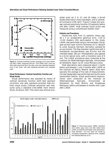
<strong>Aberrations</strong> <strong>and</strong> Visual Performance Following St<strong>and</strong>ard Laser Vision Correction/MarcosVisual Performance: Contrast Sensitivity Function <strong>and</strong>Visual AcuityVisual <strong>performance</strong> was assessed by means ofcontrast sensitivity function (with best spectaclecorrection) <strong>and</strong> best spectacle-corrected high-contrast<strong>visual</strong> acuity. Contrast sensitivity was measuredusing a st<strong>and</strong>ard CVS-1000E chart (VectorVision, Arcanum, OH). This chart uses vertical sinusoidalgrids (at 3, 6, 12, <strong>and</strong> 18 c/deg), a forceddouble-alternative choice paradigm, <strong>and</strong> a calibratedluminance of 85 cd/m 2 . Figure 2 shows the averagecontrast sensitivity function (22 patients) before<strong>and</strong> after LASIK. High contrast <strong>visual</strong> acuity wasmeasured using a conventional Snellen chart.Figure 2. Contrast sensitivity function (average of 22 eyes) before<strong>and</strong> after LASIK, measured with the CSV-1000E chart. Lines arethird order polynomial fits to the data, error bars represent st<strong>and</strong>arderror of the mean, with best spectacle-correction <strong>and</strong> undilutedpupil.Patients <strong>and</strong> ProceduresTwenty-two eyes from 12 patients (mean age,28 ± 5 yr; preoperative spherical error, -2.50 to-13.00 diopters [D]) participated in the study.LASIK was performed using a narrow-beam, flyingspotexcimer <strong>laser</strong> (Chiron Technolas 217-C; Bausch& Lomb, Surgical, Dornach, Germany), assisted byan eye-tracker. The flap diameter (performed with aHansatome microkeratome) was 8.5-mm <strong>and</strong> theintended depth 180 µm. Photoablation was appliedto a 6-mm optical zone, with a transition zone of 9mm. The LASIK procedures were conducted at theInstituto de Oftalmobiología Aplicada, Universidadde Valladolid, Spain, by Dr. Jesús Merayo-Lloves.Total aberrations were measured about 1 monthbefore <strong>and</strong> between 1 <strong>and</strong> 3 months after LASIK atthe Instituto de Optica, Madrid, Spain. Data weretypically the average of five sets of measurements.Corneal topography was performed during the sameexamination session. Visual <strong>performance</strong> measurementswere conducted at the Instituto deOftalmobiología Aplicada, Universidad deValladolid, by Raúl Martín <strong>and</strong> GuadalupeRodríguez, before <strong>and</strong> between 6 months <strong>and</strong> 1 yearafter LASIK.Figure 3. Root-mean-square (RMS) wavefronterror before (gray bars) <strong>and</strong> after (black bars)LASIK, for total third <strong>and</strong> higher order aberrations.Pupil diameter = 6.5 mm. Eyes are sorted byincreasing preoperative spherical error. Adaptedfrom Moreno-Barriuso et al. 3S598 Journal of Refractive Surgery Volume 17 September/October 2001