Physiology of the heart II. Cardiac electrophysiology The heart as a ...
Physiology of the heart II. Cardiac electrophysiology The heart as a ...
Physiology of the heart II. Cardiac electrophysiology The heart as a ...

Create successful ePaper yourself
Turn your PDF publications into a flip-book with our unique Google optimized e-Paper software.
<strong>Cardiac</strong> soundsauscultation on <strong>the</strong> chest1. (systolic) sound: contraction, valves, ejection <strong>of</strong> blood2. (di<strong>as</strong>tolic) sound: valves, can be split3. (di<strong>as</strong>tolic) sound: cuspidal valves during rapid filling4. (late di<strong>as</strong>tolic) sound: atrial contraction3<strong>The</strong> jugular pulse• Fluctuations in <strong>the</strong> right atrial pressure causepressure oscillations in <strong>the</strong> jugular vein.• Physiologically only visible during incre<strong>as</strong>edvenous pressure (weight lifting, Valsalvamanouvre).42
carotid pulsejugular pulse5Normal jugular venous pulse:A, a positive wave due to contraction <strong>of</strong> <strong>the</strong> right atrium;C, a positive deflection due to bulging <strong>of</strong> <strong>the</strong> tricuspid valve toward <strong>the</strong> atria at<strong>the</strong> onset <strong>of</strong> ventricular contraction;X, a negative deflection due to atrial relaxation during <strong>the</strong> ventricular systole;V, a positive deflection due to filling <strong>of</strong> <strong>the</strong> right atrium against <strong>the</strong> closedtricuspidal valve during ventricular contraction;Y, a negative deflection due to emptying <strong>of</strong> <strong>the</strong> right atrium upon ventricularrelaxation.63
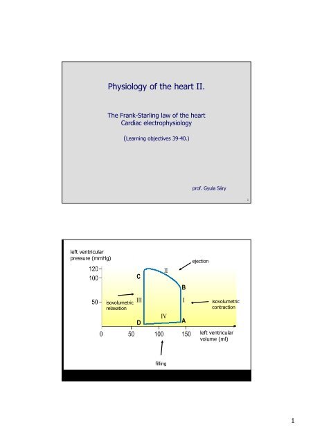
Starling’s<strong>heart</strong>-lungpreparationresistancevenous reservelungsaorta pressureventricularfilling pressureventricular volume7Starling’s <strong>heart</strong>-(lung) preparation84
ActivepressurePressureDi<strong>as</strong>tolic volumePressureActivepressureDi<strong>as</strong>tolic volume9105
11Adaptation to:incre<strong>as</strong>ed venous return (preload):change in body position, respiration…incre<strong>as</strong>ed peripheral resistance (afterload)pressure incre<strong>as</strong>es in <strong>the</strong> aort<strong>as</strong>ympa<strong>the</strong>tic stimulation (positive inotropic effect)di<strong>as</strong>tolic and systolic reserve126
Frank Starling law <strong>of</strong> <strong>the</strong> <strong>heart</strong>• stroke volume <strong>of</strong> <strong>the</strong> <strong>heart</strong> incre<strong>as</strong>es in responseto an incre<strong>as</strong>e in <strong>the</strong> volume <strong>of</strong> blood filling <strong>the</strong><strong>heart</strong> (<strong>the</strong> end di<strong>as</strong>tolic volume)or• <strong>the</strong> strength <strong>of</strong> <strong>the</strong> <strong>heart</strong>'s systolic contraction isdirectly proportional to its di<strong>as</strong>tolic expansion(end di<strong>as</strong>tolic volume)13resting length (di<strong>as</strong>tolic volume) determines <strong>the</strong> contraction force„Frank-Starling mechanism”Ca ++ sensitivity incre<strong>as</strong>es (Ca ++ channels?)works also in isolated <strong>heart</strong>contraction force may incre<strong>as</strong>e without change in length„positive inotropic effect”background: intracellular Ca ++ incre<strong>as</strong>eseffect <strong>of</strong> sympa<strong>the</strong>tic stimulation14147
Rhythmic excitation <strong>of</strong> <strong>the</strong> <strong>heart</strong>• special features: automatic and rhythmic• where is it coming from?Stannius, <strong>the</strong> ligatures and <strong>the</strong> frog <strong>heart</strong>15<strong>The</strong> Stannius ligature168
Rhythmic excitation <strong>of</strong> <strong>the</strong> <strong>heart</strong>• special features: automatic and rhythmic• Stannius and <strong>the</strong> frog <strong>heart</strong>• nomotop (natural) pacemaker (SA node)• heterotopic (ectopic) pacemakers• extr<strong>as</strong>ystole17189
19sinoatrial nodeaction potentialsatrial musclesAV nodeHis bundleTawara branchPurkinje fibresventricle muscles2010
• f<strong>as</strong>t AP (atrium, ventricle, Purkinje), more negative, steeper, largeramplitude → Na channels• slow AP (SA and AV node), less negative, less steep, smalleramplitude → no voltage dep. Na channels• conduction speed <strong>of</strong> f<strong>as</strong>t APs: 0.3-1-4 m/s,• conduction speed <strong>of</strong> slow APs: 0.02-0.1 m/s21actual pacemakerslow APthresholdmaximaldi<strong>as</strong>tolicpotentialslowdi<strong>as</strong>tolic depol.(pacemaker pot.)potencial pacemakerwhat ions ???thresholdmyocardiumf<strong>as</strong>t APrestingpot.threshold2211
23-70.0 mV2412
sympa<strong>the</strong>tic β1 receptorstimulationnormal<strong>heart</strong> rate decre<strong>as</strong>esduring vagal nervestimulationVagal stimulationnormalACh252613
Regulation <strong>of</strong> <strong>the</strong> cardiac pumping function1. Frank - Starling mechanism: intrinsic regulation2. Neural regulation• par<strong>as</strong>ympa<strong>the</strong>tic- (n. vagus – ACh - muscarinergic)negative chronotropy, dromotropy, bathmotropyinotropy (not in <strong>the</strong> ventricles)• sympa<strong>the</strong>tic- (NA- β1 receptor)positive chronotropy, dromotropy, bathmotropyinotropy, lusitropy + v<strong>as</strong>odilation3. Humoral: Adrenalin, NA4. Effect <strong>of</strong> Ca ++ and K + 2827speed <strong>of</strong> depolarization (V/s)membrane potential (mV)resting membrane potential (mV)14