RecA - Microbiology & Molecular Genetics
RecA - Microbiology & Molecular Genetics
RecA - Microbiology & Molecular Genetics

You also want an ePaper? Increase the reach of your titles
YUMPU automatically turns print PDFs into web optimized ePapers that Google loves.
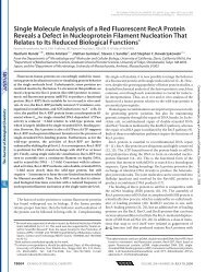
MagazineR395then most lines thereafter crossedout. Madame Auclair, the Frenchviolinist, had a gentler approach.Out of central casting, as they say:dark glasses, cigarette dangling,hoarse, accented voice. A friendwent for a special violin lesson,and asked whether he might taperecord this important event in hislife. “Of course, my boy.” He playeda bit and she said: “Very nice. Thereare some good things about yourplaying, very good. Now turn off thetape recorder.”When I am struggling overyet another of my obscurelywritten drafts I sometimes recall:amateurs play music ‘in general’;professionals play each note. Andso I present to a tough-mindedfriend one paragraph — justone — and when that is reportedto be transparent I go on to therest. But even if I have followedthe rules I mentioned above, andeven if that first paragraph seemedfine at the time, now, in view ofwhat else I have written, that firstparagraph might have to go, or beseriously recast. Each paragraphis an experiment — you might notknow for some time whether it isany good.There is a theme here, beautifullyexpressed by a friend who wasgoing through the agonies of the“just the first paragraph” methodin attempting to re-write a book. Ihadn’t heard from him in a whileand began to worry — had I beentoo tough? — and he wrote: “Theonly reason I hadn’t sent it (the newparagraph) already is that I didn’twant to disappoint you. But I realizethat the only way you can help meis if I continue to disappoint you.So here it is...” All my teachers,whatever their methods, weretrying to help me, and I love themfor it. Heifetz I wouldn’t be so sureabout. Rules are one thing, but inthe end communication is all: at theend of a pleasant interview with afine scientist of foreign extractionshe shook my hand and said: “Itsbeen a pleasure talking to me.”Reference1. Ptashne, M. (2007). On speaking, writingand inspiration. Curr. Biol. 17, R348.Sloan Kettering Institute, Memorial SloanKettering Cancer Center, 1275 York Avenue,Box 595, New York, New York 10021, USA.E-mail: m-ptashne@mskcc.orgQuick guide<strong>RecA</strong>Roberto Galletto andStephen C. KowalczykowskiWhat is <strong>RecA</strong> protein? Thebacterial <strong>RecA</strong> protein is thefounding member of a class ofproteins with homologs acrossall domains of life: RadA inarchaea, and Rad51 and Dmc1in eukaryotes. In Escherichiacoli, <strong>RecA</strong> is essential forrecombinational repair of DNAbreaks, induction of the DNAdamage-induced ‘SOS’ response,and activation of translesion DNAsynthesis. The functional form of<strong>RecA</strong> protein in these processesis the nucleoprotein filament, thestructure formed by assembly of<strong>RecA</strong> protein on DNA (Figure 1),generally single-stranded DNA.During homologous recombination,the <strong>RecA</strong> nucleoprotein filamentcatalyzes the pairing and exchangeof complementary DNA strandsbetween homologous regions ofDNA. In response to DNA damage,the <strong>RecA</strong> nucleoprotein filamentactivates the SOS response bycatalyzing the auto-cleavage(co-protease activity) of the LexArepressor, leading to derepressionof over 40 unlinked genes involvedin DNA repair, including the recAgene itself. And through bothcleavage (of the UmuD subunit)and direct binding, the <strong>RecA</strong>nucleoprotein filament activatesDNA polymerase V (UmuD′ 2 Cprotein), a lesion by-pass DNApolymerase, to synthesize DNAat otherwise irreparable lesions,resulting in a mutagenic form ofDNA repair known as translesionsynthesis.What is the <strong>RecA</strong> nucleoproteinfilament? The active form of<strong>RecA</strong> and of all its homologs isthe ATP-bound nucleoproteinfilament formed on DNA. Theprotein forms a polymorphicright-handed helix around the DNAwith approximately six monomersper turn and a pitch of ~9.5 nm,in which the DNA is extended toabout 150% of its B-form length.This quaternary organizationis responsible for the catalyticproperties of the protein. Formationof the nucleoprotein filament occursby a mechanism similar to that ofother self- associating proteins,Figure 1. Assembly and disassembly of a <strong>RecA</strong> nucleoprotein filament formed on anindividual double-stranded DNA molecule.The collage shows the major steps in the entire cycle of <strong>RecA</strong> nucleoprotein filamentassembly and disassembly as visualized by single-molecule detection. The DNA, whichis invisible in these images, is bound to a polystyrene bead (leftmost ‘spot’). The <strong>RecA</strong>is fluorescently labeled, permitting visualization of the DNA-bound protein. From top tobottom and left to right, snapshots of <strong>RecA</strong> assembly from multiple nuclei to generatecomplete filaments (left-most two columns), followed by images of disassembly promotedby ATP hydrolysis (right-most two columns).
Current Biology Vol 17 No 11R396such as actin and tubulin, exceptthat polymerization typically occurson the DNA. The first step is theformation of rate-limiting nuclei thatrequire four or five <strong>RecA</strong> monomersto achieve stability on DNA. Thisnucleus then grows via addition ofATP-<strong>RecA</strong>. Although addition at oneend (the 3′-end) of the DNA-boundcluster can be preferred, assemblyof <strong>RecA</strong> at the opposite end canalso occur, resulting in bi-directionalgrowth of the nucleoproteinfilament, albeit with a net 5′→3′assembly direction.How does it work? Kinetically,the preferred substrate fornucleoprotein filament formationis single-stranded DNA. In theprototypic DNA strand-exchangereaction, the <strong>RecA</strong> nucleoproteinfilament forms on single-strandedDNA (presynapsis). Subsequently,the nucleoprotein filamentrepeatedly binds double- strandedDNA non- specifically, whileit ‘searches’ for homologousregions within the duplex.During this homology search,the double- stranded DNA istopologically unwound, but strandseparation does not occur. Suchunwinding of the DNA likelyactivates the normally stable duplexDNA for the subsequent stepsof DNA strand exchange. Uponrecognition of a homologous regionof about 15–40 base pairs in thedouble-stranded DNA (synapsis),the <strong>RecA</strong> filament promotesswitching of base pairs, resultingin homologously paired ‘joint’DNA molecules. Continued pairingand extension of the nascentDNA heteroduplex joint (DNAheteroduplex extension) leads tothe final products of DNA strandexchange, where several thousandsof base pairs of DNA can beexchanged.Although single-stranded DNAis the preferred substrate, active<strong>RecA</strong> filaments can also be formedon double-stranded DNA. Theduplex DNA in this nucleoproteinfilament is also stretched to~150% of its B-form length andit is underwound by about 50%.This nucleoprotein filamentassembled on double- strandedDNA is active by a number ofcriteria, but the most compellingis that it can promote DNA strandexchange with single- strandedDNA; it can also promote strandexchange with single-strandedRNA. Being the directionalopposite of the canonical reaction,DNA strand exchange initiatedwith double- stranded DNAnucleoprotein filaments is called‘inverse’ DNA strand exchange.What controls <strong>RecA</strong> nucleoproteinfilament dynamics? In vitro,assembly of <strong>RecA</strong> nucleoproteinfilaments is spontaneous, anddoes not require any accessoryproteins. Regulation of the cycleof assembly and disassemblyis achieved through the bindingand hydrolysis of nucleotidecofactors. Assembly of the activenucleoprotein filaments requiresATP; both non-hydrolyzable ATPanalogs (ATPγS) and mimicsof nucleoside triphosphates(ADP•AlF 3 ) also enable formationof active nucleoprotein filaments.Thus, ATP hydrolysis is not arequisite step in the mechanismof DNA strand exchange. Thisis because the binding of ATPto <strong>RecA</strong> is sufficient to inducea state of the protein with highaffinity for DNA, and this state isactive in homologous pairing, DNAstrand exchange, and co- proteasefunction. To disassemble thefilaments, ATP hydrolysis is needed.ATP hydrolysis both destroysthe effector ligand, ATP, andproduces ADP, which destabilizesand inactivates the nucleoproteinfilament. The dynamic behaviorof <strong>RecA</strong> protein under conditionsof ATP hydrolysis is thusconceptually similar to that of otherNTP- hydrolyzing, self-associatingproteins, such as actin and tubulin.Although the rate constants foreach of the key assembly anddisassembly intermediates are stillunknown, the preferred end foraddition of ATP-<strong>RecA</strong> monomers istypically the 3′-end of the filamentand the ADP-induced dissociationof <strong>RecA</strong> commonly occurs at the5′-end. The net behavior, however,is known to be influenced by awide range of solution conditionsresulting in, at times, seeminglycontradictory reports regarding<strong>RecA</strong> protein function.Furthermore, ATP hydrolysis isrequired for dissociation of <strong>RecA</strong>from the heteroduplex product ofDNA strand exchange. Indeed, inthe absence of ATP hydrolysis — inthe presence of ATPγS orADP•AlF 3 — pairing and exchangeof DNA strands is efficient,resulting in the production of jointmolecules. The subsequent phaseof DNA heteroduplex extension is,however, blocked. Also, hydrolysisof ATP is a required step in thebypass of heterologous sequences.<strong>RecA</strong> nucleoprotein filaments cancatalyze DNA strand exchange withduplex DNA where the homologousregions are separated by up to~140 base pairs of an interveningnon-homologous sequence. Therequirement for ATP hydrolysisin DNA heterology bypass pointsto a crucial need for a dynamicnucleoprotein filament, withdissociation and redistribution ofbound <strong>RecA</strong> playing a pivotal role.How is the <strong>RecA</strong> nucleoproteinfilament regulated? The complexinterplay of ATP binding andhydrolysis provides a simple butdynamic mechanism for regulatingthe steady-state level of <strong>RecA</strong>nucleoprotein filaments. Notsurprisingly, in vivo, this assemblyprocess is regulated. The firstlevel of regulation is controlled bythe single-stranded DNA bindingprotein, SSB. SSB competeswith <strong>RecA</strong> for DNA binding, andthereby represses unwanted<strong>RecA</strong> nucleoprotein filaments; thisinhibition, for example, preventsspurious filament formation on thelagging strand single-strandedDNA gaps formed during DNAreplication. Consequently, there areproteins that promote assemblyof <strong>RecA</strong> nucleoprotein filamentson SSB–DNA complexes. Oneof the other key enzymes ofrecombinational DNA repair, theRecBCD helicase/nuclease, actually‘loads’ <strong>RecA</strong> protein onto singlestrandedDNA produced by theRecBCD enzyme. This loadingof <strong>RecA</strong> protein ensures that theprocessed broken chromosomalfragment acquires sufficient <strong>RecA</strong>to be repaired by recombination ina timely manner. A second regulatorof <strong>RecA</strong> nucleoprotein filamentformation is the RecOR complex,which mediates the exchange of theDNA-bound SSB for <strong>RecA</strong> protein.A third regulator is the RecFORcomplex; these three proteins
MagazineR397bind to the junction of duplexDNA and single-stranded DNA,and recruit <strong>RecA</strong> protein to theSSB–DNA complex. Although themechanism of these loading andrecruitment steps is not completelyunderstood, it is likely that the ratelimitingnucleation of <strong>RecA</strong> proteinis being stimulated via transient,direct interactions with theseproteins. Filament disassembly isalso regulated. For example, UvrDhelicase (helicase II) disassembles<strong>RecA</strong> nucleoprotein filaments thatformed inappropriately. Thus, theregulation of the site and timing of<strong>RecA</strong> filament dynamics is achievedvia coordination with the repairmachinery.Why does <strong>RecA</strong> form anucleoprotein filament? Thebroad spectrum of biologicalfunctions attributable to <strong>RecA</strong>protein, ranging from the homologysearch of DNA recombinationto the activation of proteins viaits co-protease activity, cannotbe attributed to the action of anindividual monomer. Rather, it isevident that the catalytic unit ofthe <strong>RecA</strong> function is the highlyorganized filament that assembleson DNA. Formation of a highlyordered filament on its DNAsubstrate provides a large surfacewhere catalysis can occur (‘surfacecatalysis’), where homologycan be measured and matchedto a potential complementarypartner, and where DNA structuraltransitions can be cooperativelytransmitted over long distances.This requirement for a regular<strong>RecA</strong>/Rad51 nucleoprotein filamentalso permits a broad spectrum ofregulatory control that is needed forbiological function.Where can I learn more?Bianco, P.R., Tracy, R.B., and Kowalczykowski,S.C. (1998). DNA strand exchange proteins:A biochemical and physical comparison.Front. Biosci. 3, D570–D603.McGrew, D.A., and Knight, K.L. (2003).<strong>Molecular</strong> design and functionalorganization of the <strong>RecA</strong> protein. Crit. Rev.Biochem. Mol. Biol. 38, 385–432.Spies, M., and Kowalczykowski, S.C.(2005). Homologous recombination byRecBCD and RecF pathways. In TheBacterial Chromosome, N.P. Higgins,ed. (Washington, D.C.: ASM Press), pp.389–403.Sections of <strong>Microbiology</strong> and of <strong>Molecular</strong>and Cellular Biology, University ofCalifornia, Davis, California 95616, USA.E-mail: sckowalczykowski@ucdavis.eduEssayMarr’s vision: Twenty-five years onAndrew GlennersterIt is twenty-five years since the posthumous publication of David Marr’sbook Vision [1]. Only 35 years old when he died, Marr had alreadydramatically influenced vision research. His book, and the series of papersthat preceded it, have had a lasting impact on the way that researchersapproach human and computer vision.A review at the time of publicationpredicted that “Even if no singleone of Marr’s detailed hypothesesultimately survives ... [his] lifeworkwill have been vindicated whenneuroscientists cannot understandhow it was ever possible to doubtthe validity of his theoreticalmaxims”. Twenty-five years on,most would agree that Marr’srecipe for investigating humanvision and, in particular, hisstrategy of dividing the probleminto different levels of analysis,has become unquestioned. Atthe time, Binford, Horn, Minsky,Papert, Rumelhart and others hadbeen advocating computationalmodelling as a key tounderstanding the brain’s operationbut Marr brought a number ofdifferent approaches together,made testable predictions,provided a framework for tacklingchallenging neuroscientificquestions and inspired a generationof young scientists to study thebrain and visual processing.Born in Essex, England, Marrstudied mathematics at TrinityCollege, Cambridge, beforedoing his Ph.D. in what wouldnow be called ‘computationalneuroscience’ with ProfessorG.F. Brindley. His doctoral work,expressed in a series of threeimportant papers [2–4], tiedtogether detailed anatomical dataon the cerebellum, neocortexand hippocampus within acomputational framework. Theseare fundamental papers in thefield, especially his paper on thecerebellum, but Marr now changedhis focus to vision. He wanted toconsider specific algorithms, andthe constraints of the real worldthat made them tractable, ratherthan the processing of neuralsignals in general.One of the central and bestknown ideas in his book isthe suggestion that the visualsystem generates a sequenceof increasingly symbolicrepresentations of a scene,progressing from a ‘primal sketch’of the retinal image, througha ‘2½D sketch’ to simplifiedthree- dimensional models ofobjects. In a paper with EllenHildreth [5], he proposed thatinformation from cells tuned todifferent spatial frequencies (orscales) is combined into ‘tokens’that are likely to correspondto real-world entities such asan edge. Although there is noconvincing evidence that theparticular type of combinationMarr advocated is carried out inthe visual system (other proposalshave more experimental support[6]), it is a good example of Marr’sapproach. “In the theory of visualprocesses, the underlying taskis to reliably derive properties ofthe world from images of it; thebusiness of isolating constraintsthat are both powerful enoughto allow a process to be definedand generally true of the world isa central theme of our inquiry” [1].Today, this approach is normalpractice in computer vision and atleast a widely accepted mantra inbiological vision research.The tokens comprising theprimal sketch were, Marr argued,then used as input to furtherprocesses such as objectrecognition [7]. Object recognitionis one of several areas in whichMarr’s specific ideas aboutimplementation have not survivedwell. The current focus in bothcomputer and biological vision ison matching of high dimensionalview-invariant descriptors ofimage features [8,9], taking a quite