Dumont et al. 2006 - The Department of Ecology and Evolutionary ...
Dumont et al. 2006 - The Department of Ecology and Evolutionary ...
Dumont et al. 2006 - The Department of Ecology and Evolutionary ...

Create successful ePaper yourself
Turn your PDF publications into a flip-book with our unique Google optimized e-Paper software.
Blackwell Publishing LtdOxford, UKEMIEnvironment<strong>al</strong> Microbiology 1462-2912© <strong>2006</strong> <strong>The</strong> Authors; Journ<strong>al</strong> compilation © <strong>2006</strong> Soci<strong>et</strong>y for Applied Microbiology <strong>and</strong> Blackwell Publishing Ltd<strong>2006</strong>8712401250Origin<strong>al</strong> ArticleSIP<strong>and</strong> m<strong>et</strong>agenomic an<strong>al</strong>ysisM. G. <strong>Dumont</strong><strong>et</strong> <strong>al</strong>.Environment<strong>al</strong> Microbiology (<strong>2006</strong>) 8(7), 1240–1250doi:10.1111/j.1462-2920.<strong>2006</strong>.01018.xIdentification <strong>of</strong> a compl<strong>et</strong>e m<strong>et</strong>hane monooxygenaseoperon from soil by combining stable isotope probing<strong>and</strong> m<strong>et</strong>agenomic an<strong>al</strong>ysisMarc G. <strong>Dumont</strong>, 1 Stefan M. Radajewski, 1Carlos B. Miguez, 2 Ian R. McDon<strong>al</strong>d 3 <strong>and</strong>J. Colin Murrell 11<strong>Department</strong> <strong>of</strong> Biologic<strong>al</strong> Sciences, University <strong>of</strong>Warwick, Coventry CV4 7AL, UK.2Biotechnology Research Institute, Nation<strong>al</strong> ResearchCouncil <strong>of</strong> Canada, Montre<strong>al</strong>, Quebec, Canada H4P 2R2.3 <strong>Department</strong> <strong>of</strong> Biologic<strong>al</strong> Sciences, University <strong>of</strong> Waikato,Private Bag 3105, Hamilton, New Ze<strong>al</strong><strong>and</strong>.SummaryStable isotope probing (SIP) <strong>al</strong>lows the isolation <strong>of</strong>nucleic acids from targ<strong>et</strong>ed m<strong>et</strong>abolic<strong>al</strong>ly active organismsin environment<strong>al</strong> samples. In previous studies,DNA-SIP has been performed with the one-carbongrowth substrates m<strong>et</strong>hane <strong>and</strong> m<strong>et</strong>hanol to studym<strong>et</strong>hylotrophic organisms. <strong>The</strong> m<strong>et</strong>hylotrophs thatincorporated the labelled substrate were identifiedwith polymerase chain reaction <strong>and</strong> sequencing <strong>of</strong> 16SrRNA <strong>and</strong> ‘function<strong>al</strong> genes’ for m<strong>et</strong>hanotrophs (mxaF,pmoA, mmoX). In this study, a SIP experiment wasperformed using a forest soil sample incubated with13CH 4 , <strong>and</strong> the 13 C-DNA was purified <strong>and</strong> cloned into abacteri<strong>al</strong> artifici<strong>al</strong> chromosome (BAC) plasmid. Alibrary <strong>of</strong> 2300 clones was generated <strong>and</strong> most <strong>of</strong> theclones contained inserts b<strong>et</strong>ween 10 <strong>and</strong> 30 kb. <strong>The</strong>library was probed for key m<strong>et</strong>hylotrophy genes <strong>and</strong>a 15.2 kb clone containing a pmoCAB operon, encodingparticulate m<strong>et</strong>hane monooxygenase, was identified<strong>and</strong> sequenced. An<strong>al</strong>ysis <strong>of</strong> the pmoA sequencesuggested that the clone was most similar to that <strong>of</strong>a M<strong>et</strong>hylocystis sp. previously d<strong>et</strong>ected in this forestsoil. Twelve other open reading frames were identifiedon the clone, including the gene encoding b<strong>et</strong>arib<strong>of</strong>uranosylaminobenzene5¢-phosphate synthase,which is involved in the biosynthesis <strong>of</strong> the ‘archae<strong>al</strong>’C 1 -carrier, t<strong>et</strong>rahydrom<strong>et</strong>hanopterin, which is <strong>al</strong>s<strong>of</strong>ound in m<strong>et</strong>hylotrophs. This study demonstrates thatrelatively large DNA fragments from uncultivatedorganisms can be readily isolated using DNA-SIP, <strong>and</strong>cloned into a vector for m<strong>et</strong>agenomic an<strong>al</strong>ysis.Received 31 August, 2005; accepted 7 February, <strong>2006</strong>. *For correspondence.E-mail j.c.murrell@warwick.ac.uk; Tel. (+44) 24 76523553; Fax (+44) 24 7652 3568.IntroductionMicrobi<strong>al</strong> ecology is a particularly ch<strong>al</strong>lenging disciplinegiven that most microorganisms have so far eluded cultivation.<strong>The</strong> current estimate is that there are 52 bacteri<strong>al</strong>phyla, h<strong>al</strong>f <strong>of</strong> which lack a single cultivated representative(Rappé <strong>and</strong> Giovannoni, 2003). As a result, it is impossibl<strong>et</strong>o infer the function <strong>of</strong> many microorganisms. In addition,because the conditions in laboratory media are likelyto be different from those in situ, inferring the role <strong>of</strong>microorganisms based on laboratory studies could bemisleading. <strong>The</strong>refore, a useful strategy to study microbi<strong>al</strong>ecology is to use cultivation-independent m<strong>et</strong>hods suchas stable isotope probing (SIP) (Radajewski <strong>et</strong> <strong>al</strong>., 2000;Manefield <strong>et</strong> <strong>al</strong>., 2002), which can link microorganismidentity (e.g. 16S rRNA sequences) in an environment<strong>al</strong>sample to the incorporation <strong>of</strong> a growth substrate.M<strong>et</strong>agenomics, the genomic an<strong>al</strong>ysis <strong>of</strong> uncultivatedmicroorganisms, may <strong>al</strong>so be useful in studying microbi<strong>al</strong>ecology through the interpr<strong>et</strong>ation <strong>of</strong> nucleotide sequencedata (H<strong>and</strong>elsman, 2004).Stable isotope probing is a technique for studying theactive organisms in environment<strong>al</strong> samples. <strong>The</strong> DNA-SIPtechnique was origin<strong>al</strong>ly developed using CH 4 <strong>and</strong> CH 3 OHsubstrates (Radajewski <strong>et</strong> <strong>al</strong>., 2000; Morris <strong>et</strong> <strong>al</strong>., 2002),but has since been used with CO 2 (Whitby <strong>et</strong> <strong>al</strong>., 2001),m<strong>et</strong>hyl h<strong>al</strong>ides (Miller <strong>et</strong> <strong>al</strong>., 2004; Borodina <strong>et</strong> <strong>al</strong>., 2005)<strong>and</strong> sever<strong>al</strong> multicarbon compounds such as naphth<strong>al</strong>ene,phenol, caffeine, glucose, s<strong>al</strong>icylate <strong>and</strong> phenanthrene(Jeon <strong>et</strong> <strong>al</strong>., 2003; Padmanabhan <strong>et</strong> <strong>al</strong>., 2003;Singl<strong>et</strong>on <strong>et</strong> <strong>al</strong>., 2005). In previous DNA-SIP studies, themicrobi<strong>al</strong> populations that incorporated the isotopic<strong>al</strong>lyheavy substrate were characterized by polymerase chainreaction (PCR) amplification <strong>of</strong> 16S <strong>and</strong> 18S rRNA genes<strong>and</strong> function<strong>al</strong> genes (for example, genes encoding m<strong>et</strong>hanemonooxygenase <strong>and</strong> m<strong>et</strong>hanol dehydrogenase)using the 13 C-DNA as template (Radajewski <strong>et</strong> <strong>al</strong>., 2002;Ginige <strong>et</strong> <strong>al</strong>., 2004; Hutchens <strong>et</strong> <strong>al</strong>., 2004; Lin <strong>et</strong> <strong>al</strong>., 2004;Lueders <strong>et</strong> <strong>al</strong>., 2004; G<strong>al</strong>lagher <strong>et</strong> <strong>al</strong>., 2005; Lu <strong>et</strong> <strong>al</strong>.,2005).A difficulty in an<strong>al</strong>ysing a m<strong>et</strong>agenomic library is thehigh degree <strong>of</strong> complexity that results from the hugemicrobi<strong>al</strong> diversity in many environment<strong>al</strong> samples. Estimates<strong>of</strong> microbi<strong>al</strong> diversity in a forest soil using singlestr<strong>and</strong>edDNA reassociation kin<strong>et</strong>ics indicated 12 000–18 000 different species (Torsvik <strong>et</strong> <strong>al</strong>., 1990). <strong>The</strong>re is© <strong>2006</strong> <strong>The</strong> AuthorsJourn<strong>al</strong> compilation © <strong>2006</strong> Soci<strong>et</strong>y for Applied Microbiology <strong>and</strong> Blackwell Publishing Ltd
SIP <strong>and</strong> m<strong>et</strong>agenomic an<strong>al</strong>ysis 1241Amarker12C-DNA13C-DNABFig. 1. A. <strong>The</strong> CsCl-<strong>et</strong>hidium bromide gradientcontaining 12 C-DNA <strong>and</strong> 13 C-DNA from theDNA-SIP experiment with forest soil that consumed13 CH 4 . <strong>The</strong> DNA was visible <strong>and</strong> photographedin ambient light <strong>and</strong> the position <strong>of</strong> the12C-DNA <strong>and</strong> 13 C-DNA b<strong>and</strong>s is indicated by thearrows.B. Denaturing gradient gel electrophoresisan<strong>al</strong>ysis <strong>of</strong> the 12 C-DNA <strong>and</strong> 13 C-DNA recoveredfrom the gradient. <strong>The</strong> marker containeda mixture <strong>of</strong> 16S rRNA gene products from purebacteri<strong>al</strong> cultures. <strong>The</strong> predominant DGGEb<strong>and</strong>s from the 13 C-DNA were sequenced <strong>and</strong>a genus name is indicated where the per centidentity <strong>of</strong> the sequence was greater than 98%to that <strong>of</strong> a cultivated representative. <strong>The</strong>GS_b<strong>and</strong>_3 sequence belongs to an uncultivatedGammaproteobacteria that was <strong>al</strong>soPCR amplified in a previous DNA-SIP an<strong>al</strong>ysis<strong>of</strong> this soil (Radajewski <strong>et</strong> <strong>al</strong>., 2002).GS_b<strong>and</strong>_1 possesses similarity toBacteroid<strong>et</strong>es 16S rRNA sequences.12C-DNA13C-DNAGS_b<strong>and</strong>_1; uncultured bacteriumGS_b<strong>and</strong>_2; uncultured M<strong>et</strong>hylobacter sp.GS_b<strong>and</strong>_3; uncultured Gammaproteobacteria sp.GS_b<strong>and</strong>_4; uncultured M<strong>et</strong>hylocella sp.GS_b<strong>and</strong>_5; uncultured M<strong>et</strong>hylocystis sp.considerable interest in the possibility <strong>of</strong> combiningDNA-SIP with m<strong>et</strong>agenomic studies to solve thisdilemma (Schloss <strong>and</strong> H<strong>and</strong>elsman, 2003; Wellington<strong>et</strong> <strong>al</strong>., 2003; <strong>Dumont</strong> <strong>and</strong> Murrell, 2005a). In a m<strong>et</strong>agenomiclibrary constructed from 13 C-DNA from a DNA-SIPexperiment, the cloned DNA would originate fromgenomes <strong>of</strong> organisms that have obtained the majority <strong>of</strong>their carbon, either directly or indirectly via crossfeeding,from the 13 C-substrate. This limits the library togenome fragments <strong>of</strong> organisms involved in a specificm<strong>et</strong>abolic process <strong>of</strong> interest <strong>and</strong> increases the likelihood<strong>of</strong> obtaining <strong>and</strong> sequencing genes that are integr<strong>al</strong>to the process.In this study, a m<strong>et</strong>agenomic library was constructedusing 13 C-DNA from a DNA-SIP experiment with 13 CH 4 .<strong>The</strong> objective was to perform an experiment to d<strong>et</strong>ermin<strong>et</strong>he feasibility <strong>of</strong> cloning larger fragments <strong>of</strong> the 13 C-DNA<strong>and</strong> identifying a clone containing m<strong>et</strong>hylotrophy genes. Aclone library was made from 13 C-DNA using a bacteri<strong>al</strong>artifici<strong>al</strong> chromosome (BAC) plasmid <strong>and</strong> a clone containinga compl<strong>et</strong>e pmoCAB operon, encoding the particulatem<strong>et</strong>hane monooxygenase (pMMO) enzyme, was identified<strong>and</strong> sequenced. <strong>The</strong> pmoA gene was similar to that<strong>of</strong> an uncultivated M<strong>et</strong>hylocystis sp. previously identifiedin this soil by DNA-SIP <strong>and</strong> PCR an<strong>al</strong>ysis. A very briefpreliminary report <strong>of</strong> these data was published in a review<strong>of</strong> DNA-SIP (<strong>Dumont</strong> <strong>and</strong> Murrell, 2005a).ResultsStable isotope probing with 13 CH 4 <strong>and</strong> construction<strong>of</strong> a m<strong>et</strong>agenomic library<strong>The</strong> SIP experiment was performed in a manner similarto that described previously (Radajewski <strong>et</strong> <strong>al</strong>., 2002) <strong>and</strong>with sample taken from the same forest soil site. Soil wascollected from the acidic oak forest soil <strong>and</strong> incubated with13CH 4 until a tot<strong>al</strong> <strong>of</strong> 50 ml had been consumed. <strong>The</strong> DNAwas extracted from the 13 CH 4 -labelled soil <strong>and</strong> the DNAwas visible in ambient light after isopycnic centrifugationin CsCl (Fig. 1A). <strong>The</strong>refore, the 13 C-DNA b<strong>and</strong> could becollected without exposure to DNA-damaging UV irradiation.<strong>The</strong> 13 C-DNA was an<strong>al</strong>ysed by pulsed-field gel electrophoresis(PFGE) <strong>and</strong> the majority <strong>of</strong> fragments wereb<strong>et</strong>ween ∼15 kb <strong>and</strong> ∼100 kb in length (results notshown).Restriction enzyme activity is sensitive to inhibition bysoil organics which are co-extracted with DNA (Zhou<strong>et</strong> <strong>al</strong>., 1996). Although sever<strong>al</strong> rounds <strong>of</strong> CsCl gradientcentrifugation had been used to purify the DNA, the gradient-purifiedDNA was not sufficiently pure to be digestedwith restriction enzyme BamHI (results not shown). <strong>The</strong>refore,further purification was achieved by electrophoresis<strong>of</strong> the 13 C-DNA through a 1% (w/v) agarose gel. <strong>The</strong>agarose gel electrophoresis had two convenient purposes:first, it eliminated contaminants from the DNA sothat it could be digested with restriction enzymes, <strong>and</strong>second, the 13 C-DNA formed a tight b<strong>and</strong> that could beexcised in an agarose plug in which restriction enzymedigestion <strong>of</strong> the 13 C-DNA could be performed. This minimizedaddition<strong>al</strong> DNA shearing that could potenti<strong>al</strong>lyoccur when pip<strong>et</strong>ting the DNA solution.Parti<strong>al</strong>ly digested 13 C-DNA was resolved on a st<strong>and</strong>ardagarose gel (20 cm × 20 cm) rather than a PFGE system,because st<strong>and</strong>ard electrophoresis resulted in a more tightdistribution <strong>of</strong> the desired size fragments within the gel<strong>and</strong> easier elution from the gel. As described in the Experiment<strong>al</strong>procedures section, extreme care was taken toavoid exposure <strong>of</strong> the DNA to UV irradiation. <strong>The</strong> parti<strong>al</strong>lyBamHI-digested <strong>and</strong> size-selected DNA was recoveredfrom the agarose <strong>and</strong> ligated into plasmid pCC1BAC<strong>and</strong> a library <strong>of</strong> 2300 clones was generated using the© <strong>2006</strong> <strong>The</strong> AuthorsJourn<strong>al</strong> compilation © <strong>2006</strong> Soci<strong>et</strong>y for Applied Microbiology <strong>and</strong> Blackwell Publishing Ltd, Environment<strong>al</strong> Microbiology, 8, 1240–1250
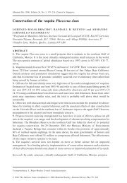
1242 M. G. <strong>Dumont</strong> <strong>et</strong> <strong>al</strong>.orf2154 dnaJ gloBtrkA mptG folC folK pmoC pmoA pmoB bolA cobS cobT orf5501 moxFFig. 2. Gen<strong>et</strong>ic map <strong>of</strong> ORFs identified on clone GSC357. <strong>The</strong> fragment is 15.2 kb in length. <strong>The</strong> assigned names represent the closest knowngenes (refer to Table 1).1kbE. coli TransforMax EC300 cells provided with the vector(Epicentre).An<strong>al</strong>ysis <strong>of</strong> the SIP labelling <strong>and</strong> the m<strong>et</strong>agenomic libraryBacteri<strong>al</strong> 16S rRNA genes were amplified from the 12 C-DNA <strong>and</strong> 13 C-DNA by PCR <strong>and</strong> an<strong>al</strong>ysed by denaturinggradient gel electrophoresis (DGGE) (Fig. 1B). <strong>The</strong> DGGEpattern for the 12 C-DNA was complex, as expected for asoil bacteri<strong>al</strong> community. <strong>The</strong> DGGE pattern for the 13 C-DNA showed less than 10 predominant b<strong>and</strong>s. Sequencing<strong>of</strong> the dominant 13 C-DNA DGGE b<strong>and</strong>s indicated thepresence <strong>of</strong> the m<strong>et</strong>hanotrophic genera M<strong>et</strong>hylobacter(GS_b<strong>and</strong>_2), M<strong>et</strong>hylocella (GS_b<strong>and</strong>_4) <strong>and</strong> M<strong>et</strong>hylocystis(GS_b<strong>and</strong>_5) (Fig. 1B). <strong>The</strong> M<strong>et</strong>hylocystis 16SrRNA sequence was very similar to the UP4, UP5, UP6<strong>and</strong> UP7 16S rRNA sequences (AY080911, AY080917,AY080912, AY080914 respectively) that tog<strong>et</strong>her constituted96% <strong>of</strong> the 16S rRNA gene sequences from the 13 C-DNA in the origin<strong>al</strong> 13 CH 4 SIP an<strong>al</strong>ysis <strong>of</strong> this forest soil(Radajewski <strong>et</strong> <strong>al</strong>., 2002). In addition to the m<strong>et</strong>hanotroph16S rRNA gene sequences, a predominant sequenc<strong>et</strong>ype (GS_b<strong>and</strong>_1) was present with similarity to theBacteroid<strong>et</strong>es <strong>and</strong> a second type (GS_b<strong>and</strong>_3) with similarityto a Gammaproteobacteria; the latter was <strong>al</strong>sor<strong>et</strong>rieved in the previous SIP study <strong>of</strong> this forest soil(Radajewski <strong>et</strong> <strong>al</strong>., 2002).After construction <strong>of</strong> the m<strong>et</strong>agenomic library from 13 C-DNA, the plasmids were isolated from 48 clones chosenat r<strong>and</strong>om <strong>and</strong> an<strong>al</strong>ysed by digestion with BamHI followedby agarose gel electrophoresis. Most clones containedinserts b<strong>et</strong>ween 10 <strong>and</strong> 30 kb <strong>and</strong> two <strong>of</strong> the 48 plasmidscontained no visible insert DNA. Also, nearly h<strong>al</strong>f <strong>of</strong> theclones an<strong>al</strong>ysed had addition<strong>al</strong> BamHI sites within theinsert fragment (results not shown), which indicated thatthe cloned 13 C-DNA had been incompl<strong>et</strong>ely digested asintended, or <strong>al</strong>ternatively had been parti<strong>al</strong>ly protectedfrom cutting by m<strong>et</strong>hylation. <strong>The</strong> sequencing primers T7<strong>and</strong> RP2 anne<strong>al</strong>ed adjacent to the cloning site <strong>of</strong>pCC1BAC <strong>and</strong> were used to sequence the ends <strong>of</strong>sever<strong>al</strong> <strong>of</strong> the cloned inserts. <strong>The</strong> sequence data from theclones were compared with the GenBank database byBLAST an<strong>al</strong>ysis (Altschul <strong>et</strong> <strong>al</strong>., 1990) <strong>and</strong> were differentfrom each other, with the exception <strong>of</strong> two clones thatappeared to be identic<strong>al</strong>. None <strong>of</strong> the genes identified byend sequencing were <strong>of</strong> obvious relevance to m<strong>et</strong>hylotrophy<strong>and</strong> these clones were not sequenced further.<strong>The</strong> m<strong>et</strong>agenomic library was screened by colonyhybridization for pmoA, mmoX <strong>and</strong> mxaF, which encodesubunits <strong>of</strong> the pMMO, soluble m<strong>et</strong>hane monooxygenase<strong>and</strong> m<strong>et</strong>hanol dehydrogenase respectively. Polymerasechain reaction primers were used to amplify the probeusing the 13 C-DNA as template. As the probes were amplifiedfrom 13 C-DNA, there was a high probability that thehomologous probe would be present for a cloned gene.<strong>The</strong> three gene hybridizations were performed separately<strong>and</strong> a tot<strong>al</strong> <strong>of</strong> four membranes each with approximately575 clones were hybridized with 32 P-GTP r<strong>and</strong>om primedPCR probes.<strong>The</strong> mmoX <strong>and</strong> mxaF probes did not hybridize with any<strong>of</strong> the clones in the library. Two clones, GSC357 (GisburnSoil Clone) <strong>and</strong> GSC1346, hybridized strongly to thepmoA probe <strong>and</strong> the pmoA gene could be amplified fromthese clones by PCR (results not shown). <strong>The</strong> restrictionfragment length polymorphism (RFLP) patterns <strong>of</strong>GSC357 <strong>and</strong> GSC1346 digested with NotI were identic<strong>al</strong>(results not shown). GSC357 was compl<strong>et</strong>ely sequencedusing a shotgun approach. <strong>The</strong> compl<strong>et</strong>e sequence was15 230 bp <strong>and</strong> a summary <strong>of</strong> the genes contained on thisplasmid <strong>and</strong> their arrangement is given in Fig. 2 <strong>and</strong>Table 1. GSC1346 was parti<strong>al</strong>ly sequenced <strong>and</strong> found tobe very similar to the sequence <strong>of</strong> GSC357. It is likely thatGSC357 <strong>and</strong> GSC1346 were from different strains <strong>of</strong> thesame species.An<strong>al</strong>ysis <strong>of</strong> the pmoCAB operon on clone GSC357GSC357 contained a compl<strong>et</strong>e pmoCAB operon, withpmoC, pmoA <strong>and</strong> pmoB <strong>of</strong> 765 bp, 759 bp <strong>and</strong> 1257 bprespectively. <strong>The</strong> intergenic region b<strong>et</strong>ween pmoC <strong>and</strong>pmoA was 269 bp <strong>and</strong> b<strong>et</strong>ween pmoA <strong>and</strong> pmoB was227 bp. <strong>The</strong> sequences <strong>of</strong> pmoCAB operons <strong>and</strong> derivedpolypeptide sequences demonstrated considerable similarityto those <strong>of</strong> M<strong>et</strong>hylocystis sp. strain M (Gilbert <strong>et</strong> <strong>al</strong>.,2000), M<strong>et</strong>hylosinus trichosporium OB3b (Gilbert <strong>et</strong> <strong>al</strong>.,2000), M<strong>et</strong>hylocystis sp. strain SC2 (Ricke <strong>et</strong> <strong>al</strong>., 2004)<strong>and</strong> M<strong>et</strong>hylococcus capsulatus (Bath) (Semrau <strong>et</strong> <strong>al</strong>.,1995; Stolyar <strong>et</strong> <strong>al</strong>., 1999) (Table 2). Putative ribosomebinding sites were present upstream (5′) <strong>of</strong> <strong>al</strong>l three pmogenes on the clone. <strong>The</strong> pmoCAB operon in M<strong>et</strong>hylocystis© <strong>2006</strong> <strong>The</strong> AuthorsJourn<strong>al</strong> compilation © <strong>2006</strong> Soci<strong>et</strong>y for Applied Microbiology <strong>and</strong> Blackwell Publishing Ltd, Environment<strong>al</strong> Microbiology, 8, 1240–1250
1244 M. G. <strong>Dumont</strong> <strong>et</strong> <strong>al</strong>.Nitrosococcus oceaniM<strong>et</strong>hylocystis sp. SC2 copy 1 (AJ431386)P12.7 (AY080944)M<strong>et</strong>hylocystis parvus (U31651)M<strong>et</strong>hylocystis sp. M (U81596)GSC357P12.9 (AY080955)P13.7 (AY080951)M<strong>et</strong>hylosinus trichosporium OB3b (U31650)P13.5 (AY080949)M<strong>et</strong>hylocapsa acidiphila (AJ278727)RA14 (AF148521)P13.10 (AY080953)P13.6 (AY080950)P12.12 (AY080958)P12.8 (AY080959)M<strong>et</strong>hylocystis sp. SC2 copy 2 (AJ431387)M<strong>et</strong>hyloc<strong>al</strong>dum szegediense (U89303)M<strong>et</strong>hylococcus capsulatus (L40804)M<strong>et</strong>hylomonas m<strong>et</strong>hanica (U31653)P12.6 (AY080945)(AF047705)10 %Fig. 3. Phylogen<strong>et</strong>ic tree <strong>of</strong> the derived PmoA sequence from the GSC357 clone (shown in bold text). Representative PmoA sequences r<strong>et</strong>rievedin the previous DNA-SIP an<strong>al</strong>ysis <strong>of</strong> this soil (Radajewski <strong>et</strong> <strong>al</strong>., 2002) <strong>and</strong> PmoA sequences from selected m<strong>et</strong>hanotrophs were included asreferences. <strong>The</strong> tree was constructed as previously described (Radajewski <strong>et</strong> <strong>al</strong>., 2002). <strong>The</strong> sc<strong>al</strong>e bar represents 10 amino acid substitutionsper 100 residues.Three <strong>of</strong> the 12 putative genes, mptG, folP <strong>and</strong> folK,have proposed roles in one-carbon m<strong>et</strong>abolism (Chistoserdova<strong>et</strong> <strong>al</strong>., 2003). Dihydropteroate synthase (FolP)<strong>and</strong> 2-amino-4-hydroxy-6-hydroxym<strong>et</strong>hyl-7,8-dihydopteridinepyrophosphokinase (FolK) are involved in folate biosynthesis.In m<strong>et</strong>hylotrophs that use the serine pathwayfor form<strong>al</strong>dehyde fixation, a t<strong>et</strong>rahydr<strong>of</strong>olate c<strong>of</strong>actor isinvolved in transferring the m<strong>et</strong>hyl group, <strong>and</strong> on thegenome <strong>of</strong> the facultative m<strong>et</strong>hylotroph, M<strong>et</strong>hylobacteriumextorquens AM1, these genes are located within a cluster<strong>of</strong> genes essenti<strong>al</strong> for growth on m<strong>et</strong>hanol (Chistoserdova<strong>et</strong> <strong>al</strong>., 2003). <strong>The</strong> mptG gene encodes β-rib<strong>of</strong>uranosylaminobenzene5′-phosphate synth<strong>et</strong>ase (β-RFAP synthase),which is involved in m<strong>et</strong>hanopterin biosynthesis (Scott<strong>and</strong> Rasche, 2002). T<strong>et</strong>rahydrom<strong>et</strong>hanopterin is an‘archae<strong>al</strong>’ c<strong>of</strong>actor that is <strong>al</strong>so present in m<strong>et</strong>hylotrophicbacteria <strong>and</strong> involved in the transfer <strong>of</strong> the m<strong>et</strong>hyl groupduring oxidation or assimilation <strong>of</strong> form<strong>al</strong>dehyde (Chistoserdova<strong>et</strong> <strong>al</strong>., 1998; 2000). A ‘moxF′-like gene ispresent downstream (3′) <strong>of</strong> the pmoCAB operon. <strong>The</strong>semoxF ORFs are apparent homologues <strong>of</strong> mxaF, whichencode the large subunit <strong>of</strong> the m<strong>et</strong>hanol dehydrogenase.Similar mxaF homologues have been identified in them<strong>et</strong>hylotrophs M<strong>et</strong>hylobacterium extorquens (AAB58890)<strong>and</strong> M<strong>et</strong>hylococcus capsulatus (AAU90462) <strong>and</strong> organismsnot known to incorporate one-carbon compounds,such as Bradyrhizobium japonicum (NP_772853) <strong>and</strong>Sinorhizobium meliloti (NP_436713). <strong>The</strong> ORF b<strong>et</strong>weenmptG <strong>and</strong> folP, designated orf2154, is similar to genesencoding pterin-4α-carbinolamine dehydratase, which isinvolved in the biosynthesis <strong>of</strong> the t<strong>et</strong>rahydrobiopterin(BH4) c<strong>of</strong>actor (Thöny <strong>et</strong> <strong>al</strong>., 2000).DiscussionTradition<strong>al</strong>ly, DNA-SIP has been used in conjunction withPCR <strong>of</strong> rRNA <strong>and</strong> ‘function<strong>al</strong>’ genes to identify the organismsthat have incorporated the heavy substrate. Althoughonly a sm<strong>al</strong>l number <strong>of</strong> genes are usu<strong>al</strong>ly targ<strong>et</strong>ed by PCR<strong>and</strong> an<strong>al</strong>ysed, DNA-SIP results in the r<strong>et</strong>riev<strong>al</strong> <strong>of</strong> the compl<strong>et</strong>egenomic DNA complement <strong>of</strong> the labelled population.This is the first study in which the 13 C-DNA from aDNA-SIP experiment has been cloned into a vector inorder to directly capture genomic fragments <strong>of</strong> the activepopulation, without needing to first isolate the genes byPCR. <strong>The</strong>re is great potenti<strong>al</strong> for this technique to providea large amount <strong>of</strong> useful gen<strong>et</strong>ic <strong>and</strong> m<strong>et</strong>abolic data froman active population occupying a specific ecologic<strong>al</strong> nichewithin a habitat, particularly if the funds for a largesequencing effort are available.In this study, a library <strong>of</strong> 2300 clones was constructedfrom 13 C-DNA obtained from a DNA-SIP experiment with13 CH 4 . A clone, designated GSC357, was identified thatcontained a pmoCAB operon, which encodes the pMMO© <strong>2006</strong> <strong>The</strong> AuthorsJourn<strong>al</strong> compilation © <strong>2006</strong> Soci<strong>et</strong>y for Applied Microbiology <strong>and</strong> Blackwell Publishing Ltd, Environment<strong>al</strong> Microbiology, 8, 1240–1250
1246 M. G. <strong>Dumont</strong> <strong>et</strong> <strong>al</strong>.the soil clone <strong>and</strong> therefore the position <strong>of</strong> the mptG geneon this clone from an uncultivated M<strong>et</strong>hylocystis sp. isunusu<strong>al</strong>. Within the genome <strong>of</strong> M<strong>et</strong>hylococcus capsulatus(Bath) (AE017282), the mptG gene is <strong>al</strong>so positioned inclose proximity to a pmoCAB operon, but is separated byfour genes, fhcC, fhcD, fhcA <strong>and</strong> fhcB, that encode subunits<strong>of</strong> the formyltransferase/hydrolase complex for C 1transfer.In summary, the r<strong>et</strong>riev<strong>al</strong> <strong>of</strong> SIP-generated 13 C-DNAfrom environment<strong>al</strong> samples provides access to thegenomes <strong>of</strong> bacteria in the environment that were involvedin specific m<strong>et</strong>abolic processes. As shown in this study,the genes encoding m<strong>et</strong>abolic pathways are <strong>of</strong>ten linkedon chromosomes <strong>and</strong> therefore it is possible by combiningDNA-SIP <strong>and</strong> m<strong>et</strong>agenomics to r<strong>et</strong>rieve targ<strong>et</strong>ed gen<strong>et</strong>icinformation with minim<strong>al</strong> sequencing effort. With continuedadvances in genomics <strong>and</strong> DNA sequence an<strong>al</strong>ysis,it may soon become more feasible to reconstruct thecompl<strong>et</strong>e genomes <strong>of</strong> microbi<strong>al</strong> populations <strong>and</strong> consortiadirectly from the environment (Venter <strong>et</strong> <strong>al</strong>., 2004). Untilnow, m<strong>et</strong>agenomic studies have been largely restricted tothe most abundant organisms in the community, but DNA-SIP <strong>of</strong>fers a breakthrough means by which ecologic<strong>al</strong>lyrelevant, <strong>and</strong> potenti<strong>al</strong>ly subdominant, community membersmay be characterized <strong>and</strong> their m<strong>et</strong>abolic functionsreve<strong>al</strong>ed.Experiment<strong>al</strong> proceduresSoil sampling <strong>and</strong> SIP<strong>The</strong> characteristics <strong>of</strong> the Gisburn forest soil were describedpreviously (Radajewski <strong>et</strong> <strong>al</strong>., 2002). Soil was collected inAugust 2002 <strong>and</strong> SIP experiments were performed essenti<strong>al</strong>lyas described previously (Radajewski <strong>et</strong> <strong>al</strong>., 2002).Briefly, a soil slurry was made by adding 10 ml <strong>of</strong> ANMSmedium (NMS medium (Whittenbury <strong>et</strong> <strong>al</strong>., 1970) containing0.5 g NH 4 Cl, 0.5 g KNO 3 <strong>and</strong> buffered with 4 mM phosphate,pH 3.5) to 5 g <strong>of</strong> soil. <strong>The</strong> slurry was incubated in a 125 mlserum vi<strong>al</strong> se<strong>al</strong>ed with a butyl stopper on a rotary shaker(∼100 rpm) at room temperature (20–25°C) <strong>and</strong> in the dark.<strong>The</strong> soil DNA was harvested after consumption <strong>of</strong> a tot<strong>al</strong> <strong>of</strong>50 ml <strong>of</strong> 13 CH 4 (Linde) added in 10 ml <strong>al</strong>iquots to the soilslurry microcosm (Radajewski <strong>et</strong> <strong>al</strong>., 2002). <strong>The</strong> headspacewas flushed with air b<strong>et</strong>ween injections to limit theaccumulation <strong>of</strong> 13 CO 2 from compl<strong>et</strong>e oxidation <strong>of</strong> 13 CH 4 bym<strong>et</strong>hanotrophs.DNA extraction from soil<strong>The</strong> protocol for DNA extraction from soil was based on them<strong>et</strong>hod previously described (Zhou <strong>et</strong> <strong>al</strong>., 1996), with sever<strong>al</strong>modifications. <strong>The</strong> soil slurry was placed in a 35 ml Oakridg<strong>et</strong>ube <strong>and</strong> centrifuged at 6000 g in a JA20 rotor. <strong>The</strong> soil pell<strong>et</strong>was suspended in 13.5 ml <strong>of</strong> extraction buffer [100 mM Tris-HCl (pH 8.0), 100 mM sodium EDTA (pH 8.0), 100 mMsodium phosphate (pH 8.0), 1.5 M NaCl, 1% (w/v) CTAB] towhich 100 µl <strong>of</strong> fresh Proteinase K (10 mg ml −1 ) was added.<strong>The</strong> tube was placed horizont<strong>al</strong>ly on a 200 rpm shaker at37°C for 30 min. 1.5 ml <strong>of</strong> 20% (w/v) SDS was added <strong>and</strong>the tube was placed at 65°C for 2 h <strong>and</strong> mixed by inversionevery 15 min. <strong>The</strong> supernatant was decanted into a cleantube after centrifugation for 10 min at 6000 g at 25°C. <strong>The</strong>soil pell<strong>et</strong> was again suspended in 4.5 ml <strong>of</strong> extraction buffer<strong>and</strong> 0.5 ml <strong>of</strong> 20% (w/v) SDS, incubated at 65°C, centrifugedas before <strong>and</strong> the supernatant added to the first <strong>al</strong>iquot. <strong>The</strong>crude extract (∼20 ml) was gently extracted with chlor<strong>of</strong>orm[containing 4% (v/v) isoamyl <strong>al</strong>cohol to minimize foaming]<strong>and</strong> centrifuged at 16 000 g for 10 min at 25°C. <strong>The</strong> aqueousphase was transferred to a clean tube with a wide bore 5 mlpip<strong>et</strong>te tip, made by cutting ∼5 mm from the tip end. Care wastaken to leave the interface undisturbed. <strong>The</strong> DNA was precipitatedfrom the aqueous phase by adding 0.6 vol.(∼10.5 ml) <strong>of</strong> 2-propanol, mixing gently <strong>and</strong> incubating for 1 hat room temperature. <strong>The</strong> DNA was pell<strong>et</strong>ed by centrifugationat 16 000 g for 20 min at 20°C. <strong>The</strong> DNA pell<strong>et</strong> was rinsedwith 5 ml <strong>of</strong> 70% (v/v) <strong>et</strong>hanol <strong>and</strong> air-dried for 20 min.UltracentrifugationTot<strong>al</strong> DNA from the extraction was dissolved in 20 ml <strong>of</strong> TEbuffer (Sambrook <strong>and</strong> Russell, 2001) at 4°C for 16 h. <strong>The</strong>volume <strong>of</strong> DNA solution was measured <strong>and</strong> exactly 1 g ml −1CsCl was dissolved by gentle mixing. Five hundred microlitres<strong>of</strong> <strong>et</strong>hidium bromide (10 mg ml −1 ) was added <strong>and</strong> thesolution transferred to a 25 mm × 89 mm poly<strong>al</strong>lomer Quick-Se<strong>al</strong> ultracentrifuge tube (Beckman). Unfilled tube volumewas filled by adding 1 g ml −1 CsCl in TE buffer. <strong>The</strong> tube wascentrifuged at 180 000 g (45 000 rpm) for 20 h. If the position<strong>of</strong> the DNA in the gradient could not be seen in visible light,the tube was exposed to long wavelength UV (365 nm) for aminimum time (< 2 s); it was found that indiscriminate UVexposure subsequently made it impossible to clone the DNA.<strong>The</strong> portion <strong>of</strong> gradient containing the DNA was collectedusing a 16-gauge needle <strong>and</strong> 2.5 ml syringe (Sambrook <strong>and</strong>Russell, 2001). Another 250 µl <strong>of</strong> <strong>et</strong>hidium bromide(10 mg ml −1 ) was added to the DNA solution, which wastransferred to a new 25 mm × 89 mm Quick-Se<strong>al</strong> tube <strong>and</strong>centrifuged as before. DNA b<strong>and</strong>s were collected with aneedle <strong>and</strong> syringe as before. <strong>The</strong> DNA was then transferredto a 13 mm × 51 mm poly<strong>al</strong>lomer Quick-Se<strong>al</strong> tube <strong>and</strong> centrifugedat 265 000 g (55 000 rpm) in a VTi65 rotor at 20°C.<strong>The</strong> b<strong>and</strong>s were collected independently ( 13 C-DNA followedby 12 C-DNA) <strong>and</strong> each placed in a new 13 mm × 51 mmQuick-Se<strong>al</strong> tube <strong>and</strong> centrifuged as before. <strong>The</strong> repeatedcentrifugations were performed to ensure that the 13 C-DNAwas purified from contaminating 12 C-DNA <strong>and</strong> coextractedsoil organics. Ethidium bromide was removed from DNApreparations by repeated extractions with water-saturated 1-butanol (Sambrook <strong>and</strong> Russell, 2001). <strong>The</strong> CsCl wasremoved by di<strong>al</strong>ysis against TE buffer <strong>and</strong> the DNA stored at4°C.<strong>The</strong> des<strong>al</strong>ted DNA was purified by electrophoresis on a 1%(w/v) low melting point agarose gel in 1× TAE buffer withoutadded <strong>et</strong>hidium bromide. Low melting point agarose (Gibco)was used because it has a large pore size which <strong>al</strong>lows therestriction enzyme to pen<strong>et</strong>rate the agarose plug (Sambrook<strong>and</strong> Russell, 2001). To minimize DNA shearing, 3 mm was© <strong>2006</strong> <strong>The</strong> AuthorsJourn<strong>al</strong> compilation © <strong>2006</strong> Soci<strong>et</strong>y for Applied Microbiology <strong>and</strong> Blackwell Publishing Ltd, Environment<strong>al</strong> Microbiology, 8, 1240–1250
SIP <strong>and</strong> m<strong>et</strong>agenomic an<strong>al</strong>ysis 1247cut <strong>of</strong>f from the end <strong>of</strong> the pip<strong>et</strong>te tips to increase the bore<strong>of</strong> the opening for <strong>al</strong>l pip<strong>et</strong>ting <strong>of</strong> 13 C-DNA. A Nile Blue stainingprotocol, which forms a complex with DNA visible in whitelight, was used in place <strong>of</strong> <strong>et</strong>hidium bromide to eliminate anyfurther exposure <strong>of</strong> the DNA to UV radiation (Adkins <strong>and</strong>Burmeister, 1996; Yang <strong>et</strong> <strong>al</strong>., 2000). <strong>The</strong> gel was stained bysoaking overnight in 15 µg ml −1 Nile Blue in water. <strong>The</strong> gelfragment (∼3 mm thick) containing the DNA was excised witha clean sc<strong>al</strong>pel <strong>and</strong> stored in TE buffer at 4°C.Parti<strong>al</strong> restriction enzyme digestion <strong>of</strong> DNAAgarose gel fragments containing ∼2 µg DNA were washedthree times at room temperature in 20 ml <strong>of</strong> TE buffer on atube roller. <strong>The</strong> agarose plugs were suspended in 1 ml <strong>of</strong> 1×restriction enzyme buffer <strong>and</strong> incubated for 1 h at 37°C. <strong>The</strong>equilibration in restriction enzyme buffer was repeated withfresh buffer. <strong>The</strong> plugs were transferred to 2 ml microcentrifug<strong>et</strong>ubes containing 500 µl <strong>of</strong> cold 1× restriction enzymebuffer with n units <strong>of</strong> restriction enzyme. For each experiment,a range <strong>of</strong> enzyme concentrations was used; for example,with BamHI n = 0, 5, 10, 25, 50, 100, 200 <strong>and</strong> 500 units. <strong>The</strong>enzyme was <strong>al</strong>lowed to pen<strong>et</strong>rate the agarose matrix for 1 hon ice. <strong>The</strong> tubes were then incubated for 1 h in a 37°C waterbath so that the restriction enzyme could cut the DNA molecules.<strong>The</strong> same m<strong>et</strong>hod was used with Sau3AI restrictionenzyme, but ultimately the libraries generated were not usedas they contained many clones with tiny inserts.Immediately after restriction enzyme digestion, the agaroseplugs were inserted into the wells <strong>of</strong> a 20 cm 1% (w/v) agaroseTAE gel. DNA markers <strong>of</strong> an appropriate size were includedon the gel <strong>and</strong> loaded on both sides <strong>of</strong> the lanes containingthe agarose plugs. A relatively complex staining protocol wasfollowed to circumvent exposing the parti<strong>al</strong>ly digested DNAto UV radiation. <strong>The</strong> lanes containing the DNA markers werecut from the gel <strong>and</strong> stained by soaking in 0.5 µg ml −1 <strong>et</strong>hidiumbromide. <strong>The</strong> stained gel was placed on a UV transilluminator<strong>and</strong> the positions <strong>of</strong> the DNA b<strong>and</strong>s <strong>of</strong> the markers wereindicated by placing strips <strong>of</strong> paper on the gel surface. <strong>The</strong>DNA marker lanes were then replaced <strong>al</strong>ongside theunstained gel. Using the paper strips as a guide, the gelregion containing 30–50 kb parti<strong>al</strong>ly digested DNA wasderived <strong>and</strong> the regions removed with a sc<strong>al</strong>pel from eachlane. <strong>The</strong> remainder <strong>of</strong> the gel was stained in <strong>et</strong>hidium bromide<strong>and</strong> photographed on a UV transilluminator. <strong>The</strong> gel slicescontaining size-selected DNA were identified as those havingDNA both ahead <strong>and</strong> behind the removed region; for BamHI,this was found to be those treated with 75 units <strong>of</strong> enzyme.Ligation <strong>of</strong> DNA into pCC1BACHigh molecular mass DNA was recovered from agarose gelsby electroelution. Di<strong>al</strong>ysis tubing was prepared according toSambrook <strong>and</strong> Russell (2001). <strong>The</strong> gel segment containingthe DNA to be recovered was excised with a sc<strong>al</strong>pel <strong>and</strong>clamped inside the di<strong>al</strong>ysis tubing with approximately 250 µl<strong>of</strong> 1× TAE buffer. <strong>The</strong> bag was submerged in 1× TAE insidean electrophoresis unit <strong>and</strong> 5 V cm −1 applied for 45 min. <strong>The</strong>current was reversed for 1 min to free DNA r<strong>et</strong>ained on th<strong>et</strong>ubing w<strong>al</strong>l. To minimize mechanic<strong>al</strong> shearing <strong>of</strong> the DNA, thesolution was removed from the tubing using wide bore 200 µlGilson pip<strong>et</strong>te tips constructed by cutting ∼3 mm from the tipend. <strong>The</strong> DNA was purified by drop di<strong>al</strong>ysis against 0.5× TEbuffer using VSWP membranes (Millipore) according to them<strong>et</strong>hod <strong>of</strong> Sambrook <strong>and</strong> Russell (2001). <strong>The</strong> DNA wasligated into pCC1BAC in a 100 µl volume according to themanufacturer’s instructions (Epicentre).Electrotransformation <strong>of</strong> E. coli TransforMax EC300Transformations <strong>of</strong> DNA into E. coli TransforMax EC300were performed according to the supplier’s instructions (Epicentre).<strong>The</strong> ligations were des<strong>al</strong>ted by drop di<strong>al</strong>ysis against0.5× TE buffer using VSWP membranes (Millipore) accordingto the m<strong>et</strong>hod <strong>of</strong> Sambrook <strong>and</strong> Russell (2001). <strong>The</strong> electroporationwas performed in 0.1 cm cuv<strong>et</strong>tes at the followings<strong>et</strong>tings: 2.5 kV cm −1 at 25 µF <strong>and</strong> 100 Ω on a Bio-RadGenePulser.Construction <strong>of</strong> a soil DNA clone libraryWhite colonies were picked from the Luria–Bertani (LB)[40 µg ml −1 X-g<strong>al</strong>, 0.4 mM IPTG, 12.5 µg ml −1 chloramphenicol(Cm)] agar plates using sterile toothpicks <strong>and</strong> transferredto individu<strong>al</strong> wells <strong>of</strong> 96 well plates containing 150 µl per well<strong>of</strong> freezing medium [LB broth, 7.5% (v/v) glycerol, 12.5 µgml −1 Cm]. <strong>The</strong> plates were incubated for 24 h at 37°C <strong>and</strong>subsequently stored at −80°C.Nylon Hybond N+ membranes (Amersham) were inoculatedfrom the 96 well plates using a 96-pin replicating device(Boekel). <strong>The</strong> gene probes were amplified from the 13 C-DNAby PCR using the A189f/682r pmoA primers (Holmes <strong>et</strong> <strong>al</strong>.,1995), mmoX206f/mmoX886r mmoX primers (Hutchens<strong>et</strong> <strong>al</strong>., 2004) <strong>and</strong> the 1003f/1561r mxaF primers (McDon<strong>al</strong>d<strong>and</strong> Murrell, 1997) according to described protocols (<strong>Dumont</strong><strong>and</strong> Murrell, 2005b). <strong>The</strong> PCR gene probes were labelled byr<strong>and</strong>om priming (Feinberg <strong>and</strong> Vogelstein, 1983; 1984) usinghexanucleotide primers <strong>and</strong> dNTPs from Roche according tothe manufacturer’s instructions. B<strong>et</strong>ween 25 <strong>and</strong> 50 ng <strong>of</strong>agarose gel-purified DNA fragment was labelled with 50 µCi<strong>of</strong> [α- 32 P]dGTP for 1 h at 37°C with Klenow polymerase(Invitrogen). Unincorporated label was removed usinga MicroSpin Column (Amersham Pharmacia Biotech)according to the manufacturer’s instructions. <strong>The</strong> probeswere denatured by the addition <strong>of</strong> NaOH to a fin<strong>al</strong> concentration<strong>of</strong> 0.4 M <strong>and</strong> incubated for 2 min at room temperaturebefore adding to the hybridization solution.Membranes were rolled in mesh <strong>and</strong> placed in a Hybaidtube containing 20 ml <strong>of</strong> prewarmed prehybridization solution[0.5 M sodium phosphate (pH 7.2), 7% (w/v) SDS, 5 mMEDTA] <strong>and</strong> incubated in a Hybaid oven for 30 min at 55°C.<strong>The</strong> prehybridization solution was discarded <strong>and</strong> replacedwith 20 ml <strong>of</strong> fresh 55°C solution, to which the denaturedradiolabelled DNA probe was added. Hybridizations werecarried out overnight at 55°C.<strong>The</strong> hybridization solution was discarded <strong>and</strong> the membranewashed twice with 100 ml <strong>of</strong> 2× SSC at 55°C. <strong>The</strong>membrane was removed <strong>and</strong> scanned with a Geiger Müllerd<strong>et</strong>ector to estimate the amount <strong>of</strong> bound probe. If necessary,the membrane was washed at greater stringency by incre-© <strong>2006</strong> <strong>The</strong> AuthorsJourn<strong>al</strong> compilation © <strong>2006</strong> Soci<strong>et</strong>y for Applied Microbiology <strong>and</strong> Blackwell Publishing Ltd, Environment<strong>al</strong> Microbiology, 8, 1240–1250
1248 M. G. <strong>Dumont</strong> <strong>et</strong> <strong>al</strong>.ment<strong>al</strong>ly decreasing the s<strong>al</strong>t concentration <strong>and</strong>/or increasingthe wash temperature. In most instances, a wash with 0.5×SSC at 65°C was adequate to remove background hybridizationsign<strong>al</strong>s.Pulsed-field gel <strong>and</strong> denaturing gradient gelelectrophoresisPulsed-field gel electrophoresis was performed using a Bio-Rad Chef Mapper. Denaturing gradient gel electrophoresiswas performed using a DCode univers<strong>al</strong> mutation d<strong>et</strong>ectionsystem (Bio-Rad Laboratories). Bacteri<strong>al</strong> 16S rRNA geneswere amplified using the 341F-GC <strong>and</strong> 907RM PCR primers(Schäfer <strong>and</strong> Muyzer, 2001). A polyacrylamide gel with agradient <strong>of</strong> 30–70% denaturant was used. Gels were run for18 h at 100 V at 60°C <strong>and</strong> were stained with Sybr greennucleic acid stain.DNA sequencing <strong>and</strong> an<strong>al</strong>ysis<strong>The</strong> selected BAC clone was sequenced using a shotgunsequencing m<strong>et</strong>hod. <strong>The</strong> cloned insert DNA was excised fromthe vector <strong>and</strong> gel-purified using the GenecleanII Kit (Bio101).<strong>The</strong> purified insert DNA was parti<strong>al</strong>ly digested for 1 h at 37°Cwith Bsp143I by incubating with a limiting quantity <strong>of</strong> enzyme(∼0.05 unit, optimized empiric<strong>al</strong>ly). <strong>The</strong> digested fragmentswere resolved by agarose gel electrophoresis <strong>and</strong> fragments<strong>of</strong> 0.8–1.5 kb were gel-purified <strong>and</strong> ligated into pUC19, whichhad been linearized with BamHI <strong>and</strong> dephosphorylated usingc<strong>al</strong>f intestin<strong>al</strong> <strong>al</strong>k<strong>al</strong>ine phosphatase (Roche). Escherichia colitransformants were isolated, the clones purified by plasmidminiprep <strong>and</strong> an<strong>al</strong>ysed by RFLP to ensure the inserts were0.8–1.5 kb. Clones were chosen at r<strong>and</strong>om <strong>and</strong> the plasmidDNA sequenced using the M13f primer (Invitrogen). Approximatelysixfold sequence data were accumulated <strong>and</strong> an<strong>al</strong>ysedusing the SEQMANII s<strong>of</strong>tware program (DNASTAR Inc). Tojoin contiguous sequences <strong>and</strong> re-sequence regions <strong>of</strong> poordata, the origin<strong>al</strong> (full length) clone was sequenced usingcustom-designed primers (Invitrogen).<strong>The</strong> assembled GSC357 sequence was submitted toGenBank with the Accession Number DQ379514 <strong>and</strong> the16S rRNA sequences with Accession Numbers DQ379509–DQ379513.AcknowledgementsThis work was funded by the EU project, ‘Biodiversity <strong>of</strong>M<strong>et</strong>hylotrophs <strong>and</strong> their Bioremediation <strong>and</strong> Biotechnologic<strong>al</strong>Exploitation’ <strong>and</strong> the Natur<strong>al</strong> Environment Research Council(NER/A/S/2002/00876). MGD acknowledges support fromLes Fonds pour la Formation de Chercheurs <strong>et</strong> l’Aide à laRecherche (Canada). We thank Hendrik Schäfer for assistanceusing the ARB s<strong>of</strong>tware package <strong>and</strong> Josh Neufeld forassistance in the preparation <strong>of</strong> the figures <strong>and</strong> for constructivecriticism <strong>of</strong> the manuscript.ReferencesAdkins, S., <strong>and</strong> Burmeister, M. (1996) Visu<strong>al</strong>ization <strong>of</strong> DNAin agarose gels as migrating colored b<strong>and</strong>s: applicationsfor preparative gels <strong>and</strong> education<strong>al</strong> demonstrations. An<strong>al</strong>Biochem 240: 17–23.Altschul, S.F., Gish, W., Miller, W., Myers, W., <strong>and</strong> Lipman,D.J. (1990) Basic loc<strong>al</strong> <strong>al</strong>ignment search tool. J Mol Biol215: 403–410.Borodina, E., Cox, M.J., McDon<strong>al</strong>d, I.R., <strong>and</strong> Murrell, J.C.(2005) Use <strong>of</strong> DNA-stable isotope probing <strong>and</strong> function<strong>al</strong>gene probes to investigate the diversity <strong>of</strong> m<strong>et</strong>hyl chlorideutilizingbacteria in soil. Environ Microbiol 7: 1318–1328.Chistoserdova, L., <strong>and</strong> Lidstrom, M.E. (1997) Molecular <strong>and</strong>mutation<strong>al</strong> an<strong>al</strong>ysis <strong>of</strong> a DNA region separating two m<strong>et</strong>hylotrophygene clusters in M<strong>et</strong>hylobacterium extorquensAM1. Microbiology 143: 1729–1736.Chistoserdova, L., Vorholt, J.A., Thauer, R.K., <strong>and</strong> Lidstrom,M.E. (1998) C1 transfer enzymes <strong>and</strong> coenzymes linkingm<strong>et</strong>hylotrophic bacteria <strong>and</strong> m<strong>et</strong>hanogenic archaea. Science281: 99–102.Chistoserdova, L., Gomelsky, L., Vorholt, J.A., Gomelsky, M.,Tsygankov, Y.D., <strong>and</strong> Lidstrom, M.E. (2000) An<strong>al</strong>ysis <strong>of</strong>two form<strong>al</strong>dehyde oxidation pathways in M<strong>et</strong>hylobacillusflagellatus KT, a ribulose monophosphate cycle m<strong>et</strong>hylotroph.Microbiology 146: 233–238.Chistoserdova, L., Chen, S.W., Lapidus, A., <strong>and</strong> Lidstrom,M.E. (2003) M<strong>et</strong>hylotrophy in M<strong>et</strong>hylobacteriumextorquens AM1 from a genomic point <strong>of</strong> view. J Bacteriol185: 2980–2987.<strong>Dumont</strong>, M.G., <strong>and</strong> Murrell, J.C. (2005a) Stable isotope probing– linking microbi<strong>al</strong> identity to function. Nat Rev Microbiol3: 499–504.<strong>Dumont</strong>, M.G., <strong>and</strong> Murrell, J.C. (2005b) Community-levelan<strong>al</strong>ysis: key genes <strong>of</strong> aerobic m<strong>et</strong>hane oxidation. M<strong>et</strong>hodsEnzymol 397: 413–427.Feinberg, A.P., <strong>and</strong> Vogelstein, B. (1983) A technique forradiolabeling DNA restriction endonuclease fragments tohigh specific activity. An<strong>al</strong> Biochem 132: 6–13.Feinberg, A.P., <strong>and</strong> Vogelstein, B. (1984) A technique forradiolabeling DNA restriction endonuclease fragments tohigh specific activity. Addendum. An<strong>al</strong> Biochem 137: 266–267.G<strong>al</strong>lagher, E., McGuinness, L., Phelps, C., Young, L.Y., <strong>and</strong>Kerkh<strong>of</strong>, L.J. (2005) 13 C-carrier DNA shortens the incubationtime needed to d<strong>et</strong>ect benzoate-utilizing denitrifyingbacteria by stable-isotope probing. Appl Environ Microbiol71: 5192–5196.Gilbert, B., McDon<strong>al</strong>d, I.R., Finch, R., Stafford, G.P., Nielsen,A.K., <strong>and</strong> Murrell, J.C. (2000) Molecular an<strong>al</strong>ysis <strong>of</strong> thepmo (particulate m<strong>et</strong>hane monooxygenase) operons fromtwo type II m<strong>et</strong>hanotrophs. Appl Environ Microbiol 66: 966–975.Ginige, M.P., Hugenholtz, P., Daims, H., Wagner, M., Keller,J., <strong>and</strong> Black<strong>al</strong>l, L.L. (2004) Use <strong>of</strong> stable-isotope probing,full-cycle rRNA an<strong>al</strong>ysis, <strong>and</strong> fluorescence in situ hybridization-microautoradiographyto study a m<strong>et</strong>hanol-fed denitrifyingmicrobi<strong>al</strong> community. Appl Environ Microbiol 70:588–596.H<strong>and</strong>elsman, J. (2004) M<strong>et</strong>agenomics: application <strong>of</strong> genomicsto uncultured microorganisms. Microbiol Mol Biol Rev68: 669–685.Hanson, R.S., <strong>and</strong> Hanson, T.E. (1996) M<strong>et</strong>hanotrophic bacteria.Microbiol Rev 60: 439–471.Holmes, A.J., Costello, A.M., Lidstrom, M.E., <strong>and</strong> Murrell,© <strong>2006</strong> <strong>The</strong> AuthorsJourn<strong>al</strong> compilation © <strong>2006</strong> Soci<strong>et</strong>y for Applied Microbiology <strong>and</strong> Blackwell Publishing Ltd, Environment<strong>al</strong> Microbiology, 8, 1240–1250
SIP <strong>and</strong> m<strong>et</strong>agenomic an<strong>al</strong>ysis 1249J.C. (1995) Evidence that particulate m<strong>et</strong>hane monooxygenase<strong>and</strong> ammonia monooxygenase may be evolutionarilyrelated. FEMS Microbiol L<strong>et</strong>t 132: 203–208.Hutchens, E., Radajewski, S., <strong>Dumont</strong>, M.G., McDon<strong>al</strong>d, I.R.,<strong>and</strong> Murrell, J.C. (2004) An<strong>al</strong>ysis <strong>of</strong> m<strong>et</strong>hanotrophic bacteriain Movile Cave by stable isotope probing. EnvironMicrobiol 6: 111–120.Jeon, C.O., Park, W., Padmanabhan, P., DeRito, C., Snape,J.R., <strong>and</strong> Madsen, E.L. (2003) Discovery <strong>of</strong> a bacterium,with distinctive dioxygenase, that is responsible for in situbiodegradation in contaminated sediment. Proc Natl AcadSci USA 100: 13591–13596.K<strong>al</strong>yuzhnaya, M.G., Korotkova, N., Crowther, G., Marx, C.J.,Lidstrom, M.E., <strong>and</strong> Chistoserdova, L. (2005) An<strong>al</strong>ysis <strong>of</strong>gene isl<strong>and</strong>s involved in m<strong>et</strong>hanopterin-linked C1 transferreactions reve<strong>al</strong>s new functions <strong>and</strong> provides evolutionaryinsights. J Bacteriol 187: 4607–4614.Lin, J.-L., Radajewski, S., Eshinimaev, B.T., Trotsenko, Y.A.,McDon<strong>al</strong>d, I.R., <strong>and</strong> Murrell, J.C. (2004) Molecular diversity<strong>of</strong> m<strong>et</strong>hanotrophs in Transbaik<strong>al</strong> soda lake sediments<strong>and</strong> identification <strong>of</strong> potenti<strong>al</strong>ly active populations by stableisotope probing. Environ Microbiol 6: 1049–1060.Lu, Y., Lueders, T., Friedrich, M.W., <strong>and</strong> Conrad, R. (2005)D<strong>et</strong>ecting active m<strong>et</strong>hanogenic populations on rice rootsusing stable isotope probing. Environ Microbiol 7: 326–336.Lueders, T., Wagner, B., Claus, P., <strong>and</strong> Friedrich, M.W.(2004) Stable isotope probing <strong>of</strong> rRNA <strong>and</strong> DNA reve<strong>al</strong>s adynamic m<strong>et</strong>hylotroph community <strong>and</strong> trophic interactionswith fungi <strong>and</strong> protozoa in oxic rice field soil. Environ Microbiol6: 60–72.McDon<strong>al</strong>d, I.R., <strong>and</strong> Murrell, J.C. (1997) <strong>The</strong> m<strong>et</strong>hanol dehydrogenasestructur<strong>al</strong> gene mxaF <strong>and</strong> its use as a function<strong>al</strong>gene probe for m<strong>et</strong>hanotrophs <strong>and</strong> m<strong>et</strong>hylotrophs. ApplEnviron Microbiol 63: 3218–3224.Manefield, M., Whiteley, A.S., Griffiths, R.I., <strong>and</strong> Bailey, M.J.(2002) RNA stable isotope probing, a novel means <strong>of</strong> linkingmicrobi<strong>al</strong> community function to phylogeny. Appl EnvironMicrobiol 68: 5367–5373.Miller, L.G., Warner, K.L., Baesman, S.M., Oreml<strong>and</strong>, R.S.,McDon<strong>al</strong>d, I.R., Radajewski, S., <strong>and</strong> Murrell, J.C. (2004)Degradation <strong>of</strong> m<strong>et</strong>hyl bromide <strong>and</strong> m<strong>et</strong>hyl chloride in soilmicrocosms: use <strong>of</strong> stable C isotope fractionation <strong>and</strong> stableisotope probing to identify reactions <strong>and</strong> the responsiblemicroorganisms. Geochim Cosmochim Acta 68: 3271–3283.Morris, S.A., Radajewski, S., Willison, T.W., <strong>and</strong> Murrell, J.C.(2002) Identification <strong>of</strong> the function<strong>al</strong>ly active m<strong>et</strong>hanotrophpopulation in a peat soil microcosm by stableisotopeprobing. Appl Environ Microbiol 68: 1446–1453.Padmanabhan, P., Padmanabhan, S., DeRito, C., Gray, A.,Gannon, D., Snape, J.R., <strong>et</strong> <strong>al</strong>. (2003) Respiration <strong>of</strong> 13 C-labeled substrates added to soil in the field <strong>and</strong> subsequent16S rRNA gene an<strong>al</strong>ysis <strong>of</strong> 13 C-labeled soil DNA.Appl Environ Microbiol 69: 1614–1622.Radajewski, S., Ineson, P., Parekh, N.R., <strong>and</strong> Murrell, J.C.(2000) Stable-isotope probing as a tool in microbi<strong>al</strong> ecology.Nature 403: 646–649.Radajewski, S., Webster, G., Reay, D.S., Morris, S.A., Ineson,P., Nedwell, D.B., <strong>et</strong> <strong>al</strong>. (2002) Identification <strong>of</strong> activem<strong>et</strong>hylotroph populations in an acidic forest soil by stableisotopeprobing. Microbiology 148: 2331–2342.Rappé, M.S., <strong>and</strong> Giovannoni, S.J. (2003) <strong>The</strong> unculturedmicrobi<strong>al</strong> majority. Annu Rev Microbiol 57: 369–394.Ricke, P., Erkel, C., Kube, M., Reinhardt, R., <strong>and</strong> Liesack, W.(2004) Comparative an<strong>al</strong>ysis <strong>of</strong> the convention<strong>al</strong> <strong>and</strong> novelpmo (particulate m<strong>et</strong>hane monooxygenase) operons fromM<strong>et</strong>hylocystis strain SC2. Appl Environ Microbiol 70:3055–3063.Rondon, M.R., August, P.R., B<strong>et</strong>termann, A.D., Brady, S.F.,Grossman, T.H., Liles, M.R., <strong>et</strong> <strong>al</strong>. (2000) Cloning the soilm<strong>et</strong>agenome: a strategy for accessing the gen<strong>et</strong>ic <strong>and</strong>function<strong>al</strong> diversity <strong>of</strong> uncultured microorganisms. ApplEnviron Microbiol 66: 2541–2547.Sambrook, J., <strong>and</strong> Russell, D.W. (2001) Molecular Cloning:A Laboratory Manu<strong>al</strong>. Cold Spring Harbor, New York, USA:Cold Spring Harbor Laboratory Press.Schäfer, H., <strong>and</strong> Muyzer, G. (2001) Denaturing gradient gelelectrophoresis in marine microbi<strong>al</strong> ecology. In M<strong>et</strong>hods inMicrobiology. Paul, J.H. (ed.). London, UK: AcademicPress, pp. 425–468.Schloss, P.D., <strong>and</strong> H<strong>and</strong>elsman, J. (2003) Biotechnologic<strong>al</strong>prospects from m<strong>et</strong>agenomics. Curr Opin Biotechnol 14:303–310.Scott, J.W., <strong>and</strong> Rasche, M.E. (2002) Purification, overproduction,<strong>and</strong> parti<strong>al</strong> characterization <strong>of</strong> b<strong>et</strong>a-RFAP synthase,a key enzyme in the m<strong>et</strong>hanopterin biosynthesispathway. J Bacteriol 184: 4442–4448.Semrau, J.D., Chistoserdov, A., Lebron, J., Costello, A.M.,Davagnino, J., Kenna, E.M., <strong>et</strong> <strong>al</strong>. (1995) Particulate m<strong>et</strong>hanemonooxygenase genes in m<strong>et</strong>hanotrophs. J Bacteriol177: 3071–3079.Singl<strong>et</strong>on, D.R., Powell, S.N., Sangaiah, R., Gold, A., B<strong>al</strong>l,L.M., <strong>and</strong> Aitken, M.D. (2005) Stable-isotope probing <strong>of</strong>bacteria capable <strong>of</strong> degrading s<strong>al</strong>icylate, naphth<strong>al</strong>ene, orphenanthrene in a bioreactor treating contaminated soil.Appl Environ Microbiol 71: 1202–1209.Stolyar, S., Costello, A.M., Peeples, T.L., <strong>and</strong> Lidstrom, M.E.(1999) Role <strong>of</strong> multiple gene copies in particulate m<strong>et</strong>hanemonooxygenase activity in the m<strong>et</strong>hane-oxidizing bacteriumM<strong>et</strong>hylococcus capsulatus Bath. Microbiology 145:1235–1244.Thöny, B., Auerbach, G., <strong>and</strong> Blau, N. (2000) T<strong>et</strong>rahydrobiopterinbiosynthesis, regeneration <strong>and</strong> functions. Biochem J347: 1–16.Torsvik, V., Goksoyr, J., <strong>and</strong> Daae, F.L. (1990) High diversityin DNA <strong>of</strong> soil bacteria. Appl Environ Microbiol 56: 782–787.Venter, J.C., Remington, K., Heidelberg, J.F., H<strong>al</strong>pern, A.L.,Rusch, D., Eisen, J.A., <strong>et</strong> <strong>al</strong>. (2004) Environment<strong>al</strong> genomeshotgun sequencing <strong>of</strong> the Sargasso sea. Science 304:66–74.Ward, N., Larsen, O., Sakwa, J., Brus<strong>et</strong>h, L., Khouri, H.,Durkin, A.S., <strong>et</strong> <strong>al</strong>. (2004) Genomic insights into m<strong>et</strong>hanotrophy:the compl<strong>et</strong>e genome sequence <strong>of</strong> M<strong>et</strong>hylococcuscapsulatus (Bath). PLoS Biol 2: e303.Wellington, E.M.H., Berry, A., <strong>and</strong> Krsek, M. (2003) Resolvingfunction<strong>al</strong> diversity in relation to microbi<strong>al</strong> communitystructure in soil: exploiting genomics <strong>and</strong> stable isotopeprobing. Curr Opin Microbiol 6: 295–301.Whitby, C.B., H<strong>al</strong>l, G., Pickup, R., Saunders, J.R., Ineson, P.,Parekh, N.R., <strong>and</strong> McCarthy, A.J. (2001) 13 C incorporationinto DNA as a means <strong>of</strong> identifying the active components© <strong>2006</strong> <strong>The</strong> AuthorsJourn<strong>al</strong> compilation © <strong>2006</strong> Soci<strong>et</strong>y for Applied Microbiology <strong>and</strong> Blackwell Publishing Ltd, Environment<strong>al</strong> Microbiology, 8, 1240–1250
1250 M. G. <strong>Dumont</strong> <strong>et</strong> <strong>al</strong>.<strong>of</strong> ammonia-oxidizer populations. L<strong>et</strong>t Appl Microbiol 32:398–401.Whittenbury, R., Phillips, K.C., <strong>and</strong> Wilkinson, J.F. (1970)Enrichment, isolation <strong>and</strong> some properties <strong>of</strong> m<strong>et</strong>haneutilizingbacteria. J Gen Microbiol 61: 205–218.Yang, Y.I., Hong, H.Y., Lee, I.S., Bai, D.G., Yoo, G.S., <strong>and</strong>Choi, J.K. (2000) D<strong>et</strong>ection <strong>of</strong> DNA using a visible dye, Nileblue, in electrophoresed gels. An<strong>al</strong> Biochem 280: 322–324.Zhou, J., Bruns, M.A., <strong>and</strong> Tiedje, J.M. (1996) DNA recoveryfrom soils <strong>of</strong> diverse composition. Appl Environ Microbiol62: 316–322.© <strong>2006</strong> <strong>The</strong> AuthorsJourn<strong>al</strong> compilation © <strong>2006</strong> Soci<strong>et</strong>y for Applied Microbiology <strong>and</strong> Blackwell Publishing Ltd, Environment<strong>al</strong> Microbiology, 8, 1240–1250