Lewis, T. C., Tocher, D. A., Acta Cryst. E, 61(4) o1023-o1025
Lewis, T. C., Tocher, D. A., Acta Cryst. E, 61(4) o1023-o1025
Lewis, T. C., Tocher, D. A., Acta Cryst. E, 61(4) o1023-o1025

Create successful ePaper yourself
Turn your PDF publications into a flip-book with our unique Google optimized e-Paper software.
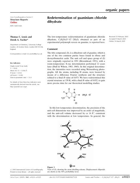
organic papers<strong>Acta</strong> <strong>Cryst</strong>allographica Section EStructure ReportsOnlineISSN 1600-5368Redetermination of guaninium chloridedihydrateThomas C. <strong>Lewis</strong> andDerek A. <strong>Tocher</strong>*Department of Chemistry, University CollegeLondon, 20 Gordon Street, London WC1H 0AJ,EnglandCorrespondence e-mail: d.a.tocher@ucl.ac.ukKey indicatorsSingle-crystal X-ray studyT = 150 KMean (C–C) = 0.002 ÅR factor = 0.032wR factor = 0.082Data-to-parameter ratio = 13.4For details of how these key indicators wereautomatically derived from the article, seehttp://journals.iucr.org/e.The low-temperature redetermination of guaninium chloridedihydrate, C 5 H 6 N 5 O + Cl 2H 2 O, obtained as part of anexperimental polymorph screen on guanine, is reported here.CommentThe title compound, (I), is a dihydrate salt of guanine, which isone of the two common purine bases found in ribose anddeoxyribonucleic acids. The unit cell and space group of (I)were originally reported in 1951 (Broomhead, 1951), with aroom-temperature X-ray determination performed 12 yearslater (Iball & Wilson, 1963, 1965). In this original determination,the intensities were recorded using Weissenberg photographs.All the atoms, including H atoms, were located bymeans of a difference Fourier synthesis and the structurerefined to a final R value of 0.073. We have redetermined thiscrystal structure at 150 K, with a final R value of 0.032, to gainmore precise data for our theoretical modelling studies.Received 25 February 2005Accepted 9 March 2005Online 18 March 2005In this low-temperature determination, the precision of theunit-cell dimensions was improved by an order of magnitude,and the unit-cell volume decreased by ca 14 Å 3 , consistentwith the determination at low temperature. In general, the# 2005 International Union of <strong>Cryst</strong>allographyPrinted in Great Britain – all rights reservedFigure 1View of (I), showing the atom labelling scheme. Displacement ellipsoidsare drawn at the 50% probability level.<strong>Acta</strong> <strong>Cryst</strong>. (2005). E<strong>61</strong>, <strong>o1023</strong>–<strong>o1025</strong> doi:10.1107/S160053680500752X <strong>Lewis</strong> and <strong>Tocher</strong> C 5 H 6 N 5 O + Cl 2H 2 O <strong>o1023</strong>
organic papersFigure 2The hydrogen-bonded (dashed lines) planar unit in (I), showing thecentrosymmetric dimer linked to four water molecules and two Cl ions.The other hydrogen bonds have been omitted for clarity.metric parameters are not significantly different, the exceptionbeing the C1—N2 bond length which is longer in the lowtemperaturestructure, while C1—N5 is shorter in the lowtemperaturestructure, both by ca 0.03 Å. The guanine moleculeis protonated at N1 and N4, with the C—N bond lengthsin the rings ranging from 1.3154 (18) to 1.3892 (18) Å, and theC2—C3, C3—C4 and N5—C1 bond lengths being 1.3797 (18),1.4202 (19) and 1.3291 (18) Å, respectively.The packing consists of centrosymmetric dimers, the twocomponents of which are linked by pairs of N—HNhydrogen bonds. These dimers are linked to four water moleculesand two Cl atoms to form a planar unit (Fig. 2). Theseplanar units are linked to one another through O—HClhydrogen bonds within the plane, forming a ribbon structure,and through N—HCl and N—HO hydrogen bonds at anangle of approximately 80 from this plane, forming a complexthree-dimensional hydrogen-bonded network (Fig. 3). Thetwo H atoms on the NH 2 group form two very dissimilarhydrogen bonds. A strong bond [N5—H7N2 iv =3.0162 (17) Å; see Table 1] is formed by one, while the second[N5—H6Cl1 i = 3.4368 (15) Å; see Table 1] is weak.The N—HN distance within the centrosymmetric dimer is3.0162 (17) Å, with the N—HO distances ranging from2.6463 (17) to 3.0348 (17) Å. The N—HCl distances are3.1281 (13) and 3.4368 (15) Å, and the O—HCl distancesrange from 3.1173 (14) to 3.1576 (13) Å. The O—HOhydrogen bond involving the carbonyl group is 2.7404 (15) Å.ExperimentalAs part of an experimental polymorph screen on guanine, (I) wasobtained from a solution of guanine in dilute hydrochloric acid whichwas allowed to evaporate at room temperature (10 ml solution, in 75 25 mm vessels), forming block-shaped crystals. If the same guaninesolution in dilute hydrochloric acid was allowed to evaporate at aslower rate by virtue of a smaller surface area, small block-likecrystals of guaninium chloride monohydrate were obtained.<strong>Cryst</strong>al dataC 5 H 6 N 5 O + Cl 2H 2 OM r = 223.63Monoclinic, P2 1 =ca = 4.8587 (11) Åb = 13.228 (3) Åc = 14.<strong>61</strong>2 (3) Å = 93.862 (4) V = 937.0 (4) Å 3Z =4D x = 1.585 Mg m 3Mo K radiationCell parameters from 2645reflections = 2.1–28.2 = 0.40 mm 1T = 150 (2) KBlock, colourless0.42 0.12 0.08 mmFigure 3The crystal packing of (I), showing the complex three-dimensionalhydrogen-bonding network (dashed lines).Data collectionBruker SMART APEXdiffractometerNarrow-frame ! scansAbsorption correction: multi-scan(SADABS; Sheldrick, 1996)T min = 0.850, T max = 0.9697902 measured reflectionsRefinementRefinement on F 2R[F 2 >2(F 2 )] = 0.032wR(F 2 ) = 0.082S = 1.042230 reflections167 parametersAll H-atom parameters refinedTable 1Hydrogen-bonding geometry (Å, ).2230 independent reflections1891 reflections with I >2(I)R int = 0.024 max = 28.2 h = 6 ! 6k = 17 ! 17l = 19 ! 19w = 1/[ 2 (F o 2 ) + (0.0433P) 2+ 0.2271P]where P =(F o 2 +2F c 2 )/3(/) max = 0.001 max = 0.34 e Å 3 min = 0.19 e Å 3D—HA D—H HA DA D—HAN1—H1Cl1 i 0.883 (18) 2.2<strong>61</strong> (19) 3.1281 (13) 167.2 (16)N3—H3O1W ii 0.87 (2) 1.83 (2) 2.6867 (16) 167.8 (19)N4—H4O2W 0.908 (19) 1.76 (2) 2.6463 (17) 164.2 (18)N5—H6O1W iii 0.8<strong>61</strong> (18) 2.518 (17) 3.0348 (17) 119.5 (13)N5—H6Cl1 i 0.8<strong>61</strong> (18) 2.682 (18) 3.4368 (15) 147.2 (14)N5—H7N2 iv 0.886 (19) 2.131 (19) 3.0162 (17) 176.9 (17)O2W—H3WCl1 0.81 (2) 2.36 (2) 3.1576 (13) 167.9 (19)O2W—H4WCl1 v 0.85 (2) 2.27 (2) 3.1173 (14) 169.2 (19)O1W—H1WCl1 0.85 (2) 2.31 (2) 3.1336 (13) 166.0 (17)O1W—H2WO4 0.85 (2) 1.93 (2) 2.7404 (15) 160 (2)Symmetry codes: (i) 1 x; y12; 1 2z; (ii) x 1; 3 2y; 1 2 þ z; (iii) x; y 12; 1 2z; (iv)1 x; 1 y; 1 z; (v) 2 x; 2 y; 1 z.H atoms were refined independently using an isotropic model.o1024 <strong>Lewis</strong> and <strong>Tocher</strong> C 5 H 6 N 5 O + Cl 2H 2 O <strong>Acta</strong> <strong>Cryst</strong>. (2005). E<strong>61</strong>, <strong>o1023</strong>–<strong>o1025</strong>
organic papersData collection: SMART (Bruker, 2000); cell refinement: SAINT(Bruker, 2000); data reduction: SAINT; program(s) used to solvestructure: SHELXS97 (Sheldrick, 1990); program(s) used to refinestructure: SHELXL97 (Sheldrick, 1997); molecular graphics:SHELXTL (Bruker, 2000) and MERCURY (Bruno et al., 2002);software used to prepare material for publication: SHELXL97.This research was supported by the EPSRC in funding astudentship for TCL. The authors acknowledge the ResearchCouncils UK Basic Technology Programme for supporting‘Control and Prediction of the Organic Solid State’. For moreinformation on this work, please visit http://www.cposs.org.uk.ReferencesBroomhead, J. (1951). <strong>Acta</strong> <strong>Cryst</strong>. 4, 92–100.Bruker (2000). SMART, SAINT and SHELXTL. Bruker AXS Inc., Madison,Wisconsin, USA.Bruno, I. J., Cole, J. C., Edgington, P. R., Kessler, M. K., Macrae, C. F., McCabe,P., Pearson, J. & Taylor, R. (2002). <strong>Acta</strong> <strong>Cryst</strong>. B58, 389–397.Iball, J. & Wilson, H. R. (1963). Nature (London), 4886, 1193–1195.Iball, J. & Wilson, H. R. (1965). Proc. R. Soc. London Ser. A, 288, 418–439.Sheldrick, G. M. (1990). <strong>Acta</strong> <strong>Cryst</strong>. A46, 467–473.Sheldrick, G. M. (1996). SADABS. University of Göttingen, Germany.Sheldrick, G. M. (1997). SHELXL97. University of Göttingen, Germany.<strong>Acta</strong> <strong>Cryst</strong>. (2005). E<strong>61</strong>, <strong>o1023</strong>–<strong>o1025</strong> <strong>Lewis</strong> and <strong>Tocher</strong> C 5 H 6 N 5 O + Cl 2H 2 O <strong>o1025</strong>