In vivo bone response and mechanical evaluation of ... - Dutch
In vivo bone response and mechanical evaluation of ... - Dutch
In vivo bone response and mechanical evaluation of ... - Dutch

You also want an ePaper? Increase the reach of your titles
YUMPU automatically turns print PDFs into web optimized ePapers that Google loves.
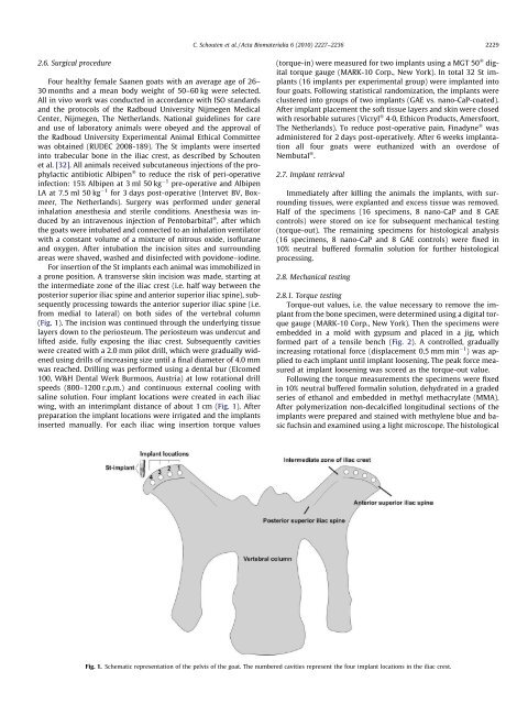
C. Schouten et al. / Acta Biomaterialia 6 (2010) 2227–2236 22292.6. Surgical procedureFour healthy female Saanen goats with an average age <strong>of</strong> 26–30 months <strong>and</strong> a mean body weight <strong>of</strong> 50–60 kg were selected.All in <strong>vivo</strong> work was conducted in accordance with ISO st<strong>and</strong>ards<strong>and</strong> the protocols <strong>of</strong> the Radboud University Nijmegen MedicalCenter, Nijmegen, The Netherl<strong>and</strong>s. National guidelines for care<strong>and</strong> use <strong>of</strong> laboratory animals were obeyed <strong>and</strong> the approval <strong>of</strong>the Radboud University Experimental Animal Ethical Committeewas obtained (RUDEC 2008-189). The St implants were insertedinto trabecular <strong>bone</strong> in the iliac crest, as described by Schoutenet al. [32]. All animals received subcutaneous injections <strong>of</strong> the prophylacticantibiotic Albipen Ò to reduce the risk <strong>of</strong> peri-operativeinfection: 15% Albipen at 3 ml 50 kg 1 pre-operative <strong>and</strong> AlbipenLA at 7.5 ml 50 kg 1 for 3 days post-operative (<strong>In</strong>tervet BV, Boxmeer,The Netherl<strong>and</strong>s). Surgery was performed under generalinhalation anesthesia <strong>and</strong> sterile conditions. Anesthesia was inducedby an intravenous injection <strong>of</strong> Pentobarbital Ò , after whichthe goats were intubated <strong>and</strong> connected to an inhalation ventilatorwith a constant volume <strong>of</strong> a mixture <strong>of</strong> nitrous oxide, is<strong>of</strong>lurane<strong>and</strong> oxygen. After intubation the incision sites <strong>and</strong> surroundingareas were shaved, washed <strong>and</strong> disinfected with povidone–iodine.For insertion <strong>of</strong> the St implants each animal was immobilized ina prone position. A transverse skin incision was made, starting atthe intermediate zone <strong>of</strong> the iliac crest (i.e. half way between theposterior superior iliac spine <strong>and</strong> anterior superior iliac spine), subsequentlyprocessing towards the anterior superior iliac spine (i.e.from medial to lateral) on both sides <strong>of</strong> the vertebral column(Fig. 1). The incision was continued through the underlying tissuelayers down to the periosteum. The periosteum was undercut <strong>and</strong>lifted aside, fully exposing the iliac crest. Subsequently cavitieswere created with a 2.0 mm pilot drill, which were gradually widenedusing drills <strong>of</strong> increasing size until a final diameter <strong>of</strong> 4.0 mmwas reached. Drilling was performed using a dental bur (Elcomed100, W&H Dental Werk Burmoos, Austria) at low rotational drillspeeds (800–1200 r.p.m.) <strong>and</strong> continuous external cooling withsaline solution. Four implant locations were created in each iliacwing, with an interimplant distance <strong>of</strong> about 1 cm (Fig. 1). Afterpreparation the implant locations were irrigated <strong>and</strong> the implantsinserted manually. For each iliac wing insertion torque values(torque-in) were measured for two implants using a MGT 50 Ò digitaltorque gauge (MARK-10 Corp., New York). <strong>In</strong> total 32 St implants(16 implants per experimental group) were implanted int<strong>of</strong>our goats. Following statistical r<strong>and</strong>omization, the implants wereclustered into groups <strong>of</strong> two implants (GAE vs. nano-CaP-coated).After implant placement the s<strong>of</strong>t tissue layers <strong>and</strong> skin were closedwith resorbable sutures (Vicryl Ò 40, Ethicon Products, Amersfoort,The Netherl<strong>and</strong>s). To reduce post-operative pain, Finadyne Ò wasadministered for 2 days post-operatively. After 6 weeks implantationall four goats were euthanized with an overdose <strong>of</strong>Nembutal Ò .2.7. Implant retrievalImmediately after killing the animals the implants, with surroundingtissues, were explanted <strong>and</strong> excess tissue was removed.Half <strong>of</strong> the specimens (16 specimens, 8 nano-CaP <strong>and</strong> 8 GAEcontrols) were stored on ice for subsequent <strong>mechanical</strong> testing(torque-out). The remaining specimens for histological analysis(16 specimens, 8 nano-CaP <strong>and</strong> 8 GAE controls) were fixed in10% neutral buffered formalin solution for further histologicalprocessing.2.8. Mechanical testing2.8.1. Torque testingTorque-out values, i.e. the value necessary to remove the implantfrom the <strong>bone</strong> specimen, were determined using a digital torquegauge (MARK-10 Corp., New York). Then the specimens wereembedded in a mold with gypsum <strong>and</strong> placed in a jig, whichformed part <strong>of</strong> a tensile bench (Fig. 2). A controlled, graduallyincreasing rotational force (displacement 0.5 mm min 1 ) was appliedto each implant until implant loosening. The peak force measuredat implant loosening was scored as the torque-out value.Following the torque measurements the specimens were fixedin 10% neutral buffered formalin solution, dehydrated in a gradedseries <strong>of</strong> ethanol <strong>and</strong> embedded in methyl methacrylate (MMA).After polymerization non-decalcified longitudinal sections <strong>of</strong> theimplants were prepared <strong>and</strong> stained with methylene blue <strong>and</strong> basicfuchsin <strong>and</strong> examined using a light microscope. The histologicalFig. 1. Schematic representation <strong>of</strong> the pelvis <strong>of</strong> the goat. The numbered cavities represent the four implant locations in the iliac crest.