Repair of amalgam restorations with conventional and ... - SciELO
Repair of amalgam restorations with conventional and ... - SciELO
Repair of amalgam restorations with conventional and ... - SciELO
Create successful ePaper yourself
Turn your PDF publications into a flip-book with our unique Google optimized e-Paper software.
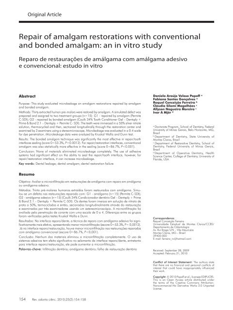
Original Article<strong>Repair</strong> <strong>of</strong> <strong>amalgam</strong> <strong>restorations</strong><strong>Repair</strong> <strong>of</strong> <strong>amalgam</strong> <strong>restorations</strong> <strong>with</strong> <strong>conventional</strong><strong>and</strong> bonded <strong>amalgam</strong>: an in vitro studyReparo de restaurações de amálgama com amálgama adesivoe convencional: estudo in vitroAbstractPurpose: This study evaluated microleakage on <strong>amalgam</strong> <strong>restorations</strong> repaired by <strong>amalgam</strong><strong>and</strong> bonded <strong>amalgam</strong>.Methods: Thirty extracted human pre-molars were restored by <strong>amalgam</strong>. A simulated defect wasprepared <strong>and</strong> assigned to two treatment groups (n=15): G1 - repaired by <strong>amalgam</strong> (PermiteC-SDI); G2 - repaired by bonded <strong>amalgam</strong> (Caulk 34% Tooth Conditioner Gel – Dentsply +Prime & Bond 2.1 – Dentsply + Permite C- SDI). The teeth were immersed in a 50% silver nitratesolution, thermocycled <strong>and</strong> then, sectioned longitudinally through the restoration center <strong>and</strong>examined by 3 examiners using a stereomicroscope. Microleakage was evaluated in a 0-4 scalefor dye penetration. Microleakage data were analyzed by Kruskal Wallis <strong>and</strong> Dunn test.Results: The bonded <strong>amalgam</strong> technique was significantly the most effective in repair/toothinterfaces sealing (score 0=53.3%, P=0.0012). For repair/restoration interfaces, <strong>conventional</strong><strong>amalgam</strong> was also statistically more effective in the sealing (score 0=86.7%, P
Pop<strong>of</strong>f et al.IntroductionDental <strong>amalgam</strong> has been used in dentistry for over a century.Despite poor esthetic characteristics, lack <strong>of</strong> adhesion<strong>and</strong> advancements in resin-based composite technology,<strong>amalgam</strong> <strong>restorations</strong> is still one <strong>of</strong> the restorative treatmentoptions in several dental practice (1-3). Such popularity canbe attributed to its good clinical performance, relativelylow cost <strong>and</strong> long-term cost-effectiveness (2). Secondarycaries <strong>and</strong> fractures are common failures related to <strong>amalgam</strong><strong>restorations</strong> <strong>and</strong> represent the main reasons for theirreplacement <strong>of</strong> defective <strong>amalgam</strong> <strong>restorations</strong> (1,3,4).Total replacement is the most common treatment fordefective <strong>amalgam</strong> <strong>restorations</strong> (1,4) <strong>and</strong> represents amajor part <strong>of</strong> restorative dental treatment. However, thisapproach contradicts the current trend for more conservativeprocedures to minimize the chances <strong>of</strong> pulpal injuries <strong>and</strong> tosave tooth structures. An aim for current restorative dentistryis to maintain <strong>restorations</strong>, i.e., to work <strong>with</strong> materials <strong>and</strong>techniques that allow the repair <strong>of</strong> localized defects (5).<strong>Repair</strong> is an alternative option for treatment <strong>of</strong> defective<strong>amalgam</strong> restoration. It involves the removal <strong>of</strong> part <strong>of</strong> therestoration <strong>and</strong> any defective tissue adjacent to the defectivearea <strong>and</strong> restoration <strong>of</strong> the prepared site (6). This procedureallows preservation <strong>of</strong> sound tooth structure (7).Marginal sealing <strong>of</strong> <strong>amalgam</strong> <strong>restorations</strong> remains achallenge in clinical practice (1,8,9). The use <strong>of</strong> <strong>amalgam</strong>bonding agents has become a popular clinical practice in therestoration <strong>of</strong> posterior teeth, showing potential advantagesincluding tooth reinforcement, decreased postoperativesensitivity, better marginal adaptation, decreased microleakage,reduced possibility <strong>of</strong> secondary caries <strong>and</strong> moreconservative preparation (10-12).Many resin adhesives have been employed <strong>and</strong> successfulreports indicate their effectiveness as <strong>amalgam</strong> bondingagents. Bond strength values <strong>and</strong> sealing data varyconsiderably among the materials employed based on theway they are applied (12), <strong>and</strong> possible influence by cavitysize, indicating that bond strength is inversely proportionalto the bonding area (3,13).Meanwhile, in a recent systematic review, authorsconcluded that there is no evidence to either claim or refutea difference in survival between bonded <strong>and</strong> non-bonded<strong>amalgam</strong> <strong>restorations</strong> (14). In view <strong>of</strong> the lack <strong>of</strong> evidenceon the additional benefit <strong>of</strong> adhesively bonding <strong>amalgam</strong> incomparison <strong>with</strong> non-bonded <strong>amalgam</strong>, it is important toinvestigate if is desirable to use this technique for specificsituations like for making repairs. Despite the limitations <strong>of</strong>in vitro studies in predicting clinical conditions, this studymay help in the construction <strong>of</strong> the scientific evidence bodyto justify or not the inclusion <strong>of</strong> bonded <strong>amalgam</strong> in thetherapeutic arsenal <strong>of</strong> dentistry.Thus, the objectives <strong>of</strong> this study were to evaluate themicroleakage <strong>of</strong> <strong>amalgam</strong> repairs when a bonding agent isapplied. The null hypothesis tested was the microleakageat <strong>amalgam</strong> repair margins is not affected by the using <strong>of</strong>bonding agents.MethodsThis study was approved by the UNIMONTES EthicsCommittee, number 105/19-07-2004.Teeth SelectionThirty non-carious human premolars freshly extracted fororthodontics purposes were employed in this study. The rootswere cleaned by scraping to remove debris <strong>and</strong> disinfectedin 0.5% thymol solution before use.Specimen PreparationClass I cavity preparations were cut on the occlusal surface,2mm wide X 4mm deep X 3mm long, using a high speedh<strong>and</strong>piece <strong>with</strong> air-water coolant <strong>and</strong> a carbide plain fissurebur # 245 (KG Sorensen Ind & Com Ltda, Barueri, SPBrazil). The burs were replaced after five cavity preparations.Preparation dimensions were measured <strong>with</strong> a periodontalprobe to maintain uniformity. One operator prepared all teethto ensure a consistent calibrated size <strong>and</strong> depth in order tominimize preparation variability.Restorative procedureThe teeth were then restored by an admixed, high copper<strong>amalgam</strong> alloy (Permite C, SDI, Bayswater, Victoria,Australia) that was h<strong>and</strong> condensed into the preparationscovering all walls <strong>and</strong> cavosurface margins, then carvedto the tooth contour <strong>with</strong> a sharp carve. Seventy two hourslater, <strong>restorations</strong> were polished <strong>and</strong> stored in saline solutionat 37º C. All restorative procedures were performed by onetrained operator.<strong>Repair</strong> procedureNew Class I cavity preparations (1mm wide X 2mm deep X3mm long) were prepared along the cavosurface margin <strong>of</strong>the <strong>amalgam</strong> <strong>restorations</strong> in order to simulate a defect. A highspeed h<strong>and</strong>piece <strong>with</strong> air-water coolant <strong>and</strong> carbide plainfissure burs # 245 (KG Sorensen Ind & Com Ltda, Barueri,SP Brazil) were used. The burs were replaced after five cavitypreparations. Preparation dimensions were measured <strong>with</strong>a periodontal probe to maintain uniformity. One operatorprepared all teeth to ensure a consistent calibrated size <strong>and</strong>depth in order to minimize preparation variability.The teeth were r<strong>and</strong>omly divided into two experimentalgroups (n=15): G1 - Amalgam repairs (Permite C - SDI,Bayswater, Victoria, Australia) <strong>and</strong> G2 - Bonded <strong>amalgam</strong>repairs (Caulk 34% Tooth Conditioner Gel - Dentsply,Milford, Delaware, USA + Prime & Bond 2.1 – Dentsply,Milford, Delaware, USA + Permite C).Thermal Cycling <strong>and</strong> Microleakage testingThe specimens were subjected to thermal cycling for 500cycles between 5ºC <strong>and</strong> 55ºC <strong>with</strong> 60 seconds dwell time.The teeth were apically obturated <strong>with</strong> glass-ionomer (KetacBond – 3M ESPE, St Paul, MN, USA) <strong>and</strong> then coated<strong>with</strong> two layers <strong>of</strong> nail varnish (Niasi, Taboão da Serra, SP,Brazil) leaving the repairs margins uncoated. They wereRev. odonto ciênc. 2010;25(2):154-158 155
<strong>Repair</strong> <strong>of</strong> <strong>amalgam</strong> <strong>restorations</strong>then immersed in a 50% silver nitrate solution for 24 hoursat 37ºC in the absence <strong>of</strong> light. Next, they were washed inrunning water <strong>and</strong> immersed into another vial <strong>with</strong> photodevelopingsolution (Decktol - Kodak, São José dos Campos,SP, Brazil) for 6 hours, under continuous illumination toreduce <strong>and</strong> precipitate silver ions. Each specimen wassectioned longitudinally by a cutting machine (Labcut 1010 -Extec Technologies Inc., USA), in a buccolingual directionthrough the restoration center. Sections were examined <strong>with</strong>a stereomicroscope (Zeiss - Oberkochen, Germany) at 50 Xmagnification by three trained examiners.Microleakage evaluationEach section was graded for microleakage at both repairtooth <strong>and</strong> repair restoration interfaces as follows (Fig. 1)0 - No dye infiltration1 - Dye penetration up to first third <strong>of</strong> the repair axial wall2 - Dye penetration up to second third <strong>of</strong> the repair axialwall3 - Dye penetration onto repair axial wall4 - Dye penetration onto pulpal wall <strong>of</strong> the repair.Statistical AnalysisThe agreement between examiners was evaluated by Cohen’sKappa test (K= 0.78 to 1.00). For the microleakage data, thescores were subjected to statistical analysis using Kruskal-Wallis <strong>and</strong> Dunn tests (P< 0.05).ResultsThe non-parametric test Kruskal-Wallis detected significantdifferences between the tested materials for both repair/tooth(P=0.000) <strong>and</strong> repair/restoration (P


