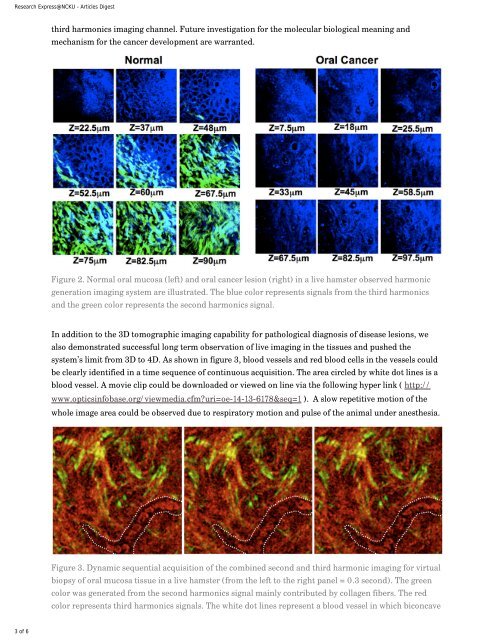
<strong>Research</strong> <strong>Express@NCKU</strong> - <strong>Articles</strong> <strong>Digest</strong>third harmonics imaging channel. Future investigation for the molecular biological meaning andmechanism for the cancer development are warranted.Figure 2. Normal oral mucosa (left) and oral cancer lesion (right) in a live hamster observed harmonicgeneration imaging system are illustrated. The blue color represents signals from the third harmonicsand the green color represents the second harmonics signal.In addition to the 3D tomographic imaging capability for pathological diagnosis of disease lesions, wealso demonstrated successful long term observation of live imaging in the tissues and pushed thesystem’s limit from 3D to 4D. As shown in figure 3, blood vessels and red blood cells in the vessels couldbe clearly identified in a time sequence of continuous acquisition. The area circled by white dot lines is ablood vessel. A movie clip could be downloaded or viewed on line via the following hyper link ( http://www.opticsinfobase.org/viewmedia.cfm?uri=oe-14-13-6178&seq=1 ). A slow repetitive motion of thewhole image area could be observed due to respiratory motion and pulse of the animal under anesthesia.Figure 3. Dynamic sequential acquisition of the combined second and third harmonic imaging for virtualbiopsy of oral mucosa tissue in a live hamster (from the left to the right panel = 0.3 second). The greencolor was generated from the second harmonics signal mainly contributed by collagen fibers. The redcolor represents third harmonics signals. The white dot lines represent a blood vessel in which biconcave3 of 6
<strong>Research</strong> <strong>Express@NCKU</strong> - <strong>Articles</strong> <strong>Digest</strong>shaped red blood cells flow through. Live images in video format are available online (http://www.opticsinfobase.org/viewmedia.cfm?uri=oe-14-13-6178&seq=1).Although THG nonlinearity exists in all bio-materials, the Gouy phase shift effect substantially limitsTHG to be observed in the vicinity of interfaces where the first order or third order susceptibilitydiscontinues. Therefore, THG is generally regarded as a morphological imaging tool due to this, withlimited capability for molecular imaging. It is thus highly desirable to develop exogenous THG contrastagents to trace the functions of a specific molecule, taking advantage of the noninvasive nature of theTHG process. Recently, noble metal nanoparticles have been proved to be able to enhance variousnonlinear optical signals through surface plasmon resonance. Hence, it should be ideal to adoptnanoparticles as molecular contrast agents of THG microscopy.With the help of surface plasmon-resonance, nanometer-sized noble metals can serve as a nanoscopicoptical resonant cavity. Metal nanoparticles with the plasmon resonance at the third harmonic of opticalexcitation, in the macroscopic point of view, is analogous to an optical third-harmonic oscillator. Wechose silver nanoparticles for its blue-violet plasmon resonance wavelengths when soaked in water. Fornonlinear biological in vivo imaging, near-infrared (NIR) femtosecond lasers as the THG excitationsources are preferred to increase the penetration depth and to reduce the potential optical damage. Inparticular, it has been demonstrated that the spectral transmission window of common biological tissuesfalls around 1200–1300 nm. With a 410-nm surface plasmon wavelength for THG resonance, thecorresponding NIR laser wavelength is 1230 nm, which concurs with the biological transparencywindow. In contrast, gold nanoparticles are with a surface plasmon resonance wavelength of 520– 560nm and the corresponding laser wavelength of 1560–1680 nm could suffer strong water absorption, thusnot suitable for in vivo biological imaging. Our recent spectral study of THG in silver nanoparticlesshowed evident THG enhancement when the third harmonic of the excitation matched the surfaceplasmon resonant frequency of silver nanoparticles, indicating that plasmon-enhanced silvernanoparticles could be an ideal contrast agent for THG microscopy.The Her2 molecule is one of the transmembrane receptor proteins for epidermal growth factors, andplays a key role in cell growth and anti-apoptotic signaling. The Her2 molecule is also important incancer development and clinical prognosis.Overexpression of Her2/ neu in cancer lesion is a predictorfor unfavorable clinical outcome with more aggressive growth behavior as well as resistance to somechemotherapy agents. To demonstrate molecular THG microscopy by using silver nanoparticles asexogenous THG contrast agents, the cultured mouse bladder carcinoma cells (MBT2) and the matchedcell line with knocked-down Her2/neu expression by RNAi were applied here. Western blot analysisrevealed the Her2/neu knocked-down cancer cell line, MBT2-KD, expressed only about 15 % the level ofthe wild type line, as shown in Figure 1a. The anti-Her2 antibodies tagged 30 nm silver nanoparticleswere then incubated with MBT2 and MBT2-KD for imaging of Her2 expression in cancer cells. Figure1b–d show the epi-THG images of the wild type MBT2 cells without and with the silver nanoparticlelabeling, and MBT2-KD cells with silver nanoparticle labeling. By comparing these THG images, brightspots on the wild type cancer cell cytoplasmic membranes can be clearly observed (Fig. 2d), in sharpcontrast to the MBT2-KD line (Fig. 1c) and in proportion to their endogenous Her-2 expression levels(Fig. 1a). Molecule- specific THG microscopy is thus successfully demonstrated by using antibodyconjugatedsilver nanoparticles as exogenous THG contrast agents.The imaged spot size of the silver nanoparticle can be found to be ~308 nm. This number is equal to theconvolution value (309 nm) of the theoretical system resolution and a 30-nm nanoparticle size. With a4 of 6