CET 20 Selecting low vision aids - part 1 - ABDO
CET 20 Selecting low vision aids - part 1 - ABDO
CET 20 Selecting low vision aids - part 1 - ABDO

Create successful ePaper yourself
Turn your PDF publications into a flip-book with our unique Google optimized e-Paper software.
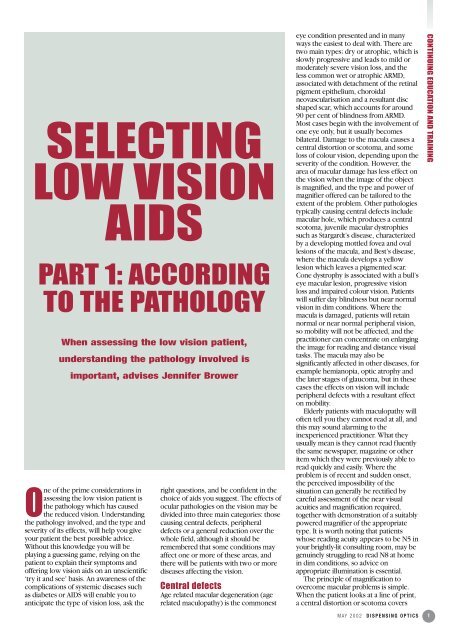
SELECTING<br />
LOW VISION<br />
AIDS<br />
PART 1: ACCORDING<br />
TO THE PATHOLOGY<br />
When assessing the <strong>low</strong> <strong>vision</strong> patient,<br />
understanding the pathology involved is<br />
important, advises Jennifer Brower<br />
One of the prime considerations in<br />
assessing the <strong>low</strong> <strong>vision</strong> patient is<br />
the pathology which has caused<br />
the reduced <strong>vision</strong>. Understanding<br />
the pathology involved, and the type and<br />
severity of its effects, will help you give<br />
your patient the best possible advice.<br />
Without this knowledge you will be<br />
playing a guessing game, relying on the<br />
patient to explain their symptoms and<br />
offering <strong>low</strong> <strong>vision</strong> <strong>aids</strong> on an unscientific<br />
‘try it and see’ basis. An awareness of the<br />
complications of systemic diseases such<br />
as diabetes or AIDS will enable you to<br />
anticipate the type of <strong>vision</strong> loss, ask the<br />
right questions, and be confident in the<br />
choice of <strong>aids</strong> you suggest. The effects of<br />
ocular pathologies on the <strong>vision</strong> may be<br />
divided into three main categories: those<br />
causing central defects, peripheral<br />
defects or a general reduction over the<br />
whole field, although it should be<br />
remembered that some conditions may<br />
affect one or more of these areas, and<br />
there will be patients with two or more<br />
diseases affecting the <strong>vision</strong>.<br />
Central defects<br />
Age related macular degeneration (age<br />
related maculopathy) is the commonest<br />
eye condition presented and in many<br />
ways the easiest to deal with. There are<br />
two main types; dry or atrophic, which is<br />
s<strong>low</strong>ly progressive and leads to mild or<br />
moderately severe <strong>vision</strong> loss, and the<br />
less common wet or atrophic ARMD,<br />
associated with detachment of the retinal<br />
pigment epithelium, choroidal<br />
neovascularisation and a resultant disc<br />
shaped scar, which accounts for around<br />
90 per cent of blindness from ARMD.<br />
Most cases begin with the involvement of<br />
one eye only, but it usually becomes<br />
bilateral. Damage to the macula causes a<br />
central distortion or scotoma, and some<br />
loss of colour <strong>vision</strong>, depending upon the<br />
severity of the condition. However, the<br />
area of macular damage has less effect on<br />
the <strong>vision</strong> when the image of the object<br />
is magnified, and the type and power of<br />
magnifier offered can be tailored to the<br />
extent of the problem. Other pathologies<br />
typically causing central defects include<br />
macular hole, which produces a central<br />
scotoma, juvenile macular dystrophies<br />
such as Stargardt’s disease, characterized<br />
by a developing mottled fovea and oval<br />
lesions of the macula, and Best’s disease,<br />
where the macula develops a yel<strong>low</strong><br />
lesion which leaves a pigmented scar.<br />
Cone dystrophy is associated with a bull’s<br />
eye macular lesion, progressive <strong>vision</strong><br />
loss and impaired colour <strong>vision</strong>. Patients<br />
will suffer day blindness but near normal<br />
<strong>vision</strong> in dim conditions. Where the<br />
macula is damaged, patients will retain<br />
normal or near normal peripheral <strong>vision</strong>,<br />
so mobility will not be affected, and the<br />
practitioner can concentrate on enlarging<br />
the image for reading and distance visual<br />
tasks. The macula may also be<br />
significantly affected in other diseases, for<br />
example hemianopia, optic atrophy and<br />
the later stages of glaucoma, but in these<br />
cases the effects on <strong>vision</strong> will include<br />
peripheral defects with a resultant effect<br />
on mobility.<br />
Elderly patients with maculopathy will<br />
often tell you they cannot read at all, and<br />
this may sound alarming to the<br />
inexperienced practitioner. What they<br />
usually mean is they cannot read fluently<br />
the same newspaper, magazine or other<br />
item which they were previously able to<br />
read quickly and easily. Where the<br />
problem is of recent and sudden onset,<br />
the perceived impossibility of the<br />
situation can generally be rectified by<br />
careful assessment of the near visual<br />
acuities and magnification required,<br />
together with demonstration of a suitably<br />
powered magnifier of the appropriate<br />
type. It is worth noting that patients<br />
whose reading acuity appears to be N5 in<br />
your brightly-lit consulting room, may be<br />
genuinely struggling to read N8 at home<br />
in dim conditions, so advice on<br />
appropriate illumination is essential.<br />
The principle of magnification to<br />
overcome macular problems is simple.<br />
When the patient looks at a line of print,<br />
a central distortion or scotoma covers<br />
MAY <strong>20</strong>02 DISPENSING OPTICS<br />
CONTINUING EDUCATION AND TRAINING<br />
1
CONTINUING EDUCATION AND TRAINING<br />
2<br />
some of the letters and the word cannot<br />
be read. When the image is magnified,<br />
the letters appear larger but the scotoma<br />
remains the same size and so has less of<br />
an impact on the words. The exact<br />
position of a scotoma will affect the<br />
amount of magnification required. If it is<br />
to the right of centre, higher<br />
magnification may be needed where the<br />
patient is reading left to right and directly<br />
into the scotoma. If the scotoma is to the<br />
left of centre, less magnification may be<br />
required. There is a wide range of<br />
magnifying <strong>aids</strong> available to help patients<br />
with central defects. These include<br />
illuminated and non-illuminated hand and<br />
stand magnifiers, spectacle magnifiers,<br />
telescopes for distance and near <strong>vision</strong>,<br />
flat field magnifiers, pocket magnifiers,<br />
bar magnifiers and clip-on loupes. CCTV<br />
systems are becoming more widely<br />
available, and there are less expensive<br />
portable systems, where the camera is<br />
linked to the patient’s own TV.<br />
Computers can be programmed with<br />
large font sizes and the print size of emails<br />
can be increased. Children with<br />
macular problems but normal<br />
accommodation may be able to achieve<br />
enough magnification by bringing the<br />
page closer and/or photocopying pages<br />
of print two or three times the size. Very<br />
young children will be unable to cope<br />
with sophisticated magnifying <strong>aids</strong>, and<br />
do not usually have to read very small<br />
print. Older children will generally use a<br />
variety of <strong>aids</strong>: a distance telescope for<br />
travelling to school by bus or train, a<br />
computer or CCTV at school, a stand<br />
magnifier for reading and writing<br />
homework and a small hand or pocket<br />
magnifier for the shops.<br />
Children generally have a positive<br />
attitude to their visual problems, and<br />
accept <strong>low</strong> <strong>vision</strong> <strong>aids</strong> with enthusiasm,<br />
although occasionally children will resist<br />
using <strong>aids</strong> because they do not want to<br />
seem ‘different’ from their normally<br />
sighted friends. A fourteen-year-old<br />
schoolgirl who needs a telescope to read<br />
bus numbers may be persuaded with a<br />
small monocular fitted with a finger ring.<br />
This can be hidden in the palm of the<br />
hand and brought up to the eye when<br />
needed. A small domed flat field<br />
magnifier can also be palmed effectively,<br />
and produced when necessary to read<br />
the price on a magazine or CD.<br />
The amount of magnification selected<br />
will vary in relation to the acuity levels<br />
and visual task, for example a patient<br />
with a reading acuity of N48 may use a<br />
6x spectacle magnifier to read a<br />
newspaper but only need 3x to read a<br />
large print book. An insulin dependent<br />
diabetic with N36 may use a 7x clip on<br />
loupe to read syringe markings but a 3x<br />
stand magnifier to fill in a crossword.<br />
Non-optical <strong>aids</strong> are also useful for<br />
patients with central defects. One widely<br />
used is a typoscope. This is a matt black<br />
card, approximately the size of a credit<br />
DISPENSING OPTICS MAY <strong>20</strong>02<br />
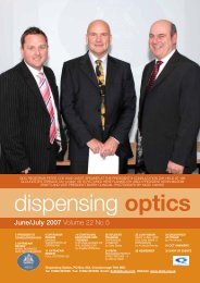
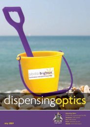
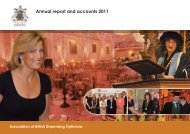
Effective use of<br />
illumination<br />
▲<br />
card, with a central horizontal slit, which<br />
can be used as a signature guide or line<br />
guide for reading. Contrast is improved<br />
by the black border surrounding the print<br />
on the white page. Larger typoscopes are<br />
available for use with pension books and<br />
writing cheques. Other non-optical <strong>aids</strong><br />
include illumination, filters, large print<br />
books, big number telephones and large<br />
print playing cards, and writing <strong>aids</strong> such<br />
as broad lined paper and thick felt pens.<br />
Where high levels of magnification are<br />
required, it is impractical to expect<br />
patients to sustain lengthy periods of<br />
reading, and <strong>aids</strong> which make use of<br />
senses other than sight can be very<br />
helpful. Some examples are ‘talking’<br />
books, newspapers and watches, ‘voices’<br />
on electronic diaries and computers, and<br />
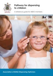
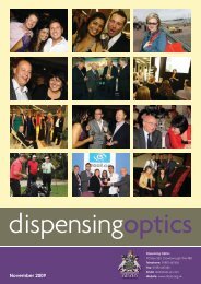
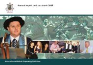
▲ Pocket magnifiers<br />
▼ A selection of filters<br />
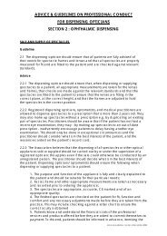
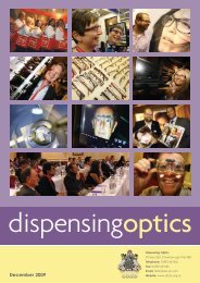
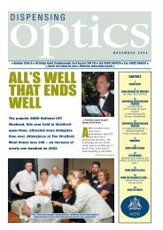
▲ Poppy - a guide dog in training (Photograph by kind permission of Guide Dogs for the Blind Association)
tactile <strong>aids</strong> as diverse as needle threaders,<br />
pavement bumps by zebra crossings,<br />
using different shaped containers for tea,<br />
coffee and sugar and rubber bands<br />
around tins of pet food. High powered<br />
magnifiers may still be given for ‘survival’<br />
reading, such as utility bills, bank<br />
statements and letters, and cooking times<br />
on food packs, and these will help the<br />
patient maintain some independence and<br />
privacy.<br />
Peripheral defects<br />
Glaucoma is the most common pathology<br />
causing loss of field and occurs from<br />
damage to the optic nerve. It is often<br />
associated with increased ocular<br />
pressure, although around 50 per cent of<br />
glaucomas are now seen in patients with<br />
normal or <strong>low</strong> ocular tensions. Acute<br />
attacks must be treated immediately. In<br />
chronic simple glaucoma, the visual field<br />
constricts, at first without the patient<br />
noticing, and if untreated, may reduce<br />
until only tiny central and temporal areas<br />
of <strong>vision</strong> remain. In some cases glaucoma<br />
may cause complete loss of <strong>vision</strong>. Other<br />
pathologies causing varying degrees of<br />
field loss are retinitis pigmentosa and<br />
other retinal dystrophies, hemianopias<br />
and optic atrophy. Magnification is not<br />
appropriate for these conditions, unless<br />
the central <strong>vision</strong> becomes involved, but<br />
the patient’s mobility may be seriously<br />
affected, so a guide cane may be<br />
provided. Where the central acuity is<br />
unaffected, a reverse telescope may be<br />
useful. By reversing a distance telescope,<br />
the image is minified but the field of view<br />
increased, so a patient looking at a notice<br />
board will be able to see more<br />
information at once. Used in this way a<br />
telescope is known as a field expander.<br />
Other types of field expander are mirror<br />
or prism systems, used in cases of<br />
hemianopia to reflect or refract more<br />
information into the patient’s field of<br />
view.<br />
Reduced <strong>vision</strong> over the<br />
whole field<br />
Cataract, corneal dystrophy and some<br />
retinal dystrophies cause a general<br />
reduction in <strong>vision</strong>. Diabetic retinopathy<br />
is associated with neovascularisation and<br />
haemorrhage – which causes patchy field<br />
loss and black spots in the visual field –<br />
traction of the vitreous – which induces<br />
apparent flashes of light – and retinal<br />
detachment. The symptoms tend to<br />
fluctuate. Diabetic retinopathy may affect<br />
the macula, although laser<br />
photocoagulation is often given as a<br />
preventative measure. New, fragile blood<br />
vessels around the macula are destroyed<br />
in an attempt to prevent invasion. High<br />
myopia has a high incidence of retinal<br />
detachment, other effects are macular<br />
change and retinal haemorrhage leading<br />
to reduced acuity and constricted visual<br />
fields. Around 75% of patients with AIDS<br />
develop ocular problems and about 40%<br />
develop cytomegalovirus retinitis (CMV).<br />
Signs include retinal haemorrhage and<br />
opacification and sometimes total retinal<br />
atrophy or detachment.<br />
Albinism is often associated with<br />
nystagmus, reduced acuity and<br />
photophobia. For severe photophobia<br />
filters are available as dark as 1% LTF, but<br />
where the acuity is seriously reduced,<br />
this should be taken into account to<br />
prevent mobility problems. An alternative<br />
solution is an opaque lens with a<br />
stenopaic slit to admit minimal light.<br />
Where there are lens and/or other media<br />
opacities, the patient may suffer a<br />
disability glare, <strong>part</strong>icularly in bright<br />
sunlight, where light entering the eye is<br />
scattered by these opacities, reducing<br />
contrast, and causing a white glare effect.<br />
Amber filters which absorb blue light can<br />
improve contrast significantly. Preventing<br />
light entering the eye is also<br />
recommended by wearing a sun visor or<br />
large brimmed hat, or tinted spectacles<br />
with sideshields. A guide cane may be<br />
given in cases of very <strong>low</strong> acuity. Some<br />
patients will also have a guide dog.<br />
Magnification can also be useful where<br />
the whole field is affected – the larger<br />
and bolder the image, the easier it is to<br />
see – but where contrast sensitivity is<br />
<strong>low</strong>, and in cases of photophobia, care<br />
must be taken to avoid flooding the eye<br />
with light, so non-illuminated magnifiers,<br />
often in conjunction with a typoscope,<br />
may prove more successful than<br />
illuminated units.<br />
The older the patient, the more likely<br />
that the ocular pathology will not be the<br />
only disease present. Some patients will<br />
be arthritic and unable to grasp a<br />
magnifier handle or focus a monocular, or<br />
have a head or hand tremor. Other<br />
patients may have hearing difficulties,<br />
general mobility problems or<br />
communication difficulties caused by a<br />
stroke. It is not unusual to see a<br />
combination of ocular pathologies –<br />
glaucoma and macular degeneration, high<br />
myopia and cataract, macular<br />
degeneration and cataract – so the type<br />
of <strong>aids</strong> offered must be carefully selected<br />
for the patient’s maximum benefit and<br />
minimum inconvenience. Regular review<br />
is important to check if the pathology has<br />
progressed and acuity levels changed,<br />
requiring different or additional <strong>aids</strong>, and<br />
children should be monitored frequently.<br />
Patients with age-related cataract,<br />
glaucoma and diabetic retinopathy are<br />
likely to have greater visual changes than<br />
a patient with dry macular degeneration<br />
but the priority in the selection of <strong>aids</strong><br />
must be to understand the pathology<br />
involved, its effects on <strong>vision</strong> and the<br />
types of <strong>aids</strong> which will best meet the<br />
needs of the individual patient.<br />
Reference<br />
J J Kanski, ‘Clinical Ophthalmology’ 4th<br />
edition, 1999, Butterworth-Heinemann.<br />
Jennifer Brower FBDO(Hons)LVA Cert Ed is a<br />
dispensing optician and <strong>low</strong> <strong>vision</strong><br />
practitioner, working in private practice<br />
and the hospital eye service. She is a tutor,<br />
theory and practical examiner for the <strong>ABDO</strong><br />
Honours Diploma in Low Visual Acuity and<br />
lectures regularly on <strong>low</strong> <strong>vision</strong>. Jennifer is<br />
a member of the GOC, chairman of the LVA<br />
committee, a <strong>CET</strong> approver and a member of<br />
Council, and is a member of the editorial<br />
advisory board of Dispensing Optics. ■<br />
MAY <strong>20</strong>02 DISPENSING OPTICS<br />
CONTINUING EDUCATION AND TRAINING<br />
3