better count fast
better count fast
better count fast
- No tags were found...

You also want an ePaper? Increase the reach of your titles
YUMPU automatically turns print PDFs into web optimized ePapers that Google loves.
SALES BROCHURE<strong>better</strong> <strong>count</strong> <strong>fast</strong>easy and reliable cell <strong>count</strong>ingCountess ® Automated Cell Counter
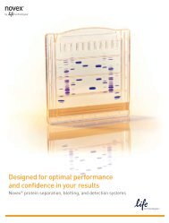
Consistent. Fast. Easy.The Countess ® Automated Cell Counter takes the subjectivity andtedium out of one of the fundamental steps of cell culture—<strong>count</strong>inglive and dead cells. By reducing user error and speeding up cell<strong>count</strong>ing, the Countess ® Automated Cell Counter has revolutionizedhow researchers have <strong>count</strong>ed cells with less sample and less hassle.Why use the Countess ® Automated Cell Counter?With the Countess ® Automated Cell Counter, producingquality reproducible data is <strong>fast</strong> and easy. It is alsoinexpensive and small, and can easily fit into a lab’sbudget and workspace.• Accurate—eliminate the subjectivity of manual cell<strong>count</strong>ing; minimize guessing and user-to-user variability• Fast—<strong>count</strong>s live and dead cells, measures viability andaverage cell size in 30 seconds with just 10 μL of sample• Convenient—no setup, cleaning, or service required• Adaptable—optimize <strong>count</strong>ing protocols for yourspecific cell line• Reliable—gating allows <strong>count</strong>ing of specific cell types inmixed populations• The small footprint (27 cm (w) x 20 cm (d) x 19 cm (h)),and data archiving function of the Countess ® AutomatedCell Counter make sharing between labs and users easy.In addition, the disposable cell <strong>count</strong>ing chamber slidesminimize exposure of users to biohazardous samples.The Countess ® Automated Cell Counter is ideal for:• Cell culture labs• Flow cytometry core facilities• HTS labs• HIV and other infectious disease labs• Stem cell research labsThe Countess ® Automated Cell Counter is easy to use (Figure1). Simply pipet the sample into the <strong>count</strong>ing slide (A), insertthe slide into the Countess ® Automated Cell Counter (B),then press “Count cells” (C); results are displayed in 30seconds (D).ABCDFigure 1. The Countess ® Automated Cell Counter workflow. Pipet the sample into the <strong>count</strong>ing slide (A); insert the slide into the Countess ®Automated Cell Counter (B); press “Count cells”(C); results are displayed in 30 seconds (D).
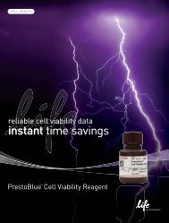
Measured concentration (cells/mL)How does the Countess ® Automated CellCounter work?The Countess ® Automated Cell Counter uses trypan bluestaining combined with a sophisticated image analysisalgorithm to produce accurate cell and viability <strong>count</strong>s inas little as 30 seconds.10 7 Countess ® Automated Cell CounterHemocytometerThe algorithm also measures the average size of live,dead, and total cells, to give you all the data you needto proceed with your experiments. The measurementrange extends from 1 x 10 4 to 1 x 10 7 cells/mL, with anoptimal range from 1 x 10 5 to 4 x 10 6 cells/mL, broaderthan that of a hemocytometer (Figure 2). The optimalcell size is between 5 μm and 60 μm.For accurate viability <strong>count</strong>s, ensure the <strong>count</strong>ing area iscovered with cell suspension and <strong>count</strong> cells within 2 minutesof mixing the cells with trypan blue solution. We highlyrecommend that you use the default setting to begin with foryour cell <strong>count</strong>ing, unless you know the default setting is notsuited to your particular cell sample. For the best data withbiological samples, we recommend <strong>count</strong>ing at least twosamples and taking an average. A handy dilution calculatorhelps you determine how to prepare your sample for yournext passage or experiment.Adjust the focus knob to optimize the image for analysis asfollows (Figure 3):• Live cells have bright centers and dark edges• Dead cells have a uniform blue color throughout the cellwith no bright centers10 610 5 10 510 610 7Cell suspension dilution (cells/mL)Figure 2. Data from the Countess ®Automated Cell Counter extendfurther into the high concentration range than hemocytometerreadings.ABDeadLiveDeadLiveCDeadLiveFigure 3. Correct and incorrect images. (A) Correct image: live cellshave bright centers and dark edges. (B) Incorrect image: dead cells havebright, blue centers and are <strong>count</strong>ed as live. (C) Incorrect image: livecells have dark centers and are <strong>count</strong>ed as dead.
How accurate is the Countess ® AutomatedCell Counter?The Countess ® Automated Cell Counter was compared with the hemocytometer and Coulter Cell and Particle Counter ®methods (Table 1, Figures 4 and 5). Download the complete technical notes including protocols and cell <strong>count</strong>ing tips atwww.invitrogen.com/<strong>count</strong>essdata.Table 1. Comparison between the Countess ® Automated Cell Counter and Coulter Cell and Particle Counter ® method of cell <strong>count</strong>ing.Feature Countess ® Automated Cell Counter Coulter Cell and Particle Counter ®Viability data Provided None providedCell size data Provided None providedResults visualization Provided None providedSetup Easy DifficultCalibration/maintenance None required Required daily/monthly1.4 x 10 61.4 x 10 51.2 x 10 61.2 x 10 5Cells/mL1.6 x 10 51.0 x 10 58 x 10 46 x 10 4Z1 SeriesBeads/mL1 x 10 6 Cell <strong>count</strong>ing method8 x 10 56 x 10 54 x 10 44 x 10 52 x 10 42 x 10 5 00Coulter Counter ®Countess ® AutomatedCell CounterCell <strong>count</strong>ing methodCountess ® GlassAutomated hemocytometerCell CounterDisposablehemocytometerBeadstandardFigure 4. Equivalent results between Countess ®Automated CellCounter and Coulter Cell and Particle Counter ®methods of cell<strong>count</strong>ing. K562 cells were <strong>count</strong>ed using the Z1 Series Coulter Counter ®Cell and Particle Counter and the Countess ® Automated Cell Counter.Cell <strong>count</strong>s from replicate samples were obtained on the Countess ®Automated Cell Counter according to the manufacturer’s instructions.Replicate samples were prepared for analysis on the Coulter Cell andParticle Counter ® instrument by diluting 100 μL of K562 cells into 10 mLof Isoton II Diluent.Figure 5. Accuracy and precision of the Countess ®AutomatedCell Counter compared to glass and disposable hemocytometermethods. A standard bead solution of known concentration (9.57 x 10 5beads/mL, Beckman Coulter) was used as the starting material for<strong>count</strong>ing measurements using the Countess ® Automated Cell Counter,Bright-Line glass hemocytometer (Hausser Scientific), and C-Chipdisposable hemocytometer (Digital Bio). The beads were mixed 1:1 with0.4% trypan blue, and triplicate samples were measured according toeach manufacturer’s instructions. Accuracy was evaluated by comparingthe mean concentration (in cells/mL) measured by each method to theexpected concentration of the standardized bead solution x 10 5 ± 10%beads/mL). Precision is indicated by the standard deviations; error barsrepresent one standard deviation. Accuracy is comparable between theCountess ® Automated Cell Counter and the disposable hemocytometer,and the Countess ® Automated Cell Counter is more precise than thehemocytometers.
What can be <strong>count</strong>ed with the Countess ®Automated Cell Counter?Table 2 lists some of the cell lines validated on theCountess ® Automated Cell Counter. Some other<strong>count</strong>ing capabilities include:White blood cellsThe Countess ® Automated Cell Counter is able to <strong>count</strong>white blood cells from lysed whole blood and Ficoll cellpreparations.Whole blood cells containing non-lysed cellsDilute the blood sample approximately 1:10,000 and <strong>count</strong> in“bead” mode. The Countess ® Automated Cell Counter cannotassess the viability of cells in a whole blood sample.PBMCs (peripheral blood mononuclear cells)The Countess ® Automated Cell Counter can <strong>count</strong> PBMCs.However, it cannot differentiate white blood cell types.RBCs (red blood cells)The Countess ® Automated Cell Counter can <strong>count</strong> RBCs.Dilute the blood sample approximately 1:10,000 and <strong>count</strong> in“bead” mode. The instrument cannot assess the viability ofred blood cells. In addition, the instrument cannot distinguishred blood cells from white blood cells.Read about how to <strong>count</strong> blood cells and download thisapplication note at www.invitrogen.com/<strong>count</strong>essdata.Bookmark this page, as additional application notes areadded regularly.The Countess ® Automated Cell Counter can accurately<strong>count</strong> clumps of cells. In some cases, the actual number ofcells in a clump will be underestimated and the cell size willsometimes be overestimated. However, we have found thatthe Countess ® Automated Cell Counter provides data at leastas accurate as <strong>count</strong>ing with a hemocytometer.Yeast cellsWe have successfully <strong>count</strong>ed Saccharomyces cerevisiae(Fleischmann’s baker’s yeast), a consumer product. However,the instrument cannot distinguish yeast viability.Plant cellsPlant cells have not been tested. If the plant cells fall withinthe optimal size range for the instrument and are notextremely clumpy, the instrument should be able to <strong>count</strong>plant cells.Elongated cellsWe have not tested very irregularly shaped or elongatedcells. If the cells are very irregular or elongated, try the“Parameters” function under the “Settings” menu tovary the circularity. This function alters the way that theimage analysis software recognizes cells. You may need toexperiment with several circularity settings until you findthe one that is perfect for your cell type.Bacterial cellsBacteria are too small to be distinguished from non-celldebris in the Countess ® Automated Cell Counter.Sperm and other <strong>fast</strong>-moving cellsIt is difficult to <strong>count</strong> cells that are moving quickly. TheCountess ® Automated Cell Counter does not <strong>count</strong> livesperm and <strong>fast</strong>-moving protozoa.HepatocytesThe instrument is able to calculate total concentration,but cannot provide viability information on the cells.Set the instrument to “bead” mode, then you canperform the cell <strong>count</strong>ing.
Table 2. Cell lines validated on the Countess ® Automated Cell Counter. For more information on our findings,visit www.invitrogen.com/<strong>count</strong>essdata.Cell type Vendor Cat. No. Animal Organ Cell size (diameter)A431 ATCC CRL-2592 Human Skin 15.5 μmADSC Invitrogen R7788-110 Human Adipose-derive stem cells 13 μmAortic smooth muscle Invitrogen C-007-5C Human Smooth muscle 20 μmBlood, whole lysed Donor NA Human Blood NACHO-M1WT2 ATCC CRL-1984 Chinese hamster Ovary NACHSE ATCC CRL-1861 Chinook salmon Embryo 16 –17 μmCOLO-205 ATCC CCL-243 Human Colon NACOS-7 ATCC CRK-1651 African monkey Kidney NAESC Millipore CMTI-1 Mouse Embryo 10 μmHEK-293 ATCC CRL-1573 Human Kidney 13 μmHEKn Invitrogen C-001-5C Human Neonatal foreskin 12.5 μmHeLa ATCC CCL-2 Human Cervix NAHepG2 ATCC CRL-10741 Human Liver 18 μmHESC WiCell NA Human Enbryo 12–13 μmHL-60 ATCC CCL-240 Human Blood NAJ774A.1 ATCC TIB-67 Mouse Blood 13–14 μmJurkat ATCC TIB-152 Human Blood 12 μmK-562 ATCC CCL-243 Human Bone marrow NAMCF7 ATCC HTB-22 Human Breast 20–24 μmMRC-5 ATCC CCL-171 Human Lung 18 μmNIH/3T3 ATCC CRL-1658 Human Embryo 18 μmNSC Invitrogen N7744-100 Rat Brain 11 μmPC-12 ATCC CRL-1721 Rat Adrenal gland 12–14 μmPulmonary artery endothelial Invitrogen C-008-5C Human Blood vessel 13 μmPulmonary artery smooth muscle Invitrogen C-009-5C Human Smooth muscle 20 μmSF-21 Invitrogen 12682-019 Insect Ovary NAU266 ATCC TIB-196 Human Blood 12–13 μmU-2 OS ATCC HTB-96 Human Bone NAUmbilical vein endothelial Invitrogen C-015-5C Human Blood vessel 17 μm
Has the Countess ® Automated Cell Counter been referenced in peer-reviewed publications?The Countess ® Automated Counter has been referenced in over 100 publications using over 120 different cell types from 18different species. For a complete list of references, visit www.invitrogen.com/<strong>count</strong>ess.References• Bovine chondrocytes, primaryMhanna RF et al. (2011) Layer-by-Layer Films Made fromExtracellular Matrix Macromolecules on Silicone Substrates.Biomacromolecules. 12:609–616.• Canine MDCK kidney epithelial cellsKakiashvili E et al. (2011) The epidermal growth factor receptor mediatesTumor Necrosis Factor-α induced activation of the ERK/GEF-H1/RhoA pathway in tubular epithelium. JBC. 286:9268-9279.• Dictyostelium discoidum, amoebaeFumanelli L et al. (2011) Mathematical modeling of bacterial virulenceand host–pathogen interactions in the Dictyostelium/Pseudomonassystem. J Theor. Bio. 270:19-24.• Human A549 Adenocarcinomic alveolar basal epithelial cellsGarcía-Álvarez I et al. (2011) Lipid and ganglioside alterations intumor cells treated with antimitotic oleyl glycoside. Mol. Biosys.7:129-138.• Human colorectal carcinoma cellsWilliams BS et al. (2010) Mediated Decay Resistant Mutations Are aSource of Expressed Mutant Proteins in Colon Cancer Cell Lines withMicrosatellite Instability. PLoS ONE doi:10.1371/journal.pone.0016012• Human DU145 prostate cancer cellsChen H-M et al. (2011) A novel synthetic protoapigenone analogue,WYC02-9, induces DNA damage and apoptosis in DU145 prostatecancer cells through generation of reactive oxygen species. Free RadBio Med. 50:1151-1162.• Human EOL-1 eosinophilic leukemic cell lineKahn J-E et al. (2011) Comparative Proteomic Analysis of BloodEosinophils Reveals Redox Signaling Modifications in Patients withFIP1L1-PDGFRA-Associated Chronic Eosinophilic Leukemia. J.Proteome Res. 10:1468–1480• Human embryonic stem cellsWilson KD et al. (2010) Effects of Ionizing Radiation on Self-Renewaland Pluripotency of Human Embryonic Stem Cells. Cancer Res.70:5539-5548.• Human fibroblasts, primaryChurko JM et al. (2011) Human dermal fibroblasts derived fromoculodentodigital dysplasia patients suggest that patients may havewound-healing defects. Human Maturation. 32:456-466.• Human H1299 lung carcinoma cell lineChen JY et al. (2010) Additive effects of C2-ceramide on paclitaxelinducedpremature senescence of human lung cancer cells. LifeSciences 87:350-357.• Human mesenchymal stem cellsGenetos DC (2010) Betacellulin inhibits osteogenic differentiation andstimulates proliferation through HIF-1α. Cell Tissue Res 340:81–89.• Human neuroblastoma cell linesBrignole C et al. (2010) Therapeutic targeting of TLR9 inhibitscell growth and induces apoptosis in neuroblastoma. Cancer Res.70:9816-9826.• Human whole bloodKoutna I et al. (2011) Proliferation and differentiation potential ofCD133+ and CD34+ populations from the bone marrow and mobilizedperipheral blood. Ann Hematol 90:127-137.• Human U-2 osteosarcoma cell lineBeacham DW et al. (2010) Cell-based potassium ion channel screeningusing the FluxOR assay. J Biomol Screen. 15:441-6.• Human U251 glioblastoma cellsAka A (2010) Label-Free Determination of the Number ofBiomolecules Attached to Cells by Measurement of the Cell’sElectrophoretic Mobility in a Microchannel. PLoS ONE 5(12): e15641.• Human WI-38T embryonic lung fibroblastsBuganim Y et al (2010) A Novel Translocation Breakpoint within theBPTF Gene Is Associated with a Pre-Malignant Phenotype. PLoS ONE5(3): e9657.• Mouse Ba/F3 pro-B cellsWasag B et al (2011) The kinase inhibitor TKI258 is active against thenovel CUX1-FGFR1 fusion detected in a patient with T-lymphoblasticleukemia/lymphoma and t(7;8)(q22;p11) Haematologica 10.3324/haematol.2010.036558• Mouse C2C12 myoblasts, human mesenchymal stem cells,bovine chondrocytesGuillaume-Gentil O et al. (2011) Electrochemically switchableplatform for the micro-patterning and release of heterotypic cellsheets. Biomed Microdevices 13:221–230.• Mouse embryonic cardiomyocytes, primaryLai D et al. (2010) Neuregulin 1 sustains the gene regulatory networkin both trabecular and nontrabecular myocardium. Circ. Res.107:715-727.• Mouse induced pluripotent stem cells (iPSC)Polo JM, et al. (2010) Cell type of origin influences the molecular andfunctional properties of mouse induced pluripotent stem cells. Nat.Biotech. 28:848–855.• Mouse melanocytes, primaryMishra PJ et al. (2010) Dissection of RAS downstream pathways inmelanomagenesis: a role for Ral in transformation. Oncogene29:2449–2456.• Rainbow trout enterocytesKwong RWM et al (2010) Molecular evidence and physiologicalcharacterization of iron absorption in isolated enterocytes of rainbowtrout (Oncorhynchus mykiss): Implications for dietary cadmium andlead absorption. Aquatic Toxicology. 99:343-350.• Rat INS-1 832/13 pancreatic cellsAltirribe J et al. (2010) The role of transmembrane protein 27(TMEM27) in islet physiology and its potential use as a beta cell massbiomarker. Diabetologia 53:1406–1414• Rat olfactory ensheathing cellsDai C et al. (2010) Survival of retinal ganglion cells in slice cultureprovides a rapid screen for olfactory ensheathing cell preparations.Brain Research. 1354:40-46.• Rat PC12 adrenal pheochromocytoma cell lineMaheshwari M et al. (2010) Hydralazine Modifies Aβ Fibril Formationand Prevents Modification by Lipids in Vitro. Biochemistry. 49:10371–10380.• Zebrafish ZEB2J blastula cellsMonaghan SR et al. (2011) In vitro growth of microsporidia Anncaliiaalgerae in cell lines from warm water fish. In Vitro Cell Dev Biol Anim47:104-113.
See what Countess ® users have to say:“The Countess ® Automated Cell Counter makes tissueculture work much easier and more productive. I wishI had one years ago!”John McGrath, Dana-Farber Cancer Institute“I am using the Countess ® Automated Cell Counter toverify cell <strong>count</strong> and viability following cell sorting by flowcytometry. I have found the output to be very useful asverification of a successful sort. Users have found theoutput to be a time-saving tool as they prepare theircells for downstream processing.”S. Schloemann, Washington University“The Countess ® Automated Cell Counter is very easy to set upand use. Great cell <strong>count</strong>ing instrument for a small lab or alarge group of users. No mess left behind when <strong>count</strong>ing isdone, unlike Coulter Counter ® where you have to blank aftereach use.”Alexandra Lin, National Institutes of Health“The Countess ® Automated Cell Counter has worked out wellfor our lab. The Countess ® intrument cell <strong>count</strong>s agree withour manual cell <strong>count</strong>s, and the Countess ® Automated CellCounter is much <strong>fast</strong>er.”Danielle Krebs, UBC Life Sciences Centre“We found the Countess ® Automated Cell Counter veryhelpful for our migration assays. Instead of spending hours<strong>count</strong>ing cells to get results, we are able to quickly quantifyour data! A definite time saver and well worth the cost!”Holly, University of Rochester“The Countess ® Automated Cell Counter has proven to be ahuge time saver. It increases productivity at all levels.”John Coburn, Massachusetts General Hospital“The Countess ® Automated Cell Counter saves us hours of timeduring experimentally intense workdays. We also appreciate theconsistency of <strong>count</strong>s even with different users.”Sarah, University of Illinois“My experiences thus far have been pleasant. The Countess ®Automated Cell Counter rivals the precision of my CoulterCounter ® .”Peter, Louisiana State UniversityRead more customer commentary atwww.invitrogen.com/<strong>count</strong>essfeedback.Ordering informationDescription Quantity Cat. No.Countess ® Automated Cell Counter each C10227Countess ® Automated Cell Counter Starter Kit1 kit C10310(includes 1 cell <strong>count</strong>er and 11 boxes of slides)Countess ® Automated Cell Counter Lab Starter Kit1 kit C10311(includes 1 cell <strong>count</strong>er and 101 boxes of slides)Countess ® Cell Counting Chamber Slides 50 slides (100 <strong>count</strong>s) C10228500 slides (1,000 <strong>count</strong>s) C103121,250 slides (2,500 <strong>count</strong>s) C103132,500 slides (5,000 <strong>count</strong>s) C103145,000 slides (10,000 <strong>count</strong>s) C10315Life Technologies offers a breadth of productsDNA | RNA | PROTEIN | CELL CULTURE | INSTRUMENTSFor Research Use Only. Not intended for any animal or human therapeutic or diagnostic use.© 2011 Life Technologies Corporation. All rights reserved. The trademarks mentioned herein are the property of Life Technologies Corporation or their respective owners.Printed in the USA. CO22830 0511Headquarters5791 Van Allen Way | Carlsbad, CA 92008 USA | Phone +1.760.603.7200 | Toll Free in the USA 800.955.6288www.lifetechnologies.com