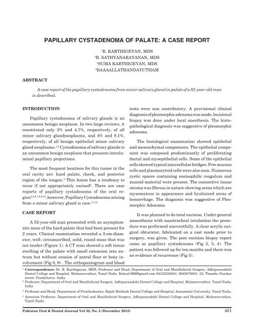
papillary cystadenoma of palate: a case report - Pakistan Oral and ...
papillary cystadenoma of palate: a case report - Pakistan Oral and ...
papillary cystadenoma of palate: a case report - Pakistan Oral and ...

You also want an ePaper? Increase the reach of your titles
YUMPU automatically turns print PDFs into web optimized ePapers that Google loves.
Papillary Cystadenoma <strong>of</strong> <strong>palate</strong>: A <strong>case</strong> ReportPAPILLARY CYSTADENOMA OF PALATE: A CASE REPORT1R. KARTHIGEYAN, MDS2R. SATHYANARAYANAN, MDS3SUMA KARTHIGEYAN, MDS4BAAAALLATHANDAYUTHAMABSTRACTA <strong>case</strong> <strong>report</strong> <strong>of</strong> the <strong>papillary</strong> <strong>cystadenoma</strong> from minor salivary gl<strong>and</strong> in <strong>palate</strong> <strong>of</strong> a 52-year-old manis described.INTRODUCTIONPapillary <strong>cystadenoma</strong> <strong>of</strong> salivary gl<strong>and</strong>s is anuncommon benign neoplasm. In two large reviews, itconstituted only 2% <strong>and</strong> 4.7%, respectively, <strong>of</strong> allminor salivary gl<strong>and</strong>neoplasms, <strong>and</strong> 4% <strong>and</strong> 8.1%,respectively, <strong>of</strong> all benign epithelial minor salivarygl<strong>and</strong> neoplasms. 1,2 Cystadenoma <strong>of</strong> salivary gl<strong>and</strong>s isan uncommon benign neoplasm that presents intraluminal<strong>papillary</strong> projections.The most frequent locations for this tumor in theoral cavity are: hard <strong>palate</strong>, cheek, <strong>and</strong> posteriorregion <strong>of</strong> the tongue. 4 This lesion has a tendency torecur if not appropriately excised 5 . There are <strong>case</strong><strong>report</strong>s <strong>of</strong> <strong>papillary</strong> <strong>cystadenoma</strong> <strong>of</strong> the oral region3,5,6,7,8,9,10 , however, Papillary Cystadenoma arisingfrom a minor salivary gl<strong>and</strong> is rare. 11,12CASE REPORTA 52-year-old man presented with an asymptomaticmass <strong>of</strong> the hard <strong>palate</strong> that had been present for2 years. Clinical examination revealed a 3-cm-diameter,well- circumscribed, solid, round mass that wasnot tender (Figure 1). A CT scan showed a s<strong>of</strong>t tissueswelling <strong>of</strong> the <strong>palate</strong> with small extension into antrumbut without erosion <strong>of</strong> antral floor or bony involvement(Fig 8, 9) . The orthopantogram <strong>and</strong> bloodtests were non contributory. A provisional clinicaldiagnosis <strong>of</strong> pleomorphic adenoma was made. Incisionalbiopsy was done under local anesthesia. The histopathologicaldiagnosis was suggestive <strong>of</strong> pleomorphicadenoma.The histological examination showed epithelial<strong>and</strong> mesenchymal components. The epithelial componentwas composed predominantly <strong>of</strong> proliferatingductal <strong>and</strong> myoepithelial cells. Some <strong>of</strong> the epithelialcells showed typical intercellular bridges. Few mucouscells <strong>and</strong> plasmacytoid cells were also seen. Numerouscystic spaces containing eosinophilic coagulum <strong>and</strong>mucoid material were present. The connective tissuestroma was fibrous in nature showing areas which aremyxomatous in appearance <strong>and</strong> hyalinised areas <strong>of</strong>hemorrhage. The diagnosis was suggestive <strong>of</strong> PleomorphicAdenoma.It was planned to do total excision. Under generalanaesthesia with nasotracheal intubation the procedurewas performed uneventfully. A clear acrylic surgicalobturator, fabricated on a cast made prior tosurgery, was given. The post excision biopsy <strong>report</strong>came as <strong>papillary</strong> <strong>cystadenoma</strong> (Fig 2, 3, 4). Thepatient was followed up for ten months <strong>and</strong> there wasno evidence <strong>of</strong> recurrence (Fig 5).1Correspondence: Dr. R. Karthigeyan, MDS, Pr<strong>of</strong>essor <strong>and</strong> Head, Department <strong>of</strong> <strong>Oral</strong> <strong>and</strong> Maxill<strong>of</strong>acial Surgery, AdhiparasakthiDental College <strong>and</strong> Hospital, Melmaruvathur, Tamil Nadu. Katcat1966@gmail.com 04132223841. 9843070021. 52, Thambu Naickerstreet. Pondicherry. India.2Pr<strong>of</strong>essor, Department <strong>of</strong> <strong>Oral</strong> <strong>and</strong> Maxill<strong>of</strong>acial Surgery, Adhiparasakthi Dental College <strong>and</strong> Hospital, Melmaruvathur, Tamil Nadu,India3Pr<strong>of</strong>essor <strong>and</strong> Head, Department <strong>of</strong> Prosthodontics, Rajah Muthiah Dental College <strong>and</strong> Hospital, Annamalai University, Tamil Nadu.4Associate Pr<strong>of</strong>essor, Department <strong>of</strong> <strong>Oral</strong> <strong>and</strong> Maxill<strong>of</strong>acial Surgery, Adhiparasakthi Dental College <strong>and</strong> Hospital, Melmaruvathur,Tamil Nadu.<strong>Pakistan</strong> <strong>Oral</strong> & Dental Journal Vol 32, No. 3 (December 2012)401
Papillary Cystadenoma <strong>of</strong> <strong>palate</strong>: A <strong>case</strong> ReportThe histologic examination showed the tumourmass to be encapsulated by thick fibrous connectivetissue <strong>and</strong> arranged in the form <strong>of</strong> lobules, sheets <strong>and</strong>ductal pattern. The mass was also composed <strong>of</strong> numerouscystic spaces lined by epithelium which are throwninto <strong>papillary</strong> projection <strong>and</strong> in few areas they weresurrounded by thickened basement membrane. Thelining epithelium comprised <strong>of</strong> cuboidal ductal cellswith vesicular nuclei <strong>and</strong> showed no atypia. Thin fibreconnective tissue core admixed with few areas <strong>of</strong>haemorrhage <strong>and</strong> hyalinization were seen. The capsulewas infiltrated in few areas by the ductal pattern<strong>of</strong> tumour mass. Final diagnosis was confirmative <strong>of</strong><strong>papillary</strong> cyst adenoma <strong>of</strong> the minor salivary gl<strong>and</strong>(Fig 8, 9).Fig. 3DISCUSSIONThe World Health Organization (WHO) 13 describedPapillary Cystadenoma as “a tumor that closely resemblesWarthin tumor but without the lymphoidFig. 4Fig. 1Fig. 2Fig. 5<strong>Pakistan</strong> <strong>Oral</strong> & Dental Journal Vol 32, No. 3 (December 2012)402
Papillary Cystadenoma <strong>of</strong> <strong>palate</strong>: A <strong>case</strong> ReportFig. 6Fig. 9elements, constituting multiple <strong>papillary</strong> projections<strong>and</strong> a greater variety <strong>of</strong> epithelial lining cells.” It isbelieved that salivary gl<strong>and</strong>s tumors are difficult todiagnose or interpret because there are many possiblepatterns <strong>of</strong> presentation. In addition, <strong>papillary</strong> <strong>cystadenoma</strong><strong>of</strong> the minor salivary gl<strong>and</strong> is rare. 11,12Fig. 7First review <strong>of</strong> the clinical, histologic, <strong>and</strong> biologicfeatures <strong>of</strong> the <strong>papillary</strong> <strong>cystadenoma</strong> 14 show that itappears to occur more frequently in women, mostpatients older than 50 years <strong>of</strong> age, with several intheir seventies. The most common sites are the <strong>palate</strong><strong>and</strong> buccal mucosa; however, tumors in the lip <strong>and</strong>tongue also have been described. The usual presentationis an asymptomatic mass.In a <strong>case</strong> series <strong>of</strong> 834 salivary gl<strong>and</strong> tumors<strong>report</strong>ed from their hospital from 1980 to 2004 , onlytwo <strong>case</strong>s <strong>of</strong> <strong>cystadenoma</strong> <strong>of</strong> a minor salivary gl<strong>and</strong>were found, in the upper lip. On microscopic examination,the neoplasm is usually well circumscribed <strong>and</strong>may be surrounded by a rim <strong>of</strong> fibrous tissue. There aresolid areas (usually limited in extent) <strong>and</strong> cystic areasinto which project papillae lined by cuboidal to columnarcells usually two layers thick. The cells usuallyhave eosinophilic cytoplasm, <strong>and</strong> goblet cells may bepresent. 16,12,11Fig. 8Perhaps the most important entity in the differentialdiagnosis <strong>of</strong> <strong>papillary</strong> <strong>cystadenoma</strong> is cystadenocarcinoma;sometimes the distinction may be difficultbecause the neoplasms have similar architecture, <strong>and</strong>also because cystadenocarcinoma <strong>of</strong>ten shows littleatypia. 16 Both neoplasms usually reveal <strong>papillary</strong> proliferation<strong>of</strong> the epithelial lining <strong>and</strong> are composed <strong>of</strong><strong>Pakistan</strong> <strong>Oral</strong> & Dental Journal Vol 32, No. 3 (December 2012)403
Papillary Cystadenoma <strong>of</strong> <strong>palate</strong>: A <strong>case</strong> Reportcells that possess bl<strong>and</strong> cytomorphologic features. Differentiation<strong>of</strong> tumour types depends largely on theidentification <strong>of</strong> actual infiltration <strong>of</strong> salivary gl<strong>and</strong>parenchyma or surrounding connective tissue by eithercystic or solid epithelium in cystadenocarcinomas.Step sections <strong>of</strong> a borderline tumour may yieldunequivocal evidence <strong>of</strong> invasion. 15 Most recent <strong>report</strong><strong>of</strong> a <strong>case</strong> involving cytological analysis <strong>and</strong> review <strong>of</strong>the literature, <strong>report</strong>ed that in the authors’ best knowledge,theirs was only the 12th <strong>case</strong> <strong>of</strong> this tumor seenin the <strong>palate</strong>. 17CONCLUSIONConservative surgical removal is the treatment <strong>of</strong>choice for Papillary Cystadenoma <strong>of</strong> minor salivarygl<strong>and</strong> <strong>and</strong> these lesions rarely recur.REFERENCES1 Alexis JB, Dembrow V. Papillary <strong>cystadenoma</strong> <strong>of</strong> a minorsalivary gl<strong>and</strong>. J <strong>Oral</strong> Maxill<strong>of</strong>ac Surg. 1995 Jan; 53(1): 70-2.2 Waldron CA, El-M<strong>of</strong>ty SK, Gnepp DR. Tumors <strong>of</strong> the intraoralminor salivary gl<strong>and</strong>s: a demographic <strong>and</strong> histologic study <strong>of</strong>426 <strong>case</strong>s. <strong>Oral</strong> Surg <strong>Oral</strong> Med <strong>Oral</strong> Pathol. 1988 Sep; 66(3):323-33.3 Kameyama Y, Okada Y, Takehana S, Mizohata M, Nishio S,Enomoto M: Papillary <strong>cystadenoma</strong>, Int J <strong>Oral</strong> Surg. 14: 556-9,1985.4 Kerpel SM, Fredman PD, Lumerman H, The <strong>papillary</strong> <strong>cystadenoma</strong><strong>of</strong> minor salivary gl<strong>and</strong> origin. <strong>Oral</strong> Surg. 46: 820-6,1978.5 Alexis J, Dewbrow V, Papillary <strong>cystadenoma</strong> <strong>of</strong> a minor salivarygl<strong>and</strong>. J <strong>Oral</strong> Maxill<strong>of</strong>ac Surg. 53:70-2, 1995.6 Akin RK, Kreller AJ, Walters PJ, Papillary <strong>cystadenoma</strong> <strong>of</strong> thelower lip -<strong>report</strong> <strong>of</strong> a <strong>case</strong>. J <strong>Oral</strong> Surg. 31: 858-60, 1973.7 Cathoun NR, Cerine FC, Mathews MJ, Papillary <strong>cystadenoma</strong><strong>of</strong> the upper lip. <strong>Oral</strong> Surg. 21: 782-5, 1966.8 Guccion JG, Redman RS, Calhoun NR, Saini N, Papillary<strong>cystadenoma</strong> <strong>of</strong> the <strong>palate</strong> - a <strong>case</strong> <strong>report</strong> <strong>and</strong> ultrastructuralstudy. J <strong>Oral</strong> Maxill<strong>of</strong>ac. Surg. 55: 59-64, 1997.9 Waldron CA, El-M<strong>of</strong>ty SK, Gnepp DR, Tumors <strong>of</strong> the intraoralminor salivary gl<strong>and</strong>s - a demographic <strong>and</strong> histologic study <strong>of</strong>426 <strong>case</strong>s. <strong>Oral</strong> Surg. 66: 323, 1988.10 Whittaker JS, Turner EP, Papillary tumours <strong>of</strong> the minorsalivary gl<strong>and</strong>s: J Clin Pathol. 29: 795-805, 1976.11 Tsurumi K, Kamiya H, Yokoi M, Kameyama Y, Papillaryoncocytic <strong>cystadenoma</strong> <strong>of</strong> palatal minor salivary gl<strong>and</strong>: a <strong>case</strong><strong>report</strong>. J <strong>Oral</strong> Maxill<strong>of</strong>ac Surg. 61: 631-3, 2003.12 Mahler V, Schell H, Papillary <strong>cystadenoma</strong>- a rare tumor <strong>of</strong> theminor salivary gl<strong>and</strong>s. Eur J Dermatol. 5: 387-9, 1999.13 Ellis GL, Auclair PL, Gnepp DR. (eds). Surgical Pathology <strong>of</strong> theSalivary Gl<strong>and</strong>s. Philadelphia, PA: Saunders; 1991.14 Collins EM. Papillary <strong>cystadenoma</strong> <strong>of</strong> accessory salivary gl<strong>and</strong>.Am J Surg. 1958 Dec; 96(6): 749-50.15 Lorena Gallego, Luis Junquera , Manuel F. Fresno, Juan Carlosde Vicente Med <strong>Oral</strong> Patol <strong>Oral</strong> Cir Bucal. 2008 Jul 1; 13(7):E460-3.16 Foss RD, Ellis GL, Auclair PL. Salivary gl<strong>and</strong> cystadenocarcinomas.A clinicopathologic study <strong>of</strong> 57 <strong>case</strong>s. Am J Surg Pathol.1996 Dec; 20(12): 1440-7.17 Lim CS, Ngu I, Collins AP, McKellar GM. Papillary <strong>cystadenoma</strong><strong>of</strong> a minor salivary gl<strong>and</strong>: <strong>report</strong> <strong>of</strong> a <strong>case</strong> involvingcytological analysis <strong>and</strong> review <strong>of</strong> the literature. <strong>Oral</strong> Surg <strong>Oral</strong>Med <strong>Oral</strong> Pathol <strong>Oral</strong> Radiol Endod. 2008 Jan; 105(1): e28-33.<strong>Pakistan</strong> <strong>Oral</strong> & Dental Journal Vol 32, No. 3 (December 2012)404