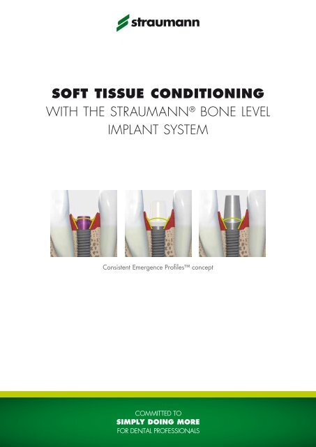
Soft tissue conditioning with the Straumann ® Bone level implant ...
Soft tissue conditioning with the Straumann ® Bone level implant ...
Soft tissue conditioning with the Straumann ® Bone level implant ...

Create successful ePaper yourself
Turn your PDF publications into a flip-book with our unique Google optimized e-Paper software.
The ITI (International Team for Implantology) is academic partner of Institut <strong>Straumann</strong> AGin <strong>the</strong> areas of research and education.
Communication is key in <strong>the</strong> dental <strong>implant</strong> treatment flowInterdisciplinary treatment planning includes <strong>the</strong> collaboration of differentparties such as:ppPatientppGeneral practitionerppSurgeonppProsthodontistppDental laboratoryIn order to reach a successful treatment outcome, communication betweenall involved is essential. The parties should be aware of <strong>the</strong> steps and interfacesand exchange <strong>the</strong>ir positions in order to be able to make <strong>the</strong> correctdecisions.A general overview of <strong>the</strong> treatment flow is depicted below, broken downinto various treatment steps. Before each decision step (lime green in <strong>the</strong> picturebelow) communication can be helpful.Surgical intervention(transmucosal healing)Case analysis andtreatment planningSurgical intervention(submucosal healing)Flap openingTreatment type1 st flap openingImplant placementImplant placementClose flapFlap re-openingHealing abutment choiceHealing abutment placementTemporizationTemporary placementin order to perform soft<strong>tissue</strong> <strong>conditioning</strong>Final restorationProcedure stepDecision/communication step1
Treatment FlowhighEs<strong>the</strong>tic complexitylowlowTreatment time & costshighI. Case analysis and treatment planning<strong>Soft</strong> <strong>tissue</strong> management starts when planning <strong>the</strong> casebecause <strong>the</strong> soft <strong>tissue</strong> shape has to be analyzed andsoft <strong>tissue</strong> <strong>conditioning</strong> is influenced by <strong>the</strong> <strong>implant</strong> placement.Each case is different; <strong>the</strong>refore accurate analysisand planning from <strong>the</strong> very beginning is helpful to reach<strong>the</strong> desired es<strong>the</strong>tic outcome. Various factors (e.g. indication,patient expectations, soft <strong>tissue</strong> shape, bone quality,site position) have an effect on <strong>the</strong> treatment outcome.The degree of complexity and risk involved in individualprocedures can be assessed, for example, <strong>with</strong> <strong>the</strong> ITISAC Assessment Tool 1 . As a general principle <strong>the</strong> <strong>level</strong> ofcomplexity rises <strong>with</strong> an increase in <strong>the</strong> number of stepsinvolved and <strong>the</strong> es<strong>the</strong>tic outcome that must be achievedto attain a satisfactory result.Increasing es<strong>the</strong>tic complexity leads in general to longertreatment time and higher costs. Informing and educating<strong>the</strong> patient at <strong>the</strong> beginning of <strong>the</strong> treatment might increasehis or her willingness to collaborate.II. Surgical treatment and healing phaseBased on <strong>the</strong> case and his or her preferences, <strong>the</strong> clinicianwill choose a submucosal or a transmucosal approach.With <strong>the</strong> submucosal approach, a closure screw is placedafter <strong>the</strong> <strong>implant</strong> placement and <strong>the</strong> flap is closed. In a secondsurgery after a first healing phase, <strong>the</strong> healing abutmentis placed and soft <strong>tissue</strong> <strong>conditioning</strong> is started.The healing abutment initiates <strong>the</strong> first phase of soft <strong>tissue</strong>contouring. <strong>Bone</strong> Level healing abutments were designedin order to correlate <strong>with</strong> <strong>the</strong> size of <strong>the</strong> abutments for <strong>the</strong>final restoration. This concept is called ‘Consistent EmergenceProfiles’.2Schematic depiction of <strong>the</strong> submucosal procedure,which requires two surgical interventions
The transmucosal approach allows a straightforward procedure,avoiding a second surgery.Schematic depiction of <strong>the</strong> transmucosal procedureThe decision, as to which abutment to use for <strong>the</strong> finalrestoration needs to be made at beginning of <strong>the</strong> treatmentso that <strong>the</strong> healing abutment can be chosen accordingly.Therefore, backward planning and good communicationbetween <strong>the</strong> treating clinicians and <strong>the</strong> dental lab areneeded. The tables on <strong>the</strong> following pages support <strong>the</strong>choice of <strong>the</strong> healing abutment by showing <strong>the</strong> best fitbetween conical healing abutment and <strong>the</strong> final restoration.There are fur<strong>the</strong>r alternatives in <strong>the</strong> healing abutmentportfolio (e.g. customizable abutments, bottle-shaped healingabutments) which are not displayed in <strong>the</strong> tables. Thesealternatives are meant to meet additional needs for individualcases and to be used based on user preferences.More details about <strong>the</strong>se solutions can be found in <strong>the</strong>brochure “Basic information on <strong>the</strong> surgical procedures”(pages 60 – 65).III. Temporization (shaping of <strong>the</strong> soft <strong>tissue</strong>)After <strong>the</strong> first healing phase, <strong>the</strong> soft <strong>tissue</strong> can be shaped<strong>with</strong> <strong>the</strong> help of temporary abutments. This additional soft<strong>tissue</strong> <strong>conditioning</strong> <strong>with</strong> <strong>the</strong> temporary abutment mayachieve an even more es<strong>the</strong>tic outcome and widen <strong>the</strong>soft <strong>tissue</strong> when needed (see cases on page 10–14). Thepapilla can be additionally shaped and adapted to <strong>the</strong> finalrestoration. For <strong>the</strong>se steps, <strong>the</strong> patient has to be madeaware about <strong>the</strong> additional effort which is needed to increase<strong>the</strong> es<strong>the</strong>tic outcome. Again, good communication<strong>with</strong> <strong>the</strong> dental lab and <strong>the</strong> treating clinicians supports <strong>the</strong>final outcome.IV. Treatment closure and follow-upOnce <strong>the</strong> pros<strong>the</strong>sis is placed, adapted and <strong>the</strong> treatmentis finished, regular follow-up visits and good oral hygienehelp to prevent any undesired effects and to keep patientsatisfaction high.3
The healing and temporary abutment portfolioIn case of submucosal healing, closure screws are availablein different heights for <strong>the</strong> different platforms (NarrowCrossFit ® [NC] and Regular CrossFit ® [RC]). These allow asimple submerged healing due to <strong>the</strong>ir reduced height anddiameter (adapted to <strong>implant</strong> diameter).Closure screws available for both platforms in 2 differen<strong>the</strong>ights.In case of transmucosal healing or after re-opening in asubmerged case, <strong>Straumann</strong> offers various healing abutmenttypes. These abutments allow <strong>the</strong> soft <strong>tissue</strong> to beshaped.Conical abutments are <strong>the</strong> straightforward solution for soft <strong>tissue</strong>management. They are available for both platforms in differentdiameters and heights.In specific cases, e.g. thin mucosa type, a bottle shape abutmentmight be beneficial. Bottle-shaped abutments are availablefor both platforms in different heights.For specific patient cases or if a cementable abutment <strong>with</strong> adiameter of 6.5 mm is used, <strong>the</strong> customizable abutment hasshown to be <strong>the</strong> ideal solution. These abutments are availablefor both connection platforms.4
In general a temporary restoration is recommended toreach a good es<strong>the</strong>tic outcome. When a temporization isrequired two types of abutments are available: PEEK temporaryand titanium temporary. These support additionalsoft <strong>tissue</strong> <strong>conditioning</strong>. One example is shown in <strong>the</strong> attachedcase.PEEK temporary abutments are available for additive andsubtractive procedures.The PEEK temporary abutment allows a subtractive and additiveprocedure to create a temporary restoration.The titanium healing abutments are designed to build upa temporary restoration by adding material on <strong>the</strong> post.Titanium temporary abutments are available for additiveprocedures (3D view needed).5
The consistent emergence profiles conceptThe healing abutments, temporary abutments and finalabutments of <strong>the</strong> <strong>Straumann</strong> ® <strong>Bone</strong> Level <strong>implant</strong> systemwere designed to have an emergence profile which is <strong>the</strong>same at all treatment steps when combining specific products.For each final standard abutment (excluding Meso,<strong>Straumann</strong> ® CARES ® and gold abutments) <strong>the</strong>re is a correspondinghealing abutment. Accordingly, <strong>the</strong> final abutmentwill possess <strong>the</strong> same emergence profile that waspreviously created by <strong>the</strong> respective healing abutment. Thepictures show examples of matching healing abutmentsand final abutments.=In <strong>the</strong> <strong>Straumann</strong> ® portfolio <strong>the</strong>re is a best fit combinationbetween one final abutment and one conical healing abutment.The examples are depicted showing a final abutment<strong>with</strong> <strong>the</strong> specific conical healing abutment which fits <strong>the</strong>best. Tables on <strong>the</strong> following pages can be consulted inorder to find <strong>the</strong> healing abutment which is <strong>the</strong> best fitfor a specific final abutment. Key properties of <strong>the</strong> finalabutment are type (e.g. Multi-base abutment), abutmentdiameter (Ø) and gingival height (GH). Abutment height(AH), angle and material do not affect <strong>the</strong> choice of <strong>the</strong>healing abutment. Once <strong>the</strong>se parameters are known acorresponding healing abutment can be chosen.The tables on page 7–9 include only <strong>the</strong> best fit between<strong>the</strong> conical healing abutment and <strong>the</strong> final abutment. Detailedinformation about <strong>the</strong> possible combinations of all<strong>Straumann</strong> ® healing abutments <strong>with</strong> <strong>the</strong> final abutments canbe found in <strong>the</strong> brochure “Basic information on <strong>the</strong> surgicalprocedures” (pages 60–65).6
Cement-retained solutionsPlatformNCTypeAnatomic abutmentCementable abutmentMaterialAngleØ (mm)GH (mm)Ti Ti IPS e.max ® IPS e.max ® Ti Ti0° 15° 0° 15° 0° 0°4.0 4.0 4.0 4.0 3.5 5.02.0 3.5 2.0 3.5 2.0 3.5 2.0 3.5 1.0 2.0 3.0 1.0 2.0 3.0GH (mm)Ø (mm)3.5 5.0 3.5 5.0 3.5 5.0 3.5 5.0 3.5 3.5 5.0 2.0 3.5 5.04.8 4.8 4.8 3.6 4.8TypeConical healing abutmentPlatformRCTypeAnatomic abutmentCementable abutmentMaterialAngleØ (mm)GH (mm)Ti Ti IPS e.max ® IPS e.max ® Ti Ti0° 15° 0° 15° 0° 0°6.5 6.5 6.5 6.5 5.0 6.52.0 3.5 2.0 3.5 2.0 3.5 2.0 3.5 1.0 2.0 3.0 1.0 2.0 3.0GH (mm)Ø (mm)4.0 6.0 4.0 6.0 4.0 6.0 4.0 6.0 2.0 4.0 6.0 2.0 4.0 6.06.0 6.0 6.0 6.0TypeConical healing abutment7
Screw-retained solutionsPlatformNCTypeAnatomic abutmentMulti-base abutmentMaterialAngleØ (mm)GH (mm)IPS e.max ® IPS e.max ® Ti Ti Ti0° 15° 0° 0° 25°4.0 4.0 3.5 4.5 4.02.0 3.5 2.0 3.5 1.0 2.5 4.0 1.0 2.5 4.0 2.5GH (mm)Ø (mm)3.5 5.0 3.5 5.0 2.0 3.5 5.0 2.0 3.5 5.0 3.54.8 3.6 4.8 4.8TypeConical healing abutmentPlatformRCTypeAnatomic abutmentMulti-base abutmentMaterialAngleØ (mm)GH (mm)IPS e.max ® IPS e.max ® Ti Ti Ti0° 15° 0° 0° 25°6.5 6.5 4.5 6.5 4.02.0 3.5 2.0 3.5 1.0 2.5 4.0 1.0 2.5 4.0 2.5GH (mm)Ø (mm)4.0 6.0 4.0 6.0 2.0 4.0 6.0 2.0 4.0 6.0 4.06.0 4.5 6.0 6.0TypeConical healing abutment8
HYBRID solutionsPlatformNCTypeMulti-base abutment Locator ®MaterialAngleØ (mm)GH (mm)Ti Ti Ti Ti alloy0° 0° 25° 0°3.5 4.5 4.0 3.81.0 2.5 4.0 2.0 2.0 3.5 2.5 2.0 3.0 4.0 5.0 6.0GH (mm)Ø (mm)2.0 3.5 5.0 2.0 3.5 5.0 3.5 2.0 3.5 5.03.6 4.8 4.8 3.6TypeConical healing abutmentPlatformRCTypeMulti-base abutment Locator ®MaterialAngleØ (mm)GH (mm)Ti Ti Ti Ti alloy0° 0° 25° 0°4.5 6.5 4.0 3.81.0 2.5 4.0 1.0 2.5 4.0 2.5 1.0 2.0 3.0 4.0 5.0 6.0GH (mm)Ø (mm)2.0 4.0 6.0 2.0 4.0 6.0 4.0 2.0 4.0 6.04.5 6.0 6.0 4.5TypeConical healing abutment9
Case example for soft <strong>tissue</strong> <strong>conditioning</strong>Every patient is an individual case and may need tobe treated in a different way. Accordingly, <strong>the</strong> followingcase shows one treatment strategy and procedure andillustrates how soft <strong>tissue</strong> <strong>conditioning</strong> can be handledto reach an es<strong>the</strong>tic result. The process in <strong>the</strong> cases correspondsto <strong>the</strong> previously explained treatment flow (page1–2) and is always <strong>the</strong> result of <strong>the</strong> collaboration of differentparties.Prof. Dr. D. Buser, Dr. J. Wittneben, CDT T. Furter (f.l.t.r.)Case: <strong>Soft</strong> <strong>tissue</strong> <strong>conditioning</strong> <strong>with</strong> <strong>the</strong> dynamiccompression technique 2(Courtesy of Dr. Wittneben)The following treatment was performed at <strong>the</strong> Universityof Berne in Switzerland. Various parties were involvedin this patient’s case. The surgery was performed byProf. Dr. D. Buser, soft <strong>tissue</strong> <strong>conditioning</strong> and <strong>the</strong> prosthodontictreatment was accomplished by Dr. J. Wittnebenand <strong>the</strong> dental laboratory work was done by CDT T.Furter from ArtDent in Berne. Extensive com municationand continuous information exchange between <strong>the</strong>treating parties were essential for <strong>the</strong> treatment. Thepatient was also involved and well-educated during <strong>the</strong>treatment.tooth 117.62 mmCase analysis and treatment planningA 21-year-old, non-smoking female patient was referredto <strong>the</strong> Oral Surgery Department from a private practicebecause tooth 11 presented an acute fistula includingan external root resorption following trauma. The patientwas referred from <strong>the</strong> Oral Surgery Departmentto <strong>the</strong> Department of Fixed Prosthodontics, University ofBerne, to perform a combined surgical-prosthodontic treatmentapproach.Figure 1: Initial situation presenting external root resorptionin position 11; thin buccal plate is visible in cone beamCT diagnosticAt <strong>the</strong> initial examination, <strong>the</strong> patient’s chief complaint wasbad odor and pus emerging from <strong>the</strong> fistula – she wantedto have a final treatment of her anterior tooth. The extraoralexamination showed no pathological findings. The intraoralexamination revealed that her teeth were all naturaland non-restored except for tooth 11, which presented <strong>with</strong>a composite restoration <strong>with</strong> discoloration and wear. In<strong>the</strong> radiographic analysis <strong>the</strong> diagnosis of a well-definedlateral root resorption could be confirmed presenting in <strong>the</strong>CT as well as in <strong>the</strong> periapical radiograph.10
Therefore, tooth 11 was considered hopeless and <strong>implant</strong><strong>the</strong>rapy was recommended as <strong>the</strong> neighboring teeth werenon-restored.Due to her young age and no previous experiences <strong>with</strong>dental treatments o<strong>the</strong>r than in area 11, <strong>the</strong> patient hadhigh es<strong>the</strong>tic expectations. At full smile, <strong>the</strong> patient presenteda high lip line, exposing all anterior maxillary teethand surrounding ginigiva.The gingival biotype was thin and highly scalloped <strong>with</strong>triangular-shaped anterior teeth. These findings led to <strong>the</strong>following SAC profile 1 : complex, because of <strong>the</strong> es<strong>the</strong>ticrisk factors involved.Figure 2: The young patient has a high lip line, thin gingivabiotype, a highly scalloped gingiva and triangular-shapedanterior teethThe oral surgeon and prosthodontist discussed <strong>the</strong> case<strong>with</strong> and <strong>with</strong>out <strong>the</strong> patient. It was important to evaluate<strong>the</strong> patient’s expectation before starting <strong>the</strong> treatment. Thepatient was also informed about <strong>the</strong> length of <strong>the</strong> treatmentand its implications. She agreed and was willing tocollaborate.The dental technician was also introduced to <strong>the</strong> patientand <strong>the</strong> final position of <strong>the</strong> <strong>implant</strong> was discussed <strong>with</strong>all clinicians on <strong>the</strong> basis of a wax-up. During <strong>the</strong> planningphase, <strong>the</strong> decision was made to resolve this case<strong>with</strong> a screw-retained ceramic abutment. This decisionwas based on <strong>the</strong> anatomical situation (frontal tooth, highlip line) and <strong>the</strong> patient’s expectations regarding <strong>the</strong> es<strong>the</strong>ticoutcome.11
Surgical treatment and healing phaseThe tooth extraction was performed carefully <strong>with</strong>out <strong>the</strong>elevation of a flap in <strong>the</strong> Oral Surgery Department by Prof.Dr. Buser. Exactly 6 weeks later (early placement concept)a <strong>Straumann</strong> ® <strong>Bone</strong> Level SLActive Implant <strong>with</strong> a diameterof 4.1 mm (RC platform) was inserted by <strong>the</strong> same clinician,including Guided <strong>Bone</strong> Regeneration (GBR). The <strong>implant</strong>was placed in a correct three-dimensional position and,when primary stability was achieved, GBR technique wasadded. The flap was closed for submucosal healing dueto simultaneous GBR procedure.Figure 3: X-ray showing ideal<strong>implant</strong> placement and <strong>the</strong> firstconical healing abutment(RC Ø 4.5 mm) placed for <strong>the</strong>healing phase 1The patient received a simple flipper as an interim provisionaldenture during <strong>the</strong> time of healing. After 10 days<strong>the</strong> denture tooth had to be modified by adding material.After <strong>the</strong> reopening procedure, a conical healing abutment(Ø 4.5 mm) for <strong>the</strong> RC platform was inserted. <strong>Soft</strong> <strong>tissue</strong><strong>conditioning</strong> starts <strong>with</strong> <strong>the</strong> first insertion of <strong>the</strong> healingabutment. After taking <strong>the</strong> impression for <strong>the</strong> provisional, alarger diameter (6 mm) was chosen to prepare <strong>the</strong> site forinsertion of <strong>the</strong> temporary abutment <strong>with</strong> <strong>the</strong> provisional.To improve <strong>the</strong> mucosa architecture surrounding <strong>the</strong> future<strong>implant</strong> crown, soft <strong>tissue</strong> <strong>conditioning</strong> <strong>with</strong> <strong>the</strong> “dynamiccompression technique” was performed.Figure 4: The narrow healing abutment has been replaced by awider healing abutment to widen <strong>the</strong> soft <strong>tissue</strong>12
<strong>Soft</strong> <strong>tissue</strong> <strong>conditioning</strong> <strong>with</strong> <strong>the</strong> “dynamic compressiontechnique”Using <strong>the</strong> early loading concept, a screw-retained <strong>implant</strong>supportedprovisional (PEEK Temporary abutment) was insertedafter 6 weeks post placement. The shape of <strong>the</strong>provisional crown was slightly overcontoured in order tocreate pressure, to form <strong>the</strong> mucosa, <strong>the</strong> emergence profileand to have an approximal contact present. As an temporarilyeffect, <strong>the</strong>re was an ischemic reaction (white soft<strong>tissue</strong>) for about 10 to 15 minutes, after this time, <strong>the</strong> soft<strong>tissue</strong> returned to its pink color.Figure 5: A slightly overcontoured provisional crown has beenplaced. An ischemic reaction can be observed, which disappeared10 to 15 minutes after crown placementDuring <strong>the</strong> following 2 weeks, selective pressure was applied(pressure phase) to <strong>the</strong> provisional crown by addingextraorally light-cured acrylic material. After 2 weeks, <strong>the</strong>shape of <strong>the</strong> provisional was modified by removing acrylicmaterial in <strong>the</strong> interproximal/cervical area in order to createspace for <strong>the</strong> papillae (release phase). This approachis performed in several steps and can be done intraorally<strong>with</strong> a fine diamond bur and <strong>the</strong>n polished <strong>with</strong> an arkansasstone – <strong>with</strong>out removing <strong>the</strong> provisional.Figure 6: After two weeks of pressure phase <strong>the</strong> release phaseis initiated by removing acrylic material intraorally13
<strong>Soft</strong> <strong>tissue</strong> <strong>conditioning</strong> is recommended for 3–5 monthsfor single edentulous gaps and for 6–8 months for adjacent<strong>implant</strong>s in <strong>the</strong> es<strong>the</strong>tic zone. In <strong>the</strong> present case,soft <strong>tissue</strong> <strong>conditioning</strong> was performed for 5 months. Thepatient is visiting <strong>the</strong> prosthodontist at a regular basis for<strong>the</strong> removal of material.Figure 7: Period after provisional crown placement: 20 daysThe created final soft <strong>tissue</strong> architecture and emergenceprofile were transferred to <strong>the</strong> final master cast by <strong>the</strong> fabricationof an individualized impression coping.Figure 8: Period after provisional crown placement: 2.5 monthsFigure 9: Period after provisional crown placement: 5 months;finalized soft <strong>tissue</strong> architecture and end of provisional phaseTreatment closure and follow-upFor <strong>the</strong> final reconstructive design, a screw-retained single<strong>implant</strong> crown was chosen and an individualized zirconiumdioxide <strong>Straumann</strong> ® CARES ® customized abutmentwas fabricated.14Figure 10: Final restoration
The planned abutment was waxed up and scanned via<strong>the</strong> <strong>Straumann</strong> ® CARES ® Visual software and a zirconiumdioxide abutment was delivered. Feldspathic ceramic washand-layered on top of <strong>the</strong> abutment. The final restorationwas inserted <strong>with</strong> 35 Ncm. The occlusion was adjusted.Figure 11: Radiograph of <strong>the</strong>final restorationThe follow-up after 1 year showed no changes in soft <strong>tissue</strong>architecture or in <strong>implant</strong> crown. This was also confirmedby a prospective clinical study on <strong>Bone</strong> Level <strong>implant</strong>s <strong>with</strong>early placement, contour augmentation and early loadingconcept in single edentulous space in <strong>the</strong> es<strong>the</strong>tic zone –<strong>the</strong>re were no changes in <strong>the</strong> overall es<strong>the</strong>tics (pink andwhite es<strong>the</strong>tic parameters) over a follow-up time of 3 years. 2Figure 12: One year after insertion of <strong>the</strong> final restoration,a follow-up visit revealed stable soft <strong>tissue</strong> and crestal boneFigure 13: One year follow-up pictureWe are thankful to Dr. Julia-Gabriela Wittneben (Universityof Berne) for providing this case and all accompanyingfigures.15
References1 Dawson A, Chen S, Buser D, Cordaro L, Martin W, Belser U. The SAC Classificationin Implant Dentistry: Straightforward – Advanced – Complex.In Dawson A. and Chen S (eds):2009:Quintessence Publishing Group.2 Buser D, Wittneben J, Bornstein MM, Grütter L, Chappuis V, Belser UC. Stabilityof contour augmentation and es<strong>the</strong>tic outcomes of <strong>implant</strong>-supported singlecrowns in <strong>the</strong> es<strong>the</strong>tic zone: 3-year results of a prospective study <strong>with</strong> early<strong>implant</strong> placement postextraction. J. Periodontol. 2011;82(3):342–9.16
International HeadquartersInstitut <strong>Straumann</strong> AGPeter Merian-Weg 12CH-4002 Basel, SwitzerlandPhone +41 (0)61 965 11 11Fax +41 (0)61 965 11 01IPS e.max ® is a registered trademark of Ivoclar Vivadent AG, Liechtenstein.LOCATOR ® is a registered trademark of Zest Anchors, Inc., USA.www.straumann.com© Institut <strong>Straumann</strong> AG, 2012. All rights reserved.<strong>Straumann</strong> ® and/or o<strong>the</strong>r trademarks and logos from <strong>Straumann</strong> ® mentioned herein are <strong>the</strong> trademarks or registered trademarks of <strong>Straumann</strong> Holding AGand/or its affiliates. All rights reserved.<strong>Straumann</strong> products are CE marked 10/11 152.533/en BG01011