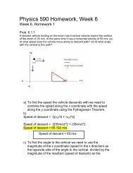
Dusty Carroll Lesson Plan 4: Getting to know Lactase Background ...
Dusty Carroll Lesson Plan 4: Getting to know Lactase Background ...
Dusty Carroll Lesson Plan 4: Getting to know Lactase Background ...

You also want an ePaper? Increase the reach of your titles
YUMPU automatically turns print PDFs into web optimized ePapers that Google loves.
<strong>Dusty</strong> <strong>Carroll</strong><br />
<strong>Lesson</strong> <strong>Plan</strong> 4: <strong>Getting</strong> <strong>to</strong> <strong>know</strong> <strong>Lactase</strong><br />
<strong>Background</strong> Information<br />
<strong>Lactase</strong> is the enzyme responsible for breaking down the lac<strong>to</strong>se in your body. Lac<strong>to</strong>se is a disaccharide<br />
that is found in milk. Human babies produce lactase in large quantities, but the production of the enzyme<br />
decreases significantly in<strong>to</strong> adulthood. Deficiency of the lactase enzyme causes many people <strong>to</strong> have<br />
trouble digesting the lac<strong>to</strong>se found in milk. This leads <strong>to</strong> the familiar problem of lac<strong>to</strong>se in<strong>to</strong>lerance. (1)<br />
<strong>Lactase</strong> can act as a catalyst for several different biological reactions. The lactase enzyme is the only<br />
human enzyme that can cleave a β-glycosidic linkage like that found in lac<strong>to</strong>se. The specific reaction that<br />
is the focus of this lesson is the breakdown of lac<strong>to</strong>se in<strong>to</strong> the two monosaccharides, galac<strong>to</strong>se and<br />
glucose as seen in the reaction below:<br />
OH<br />
CH 2OH<br />
OH<br />
O<br />
OH<br />
LACTOSE<br />
O<br />
CH 2OH<br />
OH<br />
O<br />
OH<br />
OH<br />
+ H 2O<br />
LACTASE<br />
OH<br />
OH<br />
CH 2OH<br />
OH<br />
CH 2OH<br />
OH<br />
O<br />
OH<br />
O<br />
OH<br />
OH<br />
OH<br />
GALACTOSE<br />
GLUCOSE<br />
Much research has been done on the mechanism of this reaction. In order <strong>to</strong> understand the mechanism,<br />
however, a brief overview of the structure of the enzyme is necessary.<br />
The lactase enzyme is found in humans and other organisms. In the human intestines, lactase is combined<br />
with another enzyme called phlorizin hydrolase <strong>to</strong> form a transmembrane enzyme complex called lactase-<br />
phlorizin hydrolase. It has been shown that the lactase portion of this enzyme complex is the only portion<br />
active in the breakdown of lac<strong>to</strong>se. (2) The mechanism under study for this lesson comes from the lactase<br />
found in the Escherichia coli bacteria. It was here that molecular biologists discovered the phenomenon<br />
of enzyme induction. Basically, the presence of lac<strong>to</strong>se induced the biosynthesis of an enzyme <strong>to</strong> split it.<br />
(3) The lactase-lac<strong>to</strong>se system became the focus of much research. Essentially, the lactase enzyme is<br />
genetically regulated. In other words, the action of the enzyme results from its synthesis which is<br />
regulated by the genes that code for it.<br />
The remainder of this lesson will concentrate on the independent lactase enzyme (not that complexed with<br />
the phlorizin hydrolase). The structure of lactase is rather complex. Its crystal structure contains four<br />
identical subunits. Each subunit contains a chain of 1023 amino acid residues. When this structure was<br />
determined, it was the longest polypeptide for which an a<strong>to</strong>mic structure had been obtained. (3) It is a<br />
very large enzyme and scientists continue <strong>to</strong> query about the biological reasons for such a large structure.<br />
Since each region of the enzyme seems <strong>to</strong> have a clear purpose, the common belief is that portions of the<br />
molecule were useful in certain ways and it just sort of…happened. The large size does not appear <strong>to</strong><br />
have any reason other than that resulting from the combination of all of its parts. (3,4)
Primary Structure (5)<br />
Again, the lactase enzyme consists of four identical subunits or chains. Below is the sequence of amino<br />
acids that make up just one of those chains. The sequence is shown using the one-letter abbreviations for<br />
the amino acid residues.<br />
GSHMLEDPVVLQRRDWENPGVTQLNRLAAHPPFASWRNSEEARTDRPSQQLRSLNGEWRFAWF<br />
PAPEAVPESWLECDLPEADTVVVPSNWQMHGYDAPIYTNVTYPITVNPPFVPTENPTGCYSLTFN<br />
VDESWLQEGQTRIIFDGVNSAFHLWCNGRWVGYGQDSRLPSEFDLSAFLRAGENRLAVMVLRW<br />
SDGSYLEDQDMWRMSGIFRDVSLLHKPTTQISDFHVATRFNDDFSRAVLEAEVQMCGELRDYLR<br />
VTVSLWQGETQVASGTAPFGGEIIDERGGYADRVTLRLNVENPKLWSAEIPNLYRAVVELHTAD<br />
GTLIEAEACDVGFREVRIENGLLLLNGKPLLIRGVNRHEHHPLHGQVMDEQTMVQDILLMKQNN<br />
FNAVRCSHYPNHPLWYTLCDRYGLYVVDEANIETHGMVPMNRLTDDPRWLPAMSERVTRMVQ<br />
RDRNHPSVIIWSLGNESGHGANHDALYRWIKSVDPSRPVQYEGGGADTTATDIICPMYARVDED<br />
QPFPAVPKWSIKKWLSLPGETRPLILCEYAHAMGNSLGGFAKYWQAFRQYPRLQGGFVWDWV<br />
DQSLIKYDENGNPWSAYGGDFGDTPNDRQFCMNGLVFADRTPHPALTEAKHQQQFFQFRLSGQ<br />
TIEVTSEYLFRHSDNELLHWMVALDGKPLASGEVPLDVAPQGKQLIELPELPQPESAGQLWLTVR<br />
VVQPNATAWSEAGHISAWQQWRLAENLSVTLPAASHAIPHLTTSEMDFCIELGNKRWQFNRQS<br />
GFLSQMWIGDKKQLLTPLRDQFTRAPLDNDIGVSEATRIDPNAWVERWKAAGHYQAEAALLQC<br />
TADTLADAVLITTAHAWQHQGKTLFISRKTYRIDGSGQMAITVDVEVASDTPHPARIGLNCQLA<br />
QVAERVNWLGLGPQENYPDRLTAACFDRWDLPLSDMYTPYVFPSENGLRCGTRELNYGPHQW<br />
RGDFQFNISRYSQQQLMETSHRHLLHAEEGTWLNIDGFHMGIGGDDSWSPSVSAEFQLSAGRYH<br />
YQLVWCQK<br />
From this list it is easy <strong>to</strong> see why scientists are curious about the size of the enzyme. The four chains<br />
<strong>to</strong>gether amount <strong>to</strong> 4092 amino acid residues!<br />
Secondary Structure<br />
Each of the four chains is analyzed as having five separate domains. Each domain serves a different<br />
purpose in the enzyme. Some act <strong>to</strong> help the polypeptide chain attain its tertiary structure. Some act <strong>to</strong><br />
hold one chain <strong>to</strong> another <strong>to</strong> aid in the formation of the quaternary structure. Some join with a domain on<br />
another chain <strong>to</strong> form the active site for the molecule. Below is a picture of the five domains found on<br />
chain A of lactase. These images are taken from the CATH Protein Structure Classification website. (6)<br />
Note that each domain appears <strong>to</strong> have different secondary structures. The 3 rd domain is the one where<br />
most of the helices appear. The 4 th domain has no helices and consists of only beta sheets. The other<br />
domains contain mostly beta sheets and a few sections of helices. The seemingly individual nature of<br />
each domain may be necessary <strong>to</strong> facilitate the folding of the entire chain. (4)
Tertiary Structure<br />
Together, these five domains form just one chain of the enzyme with a molecular weight of 116,570.8 D:<br />
Jacobsen, Zhang, DuBose & Matthews, in their Nature article (4) show a similar structure of the one<br />
chain with the domains labeled. The picture in the article is a stereo view of the chain.<br />
Quaternary Structure<br />
Many pictures of the full enzyme are available. (3,4,5,7) The overall structure is a homotetramer<br />
consisting of four identical chains. It is thought that the individual monomers form first, followed by<br />
formation of dimers, then dimerization of the dimers <strong>to</strong> form the tetramer. (7) Each domain has a<br />
hydrophobic core, consistent with the notion that the monomers formed individually before joining.<br />
Some of the domains then interact with each other through polar networks on their exterior surfaces.<br />
There are three major regions of interface formed from complementary regions of the monomers. There<br />
are four active sites in each tetramer. Each site is formed from the interaction of two of the monomers.<br />
Each site is also marked by two metal ligands, Na + and Mg 2+ . In the image from the Juers, et.al. article,<br />
the domains on each monomer are colored such that each domain on a given monomer is a different shade<br />
of the same color.<br />
Another image of the overall structure is found on the RSCB Protein Data Base (5) as seen below:<br />
Here you can see how the interactions of the four monomers form in<strong>to</strong> a single tetramer.
Kyte-Doolittle Hydropathy Plot (8)<br />
This plot is calculated for a single 1023 residue chain of the lactase enzyme.<br />
With a window size of 9, this plot shows many peaks below the midline. This corresponds <strong>to</strong> surface<br />
regions of the globular protein. With so many peaks, it appears that there is a large surface area which is<br />
hydrophilic. (Hydrophilic regions are given negative values.)<br />
With a window size of 19, possible transmembrane regions show as peaks above 1.8. This lactase chain<br />
shows no peaks and therefore is unlikely <strong>to</strong> have any transmembrane regions. The lactase-phlorizin<br />
hydrolase enzyme noted in the introduction is a transmembrane enzyme. According <strong>to</strong> this plot, the<br />
lactase portion of that enzyme would not be the portion spanning the interior and exterior of the<br />
membrane.<br />
Reaction Mechanism<br />
The Enzyme Commission code for lactase is 3.2.1.23.<br />
• 3 – Hydrolases<br />
o 2 – Glycosylases<br />
� 1 – Glycosidases (enzymes hydrolyzing O- and S-glycosyl compounds<br />
• 23 – lactase or beta-galac<strong>to</strong>sidase
In other words, lactase acts as a catalyst for the hydrolysis of the O-galac<strong>to</strong>sidic bond in the sugar, lac<strong>to</strong>se.<br />
The exact mechanism of the reaction has been studied and Juers, Heightman, et.al. have summarized<br />
previous work and given clarification <strong>to</strong> the proposed mechanism. (9) <strong>Lactase</strong>, aka β-galac<strong>to</strong>sidase,<br />
hydrolyzes its substrate (lac<strong>to</strong>se) while allowing the constituent monosaccharides <strong>to</strong> keep their<br />
stereochemistry. The reaction is a two step reaction. The first step is cleavage of the glycosidic bond.<br />
HO<br />
HO<br />
H<br />
OH<br />
H<br />
H<br />
H<br />
O<br />
O<br />
O<br />
OH<br />
C<br />
H<br />
H<br />
Glu 537<br />
Glu 461<br />
C<br />
O –<br />
O<br />
O<br />
HO<br />
OH<br />
OH<br />
H<br />
H<br />
H<br />
O<br />
OH<br />
H<br />
OH<br />
HO<br />
HO<br />
H<br />
OH<br />
H<br />
H<br />
H<br />
O<br />
O<br />
O<br />
OH<br />
C<br />
H<br />
Glu 537<br />
Glu 461<br />
C<br />
H<br />
O<br />
O<br />
O<br />
HO<br />
OH<br />
OH<br />
It is believed that this process takes place with a mechanism somewhere between that of an SN1 and that<br />
of an SN2. The Glu537 from the active site of the enzyme acts as a nucleophile <strong>to</strong>ward the anomeric<br />
carbon of the galac<strong>to</strong>syl group. This forms an intermediate with enzyme Glu537 in the alpha-glycosidic<br />
orientation. As seen in the above reaction, this is facilitated by a concerted pro<strong>to</strong>nation of the glycosidic<br />
oxygen. This particular step is not well-proven, yet, but it is one explanation. The acid responsible for<br />
pro<strong>to</strong>nation may be the Glu461 from the active site<br />
of the enzyme.<br />
Functional Amino Acid Residues within <strong>Lactase</strong><br />
Glutamic acid: In its anionic state (like Glu537), this acts<br />
as a nucleophile. In its neutral state (like Glu461), it acts<br />
as an acid and donates a pro<strong>to</strong>n as described in the text.<br />
H 2N CH C<br />
CH 2<br />
CH 2<br />
C<br />
OH<br />
Tyrosine: The –OH group on the side chain participates<br />
in hydrogen bonding in order <strong>to</strong> stabilize the transition<br />
state.<br />
O<br />
O<br />
O<br />
OH<br />
H<br />
H<br />
H<br />
O<br />
OH<br />
H<br />
OH<br />
HO<br />
HO<br />
H<br />
OH<br />
H<br />
H<br />
O<br />
H<br />
C<br />
O<br />
O<br />
OH<br />
Glu 537<br />
O<br />
Glu 461<br />
C<br />
O<br />
H<br />
O<br />
HO<br />
OH OH<br />
The second step is the transfer of the galac<strong>to</strong>syl<br />
product from the nucleophile of the enzyme<br />
(Glu537) <strong>to</strong> an accep<strong>to</strong>r molecule. This step is<br />
believed <strong>to</strong> occur in an SN1 release of the<br />
nucleophile. During this process, the carbocation<br />
(oxocarbenium ion) transition state is thought <strong>to</strong> be<br />
stabilized by interactions between Glu537, Tyr503<br />
and the oxygen on the galac<strong>to</strong>syl ring. Glu461<br />
abstracts a pro<strong>to</strong>n from the accep<strong>to</strong>r molecule,<br />
allowing the accep<strong>to</strong>r molecule <strong>to</strong> act as a<br />
nucleophile <strong>to</strong>ward the oxocarbenium ion.<br />
There is some debate as <strong>to</strong> whether the metal<br />
H2N CH<br />
CH2 C OH<br />
ligands in the active site play a role in the catalysis.<br />
They appear <strong>to</strong> facilitate the catalysis, but the exact<br />
mechanism for this is not yet determined. The<br />
major metal ion ligands found in the enzyme are<br />
OH<br />
magnesium and either sodium or potassium ions.<br />
An alternate theory of mechanism for the first step<br />
of this reaction involves magnesium forming a complex by direct electrophilic attack on the glycosidic<br />
oxygen. This reaction will not be further discussed, but it is important <strong>to</strong> realize that there are still several<br />
possibilities for showing the actual reaction mechanism for this catalysis.<br />
Catalytic Function<br />
Catalytic efficiency values are altered by pH and also by the absence of magnesium for the lactase<br />
enzyme. (9) Though the mechanism through which magnesium ion works is not clear, it appears that its<br />
H<br />
H<br />
H<br />
O<br />
OH<br />
H<br />
OH
presence is necessary for optimal catalysis by lactase. This may be some sort of enzyme regulation by the<br />
metal ions, but the exact parameters are still un<strong>know</strong>n. The efficiency values are listed by Juers,<br />
Heightman, et.al. as:<br />
• Kcat ≈ 60 s-1<br />
• Km ≈ 1 mM or 1 x 10 -3 M (This value is reported elsewhere as 4 mM) (10, 11)<br />
These values are used in a ratio <strong>to</strong> determine the catalytic efficiency of the enzyme as follows (1):<br />
−1<br />
−1<br />
−1<br />
kcat / Km<br />
= 60s<br />
/ 1mM<br />
= 60,<br />
000s<br />
M<br />
Enzymes whose ratios are in the range of 10 9 s -1 M -1 are considered <strong>to</strong> be the most efficient. This shows<br />
that lactase has a pretty good efficiency.<br />
Target Audience for <strong>Lesson</strong><br />
This is an interdisciplinary lesson intended for a combination AP chemistry/AP biology class. The school<br />
in which I teach puts great value on interdisciplinary lessons, so it is quite realistic <strong>to</strong> combine these two<br />
classes. It will be necessary for the chemistry teacher and the biology teacher <strong>to</strong> collaborate on this lesson.<br />
In addition <strong>to</strong> the background information in this lesson, both teachers should have a working <strong>know</strong>ledge<br />
of basic chemical kinetics.<br />
The AP biology students will have a strong background in the structure and function of enzymes. The AP<br />
chemistry students will have a strong background in the kinetics of chemical reactions.<br />
Objectives<br />
• Students will gain a basic overview of the structure and function of the lactase enzyme.<br />
• Students will describe the differences between a unimolecular process and a bimolecular process.<br />
• Students will use given data <strong>to</strong> determine the kinetic aspects of a given reaction.<br />
<strong>Lesson</strong><br />
Using the classroom projec<strong>to</strong>r, a structure of lactase will be on the screen (from the Protein Data Base<br />
website 5 ). Students will be broken in<strong>to</strong> groups of four with approximately equal numbers of chemistry<br />
and biology students per group. Students will complete the “Introduc<strong>to</strong>ry Worksheet” <strong>to</strong>gether for 5<br />
minutes.<br />
Discussion<br />
• Review the correct answers <strong>to</strong> the worksheet<br />
• Using the structure on the projec<strong>to</strong>r, point out the various aspects of structure that can be seen<br />
o The four chains are colored differently<br />
o Helices and beta sheets as symbolized<br />
o Strands that are not helices or beta sheets<br />
o Show the regions where the active sites are<br />
o Discuss the meaning of “active site” (the specific portion of the enzyme that interacts with<br />
the substrate)<br />
• Connect the idea of an enzyme <strong>to</strong> that of a catalyst (small group discussions)<br />
o Write the following on the board:<br />
� Speeds up reaction<br />
� May interact with substrate but is not used up<br />
� Lowers activation energy for reaction
o Tell groups <strong>to</strong> discuss what this means. Chemistry and biology students should have a<br />
different perspective <strong>to</strong> share with each other. Highlights should be:<br />
� Speeds a reaction<br />
• Necessary in the body because without enzymes, reactions would be <strong>to</strong>o<br />
slow<br />
� May interact with substrate but is not used up in the reaction<br />
• Substrate is the reactant that the enzyme works on<br />
� Lowers the activation energy<br />
• Provides a lower energy path for the reaction <strong>to</strong> begin, yet overall free<br />
energy of the reaction is not affected<br />
o Have groups write the overall reaction for the following:<br />
� “lac<strong>to</strong>se, a disaccharide, is broken in<strong>to</strong> galac<strong>to</strong>se and glucose by hydrolysis when<br />
the enzyme lactase is present”<br />
• Student reactions should match the reaction in the introduction. All of these<br />
structures are in any AP biology textbook and several AP chemistry<br />
textbooks. Students just need <strong>to</strong> make an equation out of it.<br />
• “lactase” should be written above the arrow; water and lac<strong>to</strong>se are the<br />
reactants; galac<strong>to</strong>se and glucose are the products; students should recognize<br />
the 1:1 s<strong>to</strong>ichiometry throughout the reaction.<br />
� Ask whether the reaction mechanism is obvious from this reaction (no).<br />
� Ask how a mechanism might be determined.<br />
• Only by experimentation<br />
• Determining the rate equations and rate constants<br />
• The exact mechanism for this process is under debate.<br />
o May be unimolecular or bimolecular<br />
� Chemistry students: explain <strong>to</strong> the bio students how this relates <strong>to</strong> the reaction order<br />
• For elementary steps, molecularity is the same as order (uni = 1 st order, etc)<br />
� Biology students explain <strong>to</strong> the chem. students how this relates <strong>to</strong> the enzyme and<br />
substrate<br />
• For a unimolecular reaction the rate depends only on the concentration of<br />
the substrate<br />
• For a bimolecular reaction the rate depends on the concentration of the<br />
enzyme and of the substrate<br />
• Summarize: More experimentation is necessary <strong>to</strong> determine the exact reaction mechanism for the<br />
lactase enzyme. This may be accomplished through kinetic studies similar <strong>to</strong> the basic kinetics we<br />
study at this level.<br />
Follow-up<br />
Complete “Putting it all <strong>to</strong>gether” sheet as a group. Each student should have the answers. Turn in the<br />
most legible copy.
AP Chem/Bio Enzyme <strong>Lesson</strong><br />
Introduc<strong>to</strong>ry Worksheet<br />
Try <strong>to</strong> answer the following questions without the help of those in your group.<br />
1. Next <strong>to</strong> each of the following descriptions or pictures, write one of the following:<br />
Primary Structure<br />
Secondary Structure<br />
Tertiary Structure<br />
Quaternary Structure<br />
Several chains of<br />
amino acids can come <strong>to</strong>gether <strong>to</strong> form a large 3dimensional<br />
structure which is made up of several<br />
units held <strong>to</strong>gether by covalent or intermolecular<br />
forces.<br />
The sequence of amino acids that make up the<br />
enzyme protein. Below are a few amino acids that<br />
may be included in the sequence:<br />
H 2N CH C<br />
CH 2<br />
CH 2<br />
C<br />
OH<br />
O<br />
OH<br />
2. What is a catalyst?<br />
3. What is an enzyme?<br />
O<br />
H 2N CH C<br />
CH 2<br />
OH<br />
O<br />
OH<br />
The flat arrows represent beta sheets<br />
The spirals represent alpha helices<br />
This shows how sections of amino acids interact<br />
with each other in long chains.<br />
The alpha helices and<br />
beta sheets can interact with each other <strong>to</strong> form a<br />
larger 3-dimensional structure that has a particular<br />
shape.
Putting It All Together<br />
Work <strong>to</strong>gether in your chem/bio groups <strong>to</strong> answer the following questions. Make sure that EACH of you<br />
<strong>know</strong>s how <strong>to</strong> answer the questions and is able <strong>to</strong> understand what you write down. This may NOT be the<br />
last time you see these questions!!!<br />
1. You have learned how the study of enzymes can depend on some chemistry principles. Using this<br />
simpler example, answer the following questions.<br />
From the 1999 AP Chemistry Test<br />
2 NO(g) + Br2(g) � 2 NOBr(g)<br />
A rate study of the reaction represented above was conducted at 25ºC. The data that were obtained<br />
are shown in the table below.<br />
Experimen<br />
t<br />
Initial<br />
[NO]<br />
(mol L –1 )<br />
Initial<br />
[Br2]<br />
(mol L -1 )<br />
Initial Rate of<br />
Appearance of<br />
NOBr (mol L –1 s –<br />
1 )<br />
1 0.0160 0.0120 3.24x10 –4<br />
2 0.0160 0.0240 6.38x10 –4<br />
3 0.0320 0.0060 6.42x10 –4<br />
(a) Calculate the initial rate of disappearance of Br2(g) in experiment 1.<br />
(b) Determine the order of the reaction with respect <strong>to</strong> each reactant, Br2(g)and NO(g). In each case, explain<br />
your reasoning.<br />
(c) For the reaction,<br />
(i) write the rate law that is consistent with the data, and<br />
(ii) calculate the value of the specific rate constant, k, and specify units.<br />
(d) The following mechanism was proposed for the reaction:<br />
Br2(g) + NO(g) � NOBr2(g) slow<br />
NOBr2(g) + NO(g) � 2 NOBr(g) fast<br />
Is this mechanism consistent with the given experimental observations? Justify your answer.<br />
2. You have learned that the rate of enzyme-catalyzed reactions may depend on various fac<strong>to</strong>rs. Think<br />
about the conditions inside your body and list several other fac<strong>to</strong>rs which may influence the rate of a<br />
reaction inside your body.<br />
3. It was mentioned that in the quaternary structure of an enzyme, the individual chains may be held<br />
<strong>to</strong>gether by covalent or intermolecular forces. What are the intermolecular forces we have discussed<br />
previously in this class?
AP Chem/Bio Enzyme <strong>Lesson</strong><br />
Introduc<strong>to</strong>ry Worksheet<br />
Try <strong>to</strong> answer the following questions without the help of those in your group.<br />
1. Next <strong>to</strong> each of the following descriptions or pictures, write one of the following:<br />
Primary Structure<br />
Secondary Structure<br />
Tertiary Structure<br />
Quaternary Structure<br />
Several chains of<br />
amino acids can come <strong>to</strong>gether <strong>to</strong> form a large 3dimensional<br />
structure which is made up of several<br />
units held <strong>to</strong>gether by covalent or intermolecular<br />
forces.<br />
The sequence of amino acids that make up the<br />
enzyme protein. Below are a few amino acids that<br />
may be included in the sequence:<br />
H 2N CH C<br />
CH 2<br />
CH 2<br />
C<br />
OH<br />
O<br />
O<br />
OH<br />
H 2N CH C<br />
CH 2<br />
OH<br />
O<br />
OH<br />
The flat arrows represent beta sheets<br />
The spirals represent alpha helices<br />
This shows how sections of amino acids interact<br />
with each other in long chains.<br />
The alpha helices and<br />
beta sheets can interact with each other <strong>to</strong> form a<br />
larger 3-dimensional structure that has a particular<br />
shape.<br />
2. What is a catalyst? Something that speeds up a chemical reaction without being used up in the<br />
process.<br />
3. What is an enzyme? A biological catalyst. An enzyme is a protein in the body that helps<br />
important biochemical reactions occur at an acceptable rate.
Putting It All Together<br />
Work <strong>to</strong>gether in your chem/bio groups <strong>to</strong> answer the following questions. Make sure that EACH of you<br />
<strong>know</strong>s how <strong>to</strong> answer the questions and is able <strong>to</strong> understand what you write down. This may NOT be the<br />
last time you see these questions!!!<br />
1. You have learned how the study of enzymes can depend on some chemistry principles. Using this<br />
simpler example, answer the following questions.<br />
From the 1999 AP Chemistry Test<br />
2 NO(g) + Br2(g) � 2 NOBr(g)<br />
A rate study of the reaction represented above was conducted at 25ºC. The data that were obtained<br />
are shown in the table below.<br />
Experimen<br />
t<br />
Initial<br />
[NO]<br />
(mol L –1 )<br />
Initial<br />
[Br2]<br />
(mol L -1 )<br />
Initial Rate of<br />
Appearance of<br />
NOBr (mol L –1 s –<br />
1 )<br />
1 0.0160 0.0120 3.24x10 –4<br />
2 0.0160 0.0240 6.38x10 –4<br />
3 0.0320 0.0060 6.42x10 –4<br />
(a) Calculate the initial rate of disappearance of Br2(g) in experiment 1.<br />
(b) Determine the order of the reaction with respect <strong>to</strong> each reactant, Br2(g)and NO(g). In each case, explain<br />
your reasoning.<br />
(c) For the reaction,<br />
(i) write the rate law that is consistent with the data, and<br />
(ii) calculate the value of the specific rate constant, k, and specify units.<br />
(d) The following mechanism was proposed for the reaction:<br />
Br2(g) + NO(g) � NOBr2(g) slow<br />
NOBr2(g) + NO(g) � 2 NOBr(g) fast<br />
Is this mechanism consistent with the given experimental observations? Justify your answer.<br />
Answer<br />
Note: Some of the equations show the infinity sign instead of the multiplication sign. My<br />
apologies…I’ll have <strong>to</strong> reload my equation edi<strong>to</strong>r.<br />
(a) Since the disappearance of 1 Br2 produces 2 NOBr, then the rate would be half as much or rate<br />
= -1.62x10 –4 .<br />
(b) With respect <strong>to</strong> Br2: rate = k [NO] m [Br2] n<br />
In expt. 2 the [Br2] is twice the concentration in expt. 1, as well, the initial rate of expt. 2 is<br />
twice the initial rate of expt. 1, while [NO] remains constant. Therefore, it is 1st order with<br />
respect <strong>to</strong> [Br2], n = 1.<br />
With respect <strong>to</strong> NO: rate = k[NO]<br />
m[Br2] 1<br />
expt. 2: 6.38x10 –4 = k (0.0160) m (0.024)
k = (0.0160)m (0.0240)<br />
6.38∞10 –4<br />
expt 3: 6.42x10 –4 = k (0.0320) m(0.0060)<br />
k = (0.0320)m (0.0060)<br />
6.42∞10 –4<br />
(0.0160) m (0.0240)<br />
6.38¥10 –4 = (0.0320)m (0.0060)<br />
6.42∞10 –4<br />
solving: m = 1.97 or m = = 2<br />
therefore, 2nd order with respect <strong>to</strong> [NO].<br />
(c) (i) rate = k[NO] 2[Br2]<br />
rate<br />
(ii) k =<br />
[NO] 2 [Br2]<br />
3.24∞10<br />
=<br />
–4 mol L –1 s –1<br />
(0.0160 mol L –1 ) 2 (0.0120 mol L –1 )<br />
= 105 L2mol –2s –1<br />
(d) No; since the rate determining step is the slowest step (and in this case, the first step), then the<br />
rate for this proposed mechanism depends only on the cencentration of the reactants in the first<br />
step and would be: rate = k[NO][Br2]<br />
2. You have learned that the rate of enzyme-catalyzed reactions may depend on various fac<strong>to</strong>rs. Think<br />
about the conditions inside your body and list several other fac<strong>to</strong>rs which may influence the rate of a<br />
reaction inside your body.<br />
Body temperature is fairly regular, but the reactions may be affected when the body has a fever or<br />
changes temperature. pH values may affect reaction rates. Depending on where the reactions are<br />
occurring, different concentrations of salts or other molecules from the diet may affect the reaction<br />
rates.<br />
3. It was mentioned that in the quaternary structure of an enzyme, the individual chains may be held<br />
<strong>to</strong>gether by covalent or intermolecular forces. What are the intermolecular forces we have discussed<br />
previously in this class?<br />
Intermolecular forces (IMF) are the forces resulting from molecules interacting with each other.<br />
*Hydrogen Bonds – the strongest of the IMF; the attraction for a very electropositive hydrogen for<br />
the lone pairs of an electronegative a<strong>to</strong>m. Results with H bonds <strong>to</strong> N, O, or F.<br />
*Dipole-Dipole Forces – the attraction of polar molecules for each other (opposite ends, of course)<br />
*Dipole-Induced dipole – a polar molecule induces a dipole moment in a nonpolar molecule,<br />
therefore causing an attraction<br />
*London Disperson Forces – Temporary fluctuations in the electron clouds surrounding molecules<br />
cause temporary dipole moments in molecules. These are then attracted.<br />
(Those forces involving ions are studied under a separate classification.)
Literature Cited<br />
1. Garrett, R.H. and Grisham, C.M., Principles of Biochemistry With a Human Focus, Brooks/Cole and<br />
Thomson Learning, 2002.<br />
2. Panzer, P., Preuss, U., Joberty, G., and Naim, H. J. Biol. Chem. 1998, 273, 13861-13869.<br />
3. Ullmann, A., “Escherichia coli Lac<strong>to</strong>se Operon”, Encyclopedia of Life Sciences, Nature Publishing<br />
Group, 2001, www.els.net<br />
4. Jacobson, R.H., Zhang, X-J, DuBose, R.F., and Matthews, B.W., Nature, 1994, 369, 761-766.<br />
5. RSCB Protein Data Bank http://www.rcsb.org/pdb/Welcome.do ID# 1DP0<br />
6. http://cathwww.biochem.ucl.ac.uk/cgi-bin/cath/SearchPdb.pl?type=PDB&query=1DP0<br />
7. Juers, D.H., Jacobson, R.H., et.al., Protein Sci. 2000, 9, 1685-1699.<br />
8. Plot rendered from http://wrpsun3.bioch.virginia.edu/fasta_www/grease.htm Kyte, J. and Doolittle, R.<br />
F., J. Mol. Biol. 1982 157, 105-132.<br />
9. Juers, D.H., Heightman, T.D., et.al., Biochemistry, 2001, 40, 14781-14794.<br />
10. http://employees.csbsju.edu/hjakubowski/classes/ch331/transkinetics/kmkcatvalues.htm<br />
11. http://www.ncbi.nlm.nih.gov/books/bv.fcgi?rid=stryer.table.1055