U - Z Violet Red Bile Agar with MUG - BVA Scientific
U - Z Violet Red Bile Agar with MUG - BVA Scientific
U - Z Violet Red Bile Agar with MUG - BVA Scientific

Create successful ePaper yourself
Turn your PDF publications into a flip-book with our unique Google optimized e-Paper software.
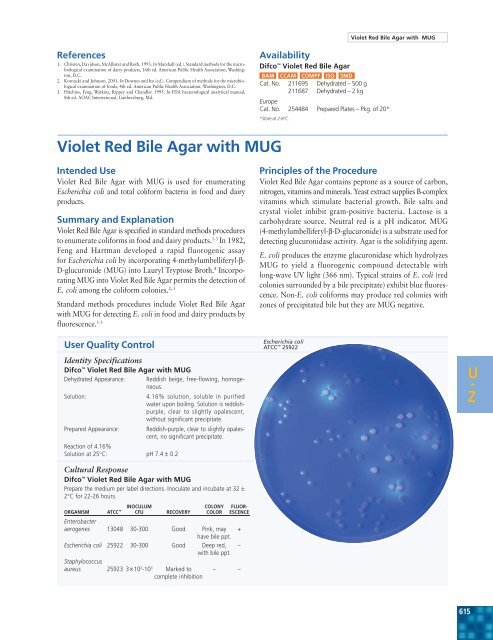
<strong>Violet</strong> <strong>Red</strong> <strong>Bile</strong> <strong>Agar</strong> <strong>with</strong> <strong>MUG</strong>References1. Christen, Davidson, McAllister and Roth. 1993. In Marshall (ed.). Standard methods for the microbiologicalexamination of dairy products, 16th ed. American Public Health Association, Washington,D.C.2. Kornacki and Johnson. 2001. In Downes and Ito (ed.). Compendium of methods for the microbiologicalexamination of foods, 4th ed. American Public Health Association, Washington, D.C.3. Hitchins, Feng, Watkins, Rippey and Chandler. 1995. In FDA bacteriological analytical manual,8th ed. AOAC International, Gaithersburg, Md.AvailabilityDifco <strong>Violet</strong> <strong>Red</strong> <strong>Bile</strong> <strong>Agar</strong>BAM CCAM COMPF ISO SMDCat. No. 211695 Dehydrated – 500 g211687 Dehydrated – 2 kgEuropeCat. No. 254484 Prepared Plates – Pkg. of 20**Store at 2-8°C.<strong>Violet</strong> <strong>Red</strong> <strong>Bile</strong> <strong>Agar</strong> <strong>with</strong> <strong>MUG</strong>Intended Use<strong>Violet</strong> <strong>Red</strong> <strong>Bile</strong> <strong>Agar</strong> <strong>with</strong> <strong>MUG</strong> is used for enumeratingEscherichia coli and total coliform bacteria in food and dairyproducts.Summary and Explanation<strong>Violet</strong> <strong>Red</strong> <strong>Bile</strong> <strong>Agar</strong> is specified in standard methods proceduresto enumerate coliforms in food and dairy products. 1-3 In 1982,Feng and Hartman developed a rapid fluorogenic assayfor Escherichia coli by incorporating 4-methylumbelliferyl-β-D-glucuronide (<strong>MUG</strong>) into Lauryl Tryptose Broth. 4 Incorporating<strong>MUG</strong> into <strong>Violet</strong> <strong>Red</strong> <strong>Bile</strong> <strong>Agar</strong> permits the detection ofE. coli among the coliform colonies. 2, 3Standard methods procedures include <strong>Violet</strong> <strong>Red</strong> <strong>Bile</strong> <strong>Agar</strong><strong>with</strong> <strong>MUG</strong> for detecting E. coli in food and dairy products byfluorescence. 1-3User Quality ControlIdentity SpecificationsDifco <strong>Violet</strong> <strong>Red</strong> <strong>Bile</strong> <strong>Agar</strong> <strong>with</strong> <strong>MUG</strong>Dehydrated Appearance: <strong>Red</strong>dish beige, free-flowing, homogeneous.Solution:4.16% solution, soluble in purifiedwater upon boiling. Solution is reddishpurple,clear to slightly opalescent,<strong>with</strong>out significant precipitate.Prepared Appearance: <strong>Red</strong>dish-purple, clear to slightly opalescent,no significant precipitate.Reaction of 4.16%Solution at 25°C: pH 7.4 ± 0.2Cultural ResponseDifco <strong>Violet</strong> <strong>Red</strong> <strong>Bile</strong> <strong>Agar</strong> <strong>with</strong> <strong>MUG</strong>Prepare the medium per label directions. Inoculate and incubate at 32 ±2°C for 22-26 hours.INOCULUM COLONY FLUOR-ORGANISM ATCC CFU RECOVERY COLOR ESCENCEEnterobacteraerogenes 13048 30-300 Good Pink, may +have bile ppt.Escherichia coli 25922 30-300 Good Deep red, –<strong>with</strong> bile ppt.Staphylococcusaureus 25923 3×10 2 -10 3 Marked to – –complete inhibitionPrinciples of the Procedure<strong>Violet</strong> <strong>Red</strong> <strong>Bile</strong> <strong>Agar</strong> contains peptone as a source of carbon,nitrogen, vitamins and minerals. Yeast extract supplies B-complexvitamins which stimulate bacterial growth. <strong>Bile</strong> salts andcrystal violet inhibit gram-positive bacteria. Lactose is acarbohydrate source. Neutral red is a pH indicator. <strong>MUG</strong>(4-methylumbelliferyl-β-D-glucuronide) is a substrate used fordetecting glucuronidase activity. <strong>Agar</strong> is the solidifying agent.E. coli produces the enzyme glucuronidase which hydrolyzes<strong>MUG</strong> to yield a fluorogenic compound detectable <strong>with</strong>long-wave UV light (366 nm). Typical strains of E. coli (redcolonies surrounded by a bile precipitate) exhibit blue fluorescence.Non-E. coli coliforms may produce red colonies <strong>with</strong>zones of precipitated bile but they are <strong>MUG</strong> negative.Escherichia coliATCC 25922U -Z615
Section IIIU-Z <strong>Violet</strong> <strong>Red</strong> <strong>Bile</strong> <strong>with</strong> <strong>MUG</strong>, cont.FormulaDifco <strong>Violet</strong> <strong>Red</strong> <strong>Bile</strong> <strong>Agar</strong> <strong>with</strong> <strong>MUG</strong>Approximate Formula* Per LiterYeast Extract .............................................................. 3.0 gPeptone ..................................................................... 7.0 g<strong>Bile</strong> Salts No. 3 ........................................................... 1.5 gLactose ..................................................................... 10.0 gSodium Chloride ........................................................ 5.0 g<strong>Agar</strong> ......................................................................... 15.0 gNeutral <strong>Red</strong> ................................................................ 0.03 gCrystal <strong>Violet</strong> .............................................................. 2.0 mg<strong>MUG</strong> (4-methylumbelliferyl-ß-D-glucuronide) ............ 0.1 g*Adjusted and/or supplemented as required to meet performance criteria.Directions for Preparation fromDehydrated Product1. Suspend 41.6 g of the powder in 1 L of purified water. Mixthoroughly.2. Heat <strong>with</strong> frequent agitation and boil for 1 minute tocompletely dissolve the powder. DO NOT AUTOCLAVE.3. Cool to 45-50°C and use immediately.4. Test samples of the finished product for performance usingstable, typical control cultures.Procedure1. Process each specimen as appropriate for that specimen. 1-32. Incubate plates at 35°C for 22-26 hours.3. Examine plates for growth and fluorescence.Expected ResultsColiform organisms form purplish-red colonies that aregenerally surrounded by a reddish zone of precipitated bile.When examined under long-wave fluorescent light, <strong>MUG</strong>positivecolonies are surrounded by a bluish fluorescent halo.<strong>MUG</strong>-negative colonies lack the fluorescent halo.E. coli colonies are red surrounded by a zone of precipitatedbile and fluoresce blue under long-wave UV light.Salmonella and Shigella strains that produce glucuronidase maybe encountered infrequently but these are generally lactosenegative and appear as colorless colonies which may fluoresce.Limitations of the Procedure1. Glucuronidase-negative strains of E. coli have been encountered.5-7 Similarly, glucuronidase-positive strains of E. colithat do not fluoresce have been reported. 82. Strains of Salmonella and Shigella that produce glucuronidasemay infrequently be encountered. 9 These strainsmust be distinguished from E. coli on the basis of otherparameters; e. g., gas production, lactose fermentation orgrowth at 44.5°C.References1. Christen, Davidson, McAllister and Roth. 1993. In Marshall (ed.). Standard methods for the microbiologicalexamination of dairy products, 16th ed. American Public Health Association, Washington,D.C.2. Kornacki and Johnson. 2001. In Downes and Ito (ed.). Compendium of methods for the microbiologicalexamination of foods, 4th ed. American Public Health Association, Washington, D.C.3. Hitchins, Feng, Watkins, Rippey and Chandler. 1995. In FDA bacteriological analytical manual,8th ed. AOAC International, Gaithersburg, Md.4. Feng and Hartman. 1982. Appl. Environ. Microbiol. 43:1320.5. Chang, Brill and Lum. 1989. Appl. Environ. Microbiol. 55:335.6. Hansen and Yourassowsky. 1984. J. Clinical Microbiol. 20:1177.7. Kilian and Bulow. 1976. Acta Pathol. Microbiol. Scand. Sect. B 84:245.8. Mates and Shaffer. 1989. J. Appl. Bacteriology 67:343.9. Damare, Campbell and Johnston. 1985. J. Food Sci. 50:1736.AvailabilityDifco <strong>Violet</strong> <strong>Red</strong> <strong>Bile</strong> <strong>Agar</strong> <strong>with</strong> <strong>MUG</strong>BAM COMPFCat. No. 229100 Dehydrated –500 gBBL <strong>Violet</strong> <strong>Red</strong> <strong>Bile</strong> <strong>Agar</strong> <strong>with</strong> <strong>MUG</strong>BAM COMPFCat. No. 299128 Prepared Bottle (200 mL) – Pkg. of 10<strong>Violet</strong> <strong>Red</strong> <strong>Bile</strong> Glucose <strong>Agar</strong>Intended Use<strong>Violet</strong> <strong>Red</strong> <strong>Bile</strong> Glucose <strong>Agar</strong> is used for detecting and enumeratingEnterobacteriaceae in foods and dairy products.Summary and ExplanationThe Enterobacteriaceae group includes lactose-fermentingcoliform bacteria, lactose-nonfermenting strains of E. coli,and lactose-nonfermenting species, such as Salmonella andShigella. When examining some foods, it is desirable to detectEnterobacteriaceae rather than the coliform bacteria. 1,2Enterobacteriaceae are glucose-fermenting bacteria. Mosselet al. 3 modified lactose-containing <strong>Violet</strong> <strong>Red</strong> <strong>Bile</strong> <strong>Agar</strong> byadding glucose to improve the recovery of Enterobacteriaceae.Later work by Mossel et al. 4,5 demonstrated that lactose couldbe omitted, resulting in the formulation known as <strong>Violet</strong> <strong>Red</strong><strong>Bile</strong> Glucose <strong>Agar</strong> (VRBGA).The Hycheck hygiene contact slide is a double-sided paddlecontaining two agar surfaces for immersing into fluids orsampling surfaces. There is one slide containing <strong>Violet</strong> <strong>Red</strong><strong>Bile</strong> Glucose <strong>Agar</strong> paired <strong>with</strong> Tryptic Soy <strong>Agar</strong>.Principles of the Procedure<strong>Violet</strong> <strong>Red</strong> <strong>Bile</strong> Glucose <strong>Agar</strong> contains peptone as a source ofcarbon, nitrogen, vitamins and minerals. Yeast extract suppliesB-complex vitamins which stimulate bacterial growth. Glucoseis a carbohydrate. <strong>Bile</strong> salts and crystal violet inhibit grampositivebacteria. Glucose fermenters produce red colonies <strong>with</strong>red-purple halos (bile precipitation) in the presence of neutralred, a pH indicator. <strong>Agar</strong> is the solidifying agent.616