M MYP Agar Antimicrobic Vial P - BVA Scientific
M MYP Agar Antimicrobic Vial P - BVA Scientific
M MYP Agar Antimicrobic Vial P - BVA Scientific

You also want an ePaper? Increase the reach of your titles
YUMPU automatically turns print PDFs into web optimized ePapers that Google loves.
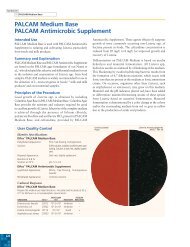
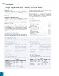
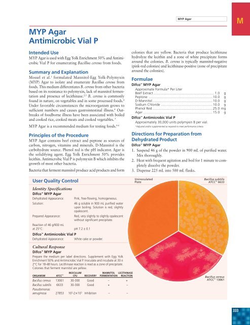
<strong>MYP</strong> <strong>Agar</strong><strong>Antimicrobic</strong> <strong>Vial</strong> PIntended Use<strong>MYP</strong> <strong>Agar</strong> is used with Egg Yolk Enrichment 50% and <strong>Antimicrobic</strong><strong>Vial</strong> P for enumerating Bacillus cereus from foods.Summary and ExplanationMossel et al. 1 formulated Mannitol-Egg Yolk-Polymyxin(<strong>MYP</strong>) <strong>Agar</strong> to isolate and enumerate Bacillus cereus fromfoods. This medium differentiates B. cereus from other bacteriabased on its resistance to polymyxin, lack of mannitol fermentationand presence of lecithinase. 2,3 B. cereus is commonlyfound in nature, on vegetables and in some processed foods. 4Under favorable circumstances the microorganism grows tosufficient numbers and causes gastrointestinal illness. 4 Outbreaksof foodborne illness have been associated with boiledand cooked rice, cooked meats and cooked vegetables. 5<strong>MYP</strong> <strong>Agar</strong> is a recommended medium for testing foods. 4-6Principles of the Procedure<strong>MYP</strong> <strong>Agar</strong> contains beef extract and peptone as sources ofcarbon, nitrogen, vitamins and minerals. D-Mannitol is thecarbohydrate source. Phenol red is the pH indicator. <strong>Agar</strong> isthe solidifying agent. Egg Yolk Enrichment 50% provideslecithin. <strong>Antimicrobic</strong> <strong>Vial</strong> P is polymyxin B which inhibits thegrowth of most other bacteria.Bacteria that ferment mannitol produce acid products and formcolonies that are yellow. Bacteria that produce lecithinasehydrolyze the lecithin and a zone of white precipitate formsaround the colonies. B. cereus is typically mannitol-negative(pink-red colonies) and lecithinase-positive (zone of precipitatearound the colonies).FormulaeDifco <strong>MYP</strong> <strong>Agar</strong>Approximate Formula* Per Liter<strong>MYP</strong> <strong>Agar</strong>Beef Extract ................................................................ 1.0 gPeptone ................................................................... 10.0 gD-Mannitol ............................................................... 10.0 gSodium Chloride ...................................................... 10.0 gPhenol Red ............................................................... 25.0 mg<strong>Agar</strong> ......................................................................... 15.0 gDifco <strong>Antimicrobic</strong> <strong>Vial</strong> PApproximately 30,000 units polymyxin B per vial.*Adjusted and/or supplemented as required to meet performance criteria.Directions for Preparation fromDehydrated ProductDifco <strong>MYP</strong> <strong>Agar</strong>1. Suspend 46 g of the powder in 900 mL of purified water.Mix thoroughly.2. Heat with frequent agitation and boil for 1 minute to completelydissolve the powder.3. Dispense 225 mL into 500 mL flasks.MUser Quality ControlIdentity SpecificationsDifco <strong>MYP</strong> <strong>Agar</strong>Dehydrated Appearance:Solution:Pink, free-flowing, homogeneous.46 g soluble in 900 mL purified waterupon boiling. Solution is red, slightlyopalescent.Prepared Appearance: Red, very slightly to slightly opalescentwithout significant precipitate.Reaction of 46 g/900 mLat 25°C: pH 7.2 ± 0.1Difco <strong>Antimicrobic</strong> <strong>Vial</strong> PDehydrated Appearance: White cake or powder.Cultural ResponseDifco <strong>MYP</strong> <strong>Agar</strong>Prepare the medium per label directions. Supplement with Egg YolkEnrichment 50% and <strong>Antimicrobic</strong> <strong>Vial</strong> P. Inoculate and incubate at 30 ±2°C for 18-48 hours. Lecithinase reaction is read as a zone of precipitate.Colonies that ferment mannitol are yellow.INOCULUM MANNITOL LECITHINASEORGANISM ATCC CFU RECOVERY FERMENTATION REACTIONBacillus cereus 13061 30-300 Good – +Bacillus subtilis 6633 30-300 Good + –Pseudomonasaeruginosa 27853 10 3 -2×10 3 Inhibition – –UninoculatedPlateBacillus subtilisATCC 6633Bacillus cereusATCC 13061333
Section IIIM <strong>MYP</strong> <strong>Agar</strong>, cont.4. Autoclave at 121°C for 15 minutes. Cool to 45-50°C.5. Aseptically add 12.5 mL Egg Yolk Enrichment 50% and4.1 mL <strong>Antimicrobic</strong> <strong>Vial</strong> P rehydrated with 5 mL sterilewater (25,000 units of polymyxin B). Mix thoroughly.Difco <strong>Antimicrobic</strong> <strong>Vial</strong> P (Polymyxin B)1. To rehydrate, aseptically add 5 mL sterile purified water(to achieve the desired concentration for <strong>MYP</strong> <strong>Agar</strong>).2. Rotate in an end-over-end motion to dissolve the contentscompletely.ProcedureConsult appropriate references. 4-6Expected ResultsConsult appropriate references. 4-6References1. Mossel, Koopman and Jongerius. 1967. Appl. Microbiol. 15:650.2. Donovan. 1958. J. Appl. Bacteriol. 21:100.3. Coliner. 1948. J. Bacteriol. 55:777.4. Rhodehamel and Harmon. 1995. FDA bacteriological analytical manual, 8th ed. AOAC International,Gaithersburg, Md.5. Bennett and Belay. 2001. In Downes and Ito (ed.), Compendium of methods for the microbiologicalexamination of foods, 4th ed. American Public Health Association, Washington, D.C.6. Andrews. 2000. In Horwitz (ed.), Official methods of analysis of AOAC International, 17th ed.,vol. I. AOAC International, Gaithersburg, Md.AvailabilityDifco <strong>MYP</strong> <strong>Agar</strong>AOAC BAM COMPF ISO USDACat. No. 281010 Dehydrated – 500 gEuropeCat. No. 257004 Prepared Plates – Pkg. of 20*JapanCat. No. 251264 Prepared Plates – Pkg. of 20*251265 Prepared Plates – Ctn. of 100*Difco <strong>Antimicrobic</strong> <strong>Vial</strong> PAOAC BAM COMPF ISO USDACat. No. 232681 <strong>Vial</strong> – 6 × 10 mL*Difco Egg Yolk Enrichment 50%AOAC BAM COMPF ISO USDACat. No. 233471 Tube – 12 × 10 mL*233472 Bottle – 6 × 100 mL**Store at 2-8°C.MacConkey <strong>Agar</strong>sMacConkey <strong>Agar</strong> • MacConkey <strong>Agar</strong> BaseMacConkey <strong>Agar</strong> without Crystal VioletMacConkey <strong>Agar</strong> without Crystal Violet or SaltMacConkey <strong>Agar</strong> without SaltIntended UseMacConkey <strong>Agar</strong> conforms with the specifications of TheUnited States Pharmacopeia (USP).MacConkey agars are slightly selective and differential platingmedia mainly used for the detection and isolation of gram-negativeorganisms from clinical, 1 dairy, 2 food, 3,4 water, 5 pharmaceutical 6and industrial 7 sources.MacConkey <strong>Agar</strong> is used for isolating and differentiating lactosefermentingfrom lactose-nonfermenting gram-negative entericbacilli.MacConkey <strong>Agar</strong> Base is used with added carbohydrate indifferentiating coliforms based on fermentation reactions.MacConkey <strong>Agar</strong> without Crystal Violet is used for isolatingand differentiating enteric microorganisms while permittinggrowth of staphylococci and enterococci. The medium canbe used also to separate Mycobacterium fortuitum andM. chelonae from other rapidly growing mycobacteria.MacConkey <strong>Agar</strong> without Crystal Violet or Salt and MacConkey<strong>Agar</strong> without Salt are used for isolating and differentiatinggram-negative bacilli while suppressing the swarming of mostProteus species.Summary and ExplanationMacConkey <strong>Agar</strong> is based on the bile salt-neutral red-lactoseagar of MacConkey. 8The original MacConkey medium was used to differentiatestrains of Salmonella typhosa from members of the coliformgroup. Formula modifications improved the growth of Shigellaand Salmonella strains. These modifications included the additionof 0.5% sodium chloride, decreased agar content, andaltered bile salts and neutral red concentrations. The formulaimprovements gave improved differential reactions betweenthese enteric pathogens and the coliform group.MacConkey <strong>Agar</strong> contains crystal violet and bile salts thatinhibit gram-positive organisms and allow gram-negativeorganisms to grow. Isolated colonies of coliform bacteria arebrick red in color and may be surrounded by a zone of precipitatedbile. This bile precipitate is due to a local pH drop aroundthe colony due to lactose fermentation. Colonies that do notferment lactose (such as typhoid, paratyphoid and dysenterybacilli) remain colorless. When lactose nonfermenters growin proximity to coliform colonies, the surrounding mediumappears as cleared areas. It is recommended in the USP for usein the performance of Microbial Limit Tests. 6334