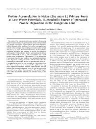
100 <strong>Plant</strong> Cell Tiss Organ Cult (2009) 99:97–108Fig. 2 Sorghum transformation, plantlet regeneration, GUS assayand leaf-painting analysis <strong>of</strong> <strong>transgenic</strong> events. a Embryos wereco-cultivated with Agrobacterium on TM for 3–5 days; b Resting onRM for additional 4 days; c Callus induction under selection pressure,herbicide-tolerance calli developed from some embryos on CM;d Shoot formation on SM; e After 2–3 weeks <strong>of</strong> being transferredonto ZM, shoots began rooting; f and g Putative <strong>transgenic</strong> plants (youngand adult) were in greenhouse; h GUS assay <strong>of</strong> embryogenic calli;i leaf painting showing susceptible response <strong>of</strong> wild-type leaves (lefttwo leaf strips) and resistance <strong>of</strong> <strong>transgenic</strong> leaves (right two strips)The embryos were transferred onto resting medium(RM) for another 4 days at 28°C in the dark (Fig. 2b) andthen transferred weekly onto freshly made callus inductionmedium (CM) with PPT 2.5 mg l -1 , incubated at 28°C for4–8 weeks (Fig. 2c). Herbicide-tolerant calli can be maintainedon CM, but untransformed calli eventually die. Oncesomatic embryogenic cells developed from transformedcalli, they were transferred onto shooting medium (SM)(Fig. 2d). Shoots (2–5 cm long) were separated and transferredinto plastic boxes (898910 cm) containing rootingmedium (ZM), and maintained at 25°C under a photoperiod<strong>of</strong> 16 h light and 8 h dark; the shoots later developed into8–10 cm tall plantlets with healthy roots (Fig. 2e). Theywere then transferred into pots containing Promix soil mixedwith Osmocote (18-6-12) in the greenhouse (Fig. 2f and g).GUS assay and leaf painting using libertyÒTo verify the expression <strong>of</strong> GUS gene, calli and plant tissueswere assayed with histochemical X-Gluc staining at 37°Cfor 24 h (Fig. 2h; Jefferson et al. 1987). The X-Gluc solutionfor staining target tissues contained: 0.1 M NaPO 4 ,pH7.0; 1.0 mM K 3 [Fe(CN) 6 ]; 10 mM EDTA; 2 mM X-Gluc;0.1% (v/v) Triton-100; 20% (v/v) methanol anhydrate.123
<strong>Plant</strong> Cell Tiss Organ Cult (2009) 99:97–108 101To confirm the functional expression <strong>of</strong> the bar gene,500 mg l -1 Liberty Ò was applied with a cotton applicatoronto the leaves <strong>of</strong> young plants (with 4–5 leaves) grown ina growth chamber at day/night temperatures 26/21°C and aphotoperiod <strong>of</strong> 16 h light/8 h dark. <strong>Plant</strong>s that did notexhibit necrosis at 5 days were scored as herbicide resistant(Fig. 2i).Polymerase chain reaction analysisPCR analysis was used for quick screening <strong>of</strong> the presence<strong>of</strong> trans-genes in all T0 and randomly chosen T1 generations.Young healthy leaf tissue was collected from herbicideresistant plantlets that had been transferred into soil.RED-Extract-N-AmpTM <strong>Plant</strong> PCR Kit (Sigma) was usedfor DNA extraction and amplification. The primer pairs forPCR were: for bar gene, forward (fwd)-bar (5 0 GTCTGCACCATCGTCAACC) and reverse (rev)-bar (5 0 GAAGTCCAGCTGCCAGAAAC); for tRNA lys , fwd-tRNA (5 0 CCGCATGCATGTATAAGTGTGTCGGAACTGG) and trev-RNA (5 0 TGCTGCAGGTTTGACTAACTAACGGGGTTGTTG); for the SKRS gene, three pairs <strong>of</strong> primers for promoter,coding and terminator regions <strong>of</strong> SKRS gene,respectively, were used for screening; pair 1, fwd-CZ19-565 (5 0 TGTGGACAATACCGAGAGGA) and rev-flag (5 0CATGATGCTTGTCATCGTCGTCCTTGTAGTC); pair2: fwd-flag (5 0 CATGGACTACAAGGACGACGATGACAAGCAT) and rev-E12-418 (5 0 TGTAAGCTTCTCCCCATTGC); pair 3, fwd-CZ19-5(5 0 ATACTGCAGTTGCCTCCTTATGCTCCTTG) and rev-CZ19-3 (5 0 ATGCGGCCGCGAATTCGATTCTTCCCATTTC. Thermal cycling conditionsincluded 45 s at 94°C for denaturalization, 1 min at58°C for annealing and 1 min at 72°C for extension with29 cycles.Southern blot analysisSouthern blot analysis (Southern 1975) was used to confirmthe integration and stable inheritance <strong>of</strong> transgene inserts.Genomic DNA was extracted from <strong>sorghum</strong> leaf tissuesusing a modified CTAB-based protocol (Dellaporta et al.1983). Twenty-five microgram <strong>of</strong> DNA were digested witha single restriction enzyme, which cuts once within one ortwo T-DNA regions, dependent on the T-DNA regions that<strong>transgenic</strong> events carried. The digested genomic DNA wasfractionated on a 1.0% agarose gel prior to transfer to aZeta-ProbeÒ GT nylon membrane (Bio-Rad, CA, USA).DNA was fixed to the membrane by UV cross-linking.Hybridization and washing conditions followed the Zeta-ProbeÒ GT manufacturer’s instructions and were at 65°C.The bar-TEV (from vector pZY101) and TC2 or SKRS(genes <strong>of</strong> interest) were used to generate a 32 P-labeledprobe by random primer synthesis incorporating 32 P-dATPutilizing the Prime-it Ò II kit (Stratagene, USA).Statistical analysisThe SAS (version 9.1) GLM (General Linear Model)program was used for variance analysis. Mean comparisonswere made using LSD at a = 0.01 level. The 2 -test wasused to test the goodness-<strong>of</strong>-fit <strong>of</strong> progeny segregation.Transformation frequency (%) was determined by thenumber <strong>of</strong> independent fertile <strong>transgenic</strong> <strong>sorghum</strong> eventsdivided by the total number <strong>of</strong> embryos inoculated. Eachindependent event was defined by its herbicide resistanceand PCR-positive plants whose transgene integrations andtransmittance were further confirmed by different bandingpatterns in Southern blot analysis and by a segregationanalysis using PCR, respectively. The frequency <strong>of</strong> independentcallus lines was calculated by the number <strong>of</strong> herbicideresistant callus lines obtained from differentembryos divided by the total number <strong>of</strong> embryosinoculated.ResultsTransformation with pZY102High quality immature embryos were harvested from plantsgrown with a modified greenhouse care procedure thatrequired little labor and which provided consistent plantmaterials (see ‘‘Materials and methods’’). A. tumefaciensstrain EHA101 carrying a standard binary vector (pZY102),which harbors the bar gene as a plant selectable <strong>marker</strong> andthe GUS-intron gene as a reporter, was used to evaluatetransformation parameters. Several different co-cultivationconditions were tested for improvement <strong>of</strong> T-DNA delivery(Table 2). These conditions included: Treatments I-3 and I-5, TM combined with 3 and 5 days co-cultivation periods,respectively; II-3 and II-5, TM supplemented with L-cysteine(0.40 g l -1 ) and DTT (0.31 g l -1 ) and co-cultivationfor 3 or 5 days; III-3, and III-5, TM supplemented withPVPP (10 g l -1 ) combined with co-cultivation periods <strong>of</strong> 3and 5 days; IV-3 and IV-5, TM supplemented with L-cysteine(0.40 g l -1 , DTT 0.31 g l -1 ) and PVPP (10 g l -1 ), forco-cultivation periods <strong>of</strong> 3 and 5 days. Following selection,a total <strong>of</strong> 104 herbicide-tolerant callus lines that expressedGUS were obtained. Frequencies <strong>of</strong> callus lines obtained atthis stage ranged from 0.21 to 4.55%. Treatment III-5exhibited the highest efficiency, followed by treatments I-3,III-3, and I-5.Statistical analysis indicates that treatments I and III aresuperior to treatments II and IV, at either 3 or 5 days.Taking into consideration that browning happened during123