INFORMATION PAPER: Hand Held Assay (HHA)
INFORMATION PAPER: Hand Held Assay (HHA)
INFORMATION PAPER: Hand Held Assay (HHA)

Create successful ePaper yourself
Turn your PDF publications into a flip-book with our unique Google optimized e-Paper software.
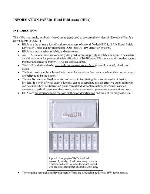
<strong>INFORMATION</strong> <strong>PAPER</strong>: <strong>Hand</strong> <strong>Held</strong> <strong>Assay</strong> (<strong>HHA</strong>)INTRODUCTIONThe <strong>HHA</strong> is a simple, antibody - based assay (test) used to presumptively identify Biological Warfare(BW) agents (Figure 1).• <strong>HHA</strong>s are the primary identification component of several fielded (BIDS, IBAD, Portal Shield,Dry Filter Units) and developmental DOD (JBPDS) BW detection systems.• <strong>HHA</strong>s are inexpensive, reliable, and easy to use.• An <strong>HHA</strong> is a one-time use capability designed to presumptively identify one agent. The currentcapability allows for presumptive identification of 10 different BW threat and 4 stimulant agents.Positive and negative trainer <strong>HHA</strong>s are also available.• The <strong>HHA</strong> is designed to be used only on non-porous surfaces (example - metal, plastic andglass)• The best results can be achieved when samples are taken from an area where the concentrationsare believed to be the highest.• The results can be utilized to advise and assist in facilitating the resolution of a biologicalincident. It is only after an agent’s identity can be ascertained that an effective outer perimetercan be established, neutralization plans formulated, decontamination procedures enacted,emergency medical treatment plans made, and environmental preservation precautions taken.• <strong>HHA</strong>s are not designed to be the sole method of identification and are not for diagnostic use.Figure 1. Photograph of JPO’s <strong>Hand</strong> <strong>Held</strong><strong>Assay</strong>s. Typically, 10 individual assays come ina cassette packaged in a white foil pouch labeledwith the assay, lot number, and manufacture date.• The ongoing research and development efforts are producing additional BW agent assays.
THE SCIENCE BEHIND THE HAND HELD ASSAYThe <strong>HHA</strong> is a form of biological assay called immunochromatography and is designed to provide aquick and accurate presumptive identification of selected biological warfare agents. The <strong>HHA</strong> works onthe principle of antigen/antibody interactions.• Antigens are any foreign substance that when introduced into the host are capable of eliciting animmune response, which ultimately results in antibody production.• Antibodies are molecules that are found in the blood and tissue fluids of mammals that areproduced in response to a given antigen. Biologically, the role of the antibody is to bind theintruding foreign substance and facilitate its removal from the body.Typically, an organism carries many different complex antigens on its surface. The differing antigensare called epitopes and it is not uncommon for manydifferent antibodies to be produced in response to aninfection (Figure 2). An epitope is unique to a givenEpitopesantigen and correlates with the genetic diversity ofvarying species of microorganisms.<strong>HHA</strong>’s exploit the exquisite sensitivity and specificityof antibodies to detect and differentiatemicroorganisms. These antibodies are able to physicallygrab on to a portion of an antigen with their antigenbindingsite. Two categories of antibodies are typically used in immunoassays:• polyclonal antibodies (PAB’s) - Polyclonals represent a population of many antibodies whichbind to numerous different antigens (epitopes) (Figure 3).• monoclonal antibodies (MAB’s) - Monoclonals represent a single type of antibody which bindto one specific antigen (epitope) (Figure 3).Polyclonal antibodies are typically used for immunoassays because of their ease of production and theirsuperior sensitivity. What makes polyclonal antibody assays more sensitive is that they can cover thesurface of a complex antigen such as a microorganism more uniformly thus improving the detectioncapability (Figure 3). Monoclonalsrepresent a single type of antibodywhich bind to one specific epitope.A high degree of sensitivity andspecificity against a particularbiological agent can be achievedby careful screening and selectionof a monoclonal antibody.However, monoclonal antibodiescan bind to only one type ofepitope on the surface of the cell,possibly reducing the level ofcoating. The potential then existsPolyclonalPolyclonals are many different B clonesCellgrouped together.AntigenFigure 2. Schematic of typical bacterial antigenillustrating the variability of surface epitopesMonoclonalMonoclonals are single cloneFigure 3. Illustration demonstrating how a polyclonal will coatthe surface of an antigen more uniformly than a polyclonaltypically will.to give up a certain level of sensitivity. Polyclonal antibodies are far easier, faster, and cheaper toproduce. However, in general, polyclonal antibodies do not have the specificity of a monoclone. Efforts
to combine monoclones are being successfully employed to improve new <strong>HHA</strong>s through balancingsensitivity and specificity.<strong>HHA</strong> COMPONENTSThe components of the <strong>HHA</strong> are as follows (Figure 4):• Sample delivery pad: When the sample is added to the sample delivery well, it contacts thesample delivery pad first. The sample delivery pad functions to filter out any large particulatematter in the sample and to hold the sample so that is can slowly wick through into the conjugaterelease pad.• Conjugate release pad: The conjugate pad contains the detector antibody which is conjugated tocolloidal gold. This allows for visualization of the antibody. If sample is added to the assay thatcontains compatable antigen, the colloidal gold labeled antibody will bind to target antigen andallow for detection of the antigen when it subsequently binds to the capture antibody.• Nitrocellulose Membrane: The sample enters the nitrocellulose membrane via capillary actiontowards the sample wicking pad. Bound to the membrane are the capture antibody and the antispeciesantibody which are sprayed in descreet lines on the membrane about halfway up theticket.• Capture antibody: The capture antibody is what makes up the test line on the ticket. The testline is adjacent to the letter “T” on the plastic cassette. The capture antibody is bound to themembrane and when antigen flows past it serves to capture the antigen.• Antispecies antibody: The anti-species antibody will bind the colloidal gold labeled antibodyregardless whether antigen is present or not. This serves as the control to indicate whether theassay is functioning properly and is adjacent to the letter “C“ on the plastic cassette. It is calledan anti-species antibody because it is made in one species of animal that has been immunizedwith the antibody from another species. For example, if the detector antibody was made in goatthen the anti-species antibdy would be a rabbit immunized with goat antibodies to produce arabbit anti-goat antibodies.• Sample wicking pad: The sample wicking pad serves as a reservoir to hold the sample after ithas wicked across the nitrocellulose membrane. The sample wicking pad will only hold thesample for a short period of time before the sample will begin to flow back across the membranetowards the sample delivery pad during which time nonspecific binding can occur producingfalse positives. That is the capture antibody and detector will adhere to each other whetherantigen is present or not. For this reason it is important to read the <strong>HHA</strong> at the 15 minute timepoint.• Tape backing: The tape backing serves simply to hold the above components in place.• <strong>HHA</strong> buffer: A component of the <strong>HHA</strong> which is not part of the <strong>HHA</strong> device, but a critical partof the kit is the <strong>HHA</strong> sample dilution buffer. The solution added to the <strong>HHA</strong> must be aqueousfor the assay to function. The <strong>HHA</strong> buffer contains PBS, Triton X-100, and sodium azide. ThePBS serves to adjust the sample to a neutral pH so that the antibodies are able to functionproperly. Any significant deviation from a pH of 7 will change the conformation of theantibodies and they will no longer have the ability to bind antigen. The Triton X-100 is asurfactant that helps to prevent aggregates from forming which do no flow well across thenitrocellulose membrane. Sodium azide acts as a preservative to prevent growth of anymicrobial contaminants during storage of the buffer.
Detector antibodybound to colloidal goldCapture antibody against specific agentSample delivery padSample wicking padConjugate padTape backingNitrocellulosemembraneAntispecies antibodyFigure 4. The <strong>HHA</strong> ticket after removal from the plastic cassette.<strong>HHA</strong> RESULTS AND LIMITATIONSWhen reading an assay, any visable test line, even a very faint one, should be considered real. There arefour potential outcomes that may be observed after running an <strong>HHA</strong>.• The first potential outcome is two red lines indicating a positive assay. This may also be a resultof matrix effects (see below) so running the sample a second time following diluting 1:10 and1:100 in <strong>HHA</strong> buffer would be prudent (Figure 5.a).• The second outcome is a single line, the control line. This may be a valid negative or may be theresult of the “hook effect” (see below). Again, running the sample a second time followingdiluting 1:10 and 1:100 in <strong>HHA</strong> buffer is advised (Figure 5.b).• The third outcome is a postitve test line but no control line. This is probably due to a faultyassay which requires running the sample again with a new set of <strong>HHA</strong>’s (Figure 5.c).• The fourth outcome is where no lines show up. This can be the result of a faulty assay, a matrixeffect, or the assay may have been exposed to moisture. The nitrocellulose membrane must bedry in order to wick the sample. If an assay has an incomplete control or positive line afterrunning, the assay is also faulty. To resolve this a new <strong>HHA</strong> is used utilizing sample dilutions of1:10 and 1:100 in <strong>HHA</strong> buffer (Figure 5.d).
Figure 5.Potential results following use of <strong>HHA</strong>’s. From left to right: Apositive <strong>Assay</strong> (a); A negative assay (b); A faulty assay (c); Afaulty assay, or potential matrix effects (d).All results whether positive, negative or inconclusive should be documented. It is important to keep inmind that no matter what the outcome of an <strong>HHA</strong>, these tests provide only a presumtive identificationand that the samples will need further evaluation at a confirmatory lab.Although <strong>HHA</strong>’s are fairly reliable, accurate, and sensitive assaying enviromental samples isexceedingly difficult and some of the technological limits may surface. An awareness of possibledeficiencies/limitations will help the operator recognize and hopefully avoid any potential problems.There are four major issues with immunochromatographic assays that could affect the accuracy of ananalysis. An understanding of these limits will help to decrease their occurance and mitigate possibledetrimental effects on the accuracy of a sample analysis.
Lack of SensitivityNot enough complex to seeDirection of flowFigure 6.The <strong>HHA</strong>’s have a lower sensitivity level that is not as low as theinfectivity dose for many pathogens• Senstivity Cutoff (Figure 6). <strong>HHA</strong>’s, like all biological assays, have a sensitivity cutoff . Thismeans that for each different agent assay there is a threshold concentration that below thisconcentartion the assay it will not be able to detect the presence of the antigen. Although <strong>HHA</strong>’sare very sensitive, the infective dose for most pathogens is far lower than the sensitivity of the<strong>HHA</strong>’s. Therefore, if a sample is tested and the result appears to be negative (false negative),there may still be enough biological agent in the sample to cause illness. You may give falseinformation if you state that the sample does not have a paticular agent in it because it may verywell have.Matrix Effect in <strong>HHA</strong>’sSomething in sample nonspecifically binds to capture antibody producing a false positiveAntigen detector antibody capture antibody complexdoes not form producing a false negative.In addition, detector antibody antispeciesantibody complex often does not form.Figure 7. Matrix effects can be cause by substances in the sample resulting infalse positives and false negatives.
• Matrix effect (Figure 7). The matrix effect is often encountered when assaying environmentalsamples in <strong>HHA</strong>’s. It can not be predetermined what type of sample will have to be analyzedprior to an incident. Sometimes a sample will not be compatible with the <strong>HHA</strong>’s. This canresult in false negatives or false positives. A false negative will occur if there is biological agentin the sample, but something else in the sample or some property of that sample prevents theantibodies from binding to the antigen. Conversly a false positive can occur if there is nobiological agent in the sample, but something else in the sample or some property of that samplecauses the detector and capture antibodies to bind together non-specifically. The <strong>HHA</strong>’s arescreened using several common matrices (dust, tap water, sewage, human sera, and soil) toensure that they will be less likely to pose a problem, but these matrices and others may still posea problem. Typically, the substace causing the matrix effect can be diluted out while leavingenough of the specific antigen to react in the <strong>HHA</strong> to see a true positive. If a matrix effect issuspected, it is recommeded that a 1:10 dilution of the sample in <strong>HHA</strong> buffer be run on a second<strong>HHA</strong>. This remedy also applies if you find the pH of your sample to be significantly above orbelow neutral (pH 7.0). It is important to note the control line when running samples. If thecontrol line does not form, there may be a sample matrix problem.Cross-Reactivity in <strong>HHA</strong>sOrganism BOrganism ACapture antibody against specific agentDetector antibodyantispecies antibody complexFigure 8.Cross reactivity occurs when two closely related species share a commonantigenic epitope• Cross-Reactivity (Figure 8). Cross-reactivity is most often seen with the use of polyclonalantibodies in <strong>HHA</strong>’s but can occur even if monoclonal antibodies are employed. Cross-reactivityusually occurs when an antibody binds to the species it was designed for but it also bindsspecifically to close relatives of that species. This occurs when two closely related species sharea common antigenic epitope allowing the antibodies in the <strong>HHA</strong> to bind to both species. It isseen most often with PAB’s because they potentially can bind to many different epitopes on agiven antigen (thus the likelyhood of crossreactivity is increased). Cross-reactivity occurs withthe Bacillus anthracis <strong>HHA</strong> in which the antibodies bind not only Bacillus anthracis but alsoother Bacillus such as Bacillus thuringinsis. Unfortunately these other Bacillus are normalconstituants of soil therefore soil is incompatible with the Anthrax <strong>HHA</strong>. At this time, there is nomonoclonal antibody in production for Bacillus anthracis.