LYTIC EPIPHYSEAL LESIONS - H.U.C.
LYTIC EPIPHYSEAL LESIONS - H.U.C.
LYTIC EPIPHYSEAL LESIONS - H.U.C.
- No tags were found...

Create successful ePaper yourself
Turn your PDF publications into a flip-book with our unique Google optimized e-Paper software.
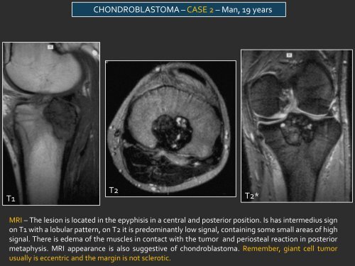
CHONDROBLASTOMA – CASE 2 – Man, 19 yearsT1T2T2*MRI – The lesion is located in the epyphisis in a central and posterior position. Is has intermedius signon T1 with a lobular pattern, on T2 it is predominantly low signal, containing some small areas of highsignal. There is edema of the muscles in contact with the tumor and periosteal reaction in posteriormetaphysis. MRI appearance is also suggestive of chondroblastoma. Remember, giant cell tumorusually is eccentric and the margin is not sclerotic.