LYTIC EPIPHYSEAL LESIONS - H.U.C.
LYTIC EPIPHYSEAL LESIONS - H.U.C.
LYTIC EPIPHYSEAL LESIONS - H.U.C.
SHOW LESS
- No tags were found...

Create successful ePaper yourself
Turn your PDF publications into a flip-book with our unique Google optimized e-Paper software.
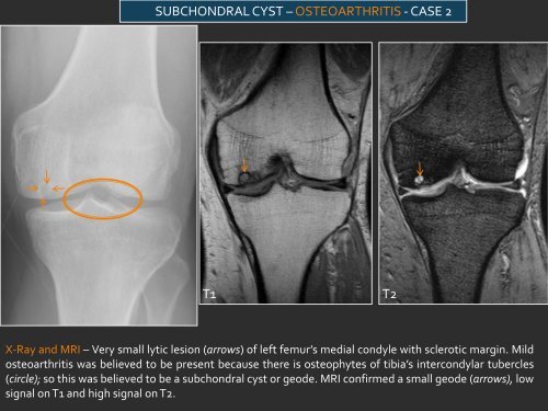
SUBCHONDRAL CYST – OSTEOARTHRITIS - CASE 2T1T2X-Ray and MRI – Very small lytic lesion (arrows) of left femur’s medial condyle with sclerotic margin. Mildosteoarthritis was believed to be present because there is osteophytes of tibia’s intercondylar tubercles(circle); so this was believed to be a subchondral cyst or geode. MRI confirmed a small geode (arrows), lowsignal on T1 and high signal on T2.