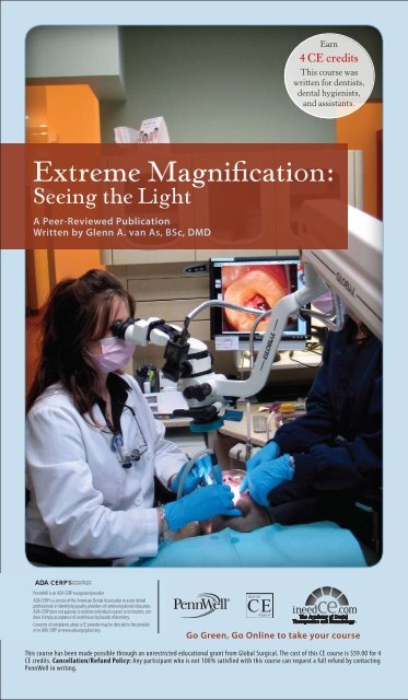
Extreme Magnification: Seeing the Light - IneedCE.com
Extreme Magnification: Seeing the Light - IneedCE.com
Extreme Magnification: Seeing the Light - IneedCE.com

You also want an ePaper? Increase the reach of your titles
YUMPU automatically turns print PDFs into web optimized ePapers that Google loves.
<strong>Extreme</strong> <strong>Magnification</strong>:<br />
<strong>Seeing</strong> <strong>the</strong> <strong>Light</strong><br />
A Peer-Reviewed Publication<br />
Written by Glenn A. van As, BSc, DMD<br />
PennWell is an ADA CERP recognized provider<br />
ADA CERP is a service of <strong>the</strong> American Dental Association to assist dental<br />
professionals in identifying quality providers of continuing dental education.<br />
ADA CERP does not approve or endorse individual courses or instructors, nor<br />
does it imply PennWell acceptance is an of credit ADA hours CERP by Recognized boards of dentistry. Provider<br />
Concerns of <strong>com</strong>plaints about a CE provider may be directed to <strong>the</strong> provider<br />
or to ADA CERP at www.ada.org/goto/cerp.<br />
Earn<br />
4 CE credits<br />
This course was<br />
written for dentists,<br />
dental hygienists,<br />
and assistants.<br />
Go Green, Go Online to take your course<br />
This course has been made possible through an unrestricted educational grant from Global Surgical. The cost of this CE course is $59.00 for 4<br />
CE credits. Cancellation/Refund Policy: Any participant who is not 100% satisfied with this course can request a full refund by contacting<br />
PennWell in writing.
Educational Objectives<br />
Upon <strong>com</strong>pletion of this course, <strong>the</strong> clinician will be able<br />
to do <strong>the</strong> following:<br />
1. Understand <strong>the</strong> evolution of <strong>the</strong> use of microscopes<br />
in dentistry.<br />
2. Know <strong>the</strong> improvements in treatment precision<br />
obtainable using a microscope.<br />
3. Understand how <strong>the</strong> integration of microscopes<br />
into <strong>the</strong> dental office can improve ergonomics and<br />
documentation, as well as aid <strong>com</strong>munication.<br />
Abstract<br />
In <strong>the</strong> late 1980s, San Diego endodontist Dr. Gary Carr<br />
concluded that <strong>the</strong> incredible magnification and illumination<br />
made possible with <strong>the</strong> microscope could be of benefit<br />
to <strong>the</strong> discipline of endodontics and he started promoting<br />
<strong>the</strong> usage of <strong>the</strong> Dental Operating Microscope (D.O.M.)<br />
as a crucial piece of <strong>the</strong> armamentarium used in <strong>the</strong> improvement<br />
of out<strong>com</strong>es of endodontic apical surgeries. By<br />
1998, <strong>the</strong> American Academy of Endodontics decided to<br />
institute <strong>the</strong> requirement that all post-graduate endodontic<br />
students from accredited programs be<strong>com</strong>e proficient<br />
in <strong>the</strong> usage of <strong>the</strong> D.O.M. in order to graduate from <strong>the</strong>ir<br />
post-doctoral program.<br />
Four basic advantages in using <strong>the</strong> operating microscope<br />
and ac<strong>com</strong>panying documentation systems (digital<br />
microphotography and videography) for private practice<br />
include improved precision of treatment, enhanced ergonomics,<br />
ease of digital documentation and <strong>the</strong> increased<br />
ability to <strong>com</strong>municate through integrated video. These<br />
four <strong>com</strong>mon advantages are witnessed in all aspects of a<br />
microscope-centered practice, regardless of <strong>the</strong> discipline<br />
involved or procedure being <strong>com</strong>pleted. The operating microscope<br />
also allows <strong>the</strong> dentist to sit in an upright, neutral,<br />
and balanced posture, and has proven to be of great value<br />
in aiding documentation.<br />
The advantages of improved precision and ergonomics,<br />
ease of documentation, and <strong>the</strong> ability to <strong>com</strong>municate with<br />
patients, staff, and colleagues are clear. As <strong>the</strong> new millennium<br />
dawned, dentists using <strong>the</strong> D.O.M. have found that<br />
<strong>the</strong> technology not only improves treatment out<strong>com</strong>es, but<br />
also increases <strong>the</strong> enjoyment of providing treatment.<br />
Introduction<br />
In 1981, Apo<strong>the</strong>ker brought <strong>the</strong> concept of extreme magnification,<br />
in <strong>the</strong> form of an operating microscope, into<br />
dentistry. The virtue of high levels of magnification in <strong>the</strong><br />
medical field had been understood for many decades. 1–7<br />
Dr. Apo<strong>the</strong>ker postulated that <strong>the</strong> tremendous improvements<br />
in visual acuity, made possible through <strong>the</strong> use of<br />
<strong>the</strong> operating microscope, would be beneficial to <strong>the</strong> discipline<br />
of endodontics. His primitive microscope required<br />
<strong>the</strong> clinician to work while standing upright and this,<br />
<strong>com</strong>bined with only a single level of magnification, made<br />
routine usage impossible. 8<br />
In <strong>the</strong> late 1980s, San Diego endodontist Dr. Gary<br />
Carr, working on TMJ dissections with Dr. Terry Tanaka<br />
in <strong>the</strong> anatomy lab, discovered how vital <strong>the</strong> operating<br />
microscope was in <strong>the</strong>se dissections. Dr. Carr concluded<br />
that <strong>the</strong> incredible magnification and illumination made<br />
possible with <strong>the</strong> microscope could be of benefit to <strong>the</strong> discipline<br />
of endodontics. He continued on with Apo<strong>the</strong>ker’s<br />
preliminary concepts, and started promoting <strong>the</strong> usage of<br />
Figure 1. Mandibular molar with three mesial canals<br />
Figure 2. Molar with fractured mesiolingual cusp<br />
Figure 3. Molar with fracture running across floor<br />
of cavity<br />
Figure 4. Caries under distal margin of crown preparation<br />
<strong>the</strong> Dental Operating Microscope (D.O.M.) as a crucial<br />
piece of <strong>the</strong> armamentarium used in <strong>the</strong> improvement<br />
of out<strong>com</strong>es of endodontic apical surgeries. 9,10 During<br />
<strong>the</strong> early 1990s, o<strong>the</strong>r endodontists, including Ruddle,<br />
Buchanan, Arens, Stropko, Kim, and o<strong>the</strong>rs, began to promote<br />
<strong>the</strong> D.O.M. for its value both in standard endodontic<br />
<strong>the</strong>rapy and for <strong>the</strong> improvements in out<strong>com</strong>es of both<br />
non-surgical retreatments as well as surgical cases. 11–14<br />
In 1998, <strong>the</strong> American Academy of Endodontics<br />
decided to institute <strong>the</strong> requirement that all post-graduate<br />
2 www.ineedce.<strong>com</strong>
endodontic students from accredited programs be<strong>com</strong>e<br />
proficient in <strong>the</strong> usage of <strong>the</strong> D.O.M. in order to graduate<br />
from <strong>the</strong>ir post-doctoral program. The literature was<br />
beginning to cite <strong>the</strong> advantages of using <strong>the</strong> microscope,<br />
<strong>com</strong>pared to no magnification or entry-level loupes, in root<br />
canal <strong>the</strong>rapy. 15–24 These advantages included <strong>the</strong> ability to<br />
use a more conservative access preparation and a higher incidence<br />
of locating extra canals, such as <strong>the</strong> second mesialbuccal<br />
(MB2) canals in maxillary molars, and mid-mesial<br />
(MM) canals in mandibular molars. (Figure 1)<br />
O<strong>the</strong>r advantages included a greater ability to detect<br />
additional canal anatomy, such as fins and isthmuses, as<br />
well as deep bifurcations before <strong>the</strong> canal curved in <strong>the</strong><br />
apical third. The improvement in visual acuity was also<br />
beneficial for <strong>the</strong> detection and removal of pulp stones.<br />
Additionally, it became apparent that <strong>the</strong> ability to diagnose<br />
cuspal and vertical fractures was greatly improved<br />
(Figures 2–4). Finally, D.O.M. use made it easier to use<br />
ultrasonics in <strong>the</strong> refinements of access preparations to<br />
provide for straight-line access into all canals. Surgical<br />
endodontics and <strong>the</strong> success rate for apicoectomies were<br />
also shown to improve with routine usage of <strong>the</strong> operating<br />
dental microscope.<br />
After <strong>the</strong> introduction of <strong>the</strong> microscope to endodontics,<br />
<strong>the</strong>re was a spike of interest in <strong>the</strong> D.O.M. for<br />
periodontics, and it was found by Shanelec, Belcher and<br />
o<strong>the</strong>rs that routine usage of <strong>the</strong> D.O.M. could provide for<br />
more delicate surgical procedures requiring microsurgical<br />
armamentarium, including smaller blades and 7–0 to 10–0<br />
sutures. These delicate surgical procedures allowed for<br />
reductions in postoperative pain and quicker healing. 25–33<br />
During <strong>the</strong> 1990s, a small group of restorative dentists,<br />
many with an active interest in endodontics, started<br />
to incorporate <strong>the</strong> microscope as an important part of <strong>the</strong><br />
armamentarium in general practice. For <strong>the</strong>se restorative<br />
dentists, <strong>the</strong> microscope became an integral part of all dental<br />
procedures, as <strong>the</strong>y discovered that <strong>the</strong> dramatic improvement<br />
in visual information provided by <strong>the</strong> D.O.M<br />
allowed for a level of precision in both diagnosis and<br />
treatment out<strong>com</strong>es that was not previously possible. It<br />
was in 1997 that this author first became intrigued with <strong>the</strong><br />
possibilities of creating a Microscope-Centered practice.<br />
The growth of <strong>the</strong> usage of surgical telescopes from a<br />
rarity to <strong>the</strong> norm in general practice increased dramatically<br />
from 1980 to 2001. In <strong>the</strong> author’s home province of British<br />
Columbia, <strong>the</strong> percentage of clinicians using any form of<br />
magnification rose from 20 percent in 1986 to 75 percent in<br />
2000. 34,35 In <strong>the</strong> 20 years following 1986, <strong>the</strong>re was an initial<br />
increase in <strong>the</strong> number of clinicians using entry-level powers<br />
of magnification (2.0– 3.0×), and a subsequent growth<br />
in those practitioners purchasing medium-powered loupes<br />
(3.0–6.0× power). As clinicians began to understand <strong>the</strong><br />
role and value that magnification could provide for all<br />
disciplines of dentistry, many purchased a second or third<br />
set of loupes that were higher in power and often used a<br />
headlight to improve <strong>the</strong> illumination of <strong>the</strong> surgical field.<br />
As this decade has progressed, <strong>the</strong> greatest increase in new<br />
users of <strong>the</strong> D.O.M. has been from those clinicians familiar<br />
with using medium-powered loupes routinely. The author<br />
started to notice this trend in <strong>the</strong> early part of this decade,<br />
and coined <strong>the</strong> term <strong>Magnification</strong> Continuum to describe<br />
<strong>the</strong> development of ever-increasing magnifications being<br />
used in dentistry. 36<br />
During <strong>the</strong> early part of this decade, and progressing<br />
to <strong>the</strong> present, evidence of <strong>the</strong> usefulness of <strong>the</strong> D.O.M. in<br />
restorative dentistry began to accumulate. The microscope<br />
offered merit in <strong>the</strong> early diagnosis of decay, especially in<br />
<strong>the</strong> area of occlusal fissures, where traditionally, <strong>the</strong> usage<br />
of an explorer and radiographs had been shown to be particularly<br />
weak. The earlier visualization of dentinal cracks<br />
both prior to and after <strong>the</strong> removal of restorative materials<br />
was again documented by Dr. Clark in his landmark study<br />
in 2003. 37 (Figures 2,3) In addition, <strong>the</strong> value of <strong>the</strong> microscope<br />
in <strong>the</strong> provision of restorative dentistry, prosthodontics,<br />
and cosmetic dentistry has been documented<br />
numerous times. 38–55<br />
Benefits of Microscope-Centered<br />
Practices<br />
The author has been using <strong>the</strong> microscope routinely for<br />
almost 100 percent of his clinical dentistry since 1997, and<br />
has identified four basic advantages in using <strong>the</strong> operating<br />
microscope and ac<strong>com</strong>panying documentation systems<br />
(digital microphotography and videography) for private<br />
practice. These benefits include:<br />
1. Improved precision of treatment<br />
2. Enhanced ergonomics<br />
3. Ease of digital documentation<br />
4. Increased ability to <strong>com</strong>municate through<br />
integrated video<br />
These four <strong>com</strong>mon advantages are witnessed in all<br />
aspects of a microscope-centered practice, regardless of <strong>the</strong><br />
discipline involved or procedure being <strong>com</strong>pleted.<br />
Improved Precision of Treatment<br />
The visual information provided by <strong>the</strong> operating microscope<br />
is, in fact, not indicative of <strong>the</strong> magnification<br />
that is being employed. The actual amount of visual information<br />
is <strong>the</strong> area under <strong>the</strong> scope and is <strong>the</strong>refore <strong>the</strong><br />
number of horizontal pixels multiplied by <strong>the</strong> number of<br />
vertical pixels.<br />
Therefore, <strong>the</strong> clinician using <strong>the</strong> <strong>com</strong>monly purchased<br />
2× magnification of entry-level loupes sees approximately<br />
four times <strong>the</strong> visual information of a dentist not using any<br />
magnification at all (i.e., with <strong>the</strong> naked eye). A set of 3×<br />
loupes provides nine times <strong>the</strong> visual information of <strong>the</strong><br />
unmagnified view and more than doubles what is seen with<br />
<strong>the</strong> typical 2× entry-level set of loupes.<br />
A microscope at 10× magnification (typical magnification<br />
used by <strong>the</strong> author for routine, single-tooth<br />
prosthodontic preparations and finishing of prosthodontic<br />
margins) provides 100 times <strong>the</strong> amount of visual information<br />
<strong>com</strong>pared to <strong>the</strong> naked-eye view (Figures 5–7). It<br />
provides twenty-five times <strong>the</strong> information <strong>com</strong>pared to<br />
that obtained through <strong>the</strong> use of entry-level loupes (2×)<br />
and over ten times that of 3× power loupes. (Table 1)<br />
There is always a price to be paid for <strong>the</strong> increased<br />
amount of visual information that <strong>the</strong> microscope<br />
provides when <strong>com</strong>pared to low- or medium-powered<br />
loupes. As magnification increases, <strong>the</strong> depth and diameter<br />
of <strong>the</strong> field-of-view of <strong>the</strong> operating field decrease.<br />
There is an increased demand at higher magnification<br />
for improved control of <strong>the</strong> micromotor muscles and<br />
joints (fingers and wrists) that can require stabilization<br />
www.ineedce.<strong>com</strong> 3
Figure 5. Removal of temporary cement from veneer<br />
preparation<br />
Figure 6. Insertion of crowns<br />
Figure 7. Inserted crowns, margins visualized using<br />
<strong>the</strong> D.O.M.<br />
of <strong>the</strong> gross motor joints (elbow and shoulder) with<br />
microsurgeon chairs. Shanelec and Tibbets reported<br />
that <strong>the</strong> medical literature showed that <strong>the</strong> clinician,<br />
working without magnification, made movements that<br />
were 1–2 mm at a time. At 20× magnification, <strong>the</strong> refinement<br />
in movements can be as little as 10–20 microns<br />
(10–20/1000 of a mm) at a time. It is useful <strong>the</strong>refore to<br />
note that <strong>the</strong> limitation to precision of treatment is not in<br />
<strong>the</strong> hands but in <strong>the</strong> eyes. 56<br />
Carr reported that <strong>the</strong> human eye, when unaided<br />
by magnification, has <strong>the</strong> inherent ability to resolve or<br />
distinguish two separate lines or entities that are at least<br />
200 microns, or 0.2 mm, apart. 57 If <strong>the</strong> lines are closer<br />
toge<strong>the</strong>r, <strong>the</strong>n even 20/20 unmagnified vision will not<br />
allow for <strong>the</strong> clinician to resolve <strong>the</strong>m as two separate<br />
entities and <strong>the</strong> objects will appear as one. As you bring<br />
magnification into <strong>the</strong> equation, <strong>the</strong> resolution of <strong>the</strong> human<br />
eye improves dramatically. (Table 2)<br />
Baldissara et al. 58 showed that <strong>the</strong> experienced<br />
clinician with a sharp, new explorer can determine marginal<br />
gaps with a tactile sense, when <strong>the</strong> gaps were of a<br />
distance of around 36 microns. Thus, it can be assumed<br />
Table 1. Visual information and magnification<br />
<strong>Magnification</strong><br />
Visual information<br />
(VI)<br />
VI Compared<br />
to 2× loupes<br />
Naked eye 1× 1/4<br />
2× loupes 4× Even<br />
3× loupes 9× 2.25<br />
4× loupes 16× 4×<br />
6× microscope 36× 9×<br />
10× microscope 100× 25×<br />
20× microscope 400× 100×<br />
Table 2. <strong>Magnification</strong> and resolution<br />
<strong>Magnification</strong><br />
system<br />
<strong>Magnification</strong><br />
Resolution<br />
(μm)<br />
Resolution<br />
(mm)<br />
Naked eye zero 200 0.2<br />
Low-power loupes 2× 100 0.1<br />
Med-power loupes 4× 50 0.05<br />
Sharp explorer zero 36 0.036<br />
Microscope,<br />
low mag<br />
6× 36 0.036<br />
Microscope,<br />
med mag<br />
10× 20 0.02<br />
Microscope,<br />
high mag<br />
20× 10 0.01<br />
that when magnification is greater than 6× power, <strong>the</strong><br />
reliance on an explorer and tactile means of inspection<br />
significantly decreases. This reliance on visual means<br />
of discovery, as opposed to tactile means, is something<br />
that <strong>the</strong> author and many o<strong>the</strong>r microscope-centered<br />
clinicians have discovered as <strong>the</strong>ir motor skills improve<br />
during <strong>the</strong> learning curve.<br />
The precision of treatment studies by Leknius and<br />
Geissberger, 59 as well as by Zaugg et al., 60 demonstrated<br />
that as magnification is incorporated, procedural errors<br />
decrease significantly. In <strong>the</strong> latter study, <strong>the</strong> inclusion of<br />
a microscope resulted in fewer errors than when a set of<br />
loupes was used.<br />
Improved Ergonomics<br />
The operating microscope allows <strong>the</strong> dentist to sit in an<br />
upright, neutral, and balanced posture. While using <strong>the</strong><br />
microscope, <strong>the</strong> clinician is able to practice while looking<br />
straight ahead without having to ei<strong>the</strong>r bend forward in an<br />
effort to see better (causing lower-back pain), or raise <strong>the</strong><br />
patient horizontally in order to bring <strong>the</strong> oral cavity closer<br />
to <strong>the</strong> clinician (causing neck pain). This neutral balanced<br />
posture, obtainable with <strong>the</strong> D.O.M., has been discussed<br />
as being helpful in preventing ergonomic issues that plague<br />
so many clinicians and which seem to be an occupational<br />
hazard. 61–62 The clinician is able to sit upright while using<br />
<strong>the</strong> microscope without fatigue, tension, or stress in <strong>the</strong><br />
neck or lower back muscles, which allows one to focus<br />
<strong>com</strong>pletely on <strong>the</strong> task at hand. The microscope allows for<br />
100 percent of <strong>the</strong> retina to be focused on <strong>the</strong> site.<br />
4 www.ineedce.<strong>com</strong>
Ease of Digital Documentation<br />
The D.O.M. can be a tremendous addition to a general<br />
practice when it <strong>com</strong>es to documenting a clinical case.<br />
With <strong>the</strong> addition of a beamsplitter that splits <strong>the</strong> light<br />
and image to two ports (sides), a dentist can use an adapter<br />
to connect a digital camera (point and shoot, or an SLR<br />
version) on one side of <strong>the</strong> microscope, and on <strong>the</strong> o<strong>the</strong>r<br />
side, connect a video camera. The addition of <strong>the</strong>se accessories<br />
allows for tremendous ease in documentation<br />
of procedures. The procedures can be quickly captured<br />
at multiple magnifications, and it is routine to shoot as<br />
many as sixty to eighty digital photos during a 1.5 hour<br />
procedure. Real-time video can be captured on hard<br />
drives, and mini DV tapes when used with Sony Handycams,<br />
or directly to DVD. The usage of documentation<br />
for medico-legal, insurance, patient <strong>com</strong>munication, and<br />
lecturing purposes, as well as for <strong>com</strong>munication with<br />
staff or colleagues, is impressive. Even <strong>the</strong> most seasoned<br />
clinician appreciates <strong>the</strong> detail that is possible when taking<br />
microphotography or videos. Carr, 63 Behle, 64 and <strong>the</strong><br />
present author 65 have all written articles discussing <strong>the</strong><br />
merits of digital documentation with <strong>the</strong> D.O.M. and <strong>the</strong><br />
advantages of doing so.<br />
Many digital cameras have been released during <strong>the</strong> last<br />
6 years; <strong>the</strong> number of mega pixels, <strong>the</strong> quality in <strong>the</strong> color<br />
of <strong>the</strong> images, <strong>the</strong> sharpness of <strong>the</strong> images, and <strong>the</strong> number<br />
of options available in <strong>the</strong>se cameras, have improved or<br />
increased, whereas <strong>the</strong> cost and weight of <strong>the</strong> cameras have<br />
dramatically decreased. Early adaptors placed lightweight<br />
point-and-shoot cameras on <strong>the</strong> microscope with immediate<br />
results that staggered <strong>the</strong> operator with <strong>the</strong>ir instant<br />
gratification. Recently, many users of D.O.M.s have opted<br />
to place Single Lens Reflex (SLR) cameras on <strong>the</strong>ir bodies,<br />
or alone on <strong>the</strong> microscope. The immediacy of <strong>the</strong> output of<br />
<strong>the</strong> photos, achieved by connecting <strong>the</strong> camera to a monitor<br />
in <strong>the</strong> operatory, has changed <strong>the</strong> means of documentation<br />
for <strong>the</strong> author. The storage of <strong>the</strong>se images on cards, to be<br />
transferred to <strong>com</strong>puters for permanent storage on hard<br />
drives or DVDs, has revolutionized <strong>the</strong> way that cases are<br />
archived. The ability to capture and quickly edit <strong>the</strong>se images,<br />
as well as <strong>the</strong> ability to present <strong>the</strong>m in a professional<br />
fashion without waiting for slides or photos to be developed,<br />
has truly changed <strong>the</strong> ability of <strong>the</strong> clinician to determine <strong>the</strong><br />
quality of <strong>the</strong> documentation as it is occurring. There is no<br />
longer disappointment when <strong>the</strong> slides or film are returned,<br />
to see that a vital step in <strong>the</strong> slides was missed. The Internet<br />
has improved <strong>the</strong> ability of clinicians to share <strong>the</strong>ir cases,<br />
getting feedback, helpful hints, or constructive criticism<br />
essentially within minutes of <strong>the</strong> case being <strong>com</strong>pleted.<br />
Videos may allow even greater ability to show multiple<br />
steps during <strong>the</strong> procedure, and perhaps <strong>the</strong> future for<br />
documentation lies in video, and <strong>the</strong> ability to quickly edit<br />
video files and integrate <strong>the</strong>m into programs, such as Windows<br />
Movie Maker and PowerPoint, for patient education,<br />
lectures, and discussions on techniques and cases. Recently,<br />
<strong>the</strong> Internet has sprouted several sites for individuals to<br />
post <strong>the</strong>ir homemade videos, and <strong>the</strong> future of <strong>the</strong> Internet<br />
does seem to be moving in <strong>the</strong> direction of streaming live<br />
video. This ability will open up <strong>the</strong> possibility of watching<br />
live procedures, documented through <strong>the</strong> microscope, on<br />
<strong>the</strong> Internet, and a whole new level of continuing education<br />
(CE) will emerge, as lectures and procedures be<strong>com</strong>e viewable<br />
via <strong>com</strong>puter from <strong>the</strong> <strong>com</strong>fort of one’s own home.<br />
Increased Ability to Communicate through<br />
Integrated Video<br />
Clinicians who have taken to adding video to <strong>the</strong> microscope<br />
have found it useful in providing information both to<br />
patients and to auxiliaries, as <strong>the</strong>y both now have <strong>the</strong> ability<br />
to observe treatment in real time. The microscope, like an<br />
intraoral camera, allows for co-observation, but it also allows<br />
patients and staff members to observe treatment and<br />
be<strong>com</strong>e involved in a particular portion of <strong>the</strong> procedure.<br />
Patients are educated on <strong>the</strong> conditions that exist in <strong>the</strong>ir<br />
mouths from <strong>the</strong> video, and this is very useful during newpatient<br />
exams and second opinions for consultations. The<br />
ability both to show patients pre-existing work, and also<br />
to allow <strong>the</strong>m to witness new dental restorations, helps<br />
create trust in <strong>the</strong> doctor-patient relationship. If a picture<br />
is worth a thousand words, <strong>the</strong>n how much is a magnified,<br />
live stream video worth?<br />
Mehrabian has shown that as much as 55 percent of<br />
<strong>the</strong> understanding that occurs in verbal <strong>com</strong>munication<br />
is through visual cues, and only 7 percent of <strong>the</strong> <strong>com</strong>prehension<br />
<strong>com</strong>es from <strong>the</strong> words we use. Stated differently,<br />
patients remember more of what <strong>the</strong>y see, and what <strong>the</strong>y<br />
see is what <strong>the</strong>y hear. Clinicians have found that <strong>the</strong> images<br />
from operating scopes are of benefit in educating <strong>the</strong>ir patients<br />
about treatment needs and in helping to get patients<br />
to accept treatment plans.<br />
Finally, <strong>the</strong> live video stream opens up tremendous<br />
abilities to share information with colleagues, ei<strong>the</strong>r in a<br />
lecture format, where live video can be transferred from <strong>the</strong><br />
scope to an LCD projector and transmitted onto a screen for<br />
<strong>the</strong> audience to see, or be captured on tape or hard drive and<br />
shared with colleagues. In over-<strong>the</strong>-shoulder workshops<br />
held in my office, colleagues have <strong>the</strong> ability to watch <strong>the</strong><br />
procedure <strong>com</strong>fortably and at high magnification, which<br />
allows for a greater learning experience.<br />
Summary<br />
The use of <strong>the</strong> operating microscope in dentistry provides<br />
for tremendous benefits for any clinician. The advantages<br />
of improved precision and ergonomics, ease of documentation,<br />
and <strong>the</strong> ability to <strong>com</strong>municate with patients, staff,<br />
and colleagues are clear. As <strong>the</strong> new millennium dawned,<br />
dentists using <strong>the</strong> D.O.M. have found that <strong>the</strong> technology<br />
not only improves treatment out<strong>com</strong>es, but also increases<br />
<strong>the</strong> enjoyment of providing <strong>the</strong> treatment.<br />
References<br />
1 Nylen O. The Microscope in Aural Surgery: Its First Use and Later Development.<br />
Acta Otolaryngol. 1921; 116–226.<br />
2 Dohlman GF. Carl Olof Nylen and <strong>the</strong> Birth of <strong>the</strong> Otomicroscope and<br />
Microsurgery. Arch Otolaryngol. 1969;90:161–165.<br />
3 Klopper P, Muller JH, van Hattum AH. Microsurgery and Wound Healing.<br />
Amsterdam, Excerpta Medica, 1979, p 280.<br />
4 Jacobsen JA, Suarez EI. Microsurgery in Anastomosis of Small Vessels. Surg<br />
Forum. 1960;11:243–245.<br />
5 Harms H, Mackensen G. Ocular Surgery under <strong>the</strong> Microscope. Yearbook<br />
Medical Publishers, Inc, Chicago, 1967.<br />
6 Banowski LH. A Review of Optical <strong>Magnification</strong> in Urological Surgery,<br />
in Microsurgery. Edited by Silber SJ; William and Wilkins, Baltimore:<br />
443–462, 1979.<br />
7 Barraquer JL. The History of <strong>the</strong> Microsurgery in Ocular Surgery. J Microsurg.<br />
1980;1:292.<br />
8 Apo<strong>the</strong>ker H. A Microscope for Use in Dentistry. J Microsurg. 1981;3:7.<br />
9 Carr GB. Microscopes in Endodontics. J Calif Dent Assoc. 1992;20(11):55–61.<br />
10 Carr GB. Common Errors in Periradicular Surgery. Endo Report.<br />
1993;8(1):12–18.<br />
11 Mounce R. Surgical Operating Microscope in Endodontics; The Paradigm Shift.<br />
Gen Dent. 1995;43:346–349.<br />
www.ineedce.<strong>com</strong> 5
12 Feldman M. Microscopic Surgical Endodontics. NY State Dent J.<br />
1994;60(8):43–45.<br />
13 Ruddle CJ. Endodontic Perforation Repair using <strong>the</strong> Surgical Operating<br />
Microscope. Dent Today. May 1994; 49–53.<br />
14 Ruddle CJ. Nonsurgical Endodontic Retreatment. J Calif Dent Assoc.<br />
1997;25(11):769–799.<br />
15 Tsesis I, Rosen E, Schwartz-Arad D, Fuss Z. Retrospective Evaluation of<br />
Surgical Endodontic Treatment: Traditional Versus Modern Technique. J Endod.<br />
2006;32(5):412–416.<br />
16 Schirrmeister JF, Hermanns P, Meyer KM, Goetz F, Hellwig E. Detectability of<br />
Residual Epiphany and Gutta-Percha after Root Canal Retreatment using a<br />
Dental Operating Microscope and Radiographs — an ex vivo study. Int Endod<br />
J. 2006;39(7):558–565.<br />
17 Sempira HN, Hartwell GR. Frequency of Second Mesiobuccal Canals in<br />
Maxillary Molars as determined by use of an Operating Microscope: a Clinical<br />
Study. J Endod. 2000;26(11):673–674.<br />
18 Schwarze T, Baethge C, Stecher T, Geurtsen W. Identification of Second Canals<br />
in <strong>the</strong> Mesiobuccal Root of Maxillary first and second Molars using Magnifying<br />
Loupes or an Operating Microscope. Aust Endod J. 2002;28(2):57–60.<br />
19 Gorduysus MO, Gorduysus M, Friedman S. Operating Microscope improves<br />
Negotiation of Second Mesiobuccal Canals in Maxillary Molars. J Endod.<br />
2001;27(11):683–686.<br />
20 de Carvalho MC, Zuolo ML. Orifice locating with a Microscope. J Endod.<br />
2000;26(9):532–534.<br />
21 Buhrley LJ. Effect of <strong>Magnification</strong> on Locating <strong>the</strong> MB2 Canal in Maxillary<br />
Molars. J Endod. 2002;28(4):324–327.<br />
22 Stropko JJ. Canal Morphology of Maxillary Molars: Clinical Observations of<br />
Canal Configurations. J Endod. 1999;25(6):446–450.<br />
23 Coutinho Filho T, La Cerda RS, Gurgel Filho ED, de Deus GA, Magalhaes KM. The<br />
Influence of <strong>the</strong> Surgical Operating Microscope in locating <strong>the</strong> Mesiolingual<br />
Canal Orifice: a laboratory analysis. Pesqui Odontol Bras. 2006;20(1):59–63.<br />
Epub 2006 May 22.<br />
24 Carr GB. Microscopes in Endodontics. J Calif Dent Assoc. 1992;20(11):55–61.<br />
25 Michaelides PL. Use of <strong>the</strong> Operating Microscope in Dentistry. J Calif Dent<br />
Assoc. 1996;24(10):9.<br />
26 Ruddle, CJ. Nonsurgical Endodontic Retreatment. J Calif Dent Assoc.<br />
1997;25(11):769–799.<br />
27 Shanelec DA. Current Trends in Soft Tissue Grafting. J Calif Dent Assoc.<br />
1991;19(12):57–60.<br />
28 Shanelec DA. Microsurgery and Gingival Grafting. J Calif Dent Assoc. 1991.<br />
29 Shanelec DA, Tibbetts LS. Periodontal Microsurgery. Perio Insights<br />
1994;3:4–7.<br />
30 Tibbets LS, Shanelec DA. An Overview of Periodontal Microsurgery. Current<br />
Science. 1994;2:187–193.<br />
31 Shanelec DA, Tibbetts LS. Current Status of Periodontal Microsurgery.<br />
Periodontics 2000. 1996;2:88–92.<br />
32 Belcher JM. A Perspective on Periodontal Microsurgery. Int J Perio Rest Dent.<br />
2001;21(2):191–196.<br />
33 Pecora G, Andreana S. Use of Dental Operating Microscope in Endodontic<br />
Surgery. Oral Surg Oral Med Oral Path. 1993;75(6):751–758.<br />
34 Burton JF, Rucker LM. The Use of <strong>Magnification</strong> Devices in Dentistry: a Survey of<br />
Dental Practitioners. Proceedings of IADR. 1983;Singapore.<br />
35 Lunn R, Sunell S. Posture, Position, and Surgical Telescopes in Dental Hygiene. J<br />
Dent Ed. 1996;60(2):122.<br />
36 van As G. <strong>Magnification</strong> and <strong>the</strong> Alternatives for Microdentistry. Compend<br />
Contin Educ Dent. 2001;22(11A):1008–1012, 1014–1016.<br />
37 Clark DJ, Sheets CG, Paquette JM. Definitive Diagnosis of Early Enamel<br />
and Dentin Cracks based on Microscopic Evaluation. J Es<strong>the</strong>t Restor Dent.<br />
2003;15(7):391–401.<br />
38 van As, GA. Using <strong>the</strong> Surgical Operating Microscope in General Practice.<br />
Contemp Es<strong>the</strong>t Rest Pract. 2000;4(1):34–40.<br />
39 van As, GA. Enhanced Acuity through <strong>Magnification</strong>: Clinical Application for<br />
Increased Visualization. Journal? 2001;1(2):40–42.<br />
40 Martignoni M, Schonenberger A. Precision Fixed Prosthodontics: Clinical and<br />
Laboratory Aspects. Quintessence Publishing Co. Inc., Chicago, 1990.<br />
41 Sheets CG, Paquette JM. Enhancing Precision Through <strong>Magnification</strong>. Dent<br />
Today 1998;17(1):44,46,48–49.<br />
42 Sheets CG, Paquette JM. The Magic of <strong>Magnification</strong>. Dent Today.<br />
1998;17(12):60–63,65–67.<br />
43 Friedman MJ, Landesman HM. Microscope-Assisted Precision (MAP) Dentistry:<br />
Advancing Excellence in Restorative Dentistry. Contemp Es<strong>the</strong>t. 1997;45–50.<br />
44 Cruci P. An Operating Microscope in General Dental Practice. Dent Pract.<br />
1999;37(9):1–5.<br />
45 Friedman MJ, Mora AF, Schmidt R. Microscope-Assisted Precision Dentistry.<br />
Compend Contin Educ Dent. 1999;20(8):723–728,730–731,735–736.<br />
46 Mora AF. Restorative Microdentistry: A New Standard for <strong>the</strong> Twenty-First<br />
Century. Pros<strong>the</strong>t Dent Rev. 1998;1(3).<br />
47 Piontkowski PK. The Renaissance of Dentistry: An Introduction to <strong>the</strong> Surgical<br />
Operating Microscope. Dent Today 1998;17(6):82–87.<br />
48 Paquette JM. The Clinical Microscope: Making Excellence Easier. Contemp<br />
Es<strong>the</strong>t Rest Pract. 1998.<br />
49 Christensen GJ. <strong>Magnification</strong> in Dentistry: Useful Tool or ano<strong>the</strong>r Gimmick? J<br />
Am Dent Assoc. 2003;134(12):1647–1650.<br />
50 Clark DJ. The Big Push to Clinical Microscopes for Es<strong>the</strong>tic Dentistry. Contemp<br />
Es<strong>the</strong>t Rest Pract. 2005; 30–33.<br />
51 Clark DJ, Kim J. Optimizing Gingival Es<strong>the</strong>tics: A Microscopic Perspective. Oral<br />
Health 2005; 116–126.<br />
52 Clark DJ. Microscope Enhanced Aes<strong>the</strong>tic Dentistry. Dent Today 2004.<br />
53 Garcia A. Dental <strong>Magnification</strong>: a Clear View of <strong>the</strong> Present and a Close-up View<br />
of <strong>the</strong> Future. Compend Contin Educ Dent. 2005;26(6A Suppl):459–63.<br />
54 van As GA. The Use of <strong>Extreme</strong> <strong>Magnification</strong> in Fixed Prosthodontics. Dent<br />
Today. 2003;22(6):93–99.<br />
55 van As GA. The Role of <strong>the</strong> Dental Operating Microscope in Fixed Prosthodontics.<br />
Oral Health 2002;11–25.<br />
56 Tibbets LS, Shanelec DA. Periodontal Microsurgery. Dent Clin North Am.<br />
1998;42:339–359.<br />
57 Carr GB. <strong>Magnification</strong> and Illumination in Endodontics. Clarks Clinical<br />
Dentistry, 1998;4:1–14.<br />
58 Baldissara P, Baldissara S, Scotti, R. Reliability of Tactile Perception Using<br />
Sharp and Dull Explorers in Marginal Opening Identification. Int J Prosth.<br />
1998;11(6):591–594.<br />
59 Leknius C, Geissberger M. The Effect of <strong>Magnification</strong> on <strong>the</strong> Performance of<br />
Fixed Prosthodontic Procedures. J Calif Dent Assoc. 1995;23(12):66–70.<br />
60 Zaugg B, Stassinakis A, Hotz P. Influence of <strong>Magnification</strong> Tools on <strong>the</strong><br />
Recognition of Simulated Preparation and Filling Errors. Schweiz Monatsschr<br />
Zahnmed. 2004;114(9):890–896.<br />
61 Valachi B, Valachi K. Mechanisms Leading to Musculoskeletal Disorders in<br />
Dentistry. J Am Dent Assoc. 2004;135(3):278.<br />
62 Valachi B, Valachi K. Preventing Musculoskeletal Disorders in Clinical Dentistry:<br />
Strategies to Address <strong>the</strong> Mechanisms Leading to Musculoskeletal Disorders. J<br />
Am Dent Assoc. 2004;135(3):278.<br />
63 Carr GB. Microscopic Photography for <strong>the</strong> Restorative Dentist. J Es<strong>the</strong>t Restor<br />
Dent. 2003;15(7):417–425.<br />
64 Behle C. Photography and <strong>the</strong> Operating Microscope in Dentistry. J Calif Dent<br />
Assoc. 2001;29(10):765–771.<br />
65 van As GA. Digital Documentation and <strong>the</strong> Dental Operating Microscope. Oral<br />
Health;91(12):19–25.<br />
Author Profile<br />
Glenn A. van As, BSc, DMD<br />
Dr. Glenn A. van As graduated from<br />
<strong>the</strong> faculty of dentistry at <strong>the</strong> University<br />
of British Columbia, Vancouver,<br />
Canada in 1987. In addition to being<br />
in full time private practice, Glenn<br />
served as an assistant clinical professor<br />
at U.B.C. from 1989-1999. His areas of interest and<br />
expertise involve <strong>the</strong> utilization of <strong>the</strong> Dental Operating<br />
Microscope. Since 1999, Glenn has lectured over 250 times<br />
internationally, provided numerous hands on workshops,<br />
and published on <strong>the</strong> value of multiple wavelengths of lasers<br />
and practicing with <strong>the</strong> dental operating microscope. Glenn<br />
is a member of many organizations including <strong>the</strong> British<br />
Columbia Dental Association, <strong>the</strong> Canadian Dental Association,<br />
<strong>the</strong> Academy of Microscope Enhanced Denistry<br />
(AMED) and <strong>the</strong> Academy of Laser Dentistry (ALD). He<br />
has obtained advanced levels of proficiency in laser usage<br />
from <strong>the</strong> Academy of Laser Dentistry (www.laserdentistry.<br />
org ), and was distinguished with <strong>the</strong> Leon Goldman award<br />
for world wide clinical excellence in <strong>the</strong> field of laser dentistry<br />
in 2006. In addition, Glenn is a founding member of<br />
<strong>the</strong> Academy of Microscope Enhanced Dentistry. Glenn is<br />
a consultant for many high technology <strong>com</strong>panies and as a<br />
reviewer of articles for dental magazines.<br />
Disclaimer<br />
The author of this course has no <strong>com</strong>mercial ties with <strong>the</strong><br />
sponsors or <strong>the</strong> providers of <strong>the</strong> unrestricted educational<br />
grant for this course.<br />
Reader Feedback<br />
We encourage your <strong>com</strong>ments on this or any PennWell course.<br />
For your convenience, an online feedback form is available<br />
at www.ineedce.<strong>com</strong>.<br />
6 www.ineedce.<strong>com</strong>
1. The concept of extreme magnification<br />
was brought to dentistry<br />
by ___________.<br />
a. Hypo<strong>the</strong>ker<br />
b. Apo<strong>the</strong>ker<br />
c. Apo<strong>the</strong>les<br />
d. none of <strong>the</strong> above<br />
2. The first Dental Operating<br />
Microscope (D.O.M.)___________.<br />
a. required <strong>the</strong> clinician to work sitting<br />
at an angle<br />
b. required <strong>the</strong> clinician to work<br />
standing up<br />
c. had a single level of magnification<br />
d. b and c<br />
3. Dr. Gary Carr promoted <strong>the</strong><br />
D.O.M. as ___________.<br />
a. an optional instrument for use<br />
in periodontics<br />
b. an optional instrument for use<br />
in endodontics<br />
c. a crucial piece of <strong>the</strong> armamentarium<br />
for improved endodontic apical<br />
surgery out<strong>com</strong>es<br />
d. all of <strong>the</strong> above<br />
4. The American Academy of<br />
Endodontics has required since<br />
___________ that all post-graduate<br />
endodontic students from<br />
accredited programs be<strong>com</strong>e<br />
proficient in <strong>the</strong> usage of <strong>the</strong><br />
D.O.M. in order to graduate.<br />
a. 1988<br />
b. 1993<br />
c. 1998<br />
d. 2001<br />
5. Advantages of <strong>the</strong> D.O.M. cited in<br />
<strong>the</strong> literature include ___________.<br />
a. <strong>the</strong> ability to use a more conservative<br />
access preparation<br />
b. a higher incidence of locating extra canals<br />
c. a greater ability to detect additional canal<br />
anatomy such as isthmuses<br />
d. all of <strong>the</strong> above<br />
6. The ability to diagnose cuspal and<br />
vertical fractures ___________ using<br />
<strong>the</strong> D.O.M.<br />
a. is lessened<br />
b. is greatly improved<br />
c. is of little importance<br />
d. none of <strong>the</strong> above<br />
7. The ability to perform more delicate<br />
surgical procedures using <strong>the</strong><br />
D.O.M ___________.<br />
a. allows for reduced pain<br />
b. makes procedures very time-consuming<br />
and extremely difficult<br />
c. allows for quicker healing<br />
d. a and c<br />
8. The use of <strong>the</strong> D.O.M. has increased<br />
dramatically, as evidenced by <strong>the</strong> fact<br />
that by 2000 <strong>the</strong> number of clinicians<br />
in British Columbia using some form<br />
of magnification was ___________.<br />
a. 55 percent<br />
b. 65 percent<br />
c. 75 percent<br />
d. 85 percent<br />
9. Entry-level powers of magnification<br />
are___________, and mediumpowered<br />
loupes have a magnification<br />
of ___________.<br />
a. 1.0– 3.0×; 2.0–6.0×<br />
b. 2.0– 3.0×; 3.0–6.0×<br />
c. 3.0–6.0×; 2.0– 3.0×<br />
d. 4.0– 3.0×; 6.0–6.0×<br />
Questions<br />
10. The greatest increase in new<br />
users of <strong>the</strong> D.O.M. has been with<br />
those clinicians familiar with using<br />
medium-powered loupes routinely.<br />
a. True<br />
b. False<br />
11. The term ___________<br />
was coined by <strong>the</strong> author to<br />
describe <strong>the</strong> development of<br />
ever-increasing magnifications<br />
being used in dentistry.<br />
a. <strong>Magnification</strong> Continuum<br />
b. Magnified Continuum<br />
c. <strong>Magnification</strong> Continuation<br />
d. none of <strong>the</strong> above<br />
12. In 2003, Dr. Clark<br />
documented ___________.<br />
a. earlier visualization of dentinal cracks<br />
prior to removal of restorative materials<br />
b. earlier visualization of dentinal cracks<br />
after <strong>the</strong> removal of restorative materials<br />
c. earlier visualization of cementum cracks<br />
d. a and b<br />
13. A basic advantage of <strong>the</strong><br />
operating microscope and ac<strong>com</strong>panying<br />
documentation systems<br />
is ___________.<br />
a. ease of digital documentation and<br />
increased ability to <strong>com</strong>municate<br />
through integrated video<br />
b. improved precision of treatment<br />
c. enhanced ergonomics<br />
d. all of <strong>the</strong> above<br />
14. The visual information provided<br />
by <strong>the</strong> operating microscope is<br />
indicative of <strong>the</strong> magnification that<br />
is being employed.<br />
a. True<br />
b. False<br />
15. The clinician using 2×<br />
magnification entry-level loupes sees<br />
approximately ___________ times<br />
<strong>the</strong> visual information <strong>com</strong>pared to<br />
what can be seen with <strong>the</strong> naked eye.<br />
a. two<br />
b. three<br />
c. four<br />
d. six<br />
16. A set of 3× loupes more than<br />
___________ what is seen with <strong>the</strong><br />
typical 2× entry-level set of loupes.<br />
a. doubles<br />
b. triples<br />
c. quadruples<br />
d. mitigates<br />
17. A microscope at 10× magnification<br />
provides 25 times <strong>the</strong> amount of<br />
information obtained using 3×<br />
power loupes.<br />
a. True<br />
b. False<br />
18. As magnification increases, <strong>the</strong><br />
___________ of <strong>the</strong> operating field of<br />
view decrease.<br />
a. depth and width<br />
b. diameter and length<br />
c. depth and diameter<br />
d. strength<br />
19. Microsurgeon chairs are used<br />
to ___________.<br />
a. relieve operator boredom<br />
b. stabilize gross motor joints during<br />
procedures using magnification<br />
c. fixate motor joints during procedures<br />
using magnification<br />
d. provide <strong>the</strong> patient with a sense<br />
of security<br />
20. At 20× magnification, <strong>the</strong> refinement<br />
in movements can be as little as<br />
___________ at a time.<br />
a. 10–20 microns<br />
b. 50–100 microns<br />
c. 100–120 microns<br />
d. none of <strong>the</strong> above<br />
21. If two separate lines are closer<br />
toge<strong>the</strong>r than 0.2 mm, even with<br />
20/20 unmagnified vision <strong>the</strong><br />
clinician will see <strong>the</strong>m as one line.<br />
a. True<br />
b. False<br />
22. ___________ demonstrated that as<br />
magnification is incorporated, procedural<br />
errors decrease significantly.<br />
a. Geissberger, as well as Zaugg et al.<br />
b. Giesenberg, as well as Zoog et al.<br />
c. Gisele, as well as Zach et al.<br />
d. none of <strong>the</strong> above<br />
23. Use of <strong>the</strong> D.O.M. ___________.<br />
a. improves ergonomics<br />
b. allows <strong>the</strong> clinician to sit in a balanced,<br />
neutral, upright position<br />
c. allows 100 percent of <strong>the</strong> retina to be<br />
focused on <strong>the</strong> site<br />
d. all of <strong>the</strong> above<br />
24. Ease of documentation with <strong>the</strong><br />
D.O.M. is enabled by ___________.<br />
a. <strong>the</strong> addition of a beamsplitter<br />
b. using an adapter to connect a digital<br />
camera on one side of <strong>the</strong> microscope<br />
c. connection of a video camera to <strong>the</strong> side<br />
opposite of a digital camera’s placement<br />
d. all of <strong>the</strong> above<br />
25. Use of <strong>the</strong> D.O.M. and imaging<br />
technology aids documentation for<br />
medico-legal purposes.<br />
a. True<br />
b. False<br />
26. Images taken using <strong>the</strong> D.O.M.<br />
and digital technology can be stored<br />
by ___________.<br />
a. transference to <strong>the</strong> hard drive of<br />
<strong>the</strong> <strong>com</strong>puter<br />
b. transference to DVDs<br />
c. transfer to cassette tapes<br />
d. a and b<br />
27. The use of a microscope allows for<br />
co-observation and ___________.<br />
a. allows patients and staff members to<br />
observe treatment<br />
b. assists in educating patients on conditions<br />
in <strong>the</strong>ir mouths<br />
c. is useful during new-patient exams<br />
d. all of <strong>the</strong> above<br />
28. Mehrabian has shown that as much<br />
as ___________ of <strong>the</strong> understanding<br />
that occurs in verbal <strong>com</strong>munication<br />
is through visual cues while only<br />
___________ of <strong>the</strong> <strong>com</strong>prehension<br />
<strong>com</strong>es from <strong>the</strong> words we use.<br />
a. 25 percent; 10 percent<br />
b. 35 percent; 9 percent<br />
c. 45 percent; 8 percent<br />
d. 55 percent; 7 percent<br />
29. Patients remember more of what<br />
<strong>the</strong>y see, and what <strong>the</strong>y see is what<br />
<strong>the</strong>y hear.<br />
a. True<br />
b. False<br />
30. In over-<strong>the</strong>-shoulder workshops,<br />
<strong>the</strong> ability to watch <strong>the</strong> procedure<br />
<strong>com</strong>fortably and at high magnification<br />
allows for ___________.<br />
a. a greater learning experience<br />
b. chatting during <strong>the</strong> procedure without<br />
losing concentration<br />
c. means no vision correction is required<br />
d. all of <strong>the</strong> above<br />
www.ineedce.<strong>com</strong> 7
ANSWER SHEET<br />
<strong>Extreme</strong> <strong>Magnification</strong>: <strong>Seeing</strong> <strong>the</strong> <strong>Light</strong><br />
Name: Title: Specialty:<br />
Address: E-mail:<br />
City: State: ZIP:<br />
Telephone: Home ( ) Office ( )<br />
Requirements for successful <strong>com</strong>pletion of <strong>the</strong> course and to obtain dental continuing education credits: 1) Read <strong>the</strong> entire course.<br />
2) Complete all information above. 3) Complete answer sheets in ei<strong>the</strong>r pen or pencil. 4) Mark only one answer for each question.<br />
5) A score of 70% on this test will earn you 4 CE credits. 6) Complete <strong>the</strong> Course Evaluation below. 7) Make check payable to<br />
PennWell Corp.<br />
Educational Objectives<br />
1. Understand <strong>the</strong> evolution of <strong>the</strong> use of microscopes in dentistry.<br />
2. Know <strong>the</strong> improvements in treatment precision obtainable using a microscope.<br />
3. Understand how <strong>the</strong> integration of microscopes into <strong>the</strong> dental office can improve ergonomics<br />
and documentation, as well as aid <strong>com</strong>munication.<br />
Course Evaluation<br />
Please evaluate this course by responding to <strong>the</strong> following statements, using a scale of Excellent = 5<br />
to Poor = 0.<br />
1. Were <strong>the</strong> individual course objectives met?<br />
Objective #1: Yes No Objective #3: Yes No<br />
Objective #2: Yes No<br />
2. To what extent were <strong>the</strong> course objectives ac<strong>com</strong>plished overall?<br />
5 4 3 2 1 0<br />
3. Please rate your personal mastery of <strong>the</strong> course objectives.<br />
5 4 3 2 1 0<br />
4. How would you rate <strong>the</strong> objectives and educational methods?<br />
5 4 3 2 1 0<br />
5. How do you rate <strong>the</strong> author’s grasp of <strong>the</strong> topic?<br />
5 4 3 2 1 0<br />
6. Please rate <strong>the</strong> instructor’s effectiveness.<br />
5 4 3 2 1 0<br />
7. Was <strong>the</strong> overall administration of <strong>the</strong> course effective?<br />
5 4 3 2 1 0<br />
8. Do you feel that <strong>the</strong> references were adequate?<br />
Yes No<br />
9. Would you participate in a similar program on a different topic?<br />
Yes No<br />
10. If any of <strong>the</strong> continuing education questions were unclear or ambiguous, please list <strong>the</strong>m.<br />
___________________________________________________<br />
11. Was <strong>the</strong>re any subject matter you found confusing? Please describe.<br />
___________________________________________________<br />
___________________________________________________<br />
For IMMedIate results, go to www.ineedce.<strong>com</strong><br />
and click on <strong>the</strong> button “take tests Online.” answer<br />
sheets can be faxed with credit card payment to<br />
(440) 845-3447, (216) 398-7922, or (216) 255-6619.<br />
Payment of $59.00 is enclosed.<br />
(Checks and credit cards are accepted.)<br />
PLEASE PHOTOCOPY ANSWER SHEET FOR ADDITIONAL PARTICIPANTS.<br />
Mail <strong>com</strong>pleted answer sheet to<br />
Academy of Dental Therapeutics and Stomatology,<br />
A Division of PennWell Corp.<br />
P.O. Box 116, Chesterland, OH 44026<br />
or fax to: (440) 845-3447<br />
If paying by credit card, please <strong>com</strong>plete <strong>the</strong><br />
following: MC Visa AmEx Discover<br />
Acct. Number: _______________________________<br />
Exp. Date: _____________________<br />
Charges on your statement will show up as PennWell<br />
12. What additional continuing dental education topics would you like to see?<br />
___________________________________________________<br />
___________________________________________________ AGD Code 734<br />
AUTHOR DISCLAIMER<br />
The author of this course has no <strong>com</strong>mercial ties with <strong>the</strong> sponsors or <strong>the</strong><br />
providers of <strong>the</strong> unrestricted educational grant for this course.<br />
SPONSOR/PROVIDER<br />
This course was made possible through an unrestricted educational<br />
grant from Global Surgical. No manufacturer or third party has<br />
had any input into <strong>the</strong> development of course content. All content<br />
has been derived from references listed, and or <strong>the</strong> opinions of<br />
clinicians. Please direct all questions pertaining to PennWell or <strong>the</strong><br />
administration of this course to Machele Galloway, 1421 S. Sheridan<br />
Rd., Tulsa, OK 74112 or macheleg@pennwell.<strong>com</strong>.<br />
COURSE EVALUATION and PARTICIPANT FEEDBACK<br />
We encourage participant feedback pertaining to all courses. Please be<br />
sure to <strong>com</strong>plete <strong>the</strong> survey included with <strong>the</strong> course. Please e-mail all<br />
questions to: macheleg@pennwell.<strong>com</strong>.<br />
INSTRUCTIONS<br />
All questions should have only one answer. Grading of this<br />
examination is done manually. Participants will receive confirmation<br />
of passing by receipt of a verification form. Verification forms will be<br />
mailed within two weeks after taking an examination.<br />
EDUCATIONAL DISCLAIMER<br />
The opinions of efficacy or perceived value of any products or<br />
<strong>com</strong>panies mentioned in this course and expressed herein are<br />
those of <strong>the</strong> author(s) of <strong>the</strong> course and do not necessarily reflect<br />
those of PennWell.<br />
Completing a single continuing education course does not provide<br />
enough information to give <strong>the</strong> participant <strong>the</strong> feeling that s/he is<br />
an expert in <strong>the</strong> field related to <strong>the</strong> course topic. It is a <strong>com</strong>bination<br />
of many educational courses and clinical experience that allows <strong>the</strong><br />
participant to develop skills and expertise.<br />
COURSE CREDITS/COST<br />
All participants scoring at least 70% (answering 21 or more questions<br />
correctly) on <strong>the</strong> examination will receive a verification form verifying<br />
4 CE credits. The formal continuing education program of this sponsor<br />
is accepted by <strong>the</strong> AGD for Fellowship/Mastership credit. Please<br />
contact PennWell for current term of acceptance. Participants are<br />
urged to contact <strong>the</strong>ir state dental boards for continuing education<br />
requirements. PennWell is a California Provider. The California<br />
Provider number is 3274. The cost for courses ranges from $49.00<br />
to $110.00.<br />
Many PennWell self-study courses have been approved by <strong>the</strong> Dental<br />
Assisting National Board, Inc. (DANB) and can be used by dental<br />
assistants who are DANB Certified to meet DANB’s annual continuing<br />
education requirements. To find out if this course or any o<strong>the</strong>r<br />
PennWell course has been approved by DANB, please contact DANB’s<br />
Recertification Department at 1-800-FOR-DANB, ext. 445.<br />
RECORD KEEPING<br />
PennWell maintains records of your successful <strong>com</strong>pletion of any<br />
exam. Please contact our offices for a copy of your continuing<br />
education credits report. This report, which will list all credits earned<br />
to date, will be generated and mailed to you within five business days<br />
of receipt.<br />
CANCELLATION/REFUND POLICY<br />
Any participant who is not 100% satisfied with this course can<br />
request a full refund by contacting PennWell in writing.<br />
© 2008 by <strong>the</strong> Academy of Dental Therapeutics and Stomatology, a<br />
division of PennWell<br />
8 www.ineedce.<strong>com</strong>