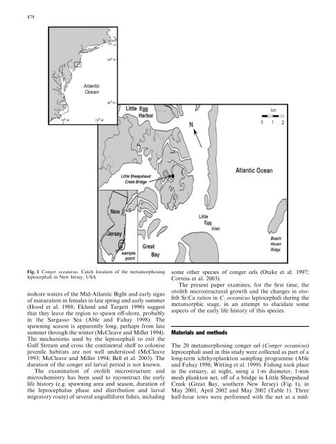
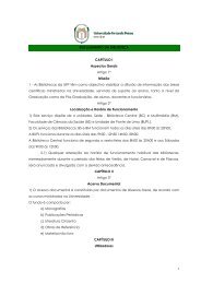
478Fig. 1 Conger oceanicus. Catch location <strong>of</strong> <strong>the</strong> metamorphosingleptocephali in New Jersey, USAinshore waters <strong>of</strong> <strong>the</strong> Mid-Atlantic Bight and early signs<strong>of</strong> maturation in females in late spring and early summer(Hood et al. 1988; Eklund and Targett 1990) suggestthat <strong>the</strong>y leave <strong>the</strong> region to spawn <strong>of</strong>f-shore, probablyin <strong>the</strong> Sargasso Sea (Able and Fahay 1998). Thespawning season is apparently long, perhaps from latesummer through <strong>the</strong> winter (McCleave and Miller 1994).The mechanisms used by <strong>the</strong> leptocephali to exit <strong>the</strong>Gulf Stream and cross <strong>the</strong> continental shelf to colonisejuvenile habitats are not well understood (McCleave1993; McCleave and Miller 1994; Bell et al. 2003). Theduration <strong>of</strong> <strong>the</strong> <strong>conger</strong> <strong>eel</strong> larval period is not known.The examination <strong>of</strong> otolith microstructure andmicrochemistry has been used to reconstruct <strong>the</strong> early<strong>life</strong> <strong>history</strong> (e.g. spawning area and season, duration <strong>of</strong><strong>the</strong> leptocephalus phase and distribution and larvalmigratory route) <strong>of</strong> several anguilliform fishes, includingsome o<strong>the</strong>r species <strong>of</strong> <strong>conger</strong> <strong>eel</strong>s (Otake et al. 1997;Correia et al. 2003).The present paper examines, for <strong>the</strong> first time, <strong>the</strong>otolith microstructural growth and <strong>the</strong> changes in otolithSr:Ca ratios in C. oceanicus leptocephali during <strong>the</strong>metamorphic stage, in an attempt to elucidate someaspects <strong>of</strong> <strong>the</strong> early <strong>life</strong> <strong>history</strong> <strong>of</strong> this species.Materials and methodsThe 20 metamorphosing <strong>conger</strong> <strong>eel</strong> (Conger oceanicus)leptocephali used in this study were collected as part <strong>of</strong> along-term ichthyoplankton sampling programme (Ableand Fahay 1998; Witting et al. 1999). Fishing took placein <strong>the</strong> estuary, at night, using a 1-m diameter, 1-mmmesh plankton net, <strong>of</strong>f <strong>of</strong> a bridge in Little SheepsheadCreek (Great Bay, sou<strong>the</strong>rn New Jersey) (Fig. 1), inMay 2001, April 2002 and May 2002 (Table 1). Threehalf-hour tows were performed with <strong>the</strong> net at a mid-
479Table 1 Conger oceanicus.Collection date, length (TL),developing stage (preanallength to total length; PAL/TL), age at recruitment to <strong>the</strong>estuary and hatching date <strong>of</strong> <strong>the</strong>metamorphosing leptocephaliNo. Collect. date TL (mm) PAL/TL Age (days) Hatching dateCO5 14 May 2001 97.0 0.74 168 28 Nov 2000CO4 96.0 0.63 179 18 Nov 2000CO2 24 May 2001 94.0 0.52 – –CO10 30 May 2001 93.0 0.58 – –CO6 84.0 0.58 – –CO8 97.0 0.55 – –CO9 90.0 0.53 – –CO7 88.0 0.53 – –CO3 91.0 0.51 – –CO11 89.0 0.47 – –CO12 4 Apr 2002 99.0 0.71 156 31 Oct 2001CO15 1 May 2002 104.0 0.68 155 28 Nov 2001CO13 100.0 0.63 182 30 Oct 2001CO14 85.0 0.67 183 31 Oct 2001CO16 8 May 2002 99.0 0.70 171 19 Nov 2001CO19 101.0 0.67 167 23 Nov 2001CO17 87.0 0.66 149 11 Dec 2001CO21 99.0 0.61 – –CO20 93.0 0.60 – –CO18 98.0 0.58 – –water depth (approximately 2 m). Temperature andsalinity were recorded at <strong>the</strong> beginning and at <strong>the</strong> end <strong>of</strong>sampling, using a field <strong>the</strong>rmometer and a hand-heldrefractometer, respectively. Water temperature andsalinity <strong>of</strong> <strong>the</strong> sampling area ranged from 8°C to17°Cand from 26 to 30 psu, respectively.After capture <strong>the</strong> leptocephali were preserved in 95%ethanol, and <strong>the</strong> general body morphology, pigmentation,morphometric and meristic characters were analysedfollowing <strong>the</strong> methodology described by Smith(1989). Measurements were made to <strong>the</strong> nearest 0.1 mm,and myomere counts were done using a dissectingmicroscope. There was no correction for shrinkagecaused by preservation.Otoliths were prepared for <strong>the</strong> examination <strong>of</strong>microstructure and microchemistry, as described inCorreia et al. (2003). Otoliths were embedded in epoxyresin, ground to expose <strong>the</strong> core with a series <strong>of</strong> gradedsilicon carbide papers (600, 1200 and 2400 grit), andfur<strong>the</strong>r polished with diamond pastes (6, 3 and 1 lm)and alumina solution (1:20). Finally, <strong>the</strong>y were cleanedin an ultrasonic bath, rinsed with deionised water andgiven a gold coating by high vacuum evaporation. Srand Ca concentrations (% dry weight) were measuredalong <strong>the</strong> longest axis <strong>of</strong> <strong>the</strong> otolith using a wavelengthdispersive X-ray electron microprobe (CAMEBAX SX50). Apatite [Ca 5 (PO 4 ) 3 ] and celestite (SrSO 4 ) were usedas standards. Accelerating voltage and beam currentwere 15 kV and 10 nA, respectively. The electron beamwas focused on a point about 2 lm in diameter, spacingmeasurements at 5-lm intervals. The microprobe measurementpoints, which were seen as burn depressions on<strong>the</strong> otolith surface, were assigned to otolith growthincrements. The averages <strong>of</strong> successive data for Sr andCa concentrations pooled for every ten successivegrowth increments were used for <strong>the</strong> <strong>life</strong>-<strong>history</strong> transectanalysis. The results are presented as <strong>the</strong> amount <strong>of</strong> Srdivided by <strong>the</strong> amount <strong>of</strong> Ca times 1000. Otolith Cacontent obtained in this study was about 10% less thanthat found in o<strong>the</strong>r previously published studies. SinceCa (and also Sr) could be measured with considerableaccuracy and precision by wavelength dispersive electronmicroprobes (Campana et al. 1997), <strong>the</strong> reason for<strong>the</strong> low Ca content in otoliths was probably <strong>the</strong> 2 years<strong>of</strong> ethanol preservation prior to elemental compositionanalysis; in at least one study, a decline <strong>of</strong> this element inotoliths has been reported as a result <strong>of</strong> this preservationmethod (Proctor and Thresher 1998). However, <strong>the</strong>overall results are still valid, since our purpose was notto analyse <strong>the</strong> absolute values <strong>of</strong> Ca and Sr, but toexamine <strong>the</strong> variation <strong>of</strong> Sr:Ca ratios (although <strong>the</strong>sevalues could be overestimated) along <strong>the</strong> otolith growthsequence, in order to reconstruct <strong>the</strong> ontogenic <strong>history</strong><strong>of</strong> <strong>the</strong> individual fishes. Following microprobe analysis,<strong>the</strong> otolith surface was repolished with alumina solution(1:20), etched for 15 s with 0.05 M HCl and vacuumcoated with gold for scanning electron microscopeobservation (SEM, Jeol JSM 630–1F) at 15 kV. Corediameter, maximum otolith diameter and maximumotolith radius were measured from <strong>the</strong> SEM photographs(at magnifications between ·300 and ·2500)according to <strong>the</strong> procedures <strong>of</strong> Correia et al. (2002a).The otolith radius and increment width were measuredalong <strong>the</strong> maximum otolith radius. The averages <strong>of</strong> everyten successive increment widths from <strong>the</strong> hatch checkto <strong>the</strong> end <strong>of</strong> <strong>the</strong> increment countable zone were used forotolith growth analysis.We assumed <strong>the</strong> growth increment in <strong>the</strong> larval otolith<strong>of</strong> <strong>conger</strong> to be daily, although daily deposition hasnot been validated in this species. We base thisassumption on <strong>the</strong> results <strong>of</strong> several related anguilliformspecies, e.g. Conger myriaster (Mochioka et al. 1989),Anguilla japonica (Tsukamoto 1989; Umezawa et al.1989; Umezawa and Tsukamoto 1991), A. rostrata(Martin 1995; Cieri and McCleave 2001), A. celebesensis(Arai et al. 2000a) and A. marmorata (Sugeha et al.