news PS - Columbia University Medical Center
news PS - Columbia University Medical Center
news PS - Columbia University Medical Center

You also want an ePaper? Increase the reach of your titles
YUMPU automatically turns print PDFs into web optimized ePapers that Google loves.
Carol Mason: Election to Institute<br />
of Medicine Recognizes Research<br />
and Commitment to Training<br />
Neuroscience Students, Postdocs<br />
By Andrea Crawford<br />
From the days of her postdoctoral work in the visual systems of cats,<br />
Carol Mason, Ph.D., has been drawn to the sense of sight as a window<br />
into the workings of the nervous system. “The visual system is intrinsically<br />
interesting and important, but I came at it because it was a way of<br />
understanding nerve cell structure and their connections” says Dr. Mason,<br />
who joined P&S in 1987 and is now professor of pathology & cell biology,<br />
neuroscience, and ophthalmic science.<br />
Over almost four decades of work – in research on axon guidance, in<br />
particular – she has indeed elucidated fundamental cellular and molecular<br />
mechanisms of the nervous system, earning her membership in the Institute<br />
of Medicine, one of the highest honors in health and medicine. IOM<br />
noted her pioneering work in the application of video microscopy and<br />
light and electron microscopy to the developing brain. With these “tools<br />
of the old anatomists – microscopes, but now laser-driven and digital,”<br />
Dr. Mason wrote in an article marking the 40th anniversary of the Society<br />
for Neuroscience in 2009, neuroscientists can “peek at living neurons<br />
behaving in their native surroundings.”<br />
Since the early 1980s, when she first showed how electron microscopy<br />
could provide a way to study synaptic connections, Dr. Mason has<br />
used evolving imaging technology to study axon guidance, the method<br />
by which neurons grow toward target cells to create the circuits of the<br />
nervous system. She has done this by looking at the optic chiasm, one of<br />
several decussating pathways, or “crossing” points, in the nervous system,<br />
of which the corpus callosum, which connects the two hemispheres<br />
of the brain, is perhaps the most commonly known.<br />
For the thalamus and, from there, the cortex to receive sensory inputs<br />
from the two retinas, the axons for retinal cells must traverse the optic<br />
chiasm, some to the same side of the brain and others to the opposite side.<br />
“People always ask me why such a ‘split’ pathway exists, and without<br />
being too teleological, it is thought to provide sensory input on both sides<br />
of the animal but also partly for behavioral escape response,” she says,<br />
explaining that if an organism gets injured in one part of its body, other<br />
parts can compensate.<br />
The number of retinal axons that cross the chiasm dictates the efficiency<br />
of an organism’s binocular vision; in healthy humans about half<br />
cross from each eye. But questions of how they do it, and what enables<br />
some to cross the midline of the brain while others are repulsed – as Dr.<br />
Mason demonstrated in 2003 when her lab identified the first gene (a<br />
transcription factor called Zic2) known to determine which axons cross<br />
by regulating a receptor that makes them turn around – have long driven<br />
her research. Over the years, she has identified the receptor, EphB1, carried<br />
by retinal ganglion cells that don’t cross at the optic chiasm, and its<br />
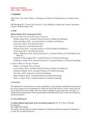
JoRG MeyeR<br />
Carol Mason, Ph.D.<br />
ligand, ephrinB2, found at the midline. Her lab is now at work to identify<br />
the molecular factors that facilitate crossing the optic chiasm midline.<br />
She believes that clues to how the retinal ganglion cells inherit the specific<br />
genes that help them find their way lie in albinos. In humans, mice, and other<br />
organisms, albinism causes a disturbance in optic chiasm crossings, which<br />
results in defects of binocular vision. Dr. Mason is trying to understand why<br />
the lack of pigment causes this imbalance. “We think that the cell layer behind<br />
the eye that makes pigment is critical for the daily visual light cycle and must<br />
send signals to the retina that control the inheritance of genes in the retinal<br />
ganglion cells for navigating the visual pathways,” she says. “If the pigment is<br />
missing, the retina does not get the right signals to develop properly.”<br />
On a winter afternoon in her laboratory, she pauses from writing the final<br />
paragraph of a paper to reflect on recent developments in her field. Neuroscientists,<br />
she notes, tend to think that the particular molecular factors<br />
and mechanisms they have discovered are the sole means of laying down<br />
the path or region under study. Their findings, individually and collectively,<br />
“are only the tip of the iceberg,” she says. “There are probably multiple programs<br />
that make a nerve cell who it is, and make the cells grow in a certain<br />
direction.” And for any one set of molecular directions for growth, there<br />
are likely multiple modes of transport and traffic signals, which she likens to<br />
New Yorkers choosing between public transport and a car.<br />
In her quest to understand the mechanisms of the nervous system, she has<br />
been surprised by how temporally important processes can be – as in the<br />
case of one receptor-ligand pair expressed in embryonic brain development<br />
for only a couple of hours – as well as by how human-like nerve cells are. A<br />
decade ago, in her studies of the developing cerebellum, she and colleagues<br />
were among the first to witness the movement of spines, tiny protrusions on<br />
the dendrites, or branches, of a neuron. “They were just incredibly motile,”<br />
she says, “They advance, withdraw, retract, move around.” The team set out<br />
to determine if the moving cells were attached to a synapse. “We found some<br />
were and some weren’t,” she says. “We still don’t understand why.”<br />
Spring 2012 <strong>Columbia</strong>Medicine 11