Western blots - Spectra Services
Western blots - Spectra Services
Western blots - Spectra Services

You also want an ePaper? Increase the reach of your titles
YUMPU automatically turns print PDFs into web optimized ePapers that Google loves.
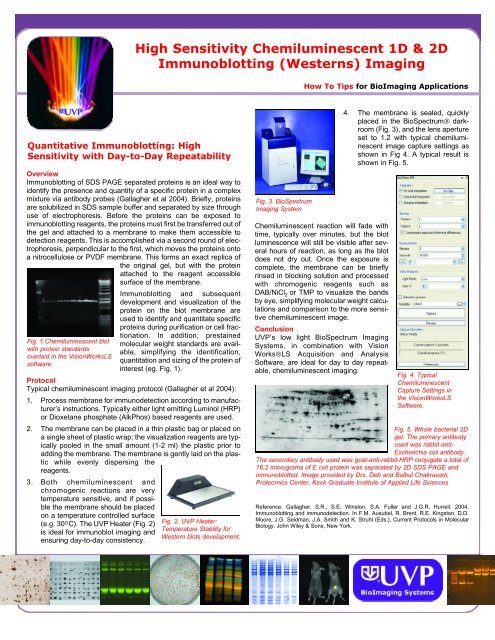
High Sensitivity Chemiluminescent 1D & 2DImmunoblotting (<strong>Western</strong>s) ImagingHow To Tips for BioImaging ApplicationsQuantitative Immunoblotting: HighSensitivity with Day-to-Day RepeatabilityOverviewImmunoblotting of SDS PAGE separated proteins is an ideal way toidentify the presence and quantity of a specific protein in a complexmixture via antibody probes (Gallagher et al 2004). Briefly, proteinsare solubilized in SDS sample buffer and separated by size throughuse of electrophoresis. Before the proteins can be exposed toimmunoblotting reagents, the proteins must first be transferred out ofthe gel and attached to a membrane to make them accessible todetection reagents. This is accomplished via a second round of electrophoresis,perpendicular to the first, which moves the proteins ontoa nitrocellulose or PVDF membrane. This forms an exact replica ofthe original gel, but with the proteinattached to the reagent accessiblesurface of the membrane.Fig. 1.Chemiluminescent blotwith protein standardsoverlaid in the VisionWorksLSsoftware.Immunoblotting and subsequentdevelopment and visualization of theprotein on the blot membrane areused to identify and quantitate specificproteins during purification or cell fractionation.In addition, prestainedmolecular weight standards are available,simplifying the identification,quantitation and sizing of the protein ofinterest (eg. Fig. 1).ProtocolTypical chemiluminescent imaging protocol (Gallagher et al 2004):1. Process membrane for immunodetection according to manufacturer’sinstructions. Typically either light emitting Luminol (HRP)or Dioxetane phosphate (AlkPhos) based reagents are used.2. The membrane can be placed in a thin plastic bag or placed ona single sheet of plastic wrap; the visualization reagents are typicallypooled in the small amount (1-2 ml) the plastic prior toadding the membrane. The membrane is gently laid on the plasticwhile evenly dispersing thereagents.3. Both chemiluminescent andchromogenic reactions are verytemperature sensitive, and if possiblethe membrane should be placedon a temperature controlled surface(e.g. 30 O C). The UVP Heater (Fig. 2)is ideal for immunoblot imaging andensuring day-to-day consistency.Fig. 2. UVP Heater:Temperature Stability for<strong>Western</strong> <strong>blots</strong> development.Fig. 3. BioSpectrumImaging SystemChemiluminescent reaction will fade withtime, typically over minutes, but the blotluminescence will still be visible after severalhours of reaction, as long as the blotdoes not dry out. Once the exposure iscomplete, the membrane can be brieflyrinsed in blocking solution and processedwith chromogenic reagents such asDAB/NiCl 2 or TMP to visualize the bandsby eye, simplifying molecular weight calculationsand comparison to the more sensitivechemiluminescent image.ConclusionUVP’s low light BioSpectrum ImagingSystems, in combination with VisionWorks®LS Acquisition and AnalysisSoftware, are ideal for day to day repeatable,chemiluminescent imaging.4. The membrane is sealed, quicklyplaced in the BioSpectrum® darkroom(Fig. 3), and the lens apertureset to 1.2 with typical chemiluminescentimage capture settings asshown in Fig 4. A typical result isshown in Fig. 5.Fig. 4. TypicalChemiluminescentCapture Settings inthe VisionWorksLSSoftware.Fig. 5. Whole bacterial 2Dgel. The primary antibodyused was rabbit-anti-Escherichia coli antibody.The secondary antibody used was goat-anti-rabbit-HRP conjugate a total of16.2 micrograms of E coli protein was separated by 2D SDS PAGE andimmunoblotted. Image provided by Drs. Deb and Bulbul Chakravarti,Proteomics Center, Keck Graduate Institute of Applied Life SciencesReference: Gallagher, S.R., S.E. Winston, S.A. Fuller and J.G.R. Hurrell. 2004.Immunoblotting and immunodetection. In F.M. Ausubel, R. Brent, R.E. Kingston, D.D.Moore, J.G. Seidman, J.A. Smith and K. Struhl (Eds.), Current Protocols in MolecularBiology. John Wiley & Sons, New York.
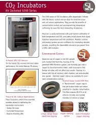
BioSpectrum Imaging System forChemiluminescent ImagingBioSpectrum Imaging System offers a multifunctional, automateddesign for a full spectrum of imaging applications.BioSpectrum 500 ImagingSystem connects to a computerinstalled withVisionWorksLS AnalysisSoftware. Imaging applicationsinclude chemiluminescence,fluorescence and colorimetric.For plant/live animalstudies, ask about theBioSpectrum 600 whichincludes the OptiChemi HRCamera for deeper coolingfor higher sensitivity andextended integration times.VisionWorksLS Acquisition and Analysis SoftwareAdvanced, yet easy to use software enables researchers toquickly capture, analyze and generate reportsControl the darkoom with a quick-link software interfaceChoose from an array of dynamic plug-ins to enhancesoftware functionalityAcquire images easily from a variety of capture andapplications templatesEnhance images with brightness/contrast, annotation,text. background correction options and moreCreate Digital Video Playback files of imagesAnalyze images with 1D Analysis, Area Density and ColonyCountingWrite macros to simplify routine proceduresGeneratecustomizablereportsBioSpectrum 500 System SpecficationsBioSpectrumDarkroomUltravioletTransilluminator*BioChemi HRCameraLensEmissionFiltersImageAcquisitionand Analysis*Optional AccessoriesComputerThermal PrinterConverter PlatesFiltersBioLite TMIlluminatorBioLite MultispectralSourceGel ToolsLight tight with wide-access doorRoll-out transilluminator trayTransilluminator timerGel view windowEpi UV, Visi-Blue and white lightDrop-down white light plateVertically adjustable chemi traySelect fromFI-26X 302nm UV, 25x26cm FirstLightLM-26E, 254/302nm UV, 21x26cmLMS-26E, 254/302/365nm UV, 21x26cmOther models available4 megapixel resolution/2048 x 204812-bit/16-bit digitizedCooled (regulated -28 o C cooling)Quantum efficiency 55% at peak 500nm,400nm 45%USB 2.0 ConnectivityMotorized 12.5 - 75mm f/1.2Ethidium Bromide, SYBR Gold/CY3SYBR Green; additional filters availableVisionWorkLS Software provides comprehensiveacquisition, analysis and documentationfunctionalityCurrent high specification computercomponents availableBlack and white digital thermal printer with256 gray scale, archive quality printsUV to White Light: 25x26cmVisi-Blue TM : 302nm to 460nm, 25x26cmUV to UV: 302 to 365nm UV, 25x26cmCustom filters availableFilter selectable visible lighttransillumination (Custom filters available)Directed visible light illumination of laboratorysamples such as plants and animalsGlowell Fluorescent Standards,Fluorescent Calibration Step Tablet, GelSentry, Gel Trays, Gel Cutter, Gel Ruler* System components may require purchase separately in someareas. Contact your UVP dealer for details.For a demonstration, quote or needs analysis, contact a UVP BioImaging Specialist or Authorized Dealer.UVP Authorized Distributor:BioImaging SystemsUVP, LLC 2066 W. 11th Street, Upland, CA 91786(800) 452-6788 | (909) 946-3197Ultra-Violet Products Ltd. Unit 1, Trinity Hall Farm Estate,Nuffield Road, Cambridge CB4 1TG UK +44(0)1223-420022BioLite and Visi-Blue are trademarks of UVP LLC. BioSpectrum andVisionWorks are registered trademarks of UVP, LLC.Specifications subject to change without notice. Reference: BioS-1Intl 107