Landry et al. 2006.pdf - Webspace
Landry et al. 2006.pdf - Webspace
Landry et al. 2006.pdf - Webspace

You also want an ePaper? Increase the reach of your titles
YUMPU automatically turns print PDFs into web optimized ePapers that Google loves.
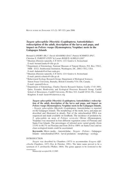
REVUE SUISSE DE ZOOLOGIE 113 (2): 307-323; juin 2006Tayg<strong>et</strong>e sphecophila (Meyrick) (Lepidoptera; Autostichidae):redescription of the adult, description of the larva and pupa, andimpact on Polistes wasps (Hymenoptera; Vespidae) nests in theG<strong>al</strong>apagos IslandsBernard LANDRY (BL) 1 , David ADAMSKI (DA) 2 , Patrick SCHMITZ (PS) 3 ,Christine E. PARENT (CEP) 4 & Lazaro ROQUE-ALBELO (LR) 51 Muséum d'histoire naturelle, C.P. 6434, 1211 Genève 6, Switzerland.E-mail: bernard.landry@ville-ge.ch2 Department of Entomology, Nation<strong>al</strong> Museum of Natur<strong>al</strong> History, P.O. Box 37012,NHB - E523, Smithsonian Institution, Washington, DC, 20013-7012, USA.E-mail: dadamski@sel.barc.usda.gov3 Muséum d'histoire naturelle, C.P. 6434, 1211 Genève 6, Switzerland.E-mail: patrick.schmitz@ville-ge.ch4 Behaviour<strong>al</strong> Ecology Research Group, Department of Biologic<strong>al</strong> Sciences,Simon Fraser University, Burnaby, British-Colombia V5A 1S6, Canada.E-mail: cparent@sfu.ca5 Department of Entomology, Charles Darwin Research Station, Casilla 17-01-3891,Quito, Ecuador; Biodiversity and Ecologic<strong>al</strong> Processes Research Group, CardiffSchool of Biosciences, Cardiff University, PO Box 915, Cardiff CF10 3TL, UnitedKingdom. E-mail: lazaro@fcdarwin.ec.orgTayg<strong>et</strong>e sphecophila (Meyrick) (Lepidoptera; Autostichidae): redescriptionof the adult, description of the larva and pupa, and impact onPolistes wasps (Hymenoptera; Vespidae) nests in the G<strong>al</strong>apagos Islands.- Tayg<strong>et</strong>e sphecophila (Meyrick) (Lepidoptera; Autostichidae) is reportedon the G<strong>al</strong>apagos Islands. The morphology of the moth, larva, and pupa aredescribed and illustrated in d<strong>et</strong>ails. Part of the mitochondri<strong>al</strong> DNA wassequenced and made available on GenBank. The incidence of predation byT. sphecophila on nests of Polistes versicolor Olivier (Hymenoptera;Vespidae) was measured in four different veg<strong>et</strong>ation zones of Floreana andSanta Cruz Islands. The percentages of infested nests varied greatly (from13.9% to 66.7% on Floreana and from 20.0 to 100% on Santa Cruz) and noclear ecologic<strong>al</strong> trends could be ascertained.Keywords: Micro moths - Autostichidae - Tayg<strong>et</strong>e - Polistes - G<strong>al</strong>apagosIslands - mitochondri<strong>al</strong> DNA - larv<strong>al</strong> predation - morphology - ecology.INTRODUCTIONTayg<strong>et</strong>e was described by Chambers (1873) to accommodate Evagora difficilisellaChambers, 1872 (Nye & Fl<strong>et</strong>cher, 1991). The latter name proved to be asynonym of T. attributella (W<strong>al</strong>ker, 1864). The genus appears to be restricted to theManuscript accepted 06.12.2005
308B. LANDRY ET AL.New World. Becker (1984) lists 13 names in this genus for the Neotropic<strong>al</strong> fauna whileHodges (1983) lists six species for the North American fauna, including five that arestated to be misplaced in this genus. BL's examination of the type specimens of theNeotropic<strong>al</strong> species at the Natur<strong>al</strong> History Museum, London, points to the possibilitythat only T. sphecophila (Meyrick, 1936) is congeneric with T. attributella in this region.However, the types of Epithectis consociata Meyrick, E. notospila Meyrick, andE. <strong>al</strong>tivola Meyrick have lost their abdomen and cannot be assigned to genus, and th<strong>et</strong>ype of E. lasciva W<strong>al</strong>singham, deposited in the USNM, Washington, could not befound.Tayg<strong>et</strong>e Chambers was considered to belong to the Gelechiidae until <strong>Landry</strong>(2002) moved it to the Autostichidae, Symmocinae sensu Hodges (1998). Tayg<strong>et</strong>esphecophila was described from three specimens bred in Trinidad from "bottom ofcells of the Hymenopteron Polistes canadensis" (Meyrick, 1936). The moth and m<strong>al</strong>egenit<strong>al</strong>ia were later illustrated with black and white photography by Clarke (1969). Onthe G<strong>al</strong>apagos Islands moths of T. sphecophila were first collected in 1989 by BL, butthe species probably arrived earlier within nests of Polistes versicolor Olivier(Vespidae).The purposes of this paper are to redescribe and illustrate the moth of T. sphecophila,to describe and illustrate the larva and pupa, to present part of its mitochondri<strong>al</strong>DNA, and to report on a few aspects of its biology, particularly with regardto the incidence of damage to P. versicolor nests by larvae.MATERIAL AND METHODSMoths of T. sphecophila were first collected at night with a mercury vapor lights<strong>et</strong> in front of a white she<strong>et</strong> and powered by a sm<strong>al</strong>l generator, and with an ultra-viol<strong>et</strong>lamp powered by a battery. Other adult specimens were reared from contained nests ofPolistes versicolor. Immature stages were found by dissecting Polistes nests and byexposing them to the sun, which causes larvae to exit nests and run away from them(Fig. 2).Specimens are deposited in the Charles Darwin Research Station (CDRS),Santa Cruz, G<strong>al</strong>apagos, Ecuador; the Canadian nation<strong>al</strong> Collection of Insects (CNC),Ottawa, Ontario, Canada; the United States Nation<strong>al</strong> Museum of Natur<strong>al</strong> History,Washington, D.C., U.S.A. (USNM), and the Muséum d'histoire naturelle (MHNG),Geneva, Switzerland.For the study of specimens using electron microscopy, larvae and pupae werefirst rinsed sever<strong>al</strong> times in water, cleaned in 10% EtOH with a camel hairbrush, andthen dehydrated in EtOH as follows: 10% EtOH for 15 minutes, 20% for 15 minutes,40% for 15 minutes, 70% for 1/2 hour, 90% for 1/2 hour, and 100% for 1/2 hour eachin two separate baths. After dehydration, specimens were critic<strong>al</strong>-point dried using aTousimis critic<strong>al</strong> point dryer, mounted on stubs, and coated with gold-p<strong>al</strong>ladium (40-60%) using a Cressington sputter coater. The ultrastructure of the larvae and pupa wasstudied with an Amray scanning electron microscope.Gross morphologic<strong>al</strong> observations and measurements of the larvae and pupaewere made using a dissecting microscope (reflected light) with a c<strong>al</strong>ibrated microm<strong>et</strong>er.
TAYGETE SPHECOPHILA IN THE GALÁPAGOS ISLANDS309FIGS 1-21, Tayg<strong>et</strong>e sphecophila, fem<strong>al</strong>e; 2, part of an abandoned nest of Polistes versicolor exposed tothe sun with at least 8 larvae of Tayg<strong>et</strong>e sphecophila exiting from it.Maps of the larv<strong>al</strong> cha<strong>et</strong>otaxy were initi<strong>al</strong>ly drawn using a WILD dissecting microscopewith a camera lucida attachment. Terminology for cha<strong>et</strong>otaxy follows Stehr(1987).
310B. LANDRY ET AL.In order to certify that the larvae corresponded to the adults found we sequenceda fragment of the mitochondri<strong>al</strong> gene Cytochrome oxidase I (COI) of both. Wholegenomic DNA was extracted using the Nucleospin kit (Macherey-Nagel). The COIgene was amplified by PCR with two primers: k698 (5’-TACAATTTATCGCC-TAAACTTCAGCC-3’), and Pat2 (5’-TCCATTACATATAATCTGCCATATTAG-3’).The therm<strong>al</strong> profile started with an initi<strong>al</strong> denaturation at 95°C for 5 min, followed by35 cycles at 94°C for 30 s, 47°C for 30 s, and 72°C for 1 min 30 s, and a fin<strong>al</strong> step at72°C for 10 min. The purified PCR product was sequenced in both directions usingfluorescent dye terminators in an ABI 377 automated sequencer. The sequence isavailable from GenBank (Accession No. DQ309437).In order to d<strong>et</strong>ermine the distribution and the density of Tayg<strong>et</strong>e sphecophila aspredator on Polistes versicolor nests, sever<strong>al</strong> study sites were selected in four of theveg<strong>et</strong>ation zones of Santa Cruz and Floreana Islands. In each veg<strong>et</strong>ation zone a seriesof quadrats of 10 m x 10 m were made at random, and the number of active andinactive nests of Polistes versicolor were counted. The delimitation of veg<strong>et</strong>ation zoneswas based on veg<strong>et</strong>ation composition (Wiggins & Porter, 1971). Nests were found byvisu<strong>al</strong>ly searching the study sites. In addition, nests found in and near Puerto Ayora, asm<strong>al</strong>l town located on the littor<strong>al</strong> and arid zones on the south coast of Santa CruzIsland, were included in the study. The presence of T. sphecophila in Polistes nests wasd<strong>et</strong>ermined by the presence of little holes on the back of the nests (Fig. 2) and distinctivebreaches on the capped cells norm<strong>al</strong>ly occupied by wasp pupae. In 1999, nests ofPolistes versicolor were monitored weekly in the area of Puerto Ayora, and nests thatwere abandoned after being infested by T. sphecophila were collected during thatperiod of time. Some adults of T. sphecophila that emerged from these nests werepreserved dry for taxonomic identification. The ecologic<strong>al</strong> observations were madeb<strong>et</strong>ween April and August 1999, February and April 2002 and 2003 on Santa CruzIsland, and b<strong>et</strong>ween April and August 1999 on Floreana Island. To test for ecologic<strong>al</strong>or insular trends in the frequency of parasitism of P. versicolor nests by T. sphecophila,we performed a G-test for goodness of fit (Sok<strong>al</strong> & Rohlf, 1995) on each island datas<strong>et</strong>using the proportion of P. versicolor nests in a given zone to infer the expectedfrequency of parasitism by T. sphecophila.TAXONOMIC TREATMENTTayg<strong>et</strong>e sphecophila (Meyrick)Epithectis sphecophila Meyrick, 1936: 624; Gaede, 1937: 113; Clarke, 1955: 290; Clarke, 1969:63, pl. 31 figs 4-4b; Makino, 1985: 25; Yamane, 1996: 85.Tayg<strong>et</strong>e sphecophila (Meyrick); Becker, 1984: 47; <strong>Landry</strong>, 1999: 68; <strong>Landry</strong>, 2002: 818-819.MATERIAL EXAMINED FOR MORPHOLOGICAL WORK: Moths (13 specimens from theG<strong>al</strong>apagos Islands, Ecuador): SANTA CRUZ: 1 , C[harles] D[arwin] R[esearch] S[tation], aridzone, 19.i.1989, M[ercury] V[apor] L[amp] (B. <strong>Landry</strong>); 4 (two dissected with genit<strong>al</strong>ia onslides CNC MIC 4586 & BL 1196, the latter with right wings on slide BL 1313), CDRS, aridzone, 3.ii.1989, MVL (B. <strong>Landry</strong>); 2 (one dissected, slide BL 1126), Barranco, ex larva ennido Polistes versicolor, 8.ii.1999 (L. Roque, No. 99.20); 1 , NNW Bella Vista, GPS: 225 melev., S 00° 41.293’, W 090° 19.665’, 18.ii.2005, u[ltra] v[iol<strong>et</strong>] l[ight] (B. <strong>Landry</strong>, P. Schmitz);1 (dissected, slide BL 1195), 2 km W Bella Vista, 27.ii.1989, MVL (B. <strong>Landry</strong>); 1 , casa L.Roque-Albelo & V. Cruz, GPS: 137 m elev., S 00° 42.595’, W 090° 19.196’, 27.ii.2005, uvl (B.
TAYGETE SPHECOPHILA IN THE GALÁPAGOS ISLANDS311FIG. 3Tayg<strong>et</strong>e sphecophila, m<strong>al</strong>e genit<strong>al</strong>ia (sizes not proportionate). 3a, dors<strong>al</strong> view of v<strong>al</strong>vae + vinculum+ juxta and ventr<strong>al</strong> view of tegumen + uncus + gnathos d<strong>et</strong>ached on right side and spreadon left side, ph<strong>al</strong>lus removed, s<strong>et</strong>ae shown on right side only; 3b, side view of ph<strong>al</strong>lus with vesicaeverted; 3c, dors<strong>al</strong> view of ph<strong>al</strong>lus, vesica inverted, sc<strong>al</strong>e = 0.1 mm; 3d, later<strong>al</strong> view of wholegenit<strong>al</strong>ia.
312B. LANDRY ET AL.<strong>Landry</strong>); 2 (dissected, slides BL 1208 & 1209), émergé d’un nid de Polistes, 1999 (C. Parent).SAN CRISTOBAL: 1 , antiguo botadero, ca. 4 km SE P[uer]to Baquerizo, GPS: 169 m elev.,S 00° 54.800’, W 089° 34.574’, 22.ii.2005, uvl (B. <strong>Landry</strong>).Larvae (166 specimens) and pupae (10 specimens) collected on Santa Cruz by P.Schmitz in 2004 and 2005.DIAGNOSIS: The presence in m<strong>al</strong>es of this species of a coremat<strong>al</strong> organ at thebase of the abdomen (Fig. 4) and a trifurcated uncus (Fig. 3a) are excellent diagnosticfeatures with regards to the rest of the G<strong>al</strong>apagos fauna. M<strong>al</strong>es of G<strong>al</strong>ag<strong>et</strong>e <strong>Landry</strong> ar<strong>et</strong>he only other G<strong>al</strong>apagos moths to share a coremat<strong>al</strong> organ, but their uncus is made ofa single projection. In fem<strong>al</strong>es the shape of segment VIII (Fig. 5), especi<strong>al</strong>ly dors<strong>al</strong>ly,will separate T. sphecophila from any other species in the G<strong>al</strong>apagos and probably therest of its range. On the archipelago, some species of G<strong>al</strong>ag<strong>et</strong>e <strong>Landry</strong> (2002) orGelechiidae may appear superfici<strong>al</strong>ly similar, especi<strong>al</strong>ly because they share a similarlyshaped hindwing, a similar wingspan, upturned labi<strong>al</strong> p<strong>al</strong>pi, and sc<strong>al</strong>es on the proboscisbas<strong>al</strong>ly, but the forewing markings of T. sphecophila (Fig. 1) are unique among thesegroups.REDESCRIPTION: Gener<strong>al</strong> appearance of moth greyish brown with dark brownmarkings on forewing (Fig. 1); sc<strong>al</strong>es usu<strong>al</strong>ly dark brown at their base and p<strong>al</strong>erapic<strong>al</strong>ly. Head sc<strong>al</strong>es longer later<strong>al</strong>ly and directed medi<strong>al</strong>ly and ventr<strong>al</strong>ly, except onocciput, directed medi<strong>al</strong>ly and dors<strong>al</strong>ly. Ocellus and cha<strong>et</strong>osema absent. Labi<strong>al</strong> p<strong>al</strong>pusgently curving upward, darker brown later<strong>al</strong>ly than medi<strong>al</strong>ly, with white rings of sc<strong>al</strong>esmostly at apex of segments; segments II and III shorter tog<strong>et</strong>her than segment I.Antenna mostly greyish brown, darker brown toward base; flagellomeres in both sexessimple and with erect sc<strong>al</strong>es ventr<strong>al</strong>ly from about middle of flagellum. Thorax concolorouswith head, som<strong>et</strong>imes darker brown at base. Foreleg mostly dark brown, withbeige sc<strong>al</strong>es at apex of tarsomere I and on <strong>al</strong>l of tarsomere V. Midleg mostly darkbrown later<strong>al</strong>ly, with p<strong>al</strong>er sc<strong>al</strong>es at apex of tarsomeres I and II, and on <strong>al</strong>l of tarsomereV, uniformly beige medi<strong>al</strong>ly on femur and tibia, <strong>al</strong>so with short tuft of dark brownsc<strong>al</strong>es dors<strong>al</strong>ly on bas<strong>al</strong> h<strong>al</strong>f of tibia. Hindleg p<strong>al</strong>er than other legs, with some darkbrown later<strong>al</strong>ly on femur and tibia, mostly dark brown on tibi<strong>al</strong> spines and at base oftarsomeres I-IV, <strong>al</strong>so with tuft of long dirty white sc<strong>al</strong>es on dors<strong>al</strong> margin of tibia.Wingspan: 7.5-9.0 mm. Forewing mostly greyish brown, with three dark brown triangularmarkings on costa, largest marking at base, reaching inner margin, sm<strong>al</strong>lest submedi<strong>al</strong>lysituated, barely reaching cell, third marking large, reaching middle of wing;with dark brown sc<strong>al</strong>ing <strong>al</strong>so at apex and as 1-3 sm<strong>al</strong>l patches of 10 sc<strong>al</strong>es or less belowpostmedian cost<strong>al</strong> marking; <strong>al</strong>so with variable amounts of yellowish-orange to rustybrownsc<strong>al</strong>es usu<strong>al</strong>ly within bas<strong>al</strong> dark brown marking, below postmedian marking,and toward apex; fringe dark brown at apex, more greyish brown elsewhere. Hindwinggreyish brown without markings, with concolorous fringe. Wing venation (based onslide BL 1313, fem<strong>al</strong>e) (Fig. 6): Forewing Sc to about 2/5 wing length; R1 from aboutmiddle of cell; R2 and R3 separate, both from before upper angle of cell; R4, R5, andM1 from upper angle of cell, connected, R4 and R5 directed toward costa before apex,M1 directed toward outer margin below apex; M2 and M3 separate, M2 from lowerangle of cell, M3 from shortly before lower angle of cell; CuA1 and CuA2 separate,both from shortly before lower angle of cell; CuP absent; cell a little more than h<strong>al</strong>f
TAYGETE SPHECOPHILA IN THE GALÁPAGOS ISLANDS313FIGS 4-5Tayg<strong>et</strong>e sphecophila. 4, ventr<strong>al</strong> view of first abdomin<strong>al</strong> segment; 5, ventr<strong>al</strong> view of fem<strong>al</strong>egenit<strong>al</strong>ia, s<strong>et</strong>ae shown on right side only.wing length; A1 and A2 joined at about 1/5 their lengths. Fem<strong>al</strong>e forewing r<strong>et</strong>inaculumconsisting of anteriorly directed sc<strong>al</strong>es at base of cubit<strong>al</strong> stem and posteriorly directedsc<strong>al</strong>es at base of Sc. Hindwing Sc closely following costa, reaching it at about 3/5 winglength; Rs connected with M1 after upper angle of cell, Rs reaching costa at about 4/5
314B. LANDRY ET AL.wing length, M1 directed toward apex; M2 from slightly above lower angle of cell,reaching outer margin below middle; M3 and CuA1 connected for about 1/2 theirlengths after lower angle of cell, M3 to tornus, CuA1 to inner margin shortly befor<strong>et</strong>ornus; CuA2 from about 2/3 cell to inner margin at 7/10; CuP and an<strong>al</strong> veins indistinct;apex distinctly produced; outer margin distinctly concave; fem<strong>al</strong>e frenulum with2 acanthae. Abdomen dors<strong>al</strong>ly mostly dark greyish brown, with dirty white sc<strong>al</strong>es atapex of <strong>al</strong>l segments except last; ventr<strong>al</strong>ly dark brown on each side of large dirty whiteband except for last segment, mostly concolorous, greyish brown; m<strong>al</strong>e first abdomin<strong>al</strong>segment (Fig. 4) ventr<strong>al</strong>ly with an invaginated pouch containing a membranousstructure bearing sc<strong>al</strong>es (see Note below).M<strong>al</strong>e genit<strong>al</strong>ia (Fig. 3). Uncus moderately long, with pair of fixed later<strong>al</strong>,pointed and gently tapering glabrous projections; <strong>al</strong>so with movable median projection,slightly longer than later<strong>al</strong> projections, enlarged at apex and bifid, with each endbulbous and s<strong>et</strong>ose, <strong>al</strong>so slightly s<strong>et</strong>ose at base later<strong>al</strong>ly. Gnathos a long curved rodpointing posteriorly, apic<strong>al</strong>ly more heavily sclerotized, tapered, glabrous, and rounded.Tegumen broad medi<strong>al</strong>ly, with moderately narrow pedunculi. V<strong>al</strong>va with unsclerotizeds<strong>et</strong>ose cucullus, tapering, rounded apic<strong>al</strong>ly, with slightly sclerotized s<strong>et</strong>ose ridge atbase on inner side, <strong>al</strong>so with medium sized apodemes directed anteriorly from base ofcosta; sacculus with pair of short, narrow, s<strong>et</strong>ose, and apic<strong>al</strong>ly rounded projections,dors<strong>al</strong> projection curved and directed dors<strong>al</strong>ly, ventr<strong>al</strong> one straight and directedposteriorly. Vinculum narrow, slightly projected anteriorly and upturned. Juxta poorlydeveloped, sm<strong>al</strong>l, b<strong>et</strong>ter sclerotized at posterior edge around ph<strong>al</strong>lus. Ph<strong>al</strong>lus (= aedeagusof authors, but see Kristensen, 2003) narrow, with shaft flattened dorsoventr<strong>al</strong>lybeyond middle, b<strong>et</strong>ter sclerotized on left side in narrow band, slightly upturnedapic<strong>al</strong>ly; coecum penis medium-sized with pair of very sm<strong>al</strong>l peduncles later<strong>al</strong>ly;vesica with minute scobination.Fem<strong>al</strong>e genit<strong>al</strong>ia (Fig. 5). Papillae an<strong>al</strong>es large and long, moderately s<strong>et</strong>ose,sclerotized dors<strong>al</strong>ly and later<strong>al</strong>ly at base. Posterior apophyses slightly curved apic<strong>al</strong>ly,slightly longer than papillae. Tergum VIII well sclerotized, with few long s<strong>et</strong>ae especi<strong>al</strong>lyon margin, with deep rounded concavity in middle apic<strong>al</strong>ly; middle of concavitywith posteriorly directed projection variable in length and bearing two s<strong>et</strong>ae. Anteriorapophyses straight, slightly enlarged apic<strong>al</strong>ly, about as long as papillae. Sternum VIIIwith apic<strong>al</strong> margin bell shaped, well sclerotized, with few long s<strong>et</strong>ae mostly posteriorly<strong>al</strong>ong margin and midventr<strong>al</strong>ly. Intersegment<strong>al</strong> membrane b<strong>et</strong>ween sternites VII andVIII slightly sclerotized on each side of midventr<strong>al</strong> line and with pair of short projectionsinside body at apic<strong>al</strong> margin. Ostium bursae in middle of sternite VIII, ventr<strong>al</strong>lyprotected by slightly protruding crescent of sclerotization. Ductus bursae short,gradu<strong>al</strong>ly enlarging, bas<strong>al</strong> h<strong>al</strong>f well sclerotized, dist<strong>al</strong> h<strong>al</strong>f spiculose and with wrinklespatterned like brood cells in bee hive. Corpus bursae slightly longer than wide, spiculose,with one large, spiny, curved, and pointed cornutus; latter s<strong>et</strong> in sm<strong>al</strong>l sclerotizedpatch with pair of bumps on each side of its base.DESCRIPTION OF THE LARVA AND PUPA: Larva. (Figs 7-17): Length 5.0-8.2 mm (n= 72), < 5.0 mm (n = 94). Body p<strong>al</strong>e gray, textured with microconvolutions; headcapsule amber; prothoracic shield amber, gradu<strong>al</strong>ly darkening posteriorly; pinaculap<strong>al</strong>e brown; an<strong>al</strong> plate p<strong>al</strong>e amber; s<strong>et</strong>ae with widened, circular, and slightly raised
TAYGETE SPHECOPHILA IN THE GALÁPAGOS ISLANDS315FIG. 6Wing venation of Tayg<strong>et</strong>e sphecophila.sock<strong>et</strong>s. Head (Figs 7-10, 17): hypognathous, textured with slightly raised, confluent,polygon<strong>al</strong> ridges except on area b<strong>et</strong>ween adfront<strong>al</strong> sclerites (Figs 7-8); adfront<strong>al</strong> scleriteswidened dist<strong>al</strong>ly, front<strong>al</strong> s<strong>et</strong>ae about equ<strong>al</strong> in length, AF2 above apex of frons,AF1 below; F1 slightly closer to AF1 than to C1; C2 at least 2 1/2 times longer thanC1; clypeus with 6 pairs of s<strong>et</strong>ae, 3 pairs on medi<strong>al</strong> h<strong>al</strong>f, 3 on dist<strong>al</strong> h<strong>al</strong>f; mandibleangular (Fig. 17), sh<strong>al</strong>lowly notched subapic<strong>al</strong>ly forming sm<strong>al</strong>l apic<strong>al</strong> dentition,bearing pair of subequ<strong>al</strong> s<strong>et</strong>ae on outer surface near condyle, and with 1 large dentitionon inner surface; sensilla types and arrangement on antenna (Fig. 9) and on maxillaryp<strong>al</strong>pi (Fig. 10) similar to those of other Gelechioidea studied by Adamski & Brown(1987), Adamski (1999), Adamski & Pellmyr (2003), <strong>Landry</strong> & Adamski (2004), andWagner <strong>et</strong> <strong>al</strong>. (2004), and other Lepidoptera studied by Adamski & Brown (2001),Albert (1980), Avé (1981), Grimes & Neunzig (1986a, b), and Schoonhoven & D<strong>et</strong>hier(1966). Three stemmata in gen<strong>al</strong> area, 1 approximate pair above antenna, and 1 stemmabelow antenna; substemmat<strong>al</strong> s<strong>et</strong>ae about equ<strong>al</strong> in length, arranged as in Fig. 8; S3 andS1 elongate and about equ<strong>al</strong> in length, S2 short; S3 lateroventr<strong>al</strong> to S2, S2 approximat<strong>et</strong>o stemma 3, and S1 approximate to stemma 5 (stemmata 1, 2, and 6 absent); A-groups<strong>et</strong>ae above gena, mes<strong>al</strong> to L1; P1 dorsolater<strong>al</strong> to AF2, P2 dorsomes<strong>al</strong> to P1. Thorax(Figs 11, 14): T1 with L-group tris<strong>et</strong>ose, on large pinaculum extending beneath andposterior of spiracle; s<strong>et</strong>ae anterior to spiracle; L1 approximate and posteroventr<strong>al</strong> toL2, about 2 1/2 times lengths of L2 and L3; SV-group s<strong>et</strong>ae on anterior part of elongatepinaculum; SV1 about 1/3 longer than SV2; coxae nearly touching, V1s veryapproximate (not shown); segments of leg textured with slightly elongate ridges, manyproduced dist<strong>al</strong>ly into hairlike spines, claw single (Fig. 11); shield with SD1 slightlyposterior to and about 1/3 longer than XD2 and XD1; XD2, XD1, D1, and SD2 about
316B. LANDRY ET AL.FIGS 7-13Scanning electron micrographs of larva of Tayg<strong>et</strong>e sphecophila. 7, Frontolater<strong>al</strong> view of headcapsule, sc<strong>al</strong>e = 100 µ; 8, Ventrolater<strong>al</strong> view of head capsule, sc<strong>al</strong>e = 100 µ; 9, Sensilla ofantenna: 1 = sensilla basiconica, 2 = sensillum cha<strong>et</strong>ica, 3 = sensillum styloconicum, 4 = sensillumtrichodeum, sc<strong>al</strong>e = 10 µ; 10, Sensilla of maxillary p<strong>al</strong>pus: A2 = sensillum styloconicum,A1, A3, M1-2, L1-3 = sensilla basiconica, SD = sensillum digitiform, sc<strong>al</strong>e = 10 µ; 11, Dist<strong>al</strong>portion of left prothoracic leg showing claw, sc<strong>al</strong>e = 10 µ; 12, Left proleg on A4, sc<strong>al</strong>e = 100 µ;13, An<strong>al</strong> plate of A10, sc<strong>al</strong>e = 100 µ.
TAYGETE SPHECOPHILA IN THE GALÁPAGOS ISLANDS317equ<strong>al</strong> in lengths, XD2 about twice distance from XD1 than from SD1; D1 in straightline with XD1, slightly posterior to SD2 and D2; D2 about same length as SD1, instraight line with SD2. T2-T3 (Fig. 14): D2 about 2 times length of D1, both on sm<strong>al</strong>lpinaculum; SD1 about 2 times length of SD2, both on sm<strong>al</strong>l pinaculum; L1 about 1/3longer than L2, both on sm<strong>al</strong>l pinaculum, L3 slightly shorter than L2, posterior to or invertic<strong>al</strong> line with SV1; MV1 on anterior margin b<strong>et</strong>ween T2-T3, slightly above SV1(hard to see); V1s on T2-T3 about equ<strong>al</strong> distance apart, at least 4 times distanceb<strong>et</strong>ween V1s on T1. Abdomen (Figs 12, 13, 15, 16): A1-A2 (Fig. 15): D2 and D1 equ<strong>al</strong>in lengths or D2 slightly longer, MD1 on anterior part of segment anteroventr<strong>al</strong> to D1;SD1 above spiracle, about 1/3 longer than D2, with minute SD2 (anterior part of pinaculum);sm<strong>al</strong>l opening on ventroposterior margin of pinaculum bearing SD1 and SD2;spiracle on A1 slightly larger than those on A2-A7; L1 2 times length of L2, both onsame pinaculum, slightly anterior of spiracle; L3 about same length as L2, anterior to,in vertic<strong>al</strong> line with, or posterior to D2; SV-group bis<strong>et</strong>ose on A1, tris<strong>et</strong>ose on A2, onsame pinaculum; V1s equ<strong>al</strong> distance apart (not shown). A3-A10 (Figs 12, 13, 16): A3-A6 with 4 pairs of protuberant prolegs, croch<strong>et</strong>s biordin<strong>al</strong>, in circle (Fig. 12); s<strong>et</strong>ae asabove; A7 as above except, SV-group bis<strong>et</strong>ose and on same pinaculum; A8 as aboveexcept with spiracle slightly larger than on previous segments and SV-group unis<strong>et</strong>ose;A9 with D2 about 2-2 1/2 times longer than D1; D1 anterior to D2 and SD1, equidistantto both s<strong>et</strong>ae; SD1 about same length as D1; L-group s<strong>et</strong>ae slightly anterior toD1; L1 about 3 times length of L2, on same pinaculum; L3 slightly longer than L2;SV1 slightly shorter than L1; V1s as previous segments; A10 (Figs 13, 16): an<strong>al</strong> platewith SD2 and SD1 equ<strong>al</strong> in lengths, about twice length of D2; D1 slightly shorter thanD2; croch<strong>et</strong>s of proleg biordin<strong>al</strong>, in semicircle, gradu<strong>al</strong>ly shortened mes<strong>al</strong>ly andlater<strong>al</strong>ly.Pupa. (Figs 18-21): Length 3.6-4.6 (n = 10): amber, smooth, spiracles protuberant;<strong>al</strong>l dors<strong>al</strong> s<strong>et</strong>ae apic<strong>al</strong>ly hooked except long s<strong>et</strong>a associated with axillarytubercle (Figs 19-20). Sclerites of antennae annulated, widely separated anteriorly,gradu<strong>al</strong>ly convergent from beyond bas<strong>al</strong> 1/3 of sclerites of maxillae, fused for shortdistance beyond dist<strong>al</strong> apices of sclerites of maxillae, gradu<strong>al</strong>ly divergent posteriorly,exposing dist<strong>al</strong> part of sclerites of hindlegs; sclerites of midleg not fused dist<strong>al</strong>ly;paired nodular scars of prolegs on A5-A6 (Fig. 18); A6-A10 fused, rotating as unit;cremaster dorsolater<strong>al</strong>ly flattened, trapezoid<strong>al</strong> bas<strong>al</strong>ly, extending posterolater<strong>al</strong>ly into 2slightly divergent and elongate spine-like processes (Fig. 21).DISTRIBUTION AND PHENOLOGY: The species was described from Trinidad(Meyrick, 1936) and never mentioned from anywhere else subsequently. In theG<strong>al</strong>apagos Islands it has been found on Floreana (from the littor<strong>al</strong> to the humid zones),San Cristob<strong>al</strong> (in the arid zone), and Santa Cruz (from the littor<strong>al</strong> to the humid zones).In the G<strong>al</strong>apagos we have collected live moths of this species in January, February,March, April, September, November, and December.NOTES: Preliminary phylogen<strong>et</strong>ic an<strong>al</strong>yses, both morphologic<strong>al</strong> and molecular,support the placement of Tayg<strong>et</strong>e sphecophila within Autostichidae (PS, unpublisheddata). For example, Kaila’s (2004) matrix was rean<strong>al</strong>yzed with T. sphecophila data, andthe species clusters in Kaila’s autostichid assemblage with G<strong>al</strong>ag<strong>et</strong>e <strong>Landry</strong>.
318B. LANDRY ET AL.FIGS 14-17Larva of Tayg<strong>et</strong>e sphecophila. 14, Cha<strong>et</strong>otaxy of thorax; 15, Cha<strong>et</strong>otaxy of abdomin<strong>al</strong> segments1-2; 16, Cha<strong>et</strong>otaxy of abdomin<strong>al</strong> segments 6-10; 17, Mandible.A comparison of a 1283 base pairs fragment (consisting of most of the COI geneexcept the first 254 base pairs) sequenced for a larva and an adult of Tayg<strong>et</strong>e sphecophilashowed no substitution, which clearly indicates conspecificity.The larva has only three stemmata, a condition that is highly unusu<strong>al</strong> inGelechioidea and that may be due to the unique host relationship.It proved impossible to evaginate the ventro-abdomin<strong>al</strong> pouch (Fig. 4) insever<strong>al</strong> m<strong>al</strong>e specimens of this species. However, BL was able to evaginate this coremat<strong>al</strong>organ from a specimen of Tayg<strong>et</strong>e attributella (W<strong>al</strong>ker). The organ consists of anarrow membranous tube, <strong>al</strong>most as long as the abdomen, on which narrow sc<strong>al</strong>es areconnected <strong>al</strong>l around. The membrane of the tube is very thin and the tube collapsed assoon as specimens were transferred to lactic acid for temporary storage. An illustrationof this structure for the closely related G<strong>al</strong>ag<strong>et</strong>e turritella <strong>Landry</strong> is provided by<strong>Landry</strong> (2002: Figs 17, 18).
TAYGETE SPHECOPHILA IN THE GALÁPAGOS ISLANDS319FIGS 18-21Pupa of Tayg<strong>et</strong>e sphecophila. 18, Ventr<strong>al</strong> view; 19, Dors<strong>al</strong> view; 20, Segments 8-10; 21, Later<strong>al</strong>view.ECOLOGICAL STUDYPATTERNS OF PREDATIONAlthough egg-laying was never observed, it is possible that the fem<strong>al</strong>e mothslay their eggs within the pup<strong>al</strong> cells of P. versicolor through numerous sm<strong>al</strong>l holes of1-2 mm in diam<strong>et</strong>er that we observed on the back of the nests. In a sample of 25P. versicolor nests, the number of T. sphecophila moths found per nest varied b<strong>et</strong>ween3 and 13. However, 42 T. sphecophila larvae were recovered by PS from a rather sm<strong>al</strong>lnest collected on Santa Cruz in 2004. The food source needed for the development ofthe moth’s larvae are the wasps’ pupae which are defenseless because of their isolationin their capped cells. When ready to emerge from the wasp's cell, the moth makes adistinctive breach through the cap covering the top of the cell.DISTRIBUTION OF INFESTED NESTSThe level of T. sphecophila infestation could only be assessed for nests ofP. versicolor that were abandoned. A tot<strong>al</strong> of 103 such nests were found on the differentstudy sites on Santa Cruz Island b<strong>et</strong>ween 1999 and 2003, and 141 nests on FloreanaIsland in 1999. The percentages of nests that presented signs of predation by T. sphecophilaare given in Table 1, <strong>al</strong>ong with the veg<strong>et</strong>ation zones in which they were found.
320B. LANDRY ET AL.TABLE 1. Percentage of Polistes versicolor nests found in four different veg<strong>et</strong>ation zones of SantaCruz and Floreana Islands presenting signs of Tayg<strong>et</strong>e sphecophila predation. Number of nestsper sample are in parentheses.Veg<strong>et</strong>ation Zone% of nests with T. sphecophila predationSanta Cruz IslandFloreana IslandLittor<strong>al</strong> 35.3 (n=17) 40.0 (n=5)Arid 43.0 (n=79) 13.9 (n=101)Transition 20.0 (n=5) 66.7 (n=6)Humid 100.0 (n=2) 51.7 (n=29)On Santa Cruz island the arid zone was the area of highest abundance of nests.This result is similar to that obtained by Roque-Albelo & Causton (1999) for abundanceof adult foragers. The percentages of infestation varied b<strong>et</strong>ween zones (Table 1).However, very few nests were collected in the littor<strong>al</strong>, transition, and humid zones.Nests of P. versicolor were again more common in the arid zone of Floreana. However,only 13.9% of them were infested by T. sphecophila in this zone. In contrast to SantaCruz, on Floreana nests <strong>al</strong>so were abundant in the humid zone, where 51.7 % of themwere infested.The results of the G-test for goodness of fit <strong>al</strong>low us to test for ecologic<strong>al</strong> trendin nest infestation according to veg<strong>et</strong>ation zonation. The proportion of parasitism inPolistes versicolor nests in the four veg<strong>et</strong>ation zones on Santa Cruz Island does notshow deviation from the expected (based on the proportion of P. versicolor nests foundin each veg<strong>et</strong>ation zone; G = 4.806, df = 3, P > 0.05). However, the situation onFloreana Island appears different as P. versicolor nests found in the arid zone areinfested by T. sphecophila less than expected, and nests found in the transition andhumid zones are infested more than expected (G = 15.482, df = 3, P < 0.01).DISCUSSIONDifferent factors, including climatic conditions, infestation by nest scavengersand parasitoids, and predation affect the wasp colony cycle (Yamane, 1996). Across itsrange of distribution, from Costa Rica to Southern Argentina, P. versicolor seems toprefer dry forest habitats (Richards, 1978). Data from previous studies suggest that inthe G<strong>al</strong>apagos the wasps are more abundant in the arid zone of the islands (Roque-Albelo & Causton, 1999; Lasso, 1997). This preference in distribution could be associatedwith climatic conditions (Parent, 2000). In the G<strong>al</strong>apagos the higher zones of theislands are cooler and receive more rainf<strong>al</strong>l than lower zones, particularly on thesouthern slopes, and this factor probably affects nest development. Collection data ofT. sphecophila suggest a similar pattern of distribution. Most moth specimens werecollected in the dryer zones of the islands suggesting a close correlation with nestabundance.On Santa Cruz Island the occurrence of T. sphecophila in different veg<strong>et</strong>ationzones is a reflection of the frequency of P. versicolor nests. However, T. sphecophilaseems to be more abundant than expected in the transition and humid zones of FloreanaIsland and less frequent in the arid zone. Therefore, T. sphecophila’s occurrence onFloreana Island is not strictly a reflection of the abundance of P. versicolor nests,
TAYGETE SPHECOPHILA IN THE GALÁPAGOS ISLANDS321suggesting that other ecologic<strong>al</strong> or climatic factors might influence its distribution. Itis not clear why there is such a difference b<strong>et</strong>ween Floreana and Santa Cruz islands,but one possible hypothesis is that T. sphecophila has colonized these two islands atdifferent points in time, so that populations on one of the island have had more time toadapt to the island’s ecologic<strong>al</strong> and climatic context.Polistes nests, as in many other soci<strong>al</strong> wasps, are scavenged and parasitized byvarious insects including more than 11 moth species from four families (Makino,1985). Only Tayg<strong>et</strong>e sphecophila was found in the G<strong>al</strong>apagos, where the speciesapparently prefers to attack large nests, and <strong>al</strong>l infested nests collected were largeenough to presume that they were in an advanced stage of the reproductive phase. Ifpredation by T. sphecophila is restricted to this stage of the wasp colonies the probabilitiesfor this moth species to be an effective agent of biologic<strong>al</strong> control are reduced.These results support the idea of Miyano (1980) that parasitic and scavengingLepidoptera reduce notably the colony’s productivity but are not thought to be a directcause of colony failure. However, the possibility to use T. sphecophila as a biologic<strong>al</strong>control agent against P. versicolor needs to be ev<strong>al</strong>uated.We believe that the first individu<strong>al</strong>s of Tayg<strong>et</strong>e sphecophila probably arrivedwithin a nest of P. versicolor built on some human-made structure that would hav<strong>et</strong>raveled by boat from the continent. It is actu<strong>al</strong>ly quite possible that both anim<strong>al</strong>sarrived tog<strong>et</strong>her on the G<strong>al</strong>apagos. The wasp was first d<strong>et</strong>ected in 1988 on Floreana,and is thought to have arrived with a shipment of bananas (Abedrabbo, 1991), butEduardo Vilema, resident of Santa Cruz, says that he first saw a nest of Polistes versicolornear Bella Vista, on Santa Cruz, in 1984 or 1985 (pers. comm. to BL in 2004).And we think it unlikely that the wasps came on banana regimes as they are not knownto build their nests there.ACKNOWLEDGEMENTSBL would like to thank the authorities of the CDRS and G<strong>al</strong>apagos Nation<strong>al</strong>Park Service (GNPS) for providing authorizations and logistic<strong>al</strong> support for samplingmicro moths in 1989, 1992, 2002, 2004, and 2005. The first two expeditions wereorganized by Stewart B. Peck, Carl<strong>et</strong>on University, Ottawa, and benefited fromfinanci<strong>al</strong> support of the Nation<strong>al</strong> Sciences and Engineering Council of Canada.Professor Peck, as well as Bradley J. Sinclair and sever<strong>al</strong> others were excellent fieldcompanions. BL is <strong>al</strong>so grateful to Jean-François <strong>Landry</strong>, Agriculture & Agri-FoodCanada, Ottawa, for providing lab space, equipment and technic<strong>al</strong> support in 1999-2000. We are thankful to curator Kevin Tuck of the Natur<strong>al</strong> History Museum, London,for his kind help to BL in London and for providing information on referencesregarding T. sphecophila and on specimens of other species. Financi<strong>al</strong> support for BL’sinvestigations in London in 2000 were provided by the G<strong>al</strong>apagos Conservation Trust,United Kingdom, and the Charles Darwin Foundation.PS would like to thank the CDRS and GNPS for <strong>al</strong>lowing field work andproviding logistic<strong>al</strong> support in the G<strong>al</strong>apagos in 2004 and 2005. Research funds to PSwere provided by the A. Lombard Foundation, the E. & L. Schmidheiny Foundation,the Swiss Academy of Sciences, and the Department of cultur<strong>al</strong> affairs of the city ofGeneva.
322B. LANDRY ET AL.CEP would like to thank the CDRS and GNPS for providing logistic<strong>al</strong> supportand required permits to conduct field work in G<strong>al</strong>apagos in 1999. Research funds toCEP were provided by CIDA, the Canadian Internation<strong>al</strong> Development Agency.DA thanks Diana Marques, scientific illustrator, for the illustrations (Figs14-21) of the larva and pupa and the digit<strong>al</strong> production of the plates, and Scott D.Whittaker, Manager of the Scanning Electron Microscopy Laboratory, SmithsonianInstitution, Washington, DC, for the help with the preparation of the larv<strong>al</strong> specimensfor study.LR thanks Henry Herrera of CDRS for his help in the field and in the lab.Fin<strong>al</strong>ly, we are grateful to Lauri Kaila and Klaus Sattler for reviewing themanuscript.REFERENCESABEDRABBO, S. 1991. Nueva avispa introducida en las islas. Carta Informativa 31: 4.ADAMSKI, D. 1999. Blastobasis graminea, new species (Lepidoptera: Gelechioidea: Coleophoridae:Blastobasinae): A stem borer of sugar cane in Colombia and Venezuela.Proceedings of the Entomologic<strong>al</strong> Soci<strong>et</strong>y of Washington 101: 164-174.ADAMSKI, D. & BROWN, R. L. 1987. A new Nearctic Glyphidocera, with descriptions of <strong>al</strong>l stages(Lepidoptera: Blastobasidae: Symmocinae). Proceedings of the Entomologic<strong>al</strong> Soci<strong>et</strong>yof Washington 89: 329-343.ADAMSKI, D. & BROWN, J. W. 2001. Systematic review of the Ecdytolopha group of genera(Lepidoptera: Tortricidae: Grapholitini). Entomologica Scandinavica, Suppl. 58: 1-86.ADAMSKI, D. & PELLMYR, O. 2003. Immature stages and redescription of Blastobasis yuccaecolellaDi<strong>et</strong>z (Lepidoptera: Gelechioidea: Coleophoridae: Blastobasini), with notes onits biology. Proceedings of the Entomologic<strong>al</strong> Soci<strong>et</strong>y of Washington 105: 388-396.ALBERT, P. J. 1980. Morphology and innervation of mouthpart sensilla in larvae of the sprucebudworm, Choristoneura fumiferana (Clem.) (Lepidoptera: Tortricidae). CanadianJourn<strong>al</strong> of Zoology 58: 842-851.AVÉ, D. A. 1981. Induction of changes in the gustatory response by individu<strong>al</strong> secondary plantcompounds in larvae of Heliothis zea (Boddie) (Lepidoptera, Noctuidae). Ph.D. dissertation,Department of Entomology, Mississippi State University, 89 pp., 4 tables, 37 figs.BECKER, V. O. 1984. Gelechiidae (pp. 44-53). In: HEPPNER, J. B. (ed.). Atlas of Neotropic<strong>al</strong>Lepidoptera, Checklist: Part 1, Micropterigoidea - Immoidea. Dr. W. Junk Publishers,The Hague, xxvii + 112 pp.CHAMBERS, V. T. 1873. Micro-Lepidoptera. The Canadian Entomologist 5: 221-232.CLARKE, J. F. G. 1955. Cat<strong>al</strong>ogue of the Type Specimens of Microlepidoptera in the BritishMuseum (Natur<strong>al</strong> History) described by Edward Meyrick. Volume 1. Trustees of theBritish Museum, London, vii + 332 pp.CLARKE, J. F. G. 1969. Cat<strong>al</strong>ogue of the Type Specimens of Microlepidoptera in the BritishMuseum (Natur<strong>al</strong> History) described by Edward Meyrick. Volume 7. Trustees of theBritish Museum (Natur<strong>al</strong> History), London, 531 pages.GAEDE, M. 1937. Familia: Gelechiidae (pp. 1-630). In: WAGNER, H., STRAND, E. & BRYK, F.(eds). Lepidopterorum Cat<strong>al</strong>ogus, part 79, Vol. IV. Dr. W. Junk, Berlin.GRIMES, L. R. & NEUNZIG, H. H. 1986a. Morphologic<strong>al</strong> survey of the maxillae in last-stagelarvae of the suborder Ditrysia (Lepidoptera): P<strong>al</strong>pi. Ann<strong>al</strong>s of the Entomologic<strong>al</strong> Soci<strong>et</strong>yof America 79: 491-509.GRIMES, L. R. & NEUNZIG, H. H. 1986b. Morphologic<strong>al</strong> survey of the maxillae in last-stagelarvae of the suborder Ditrysia (Lepidoptera): Mes<strong>al</strong> lobes (Laciniog<strong>al</strong>eae). Ann<strong>al</strong>s of theEntomologic<strong>al</strong> Soci<strong>et</strong>y of America 79: 510-526.
TAYGETE SPHECOPHILA IN THE GALÁPAGOS ISLANDS323HODGES, R. W. 1983. Gelechiidae (pp. 19-25). In: HODGES, R. W. <strong>et</strong> <strong>al</strong>. (eds). Check List of theLepidoptera of America North of Mexico. E. W. Classey Ltd. and the Wedge Entomologic<strong>al</strong>Research Foundation, London, xxiv + 284 pp.HODGES, R. W. 1998. The Gelechioidea (pp. 131-158). In:KRISTENSEN, N. P. (ed.). Handbook ofZoology, Lepidoptera, Moths and Butterflies, Vol. 1: Evolution, Systematics, and Biogeography.W<strong>al</strong>ter de Gruyter, Berlin & New York, x + 491 pp.KAILA, L. 2004. Phylogeny of the superfamily Gelechioidea (Lepidoptera: Ditrysia): anexemplar approach. Cladistics 20: 303-340.KRISTENSEN, N. P. 2003. Skel<strong>et</strong>on and muscles: adults (pp. 39-131). In: KRISTENSEN, N. P. (ed.).Handbook of Zoology, Lepidoptera, Moths and Butterflies, Vol. 2: Morphology, Physiology,and Development. W<strong>al</strong>ter de Gruyter, Berlin & New York, x + 564 pp.LANDRY, B. 1999. Quatre premières mentions de Lépidoptères pour la province de Québec(Opostegidae, Gelechiidae <strong>et</strong> Tortricidae). Fabreries 24: 66-72.LANDRY, B. 2002. G<strong>al</strong>ag<strong>et</strong>e, a new genus of Autostichidae representing the first case of anextensive radiation of endemic Lepidoptera in the G<strong>al</strong>ápagos Islands. Revue suisse deZoologie 109: 813-868.LANDRY, J.-F. & ADAMSKI, D. 2004. A new species of Nanodacna Clarke (Lepidoptera:Elachistidae: Agonoxeninae) feeding on the seeds of Austrocedrus chilensis (Cupressaceae)in andean Argentina. Journ<strong>al</strong> of the Lepidopterists’ Soci<strong>et</strong>y 58: 100-113.LASSO, T. L. 1997. Ecologia e impacto de la avispa introducida (Polistes versicolor, Vespidae-Hymenoptera) en las Islas Floreana y Santa Cruz, G<strong>al</strong>apagos Ecuador. B.Sc. Thesis,Universidad Centr<strong>al</strong> del Ecuador, Quito, 211 pp.MAKINO, S. 1985. List of parasitoides [sic] of Polistine wasps. Sphecos 10: 19-25.MEYRICK, E. 1936. Exotic Microlepidoptera 4: 609-642.MIYANO, S. 1980 Life tables of colonies of workers in a paper wasp, Polistes chinensis antenn<strong>al</strong>is,in centr<strong>al</strong> Japan (Hymenoptera: Vespidae). Research in Population Ecology 22:69-88.NYE, I. W. B. & FLETCHER, D. S. 1991. The generic names of moths of the World. Volume 6.Microlepidoptera. Natur<strong>al</strong> History Museum Publications, London, xxix + 368 pp.PARENT, C. 2000. Life cycle and ecologic<strong>al</strong> impact of Polistes versicolor versicolor (Olivier)(Hymenoptera: Vespidae), an introduced predatory wasp on the G<strong>al</strong>apagos Islands,Ecuador. M.Sc. Thesis, Carl<strong>et</strong>on University, Ottawa, Canada, 139 pp.RICHARDS, O. W. 1978. The soci<strong>al</strong> wasps of the Americas excluding the Vespinae. Trustees of theBritish Museum (Natur<strong>al</strong> History), London, Publication No. 785, vii + 580 pp. + 4 pls.ROQUE-ALBELO, L. & CAUSTON, C. 1999. Monitoreo de avispas introducidas en las IslasG<strong>al</strong>ápagos. Charles Darwin Research Station report, 12pp.SCHOONHOVEN, L. M. & DETHIER, V. G. 1966. Sensory aspects of host-plant discrimination ofLepidopterous larvae. Archives néerlandaises de zoologie 16: 497-530.SOKAL, R. R. & ROHLF, F. J. 1995. Biom<strong>et</strong>ry (3rd ed.). W. H. Freeman and Company, New York,887 pp.STEHR, F. 1987. Cosmopterigidae (Gelechioidea) (pp. 391-392). In:STEHR, F. (ed.). Immature insects,Volume 1. Kend<strong>al</strong>l/Hunt Publishing Company, Dubuque, Iowa, 754 pp.WAGNER, D. L., ADAMSKI, D. & BROWN, R. L. 2004. A new species of Mompha Hbn. fromMississippi (Lepidoptera: Gelechioidea). Proceedings of the Entomologic<strong>al</strong> Soci<strong>et</strong>y ofWashington 106: 1-18.WIGGINS, I. L. & PORTER, D. M. 1971. Flora of the G<strong>al</strong>ápagos Islands. Stanford University Press,Stanford, xx + 998 pp.YAMANE S. 1996. Ecologic<strong>al</strong> factors influencing the colony cycle of Polistes wasps (pp. 75-97).In: TURILLAZZY, S. & WEST-EBERHARD, M. J. (eds). Natur<strong>al</strong> History and evolution ofpaper-wasps. Oxford University Press, xiv + 400 pp.