FreeSurfer Course - 3D Slicer
FreeSurfer Course - 3D Slicer
FreeSurfer Course - 3D Slicer

Create successful ePaper yourself
Turn your PDF publications into a flip-book with our unique Google optimized e-Paper software.
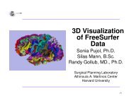
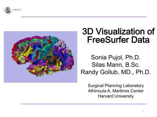
<strong>3D</strong> Visualization of<br />
<strong>FreeSurfer</strong> Data<br />
Sonia Pujol, Ph.D.<br />
Silas Mann, B.Sc.<br />
Randy Gollub, MD., Ph.D.<br />
Surgical Planning Laboratory<br />
Athinoula A. Martinos Center<br />
Harvard University<br />
Pujol S et al.<br />
National Alliance for Medical Image Computing http://na-mic.org ©2010,ARR
National Alliance for Medical Image Computing<br />
NIH U54EB005149<br />
Neuroimage Analysis Center<br />
NIH P41RR013218<br />
Morphometry Biomedical Informatics Research Network<br />
NIH U24RRO21382<br />
Surgical Planning Laboratory (BWH)<br />
Thanks to Nicole Aucoin<br />
Acknowledgements<br />
Center for Functional Neuroimaging Technology<br />
NIH P41RR14075<br />
Pujol S et al.<br />
National Alliance for Medical Image Computing http://na-mic.org ©2010,ARR
Learning Objective<br />
Guide you step-by-step through<br />
the process of loading and viewing<br />
<strong>FreeSurfer</strong> segmentations,<br />
surface reconstructions, and<br />
parcellation results within <strong>Slicer</strong>3.<br />
Pujol S et al.<br />
National Alliance for Medical Image Computing http://na-mic.org ©2010,ARR
Prerequisites<br />
This tutorial assumes that you have completed the course<br />
<strong>Slicer</strong>3Visualization Tutorial.<br />
Tutorials for <strong>Slicer</strong>3.6 are available on the <strong>Slicer</strong>101 page:<br />
http://www.slicer.org/slicerWiki/index.php/<strong>Slicer</strong>3.6:Training#Software_tutorials<br />
Pujol S et al.<br />
National Alliance for Medical Image Computing http://na-mic.org ©2010,ARR
Prerequisites<br />
This tutorial assumes a working<br />
knowledge of how to use <strong>FreeSurfer</strong><br />
to generate segmentation and surface<br />
files.<br />
Tutorials for <strong>FreeSurfer</strong> are available<br />
at the following location:<br />
http://surfer.nmr.mgh.harvard.edu/fswiki/Tutorials/<br />
Pujol S et al.<br />
National Alliance for Medical Image Computing http://na-mic.org ©2010,ARR
Materials<br />
This tutorial requires the installation of the <strong>Slicer</strong>3.6-<br />
2010-06-10 release software and the tutorial dataset.<br />
These materials are available at the following locations:<br />
• <strong>Slicer</strong>3.6 release version download page:<br />
http://www.slicer.org/pages/Special:<strong>Slicer</strong>Downloads<br />
Disclaimer: It is the responsibility of the user of <strong>Slicer</strong> to comply with both the terms<br />
of the license and with the applicable laws, regulations, and rules.<br />
Pujol S et al.<br />
National Alliance for Medical Image Computing http://na-mic.org ©2010,ARR
Materials<br />
This tutorial makes use of the same T1 weighted image dataset (bert) that is<br />
used for the <strong>FreeSurfer</strong> tutorial available at the following location:<br />
http://surfer.nmr.mgh.harvard.edu/fswiki/FsTutorial<br />
If you already have the <strong>FreeSurfer</strong> subject ‘bert’ on your computer, then just<br />
download the file ‘slicerGenericScene.mrml’<br />
http://www.na-mic.org/Wiki/index.php/Image:<strong>Slicer</strong>GenericScene.mrml<br />
If you don’t have the <strong>FreeSurfer</strong> tutorial dataset known as ‘bert’ on your<br />
computer, then download the archive below:<br />
http://www.na-mic.org/Wiki/index.php/Image:<strong>FreeSurfer</strong>Data.tar.gz<br />
Pujol S et al.<br />
National Alliance for Medical Image Computing http://na-mic.org ©2010,ARR
From <strong>FreeSurfer</strong>, <strong>Slicer</strong>3 can load:<br />
• Brain volumes . . . . . . . . . . . . . . . . . . . . . . . . .<br />
Overview<br />
• ASEG volumes . . . . . . . . . . . . . . . . . . . . . . . . . . .<br />
• Surfaces . . . . . . . . . . . . . . . . . . . . . . . . . . . . . . . . . .<br />
• Parcellation Maps . . . . . . . . . . . . . . . . . . . . . . . . . . . .<br />
• All of the above, via a scene file. . . . . . . . . . . . . . .<br />
Pujol S et al.<br />
National Alliance for Medical Image Computing http://na-mic.org ©2010,ARR
•Part 1: Loading and Visualizing<br />
<strong>FreeSurfer</strong> Volumes<br />
•Part 2: Building <strong>3D</strong> Models<br />
•Part 3: Loading <strong>FreeSurfer</strong> Surfaces<br />
and Visualizing Parcellation Maps<br />
•Part 4:Automatic Data Loading via a<br />
Generic Scene File<br />
Overview<br />
Pujol S et al.<br />
National Alliance for Medical Image Computing http://na-mic.org ©2010,ARR
Part 1: Loading and<br />
Visualizing <strong>FreeSurfer</strong><br />
Volumes<br />
Pujol S et al.<br />
National Alliance for Medical Image Computing http://na-mic.org ©2010,ARR
<strong>FreeSurfer</strong> pipeline<br />
Loading a Brain File<br />
Intensity corrected<br />
T1 volume<br />
Skull Stripping and<br />
Noise Filtering<br />
Watershed Algorithm<br />
brain.mgz<br />
Pujol S et al.<br />
National Alliance for Medical Image Computing http://na-mic.org ©2010,ARR
Loading a Brain File<br />
Select Volumes from<br />
the Module menu<br />
Pujol S et al.<br />
National Alliance for Medical Image Computing http://na-mic.org ©2010,ARR
Loading a Brain File<br />
Click on Select Volume File<br />
Pujol S et al.<br />
National Alliance for Medical Image Computing http://na-mic.org ©2010,ARR
Loading a Brain File<br />
Browse to find the<br />
dataset brain.mgz<br />
located in the<br />
directory /bert/mri/<br />
and click on Open<br />
Pujol S et al.<br />
National Alliance for Medical Image Computing http://na-mic.org ©2010,ARR
Loading a Brain File<br />
Choose Image<br />
Origin: Centered<br />
and click Apply<br />
Pujol S et al.<br />
National Alliance for Medical Image Computing http://na-mic.org ©2010,ARR
Loading a Brain File<br />
The volume brain.mgz<br />
appears in the Slice<br />
Viewer<br />
Pujol S et al.<br />
National Alliance for Medical Image Computing http://na-mic.org ©2010,ARR
Loading a Brain File<br />
Click on the links<br />
icon to link the<br />
three anatomical<br />
slices.<br />
Click on the Slice<br />
Visibility icon to<br />
display the slices in<br />
the <strong>3D</strong> Viewer<br />
Pujol S et al.<br />
National Alliance for Medical Image Computing http://na-mic.org ©2010,ARR
The three anatomical<br />
slices appear in the<br />
<strong>3D</strong> Viewer<br />
Loading a Brain File<br />
Pujol S et al.<br />
National Alliance for Medical Image Computing http://na-mic.org ©2010,ARR
<strong>FreeSurfer</strong> pipeline<br />
Loading an ASEG File<br />
Intensity corrected<br />
T1 volume<br />
Subcortical processing<br />
Segmentation<br />
aseg.mgz<br />
Pujol S et al.<br />
National Alliance for Medical Image Computing http://na-mic.org ©2010,ARR
Loading an ASEG File<br />
Click on Select Volume File, and browse to find<br />
the dataset aseg.mgz located in the directory<br />
/bert/mri/ and click on Open<br />
Pujol S et al.<br />
National Alliance for Medical Image Computing http://na-mic.org ©2010,ARR
Loading an ASEG File<br />
Select Label Map and<br />
click on Apply<br />
Pujol S et al.<br />
National Alliance for Medical Image Computing http://na-mic.org ©2010,ARR
The volume aseg.mgz<br />
appears in the Viewer<br />
The labels are<br />
superimposed on the<br />
gray brain images<br />
Loading an ASEG File<br />
Pujol S et al.<br />
National Alliance for Medical Image Computing http://na-mic.org ©2010,ARR
Loading Loading an an ASEG ASEG File File<br />
To change the lookup<br />
table for your aseg file,<br />
click on Display with<br />
aseg.mgz as the<br />
Active Volume<br />
For this tutorial, we will select<br />
Freesurfer → FreesurferLabels<br />
as our lookup table<br />
Pujol S et al.<br />
National Alliance for Medical Image Computing http://na-mic.org ©2010,ARR
Loading an ASEG File<br />
Mouse over the labels<br />
in the Axial view<br />
Pujol S et al.<br />
National Alliance for Medical Image Computing http://na-mic.org ©2010,ARR
Overlay Overlay Brain Brain & & Segmentation<br />
Segmentation<br />
The names of the<br />
labels appear in the<br />
window<br />
Pujol S et al.<br />
National Alliance for Medical Image Computing http://na-mic.org ©2010,ARR
Overlay Brain & Segmentation<br />
Find the labels corresponding to the<br />
Left Thalamus Proper, the Left<br />
Caudate, and the Left Putamen in<br />
the three anatomical views<br />
Pujol S et al.<br />
National Alliance for Medical Image Computing http://na-mic.org ©2010,ARR
Overlay Brain & Segmentation<br />
Left Thalamus Proper = #10<br />
Left Caudate = #11<br />
Left Putamen = #12<br />
Pujol S et al.<br />
National Alliance for Medical Image Computing http://na-mic.org ©2010,ARR
Part 2: Building<br />
<strong>3D</strong> Models<br />
Pujol S et al.<br />
National Alliance for Medical Image Computing http://na-mic.org ©2010,ARR
• Building a Single Model<br />
• Building Multiple Models<br />
Building <strong>3D</strong> Models<br />
Pujol S et al.<br />
National Alliance for Medical Image Computing http://na-mic.org ©2010,ARR
Building a Single Model<br />
Select the module<br />
Model Maker from the<br />
category Surface<br />
Models<br />
Pujol S et al.<br />
National Alliance for Medical Image Computing http://na-mic.org ©2010,ARR
Building a Single Model<br />
Choose Input Volume:<br />
aseg.mgz<br />
Select Models: Create New<br />
ModelHierarchy<br />
Pujol S et al.<br />
National Alliance for Medical Image Computing http://na-mic.org ©2010,ARR
Building a Single Model<br />
Enter right-hippocampus as<br />
Model Name and uncheck the<br />
box for Generate All Models<br />
Pujol S et al.<br />
National Alliance for Medical Image Computing http://na-mic.org ©2010,ARR
Building a Single Model<br />
In the Model Maker<br />
Parameters tab, type<br />
in label #53, which<br />
corresponds to the<br />
label for the Right<br />
Hippocampus<br />
Pujol S et al.<br />
National Alliance for Medical Image Computing http://na-mic.org ©2010,ARR
Building a Single Model<br />
Turn off Slice visibility<br />
and Click Apply<br />
Pujol S et al.<br />
National Alliance for Medical Image Computing http://na-mic.org ©2010,ARR
The 3-dimensional<br />
model of the Right<br />
Hippocampus appears<br />
in the <strong>3D</strong> Viewer<br />
Building a Single Model<br />
Pujol S et al.<br />
National Alliance for Medical Image Computing http://na-mic.org ©2010,ARR
• Building a Single Model<br />
• Building Multiple Models<br />
Building <strong>3D</strong> Models<br />
Pujol S et al.<br />
National Alliance for Medical Image Computing http://na-mic.org ©2010,ARR
Building Multiple Models<br />
Delete the Model Name.<br />
Delete label #53, and set the<br />
Start Label to label #10, which<br />
corresponds to the Left<br />
Thalamus Proper<br />
Set the End Label to label #13,<br />
which corresponds to the Left<br />
Pallidum<br />
Check Joint Smoothing, then<br />
click Apply.<br />
Pujol S et al.<br />
National Alliance for Medical Image Computing http://na-mic.org ©2010,ARR
The 3-dimensional models of<br />
the Left Thalamus Proper<br />
(label #10), Left Caudate (label<br />
#11), Left Putamen (label #12),<br />
and Left Pallidum (label #13)<br />
appear in the <strong>3D</strong> Viewer<br />
Building Multiple Models<br />
Pujol S et al.<br />
National Alliance for Medical Image Computing http://na-mic.org ©2010,ARR
Part 3: Loading<br />
<strong>FreeSurfer</strong> Surfaces<br />
and Visualizing<br />
Parcellation Maps<br />
Pujol S et al.<br />
National Alliance for Medical Image Computing http://na-mic.org ©2010,ARR
Building Multiple Models<br />
Select the module<br />
Models from the<br />
Module menu<br />
Pujol S et al.<br />
National Alliance for Medical Image Computing http://na-mic.org ©2010,ARR
Click on Add <strong>3D</strong> model or a<br />
model directory, and click<br />
Select Model.<br />
Loading Surfaces<br />
Pujol S et al.<br />
National Alliance for Medical Image Computing http://na-mic.org ©2010,ARR
Loading Surfaces<br />
Browse to find the surface lh.white located in<br />
the directory /bert/surf/<br />
Click on Open<br />
Pujol S et al.<br />
National Alliance for Medical Image Computing http://na-mic.org ©2010,ARR
The surface of the<br />
White Matter of the<br />
Left Hemisphere<br />
appears in the <strong>3D</strong><br />
Viewer<br />
Loading Surfaces<br />
Pujol S et al.<br />
National Alliance for Medical Image Computing http://na-mic.org ©2010,ARR
Visualizing Parcellation Maps<br />
Click on Add scalar<br />
overlay<br />
Pujol S et al.<br />
National Alliance for Medical Image Computing http://na-mic.org ©2010,ARR
Visualizing Parcellation Maps<br />
Click on Select model<br />
for overlay and select<br />
lh.white. Click Select<br />
a scalar overlay.<br />
Pujol S et al.<br />
National Alliance for Medical Image Computing http://na-mic.org ©2010,ARR
Visualizing Parcellation Maps<br />
Browse to find the Parcellation Map<br />
lh.aparc.annot located in the directory<br />
/bert/label/ and click on Open<br />
Pujol S et al.<br />
National Alliance for Medical Image Computing http://na-mic.org ©2010,ARR
Visualizing Parcellation Maps<br />
The Parcellation Map<br />
is overlaid on the<br />
White Matter surface<br />
in the <strong>3D</strong> Viewer<br />
Pujol S et al.<br />
National Alliance for Medical Image Computing http://na-mic.org ©2010,ARR
Part 4: Automatic<br />
Data Loading via a<br />
Generic Scene File<br />
Pujol S et al.<br />
National Alliance for Medical Image Computing http://na-mic.org ©2010,ARR
Loading a Generic Scene File<br />
Click on Close Scene in<br />
the File menu to close<br />
the current scene<br />
Click OK to confirm<br />
Pujol S et al.<br />
National Alliance for Medical Image Computing http://na-mic.org ©2010,ARR
Loading a Generic Scene File<br />
• The generic scene file works by looking in the subject<br />
directory created by <strong>FreeSurfer</strong>, and loading all available<br />
volumes and models based on known subdirectory<br />
names and filenames.<br />
• The file slicerGenericScene.mrml will work properly if<br />
the subdirectory names and filenames have not been<br />
changed by the user.<br />
Pujol S et al.<br />
National Alliance for Medical Image Computing http://na-mic.org ©2010,ARR
Loading a Generic Scene File<br />
Copy the file slicerGenericScene.mrml into the<br />
directory /subjects/ of our tutorial dataset.<br />
/subjects/<br />
Pujol S et al.<br />
National Alliance for Medical Image Computing http://na-mic.org ©2010,ARR
Loading a Generic Scene File<br />
Copy the file slicerGenericScene.mrml located in the<br />
tutorial directory, into the directory /bert/ of our sample<br />
subject.<br />
Pujol S et al.<br />
National Alliance for Medical Image Computing http://na-mic.org ©2010,ARR
Loading a Generic Scene File<br />
Rename the file ‘slicerGenericScene.mrml’ located in<br />
the directory /bert/ ‘slicerBertScene.mrml’<br />
Pujol S et al.<br />
National Alliance for Medical Image Computing http://na-mic.org ©2010,ARR
Loading a Generic Scene File<br />
Click on Load Scene in the File<br />
menu, and select the scene<br />
slicerBertScene.mrml located in<br />
the directory /bert/<br />
Pujol S et al.<br />
National Alliance for Medical Image Computing http://na-mic.org ©2010,ARR
Loading a Generic Scene File<br />
The scene appears with a<br />
list of files which have<br />
been automatically loaded<br />
from the subject directory<br />
bert.<br />
Pujol S et al.<br />
National Alliance for Medical Image Computing http://na-mic.org ©2010,ARR
Loading a Generic Scene File<br />
Select the mode <strong>3D</strong> only layout<br />
from the Viewer menu<br />
Pujol S et al.<br />
National Alliance for Medical Image Computing http://na-mic.org ©2010,ARR
Loading a Generic Scene File<br />
Select the module<br />
Models, and expand<br />
the tab Hierarchy &<br />
Display to display the<br />
list of models that<br />
were loaded<br />
Pujol S et al.<br />
National Alliance for Medical Image Computing http://na-mic.org ©2010,ARR
Loading a Generic Scene File<br />
Select the surface lh_pial,<br />
and turn on the visibility of<br />
the model<br />
Pujol S et al.<br />
National Alliance for Medical Image Computing http://na-mic.org ©2010,ARR
Loading a Generic Scene File<br />
<strong>Slicer</strong> displays the left<br />
hemisphere pial surface in<br />
the <strong>3D</strong> viewer<br />
Pujol S et al.<br />
National Alliance for Medical Image Computing http://na-mic.org ©2010,ARR
Loading a Generic Scene File<br />
The generic scene includes<br />
three snapshots that provide<br />
a variety of scene setups:<br />
-Left and Right Annotations<br />
-Left and Right Pial curve<br />
-Left and right white sulc<br />
Pujol S et al.<br />
National Alliance for Medical Image Computing http://na-mic.org ©2010,ARR
Loading a Generic Scene File<br />
Click on the restore snapshots<br />
icon, select the snapshot Left<br />
and right white sulc, and<br />
click on restore<br />
Pujol S et al.<br />
National Alliance for Medical Image Computing http://na-mic.org ©2010,ARR
Loading a Generic Scene File<br />
The snapshot displays the<br />
left and right sulci using the<br />
GreenRed color scheme.<br />
Pujol S et al.<br />
National Alliance for Medical Image Computing http://na-mic.org ©2010,ARR
From <strong>FreeSurfer</strong>, <strong>Slicer</strong>3 can load:<br />
• Brain volumes . . . . . . . . . . . . . . . . . . . . . . . . .<br />
Summary<br />
• ASEG volumes . . . . . . . . . . . . . . . . . . . . . . . . . . .<br />
• Surfaces . . . . . . . . . . . . . . . . . . . . . . . . . . . . . . . . . .<br />
• Parcellation Maps . . . . . . . . . . . . . . . . . . . . . . . . . . . .<br />
• All of the above, via a scene file. . . . . . . . . . . . . . .<br />
Pujol S et al.<br />
National Alliance for Medical Image Computing http://na-mic.org ©2010,ARR
• <strong>3D</strong> visualization of brain segmented surfaces<br />
and parcellation maps<br />
• Intuitive graphical user interface to interact with<br />
<strong>FreeSurfer</strong> data<br />
• Multi platforms open-source environment<br />
spujol@bwh.harvard.edu<br />
Conclusion<br />
Pujol S et al.<br />
National Alliance for Medical Image Computing http://na-mic.org ©2010,ARR