Hypersensitivity reactions
Hypersensitivity reactions
Hypersensitivity reactions
- No tags were found...
Create successful ePaper yourself
Turn your PDF publications into a flip-book with our unique Google optimized e-Paper software.
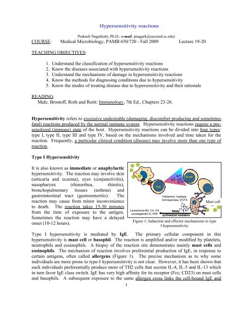
Prakash Nagarkatti, Ph.D.; e-mail: pnagark@uscmed.sc.edu)COURSE: Medical Microbiology, PAMB 650/720 - Fall 2009 Lecture 19-20TEACHING OBJECTIVES:<strong>Hypersensitivity</strong> <strong>reactions</strong>1. Understand the classification of hypersensitivity <strong>reactions</strong>2. Know the diseases associated with hypersensitivity <strong>reactions</strong>3. Understand the mechanisms of damage in hypersensitivity <strong>reactions</strong>4. Know the methods for diagnosing conditions due to hypersensitivity5. Know the modes of treating disease due to hypersensitivity and their rationaleREADING:Male, Brostoff, Roth and Roitt: Immunology, 7th Ed., Chapters 23-26.<strong>Hypersensitivity</strong> refers to excessive undesirable (damaging, discomfort producing and sometimesfatal) <strong>reactions</strong> produced by the normal immune system. <strong>Hypersensitivity</strong> <strong>reactions</strong> require a presensitized(immune) state of the host. <strong>Hypersensitivity</strong> <strong>reactions</strong> can be divided into four types:type I, type II, type III and type IV, based on the mechanisms involved and time taken for thereaction. Frequently, a particular clinical condition (disease) may involve more than one type ofreaction.Type I <strong>Hypersensitivity</strong>It is also known as immediate or anaphylactichypersensitivity. The reaction may involve skin(urticaria and eczema), eyes (conjunctivitis),nasopharynx (rhinorrhea, rhinitis),bronchopulmonary tissues (asthma) andgastrointestinal tract (gastroenteritis). Thereaction may cause from minor inconvenienceto death. The reaction takes 15-30 minutesfrom the time of exposure to the antigen.Sometimes the reaction may have a delayedonset (10-12 hours).TH2Leukotriene-B4, C4, D4,prostaglandin D, PAFIL13Histamine, tryptase,kininegenase, ECFAB cellNewlysynthesized mediatorsMast cellFigure 1: Induction and effector mechanisms in typeI hypersensitivityType I hypersensitivity is mediated by IgE. The primary cellular component in thishypersensitivity is mast cell or basophil. The reaction is amplified and/or modified by platelets,neutrophils and eosinophils. A biopsy of the reaction site demonstrates mainly mast cells andeosinophils. The mechanism of reaction involves preferential production of IgE, in response tocertain antigens, often called allergens (Figure 1). The precise mechanism as to why someindividuals are more prone to type-I hypersensitivity is not clear. However, it has been shown thatsuch individuals preferentially produce more of TH2 cells that secrete IL-4, IL-5 and IL-13 whichin turn favor IgE class switch. IgE has very high affinity for its receptor (Fcε; CD23) on mast cellsand basophils. A subsequent exposure to the same allergen cross links the cell-bound IgE and1
triggers the release of various pharmacologically active substances (Figure 1). Cross-linking ofIgE Fc-receptor is important in mast cell triggering. Mast cell degranulation is preceded byincreased Ca++ influx, which is a crucial process; ionophores which increase cytoplasmic Ca ++also promote degranulation of mast cells, whereas, agents which deplete cytoplasmic Ca ++suppress degranulation.The agents released from mast cells and their effects are listed in Table 1. Mast cells may betriggered by other stimuli such as exercise, emotional stress, chemicals (e.g., photographicdeveloping medium, calcium ionophores, codeine, etc.), anaphylotoxins (e.g., C4a, C3a, C5a,etc.). These <strong>reactions</strong> mediated by agents without IgE-allergen interaction are not typicalhypersensitivity <strong>reactions</strong>, although they produce the same symptoms.Table 1. Pharmacologic Mediators of Immediate <strong>Hypersensitivity</strong>mediatorhistaminetryptasekininogenaseECF-A(tetrapeptides)Physiological effectpreformed mediators in granulesbronchoconstriction, mucus secretion, vasodialatation, vascularpermeabilityproteolysiskinins and vasodialatation, vascular permeability, edemaattract eosinophil and neutrophilsnewly formed mediatorsleukotriene B 4leukotriene C 4 , D 4prostaglandins D 2PAFbasophil attractantsimilar to histamine but 1000x more potentEosinophil and basophil chemotactic, histamine-like but more potentedema and painplatelet aggregation and heparin release: microthrombiThe reaction is amplified by PAF (platelet activation factor) that causes platelet aggregation andrelease of histamine, heparin and vasoactive amines. Eosinophil chemotactic factor of anaphylaxis(ECF-A) and neutrophil chemotactic factors attract eosinophils and neutrophils, respectively,which release various hydrolytic enzymes that cause necrosis. Eosinophil may also control thelocal reaction by releasing arylsulphatase, histaminase, phospholipase-D and prostaglandin-E,although this role of eosinophils is now in question.2
infections and foreign antigens. Another form of delayed hypersensitivity is contact dermatitis(poison ivy, chemicals, heavy metals, etc.) in which the lesions are more papular. Type IVhypersensitivity can be classified into three categories depending on the time of onset and clinicaland histological presentation (Table 3).Mechanisms of damage in delayed hypersensitivity include T lymphocytes and monocytes and/ormacrophages. The pathogenesis is triggered primarily by helper T (TH1) cells that secretecytokines that activate and recruit macrophages, which cause the bulk of the damage (Figure 3).The delayed hypersensitivity lesions mainly contain monocytes and T cells.Major lymphokines involved in delayed hypersensitivity reaction include monocyte chemotacticfactor, interleukin-2, interferon-γ, TNF α, etc.Table 3. Delayed hypersensitivity <strong>reactions</strong>typereactiontimeclinicalappearancecontact 48-72 hr eczematuberculingranuloma48-72 hr21-28dayslocalindurationhardeninghistologylymphocytes, followed bymacrophages; edema ofepidermislymphocytes, monocytes,macrophagesmacrophages, epitheloidand giant cells, fibrosisantigen and siteepidermal ( organicchemicals, poison ivy,heavy metals, etc.)intradermal (tuberculin,lepromin, etc.)persistent antigen orforeign body presence(tuberculosis, leprosy, etc.)Diagnostic tests in vivo include delayed cutaneous reaction (e.g. Montoux test) and patch test (forcontact dermatitis). In vitro tests for delayed hypersensitivity include mitogenic response,lympho-cytotoxicity and IL-2 production.Corticosteroids and other immunosuppressive agents are used in treatment.A comparative summary of all four types of hypersensitivity <strong>reactions</strong> have been presented inTable 4 below.6
Table 4. Comparison of Different Types of hypersensitivitycharacteristicstype-I(anaphylactic)type-II(cytotoxic)type-III(immunecomplextype-IV(delayed type)antibodyIgEIgG, IgMIgG, IgMNoneantigenexogenouscell surfacesolubletissues & organsresponse time15-30 minutesminutes-hours3-8 hours48-72 hoursappearancewheal & flarelysis and necrosiserythema andedema, necrosiserythema andindurationhistologybasophils andeosinophilantibody andcomplementcomplement andneutrophilsmacrophages andT cellstransferred withantibodyantibodyantibodyT-cellsexamplesallergic asthma,hay fevererythroblastosisfetalis, Goodpasture’snephritisSLE, farmer’slung diseasetuberculin test,poison ivy,granuloma7


