PeriLoc VLP Technique.pdf - Bonerepmedical.com
PeriLoc VLP Technique.pdf - Bonerepmedical.com
PeriLoc VLP Technique.pdf - Bonerepmedical.com

Create successful ePaper yourself
Turn your PDF publications into a flip-book with our unique Google optimized e-Paper software.
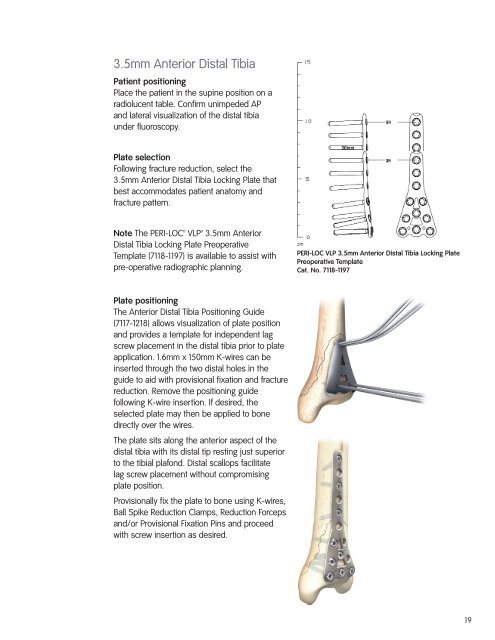
3.5mm Anterior Distal TibiaPatient positioningPlace the patient in the supine position on aradiolucent table. Confirm unimpeded APand lateral visualization of the distal tibiaunder fluoroscopy.Plate selectionFollowing fracture reduction, select the3.5mm Anterior Distal Tibia Locking Plate thatbest ac<strong>com</strong>modates patient anatomy andfracture pattern.Note The PERI-LOC <strong>VLP</strong> 3.5mm AnteriorDistal Tibia Locking Plate PreoperativeTemplate (7118-1197) is available to assist withpre-operative radiographic planning.PERI-LOC <strong>VLP</strong> 3.5mm Anterior Distal Tibia Locking PlatePreoperative TemplateCat. No. 7118-1197Plate positioningThe Anterior Distal Tibia Positioning Guide(7117-1218) allows visualization of plate positionand provides a template for independent lagscrew placement in the distal tibia prior to plateapplication. 1.6mm x 150mm K-wires can beinserted through the two distal holes in theguide to aid with provisional fixation and fracturereduction. Remove the positioning guidefollowing K-wire insertion. If desired, theselected plate may then be applied to bonedirectly over the wires.The plate sits along the anterior aspect of thedistal tibia with its distal tip resting just superiorto the tibial plafond. Distal scallops facilitatelag screw placement without <strong>com</strong>promisingplate position.Provisionally fix the plate to bone using K-wires,Ball Spike Reduction Clamps, Reduction Forcepsand/or Provisional Fixation Pins and proceedwith screw insertion as desired.19