dissection of frog, perch, worm lab.pdf
dissection of frog, perch, worm lab.pdf
dissection of frog, perch, worm lab.pdf
- No tags were found...

You also want an ePaper? Increase the reach of your titles
YUMPU automatically turns print PDFs into web optimized ePapers that Google loves.
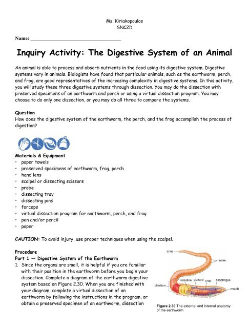
Name: ____________________________________Ms. KiriakopoulosSNC2DInquiry Activity: The Digestive System <strong>of</strong> an AnimalAn animal is able to process and absorb nutrients in the food using its digestive system. Digestivesystems vary in animals. Biologists have found that particular animals, such as the earth<strong>worm</strong>, <strong>perch</strong>,and <strong>frog</strong>, are good representatives <strong>of</strong> the increasing complexity in digestive systems. In this activity,you will study these three digestive systems through <strong>dissection</strong>. You may do the <strong>dissection</strong> withpreserved specimens <strong>of</strong> an earth<strong>worm</strong> and <strong>perch</strong> or using a virtual <strong>dissection</strong> program. You maychoose to do only one <strong>dissection</strong>, or you may do all three to compare the systems.QuestionHow does the digestive system <strong>of</strong> the earth<strong>worm</strong>, the <strong>perch</strong>, and the <strong>frog</strong> accomplish the process <strong>of</strong>digestion?Materials & Equipment• paper towels• preserved specimens <strong>of</strong> earth<strong>worm</strong>, <strong>frog</strong>, <strong>perch</strong>• hand lens• scalpel or dissecting scissors• probe• dissecting tray• dissecting pins• forceps• virtual <strong>dissection</strong> program for earth<strong>worm</strong>, <strong>perch</strong>, and <strong>frog</strong>• pen and/or pencil• paperCAUTION: To avoid injury, use proper techniques when using the scalpel.ProcedurePart 1 — Digestive System <strong>of</strong> the Earth<strong>worm</strong>1. Since the organs are small, it is helpful if you are familiarwith their position in the earth<strong>worm</strong> before you begin your<strong>dissection</strong>. Complete a diagram <strong>of</strong> the earth<strong>worm</strong> digestivesystem based on Figure 2.30. When you are finished withyour diagram, complete a virtual <strong>dissection</strong> <strong>of</strong> anearth<strong>worm</strong> by following the instructions in the program, orobtain a preserved specimen <strong>of</strong> an earth<strong>worm</strong>, <strong>dissection</strong>Figure 2.30 The external and internal anatomy<strong>of</strong> the earth<strong>worm</strong>
Ms. KiriakopoulosSNC2Dtools, and <strong>dissection</strong> pan. Rinse your specimen with water and pat dry.2. Using the hand lens, examine the external structure <strong>of</strong> the earth<strong>worm</strong> so that you can identifythe prostomium, clitellum, setae, and anus. The prostomium is in front <strong>of</strong> the mouth. The clitellumlooks like a saddle and is on the dorsal side <strong>of</strong> the earth<strong>worm</strong>. The setae are tiny bristles found onthe ventral side. The anus is found on the ventral side <strong>of</strong> the last segment <strong>of</strong> the <strong>worm</strong>.3. Hold the earth<strong>worm</strong> in your hand so that the dorsal side is facing up. Using your scissors, make ashallow cut on the dorsal side from the clitellum to the prostomium.4. Separate the tissue and use dissecting pins to pin the body wall down to the tray. You may need tocut through the tissue that holds the body wall.5. Locate the mouth, esophagus, crop, gizzard, intestine, and anus using Figure 2.30.6. Clean up your work area. Make sure to follow your teacher’s directions for safe disposal <strong>of</strong>materials. Wash your hands thoroughly.Part 2 — Digestive System <strong>of</strong> the Perch7. Complete a diagram <strong>of</strong> the <strong>perch</strong> digestive system based on Figure 2.31. When you are finishedwith your diagram, complete a virtual <strong>dissection</strong> <strong>of</strong> a <strong>perch</strong> or obtain a preserved specimen <strong>of</strong> a<strong>perch</strong>, <strong>dissection</strong> tools, and <strong>dissection</strong> pan. Rinse your specimen with water and pat dry.8. Observe the external structure <strong>of</strong> the <strong>perch</strong>. Note the position and number <strong>of</strong> fins. Find thelateral line, and locate the gill cover and anal opening.9. Examine the mouth <strong>of</strong> the <strong>perch</strong>.10.Create a flap through the muscle wall.Make an incision from the bottom <strong>of</strong> the gillcover along the ventral side to the analopening. Continue the incision up from theanal opening to the lateral line and thenalong that line to the head <strong>of</strong> the fish. Finishyour flap by extending your incision back tothe base <strong>of</strong> the gill cover.Figure 2.31 The external and internal anatomy <strong>of</strong> the <strong>perch</strong>
Ms. KiriakopoulosSNC2D11.Lift the flap <strong>of</strong> muscle wall to look at the organs <strong>of</strong> the <strong>perch</strong>. If you have a female <strong>perch</strong>, the areamay be filled with eggs. If this is the case, you should remove the mass <strong>of</strong> eggs before proceeding.If the <strong>perch</strong> is male, the testes will be smaller and lighter in colour. Locate the liver (light brown),gall bladder (olive colour), esophagus, stomach, pyloric caeca, and intestines.12.Clean up your work area. Make sure to follow your teacher’s directions for safe disposal <strong>of</strong> materials.Wash your hands thoroughly.Part 3 — Digestive System <strong>of</strong> the FrogMale or Female?1. Place a <strong>frog</strong> on a <strong>dissection</strong> tray. To determine the<strong>frog</strong>’s sex, look at the hand digits, or fingers, on itsforelegs. A male <strong>frog</strong> usually has thick pads on its"thumbs," which is one external difference betweenthe sexes, as shown in the diagram below. Male <strong>frog</strong>sare also usually smaller than female <strong>frog</strong>s. Observeseveral <strong>frog</strong>s to see the difference between malesand females.What is the sex <strong>of</strong> your <strong>frog</strong>? _________Anatomy <strong>of</strong> the Frog’s MouthProcedure: Pry the <strong>frog</strong>’s mouth open and use scissors tocut the angles <strong>of</strong> the <strong>frog</strong>’s jaws open. Cut deeply enoughso that the <strong>frog</strong>’s mouth opens wide enough to view thestructures inside.1. Locate the tongue.2. In the center <strong>of</strong> the mouth, toward the back is a singleround opening. This is the esophagus. This tube leads tothe stomach. Use a probe to poke into the esophagus.3. Close to the angles <strong>of</strong> the jaw are two openings, one oneach side. These are the Eustachian tubes. They are used to equalize pressure in the inner ear whilethe <strong>frog</strong> is swimming. Find the Eustachian tubes.4. Just behind the tongue, and before you reach the esophagus is a slit like opening. (You may need touse your probe to get it to open up). This slit is the glottis, and it is the opening to the lungs. The <strong>frog</strong>breathes and vocalizes with the glottis. Find the glottis.5. The <strong>frog</strong> has two sets <strong>of</strong> teeth. The vomarine teeth are found on the ro<strong>of</strong> <strong>of</strong> the mouth. Themaxillary teeth are found around the edge <strong>of</strong> the mouth. Both are used for holding prey, <strong>frog</strong>s swallowtheir meals whole and do NOT chew. Locate both sets <strong>of</strong> teeth.6. On the ro<strong>of</strong> <strong>of</strong> the mouth, you will find two tiny openings, if you put your probe into those openings,you will find they exit on the outside <strong>of</strong> the <strong>frog</strong>. These are the nostrils.
Frog Internal AnatomyMs. KiriakopoulosSNC2D1. Place the <strong>frog</strong> in the dissecting pan on its back (dorsal surface)2. Spread out the limbs3. Use T pins to fasten the <strong>frog</strong> to the wax in the tray.4. With forceps gently lift the lose skin away from the body Usingscissors, make a small cut in the middle <strong>of</strong> the abdomen5. Cut along the midline <strong>of</strong> the body from rear to the head.6. Make transverse (horizontal) cuts near the arms and legs.7. Life the flaps <strong>of</strong> the body wall and pin back.8. Repeat steps 5-7 but cut through the muscle tissue this timeAnatomy Check ListLocate each <strong>of</strong> the organs by using the descriptions and pictures.Liver--The largest structure <strong>of</strong> the body cavity.This brown colored organ is composed <strong>of</strong> threeparts, called lobes. The liver is not primarily anorgan <strong>of</strong> digestion; it does secrete a digestive juicecalled bile. Bile is needed for the digestion <strong>of</strong> fats.Heart - at the top <strong>of</strong> the liver, the heart is atriangular structure. The left and right atrium can befound at the top <strong>of</strong> the heart. A single ventriclelocated at the bottom <strong>of</strong> the heart.Lungs - Locate the lungs by looking underneath andbehind the heart and liver. They are two spongy organs.Gall bladder--Lift the lobes <strong>of</strong> the liver, there willbe a small green sac under the liver. This is the gallbladder, which stores bile.Stomach--Curving from underneath the liver is the stomach. The stomach is the first major site <strong>of</strong>chemical digestion. Frogs swallow their meals whole. Follow the stomach to where it turns into thesmall intestine. You can also open the stomach to see what the <strong>frog</strong>’s last meal was.Small Intestine--Leading from the stomach, a small curly structure. Pull it out to see how long it is.Eventually it will widen to become the large intestine. Nutrients are absorbed in the small intestine.Large Intestine--As you follow the small intestine down, it will widen into the large intestine. Thelarge intestine is also known as the cloaca in the <strong>frog</strong>. The cloaca is the last stop before wastes, sperm,or urine exit the <strong>frog</strong>'s body. (The word "cloaca" means sewer.)
Ms. KiriakopoulosSNC2DSpleen--Return to the folds <strong>of</strong> the mesentery, this dark red spherical object serves as a holding areafor blood, where harmful particles can be filtered out for the immune system.Esophagus--Return to the stomach and follow it upward, where it gets smaller is the beginning <strong>of</strong> theesophagus. The esophagus is the tube that leads from the <strong>frog</strong>’s mouth to the stomach. Open the <strong>frog</strong>’smouth and find the esophagus, poke your probe into it and see where it leads.Analyzing and Interpreting1. How is the mouth specialized in each <strong>of</strong> the three specimens?2. Explain how the structure <strong>of</strong> the intestines is related to their role in digestion.3. What is the function <strong>of</strong> the liver in digestion?4. What is the function <strong>of</strong> the gall bladder?5. Why do you think the gall bladder is located so close to the liver? Explain your answer.6. Trace the path <strong>of</strong> food through the digestive tract in the earth <strong>worm</strong>, <strong>frog</strong> and <strong>perch</strong> on aseparate piece <strong>of</strong> paper.7. Describe one problem that you encountered in performing the <strong>dissection</strong> and explain how yousolved the problem.8. How is the digestive system <strong>of</strong> the <strong>worm</strong>, the <strong>perch</strong>, and the <strong>frog</strong> each suited to its habitat?
Ms. KiriakopoulosSNC2DAnatomy <strong>of</strong> a Frog