A Case Report - OMJ
A Case Report - OMJ
A Case Report - OMJ

You also want an ePaper? Increase the reach of your titles
YUMPU automatically turns print PDFs into web optimized ePapers that Google loves.
<strong>Case</strong> <strong>Report</strong><br />
Abstract<br />
<strong>Case</strong> <strong>Report</strong><br />
Encysted Hydrocele of Cord in an Adult Misdiagnosed as<br />
Irreducible Hernia: A <strong>Case</strong> <strong>Report</strong><br />
Imtiaz Wani, Muddasir Rather, Gulam Naikoo, Imran Gul,<br />
Zubair Bhat, Aejaz Baba<br />
A number of pathologies can present as groin swellings in<br />
adults.Among these, encysted hydrocele of the cord presenting<br />
as swelling in an adult is a rare. A case of encysted hydrocele of<br />
cord in 36 year old male mimicking as as an irreducible hernia is<br />
reported. The diagnosis of hydrocele was made intraoperatively.<br />
An excision of the sac was performed.<br />
Wani I, et al. <strong>OMJ</strong>. 24, 218-219 (2009); doi:10.5001/omj.2009.42<br />
A 36 year old male was presented with swelling in the right<br />
groin for a 12 days duration. For the last 2 days, the patient had<br />
severe progressive pain in groin area and low grade fever for which<br />
he reported to the emergency services. There was no history<br />
of constipation, loose motions or vomiting. General physical<br />
examination as well as systemic examination were normal.<br />
Abdominal examination was normal, with no organomegaly and<br />
normal bowel sounds were present.<br />
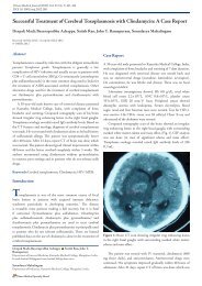
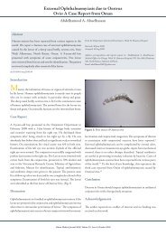
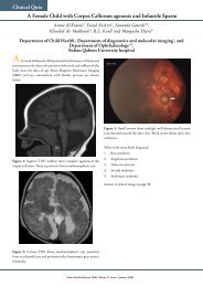
On local examination, a globular, soft, tender swelling<br />
measuring 5×2.3×1 centimeter, with negative cough impulse was<br />
present in the right inguinal region (Fig. 1).<br />
Figure 1: Swell in Right Groin<br />
Oman Medical Journal 2009, Volume 24, Issue 3, July 2009<br />
Department of surgery ,S.M.H.S Hospital /SKIMS Srinagar<br />
Received: 27 Feb 2009<br />
Accepted: 16 Mar 2009<br />
Author for correspondence: Imtiaz Wani, Amira Kadal, Srinagar, Kashmir, India.<br />
e-mail:imtazwani@gmail.com<br />
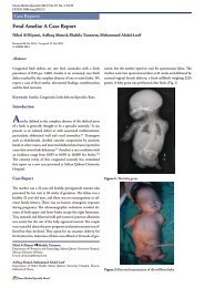
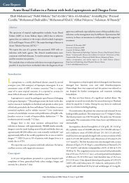
Figure 2: Sac in Right Groin<br />
The swelling could be felt completely separate from the testicle.<br />
Transillumination test as well as traction test was negative.<br />
Genitilia examination was normal. Per rectal examination was<br />
unremerkable. All baseline investigations were normal with a<br />
hemoglobin level of 13 gm/dl., total leucocyte count of 7,500/ mm 3 ,<br />
and normal electrolytes. An x-ray of the abdomen did not reveal<br />
any evidence of intestinal obstruction. Ultrasongraphy of the<br />
abdomen was normal. Scrotal ultrasound showed an oval anechoic<br />
mass in the groin. The patient was managed by intravenous fluids,<br />
antibiotics and pain killers but had mild relief of symptoms<br />
and diagnosis of irreducible hernia was made. The patient had<br />
exploration of the right groin and a sac was found abutting the<br />
spermatic cord having flimsy adhesions with the surrounding<br />
tissues (Fig. 2). The sac was abutting spermatic cord at the<br />
proximal end of the sac starting about 2 centimeters from the deep
inguinal ring with no scrotal extension observed. Aspiration of the<br />
contents of the sac revealed an amber colored fluid. An excision<br />
of the sac was performed. Fluid analysis was consistent with that<br />
of hydrocele fluid. Histopathological examination of the cyst wall<br />
showed collagenous material. Postoperative period was uneventful<br />
and the patient is regularly attending follow up clinics visits.<br />
Discussion<br />
The main pathological conditions manifesting as masses in the<br />
groin fall into five major groups: congenital abnormalities, noncongenital<br />
hernias, vascular conditions, infectious or inflammatory<br />
processes, and neoplasms. 1 Inflammatory swellings of the groin<br />
are common, and the changes are often attributed to infection<br />
and are often inflammatory swellings secondary to groin hernia. 2<br />
However, painful spermatic encysted hydrocele presenting as a<br />
groin swelling is rare.<br />
An encysted hydrocele or a non-communicating type of<br />
inguinal hydrocoele, is a loculated fluid collection along the<br />
spermatic cord, separated from and located above the testicle and<br />
the epididymis, as a result of aberrant closure of the processus<br />
vaginalis. This is idiopathic in most cases but in some cases it<br />
may be secondary to testicular torsion, tumour or trauma, and in<br />
infections, as in, orchitis, epididymitis, tuberculosis or filariasis. 3<br />
Rarely, hydrocele of pancreatic origin have been reported to<br />
occur. 4 Encysted hydrocele of the cord remains asymptomatic or is<br />
detected incidentally during evaluation during the course of other<br />
disease. 5<br />
Diagnosis is clinicaly essential but where doubt exists, scrotal<br />
ultrasound can be used to differentiate it from other scrotal<br />
lesions. Diagnosis can also be confirmed by computed tomography<br />
scan or intraoperatively. Spermatic cord hydrocele is effectively<br />
diagnosed by ultrasonography based on its specific location and<br />
shape. Ultrasonography is useful to exclude hernia, enlargement<br />
of the lymph node, or other solid masses. 6 A typical finding on<br />
ultrasonography of spermatic cord hydrocele is its avascular<br />
anoechoic structure.<br />
Excision is the treatment of choice and the excision under<br />
local anesthesia in adult patiens is well studied. 7 Fluid analysis<br />
of the hydrocele fluid showed amber color and sterile in nature<br />
Specific gravity of the fluid was 1.02. Microscopically, cholesterol<br />
crystals were isolated with tests positive for presence of albumin<br />
and fibrinogen. Histopathological examination of the cyst wall<br />
shows collagenous material. Encysted type can be misdiagnosed<br />
as hernia, lymphagiomatous cyst or cystic teratoma, inguinal<br />
lymphadenopathy, lipoma of cord ,or other tumours of the cord.<br />
Rarely, ileo femoral aneurysm, appendicular pathology, or a<br />
hematoma present as an inflammatory swelling in the groin. 8<br />
Encysted Hydrocele of Cord... Wani et al.<br />
Conclusion<br />
Oman Medical Journal 2009, Volume 24, Issue 3, July 2009<br />
Encysted hydrocele of the cord in an adult is a rare condition .It<br />
may mimic an irreducible hernia at times. Excision remains the<br />
treatment of choice.<br />
Acknowledgements<br />
The authors reported no conflict of interest and no funding has<br />
been received on this work.<br />
References<br />
1. Shadbolt CL, Heinze SBJ, Dietrich RB. Imaging of Groin Masses: inguinal<br />
anatomy and pathologic conditions revisited. RadioGraphics. 2001;21:s261–<br />
71.<br />
2. Maheswaran P, Stephen D. A rare presentation of appendicitis as groin<br />
swelling: a case report. <strong>Case</strong>s J. 2009; 2: 53.<br />
3. Ku HJ, Kim ME, Lee NK, Park YH. The excisional, placation and internal<br />
draingetechniques: a comparison of the results for idiopathic hydrocele. BJU<br />
Int. 2001;87:82–84.<br />
4. Delamarre J, Descombes P, Grillot G, Deschepper B, Deramond H.<br />
Hydrocele of pancreatic origin. X-ray computed tomographic study of an<br />
intrascrotal collection in an acute outbreak of chronic pancreatitis. J Radiol.<br />
1988;69:689-90.<br />
5. Busigo J. P, Eftekharif F, Hendell H .Encysted Spermatic Cord Hydroceles<br />
: A <strong>Report</strong> of Three <strong>Case</strong>s in Adults and a Review of the Literature. Acta<br />
Radiologica. 2007; 48:1138-1142.<br />
6. Han BH, Cho JY, Cho BJ, Ki WW Hydrocele of the Spermatic Cord:<br />
Ultrasonograhic Findings. J Korean Soc Med Ultrasound. 2002 ;21:129-<br />
133.<br />
7. Agbakwuru A , Salako A, Olajide A, Takure AO, Eziyi A. Hydrocelectomy<br />
under local anaesthesia in a Nigerian adult population Afr Health Sci. 2008;<br />
8: 160–162.<br />
8. Apostolidis S, Papavramidis S, Michalopoulos A, Papadopoulos N,<br />
Paramythiotis D, Harlaftis N. Groin Swelling, the Anatomic Way Out of<br />
Abdominal Haematomas Acta Chir Belg, 2008, 108, 251-253.