Overview of Biomaterials and Their Use in ... - ASM International
Overview of Biomaterials and Their Use in ... - ASM International
Overview of Biomaterials and Their Use in ... - ASM International

You also want an ePaper? Increase the reach of your titles
YUMPU automatically turns print PDFs into web optimized ePapers that Google loves.
© 2003 <strong>ASM</strong> <strong>International</strong>. All Rights Reserved.<br />
H<strong>and</strong>book <strong>of</strong> Materials for Medical Devices (#06974G)<br />
CHAPTER 1<br />
<strong>Overview</strong> <strong>of</strong> <strong>Biomaterials</strong><br />
<strong>and</strong> <strong>Their</strong> <strong>Use</strong> <strong>in</strong> Medical Devices<br />
A BIOMATERIAL, as def<strong>in</strong>ed <strong>in</strong> this h<strong>and</strong>book,<br />
is any synthetic material that is used to<br />
replace or restore function to a body tissue <strong>and</strong><br />
is cont<strong>in</strong>uously or <strong>in</strong>termittently <strong>in</strong> contact with<br />
body fluids (Ref 1). This def<strong>in</strong>ition is somewhat<br />
restrictive, because it excludes materials used<br />
for devices such as surgical or dental <strong>in</strong>struments.<br />
Although these <strong>in</strong>struments are exposed<br />
to body fluids, they do not replace or augment<br />
the function <strong>of</strong> human tissue. It should be noted,<br />
however, that materials for surgical <strong>in</strong>struments,<br />
particularly sta<strong>in</strong>less steels, are reviewed<br />
briefly <strong>in</strong> Chapter 3, “Metallic Materials,” <strong>in</strong><br />
this h<strong>and</strong>book. Similarly, sta<strong>in</strong>less steels <strong>and</strong><br />
shape memory alloys used for dental/endodontic<br />
<strong>in</strong>struments are discussed <strong>in</strong> Chapter 10,<br />
“<strong>Biomaterials</strong> for Dental Applications.”<br />
Also excluded from the aforementioned def<strong>in</strong>ition<br />
are materials that are used for external<br />
prostheses, such as artificial limbs or devices<br />
such as hear<strong>in</strong>g aids. These materials are not<br />
exposed to body fluids.<br />
Exposure to body fluids usually implies that<br />
the biomaterial is placed with<strong>in</strong> the <strong>in</strong>terior <strong>of</strong><br />
the body, <strong>and</strong> this places several strict restrictions<br />
on materials that can be used as a biomaterial<br />
(Ref 1). First <strong>and</strong> foremost, a biomaterial<br />
must be biocompatible—it should not elicit an<br />
adverse response from the body, <strong>and</strong> vice versa.<br />
Additionally, it should be nontoxic <strong>and</strong> noncarc<strong>in</strong>ogenic.<br />
These requirements elim<strong>in</strong>ate many<br />
eng<strong>in</strong>eer<strong>in</strong>g materials that are available. Next,<br />
the biomaterial should possess adequate physical<br />
<strong>and</strong> mechanical properties to serve as augmentation<br />
or replacement <strong>of</strong> body tissues. For<br />
practical use, a biomaterial should be amenable<br />
to be<strong>in</strong>g formed or mach<strong>in</strong>ed <strong>in</strong>to different<br />
shapes, have relatively low cost, <strong>and</strong> be readily<br />
available.<br />
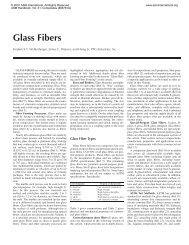
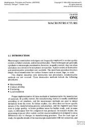
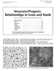
Figure 1 lists the various material requirements<br />
that must be met for successful total jo<strong>in</strong>t<br />
replacement. The ideal material or material<br />
comb<strong>in</strong>ation should exhibit the follow<strong>in</strong>g properties:<br />
• A biocompatible chemical composition to<br />
avoid adverse tissue reactions<br />
• Excellent resistance to degradation (e.g., corrosion<br />
resistance for metals or resistance to<br />
biological degradation <strong>in</strong> polymers)<br />
• Acceptable strength to susta<strong>in</strong> cyclic load<strong>in</strong>g<br />
endured by the jo<strong>in</strong>t<br />
• A low modulus to m<strong>in</strong>imize bone resorption<br />
• High wear resistance to m<strong>in</strong>imize weardebris<br />
generation<br />
<strong>Use</strong>s for <strong>Biomaterials</strong> (Ref 3)<br />
www.asm<strong>in</strong>ternational.org<br />
One <strong>of</strong> the primary reasons that biomaterials<br />
are used is to physically replace hard or s<strong>of</strong>t tissues<br />
that have become damaged or destroyed<br />
through some pathological process (Ref 3).<br />
Although the tissues <strong>and</strong> structures <strong>of</strong> the body<br />
perform for an extended period <strong>of</strong> time <strong>in</strong> most<br />
people, they do suffer from a variety <strong>of</strong> destructive<br />
processes, <strong>in</strong>clud<strong>in</strong>g fracture, <strong>in</strong>fection, <strong>and</strong><br />
cancer that cause pa<strong>in</strong>, disfigurement, or loss <strong>of</strong><br />
function. Under these circumstances, it may be<br />
possible to remove the diseased tissue <strong>and</strong><br />
replace it with some suitable synthetic material.<br />
Orthopedics. One <strong>of</strong> the most prom<strong>in</strong>ent<br />
application areas for biomaterials is for orthopedic<br />
implant devices. Both osteoarthritis <strong>and</strong><br />
rheumatoid arthritis affect the structure <strong>of</strong> freely
© 2003 <strong>ASM</strong> <strong>International</strong>. All Rights Reserved.<br />
H<strong>and</strong>book <strong>of</strong> Materials for Medical Devices (#06974G)<br />
2 / H<strong>and</strong>book <strong>of</strong> Materials for Medical Devices<br />
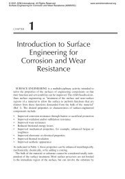
movable (synovial) jo<strong>in</strong>ts, such as the hip, knee,<br />
shoulder, ankle, <strong>and</strong> elbow (Fig. 2). The pa<strong>in</strong> <strong>in</strong><br />
such jo<strong>in</strong>ts, particularly weight-bear<strong>in</strong>g jo<strong>in</strong>ts<br />
such as the hip <strong>and</strong> knee, can be considerable,<br />
<strong>and</strong> the effects on ambulatory function quite<br />
devastat<strong>in</strong>g. It has been possible to replace these<br />
jo<strong>in</strong>ts with prostheses s<strong>in</strong>ce the advent <strong>of</strong> anesthesia,<br />
antisepsis, <strong>and</strong> antibiotics, <strong>and</strong> the relief<br />
<strong>of</strong> pa<strong>in</strong> <strong>and</strong> restoration <strong>of</strong> mobility is well<br />
known to hundreds <strong>of</strong> thous<strong>and</strong>s <strong>of</strong> patients.<br />
The use <strong>of</strong> biomaterials for orthopedic<br />
implant devices is one <strong>of</strong> the major focal po<strong>in</strong>ts<br />
<strong>of</strong> this h<strong>and</strong>book. In fact, Chapters 2 through 7<br />
<strong>and</strong> Chapter 9 (refer to Table <strong>of</strong> Contents) all<br />
deal with the materials <strong>and</strong> performance associated<br />
with orthopedic implants. As shown <strong>in</strong><br />
Table 1, a variety <strong>of</strong> metals, polymers, <strong>and</strong><br />
ceramics are used for such applications.<br />
Cardiovascular Applications. In the cardiovascular,<br />
or circulatory, system (the heart<br />
<strong>and</strong> blood vessels <strong>in</strong>volved <strong>in</strong> circulat<strong>in</strong>g blood<br />
throughout the body), problems can arise with<br />
heart valves <strong>and</strong> arteries, both <strong>of</strong> which can be<br />
successfully treated with implants. The heart<br />
valves suffer from structural changes that prevent<br />
the valve from either fully open<strong>in</strong>g or fully<br />
clos<strong>in</strong>g, <strong>and</strong> the diseased valve can be replaced<br />
with a variety <strong>of</strong> substitutes. As with orthopedic<br />
implants, ceramics (carbons, as described <strong>in</strong><br />
Chapter 6, “Ceramic Materials,” <strong>in</strong> this h<strong>and</strong>book),<br />
metals, <strong>and</strong> polymers are used as materials<br />
<strong>of</strong> construction (Table 1).<br />
Arteries, particularly the coronary arteries<br />
<strong>and</strong> the vessels <strong>of</strong> the lower limbs, become<br />
blocked by fatty deposits (atherosclerosis), <strong>and</strong><br />
it is possible <strong>in</strong> some cases to replace segments<br />
a.<br />
b.<br />
c.<br />
Fig. 1 Implant material requirements <strong>in</strong> orthopedic applications. Source: Ref 2<br />
www.asm<strong>in</strong>ternational.org<br />
with artificial arteries. As shown <strong>in</strong> Table 1,<br />
polymers are the material <strong>of</strong> choice for vascular<br />
prostheses (see Chapter 7, “Polymeric Materials,”<br />
<strong>in</strong> this h<strong>and</strong>book for further details).<br />
Ophthalmics. The tissues <strong>of</strong> the eye can<br />
suffer from several diseases, lead<strong>in</strong>g to reduced<br />
vision <strong>and</strong> eventually, bl<strong>in</strong>dness. Cataracts, for<br />
example, cause cloud<strong>in</strong>ess <strong>of</strong> the lens. This may<br />
be replaced with a synthetic (polymer) <strong>in</strong>traocular<br />
lens (Table 1). Materials for contact lenses,<br />
because they are <strong>in</strong> <strong>in</strong>timate contact with the tissues<br />
<strong>of</strong> the eye, are also considered biomaterials.<br />
As with <strong>in</strong>traocular lenses, they too are used<br />
to preserve <strong>and</strong> restore vision (see Chapter 7,<br />
“Polymeric Materials,” <strong>in</strong> this h<strong>and</strong>book for<br />
details).<br />
Dental Applications. With<strong>in</strong> the mouth,<br />
both the tooth <strong>and</strong> support<strong>in</strong>g gum tissues can<br />
be readily destroyed by bacterially controlled<br />
diseases. Dental caries (cavities), the dem<strong>in</strong>eralization<br />
<strong>and</strong> dissolution <strong>of</strong> teeth associated with<br />
the metabolic activity <strong>in</strong> plaque (a film <strong>of</strong> mucus<br />
that traps bacteria on the surface <strong>of</strong> the teeth),<br />
can cause extensive tooth loss. Teeth <strong>in</strong> their<br />
entirety <strong>and</strong> segments <strong>of</strong> teeth both can be<br />
replaced or restored by a variety <strong>of</strong> materials<br />
(Table 1). A thorough review <strong>of</strong> these materials<br />
can be found <strong>in</strong> Chapter 10, “<strong>Biomaterials</strong> for<br />
Dental Applications,” <strong>in</strong> this h<strong>and</strong>book.<br />
Wound Heal<strong>in</strong>g. One <strong>of</strong> the oldest uses <strong>of</strong><br />
implantable biomaterials can be traced back to<br />
the <strong>in</strong>troduction <strong>of</strong> sutures for wound closure.<br />
The ancient Egyptians used l<strong>in</strong>en as a suture as<br />
far back as 2000 b.c. Synthetic suture materials<br />
<strong>in</strong>clude both polymers (the most widely synthetic<br />
suture material) <strong>and</strong> some metals (e.g.,
© 2003 <strong>ASM</strong> <strong>International</strong>. All Rights Reserved.<br />
www.asm<strong>in</strong>ternational.org<br />
H<strong>and</strong>book <strong>of</strong> Materials for Medical Devices (#06974G)<br />
Chapter 1: <strong>Overview</strong> <strong>of</strong> <strong>Biomaterials</strong> <strong>and</strong> <strong>Their</strong> <strong>Use</strong> <strong>in</strong> Medical Devices / 3<br />
sta<strong>in</strong>less steels <strong>and</strong> tantalum). Chapter 7, “Polymeric<br />
Materials,” <strong>in</strong> this h<strong>and</strong>book discusses the<br />
characteristics <strong>and</strong> properties <strong>of</strong> synthetic suture<br />
materials.<br />
Table 1 Examples <strong>of</strong> medical <strong>and</strong> dental<br />
materials <strong>and</strong> their applications<br />
Material Pr<strong>in</strong>cipal applications<br />
Metals <strong>and</strong> alloys<br />
316L sta<strong>in</strong>less steel Fracture fixation, stents, surgical<br />
<strong>in</strong>struments<br />
CP-Ti, Ti-Al-V, Ti-Al-Nb, Ti- Bone <strong>and</strong> jo<strong>in</strong>t replacement,<br />
13Nb-13Zr, Ti-Mo-Zr-Fe fracture fixation, dental<br />
implants, pacemaker<br />
encapsulation<br />
Co-Cr-Mo, Cr-Ni-Cr-Mo Bone <strong>and</strong> jo<strong>in</strong>t replacement,<br />
dental implants, dental<br />
restorations, heart valves<br />
Ni-Ti Bone plates, stents, orthodontic<br />
wires<br />
Gold alloys Dental restorations<br />
Silver products Antibacterial agents<br />
Plat<strong>in</strong>um <strong>and</strong> Pt-Ir Electrodes<br />
Hg-Ag-Sn amalgam Dental restorations<br />
Ceramics <strong>and</strong> glasses<br />
Alum<strong>in</strong>a Jo<strong>in</strong>t replacement, dental<br />
implants<br />
Zirconia Jo<strong>in</strong>t replacement<br />
Calcium phosphates Bone repair <strong>and</strong> augmentation,<br />
surface coat<strong>in</strong>gs on metals<br />
Bioactive glasses Bone replacement<br />
Porcela<strong>in</strong> Dental restorations<br />
Carbons Heart valves, percutaneous<br />
devices, dental implants<br />
Polymers<br />
Polyethylene Jo<strong>in</strong>t replacement<br />
Polypropylene Sutures<br />
PET Sutures, vascular prosthesis<br />
Polyamides Sutures<br />
PTFE S<strong>of</strong>t-tissue augmentation,<br />
vascular prostheses<br />
Polyesters Vascular prostheses, drugdelivery<br />
systems<br />
Polyurethanes Blood-contact<strong>in</strong>g devices<br />
PVC Tub<strong>in</strong>g<br />
PMMA Dental restorations, <strong>in</strong>traocular<br />
lenses, jo<strong>in</strong>t replacement<br />
(bone cements)<br />
Silicones S<strong>of</strong>t-tissue replacement,<br />
ophthalmology<br />
Hydrogels Ophthalmology, drug-delivery<br />
systems<br />
Composites<br />
BIS-GMA-quartz/silica filler Dental restorations<br />
PMMA-glass fillers Dental restorations (dental<br />
cements)<br />
Abbreviations: CP-Ti, commercially pure titanium; PET, polyethylene terephthalates<br />
(Dacron, E.I. DuPont de Nemours & Co.); PTFE, polytetra fluoroethylenes<br />
(Teflon, E.I. DuPont de Nemours & Co.); PVC, polyv<strong>in</strong>yl chlorides;<br />
PMMA, polymethyl methacrylate; BIS-GMA, bisphenol A-glycidyl. Source:<br />
Adapted from Ref 3<br />
Another important wound-heal<strong>in</strong>g category<br />
is that <strong>of</strong> fracture fixation devices. These <strong>in</strong>clude<br />
bone plates, screws, nails, rods, wires, <strong>and</strong><br />
other devices used for fracture treatment.<br />
Although some nonmetallic materials (e.g., carbon-carbon<br />
composite bone plates) have been<br />
<strong>in</strong>vestigated, almost all fracture fixation devices<br />
used for orthopedic applications are made from<br />
metals, most notably sta<strong>in</strong>less steels (see Chapter<br />
3, “Metallic Materials,” <strong>in</strong> this h<strong>and</strong>book for<br />
details).<br />
Drug-Delivery Systems. One <strong>of</strong> the fastest<br />
grow<strong>in</strong>g areas for implant applications is for<br />
devices for controlled <strong>and</strong> targeted delivery <strong>of</strong><br />
drugs. Many attempts have been made to <strong>in</strong>corporate<br />
drug reservoirs <strong>in</strong>to implantable devices<br />
for a susta<strong>in</strong>ed <strong>and</strong> preferably controlled release.<br />
Some <strong>of</strong> these technologies use new polymeric<br />
materials as vehicles for drug delivery.<br />
Chapters 7, “Polymeric Materials,” <strong>and</strong> 9,<br />
“Coat<strong>in</strong>gs,” <strong>in</strong> this h<strong>and</strong>book describe these<br />
materials.<br />
Types <strong>of</strong> <strong>Biomaterials</strong> (Ref 1)<br />
Most synthetic biomaterials used for implants<br />
are common materials familiar to the average<br />
materials eng<strong>in</strong>eer or scientist (Table 1). In general,<br />
these materials can be divided <strong>in</strong>to the follow<strong>in</strong>g<br />
categories: metals, polymers, ceramics,<br />
<strong>and</strong> composites.<br />
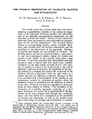
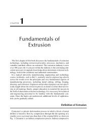
Fig. 2<br />
Schematic show<strong>in</strong>g key components <strong>of</strong> a natural synovial<br />
jo<strong>in</strong>t. It consists <strong>of</strong> layers <strong>of</strong> bear<strong>in</strong>g material<br />
(articular cartilage) mounted on relatively hard bones form<strong>in</strong>g<br />
the skeletal frame. The synovial fluid acts as a lubricant. In an<br />
artificial jo<strong>in</strong>t, lubrication is supplied by low-friction polymeric<br />
bear<strong>in</strong>g materials. Source: Ref 4
© 2003 <strong>ASM</strong> <strong>International</strong>. All Rights Reserved.<br />
H<strong>and</strong>book <strong>of</strong> Materials for Medical Devices (#06974G)<br />
4 / H<strong>and</strong>book <strong>of</strong> Materials for Medical Devices<br />
Metals. As a class <strong>of</strong> materials, metals are<br />
the most widely used for load-bear<strong>in</strong>g implants.<br />
For <strong>in</strong>stance, some <strong>of</strong> the most common orthopedic<br />
surgeries <strong>in</strong>volve the implantation <strong>of</strong><br />
metallic implants. These range from simple<br />
wires <strong>and</strong> screws to fracture fixation plates <strong>and</strong><br />
total jo<strong>in</strong>t prostheses (artificial jo<strong>in</strong>ts) for hips,<br />
knees, shoulders, ankles, <strong>and</strong> so on. In addition<br />
to orthopedics, metallic implants are used <strong>in</strong><br />
maxill<strong>of</strong>acial surgery, cardiovascular surgery,<br />
<strong>and</strong> as dental materials. Although many metals<br />
<strong>and</strong> alloys are used for medical device applications,<br />
the most commonly employed are sta<strong>in</strong>less<br />
steels, commercially pure titanium <strong>and</strong> titanium<br />
alloys, <strong>and</strong> cobalt-base alloys (Table 1).<br />
The use <strong>of</strong> metals for implants is reviewed <strong>in</strong><br />
Chapter 3, “Metallic Materials,” <strong>in</strong> this h<strong>and</strong>book.<br />
Dental alloys are discussed <strong>in</strong> Chapters<br />
10, “<strong>Biomaterials</strong> for Dental Applications,” <strong>and</strong><br />
11, “Tarnish <strong>and</strong> Corrosion <strong>of</strong> Dental Alloys.”<br />
Polymers. A wide variety <strong>of</strong> polymers are<br />
used <strong>in</strong> medic<strong>in</strong>e as biomaterials. <strong>Their</strong> applications<br />
range from facial prostheses to tracheal<br />
tubes, from kidney <strong>and</strong> liver parts to heart components,<br />
<strong>and</strong> from dentures to hip <strong>and</strong> knee<br />
jo<strong>in</strong>ts (Tables 1, 2). Chapters 7, “Polymeric<br />
Materials,” <strong>and</strong> 10, “<strong>Biomaterials</strong> for Dental<br />
Applications,” <strong>in</strong> this h<strong>and</strong>book review the use<br />
<strong>of</strong> polymers for these applications.<br />
Polymeric materials are also used for medical<br />
adhesives <strong>and</strong> sealants <strong>and</strong> for coat<strong>in</strong>gs that<br />
serve a variety <strong>of</strong> functions (see Chapters 8,<br />
“Adhesives,” <strong>and</strong> 9, “Coat<strong>in</strong>gs,” <strong>in</strong> this h<strong>and</strong>book<br />
for details).<br />
Ceramics. Traditionally, ceramics have<br />
seen widescale use as restorative materials <strong>in</strong><br />
Table 2 Examples <strong>of</strong> polymers used as<br />
biomaterials<br />
Application Polymer<br />
Knee, hip, shoulder jo<strong>in</strong>ts Ultrahigh molecular weight<br />
polyethylene<br />
F<strong>in</strong>ger jo<strong>in</strong>ts Silicone<br />
Sutures Polylactic <strong>and</strong> polyglycolic acid,<br />
nylon<br />
Tracheal tubes Silicone, acrylic, nylon<br />
Heart pacemaker Acetal, polyethylene,<br />
polyurethane<br />
Blood vessels Polyester, polytetrafluoroethylene,<br />
PVC<br />
Gastro<strong>in</strong>test<strong>in</strong>al segments Nylon, PVC, silicones<br />
Facial prostheses Polydimethyl siloxane,<br />
polyurethane, PVC<br />
Bone cement Polymethyl methacrylate<br />
PVC, polyv<strong>in</strong>yl chloride. Source: Ref 1<br />
www.asm<strong>in</strong>ternational.org<br />
dentistry. These <strong>in</strong>clude materials for crowns,<br />
cements, <strong>and</strong> dentures (see Chapter 10, “<strong>Biomaterials</strong><br />
for Dental Applications,” <strong>in</strong> this h<strong>and</strong>book<br />
for details). However, their use <strong>in</strong> other<br />
fields <strong>of</strong> biomedic<strong>in</strong>e has not been as extensive,<br />
compared to metals <strong>and</strong> polymers. For example,<br />
the poor fracture toughness <strong>of</strong> ceramics severely<br />
limits their use for load-bear<strong>in</strong>g applications.<br />
As shown <strong>in</strong> Table 1, some ceramic materials<br />
are used for jo<strong>in</strong>t replacement <strong>and</strong> bone<br />
repair <strong>and</strong> augmentation. Chapters 6, “Ceramic<br />
Materials,” <strong>and</strong> 9, “Coat<strong>in</strong>gs,” <strong>in</strong> this h<strong>and</strong>book<br />
review the uses <strong>of</strong> ceramics for nondental biomedical<br />
applications.<br />
Composites. As shown <strong>in</strong> Table 1, the most<br />
successful composite biomaterials are used <strong>in</strong><br />
the field <strong>of</strong> dentistry as restorative materials or<br />
dental cements (see Chapter 10, “<strong>Biomaterials</strong><br />
for Dental Applications,” <strong>in</strong> this h<strong>and</strong>book for<br />
details). Although carbon-carbon <strong>and</strong> carbonre<strong>in</strong>forced<br />
polymer composites are <strong>of</strong> great<br />
<strong>in</strong>terest for bone repair <strong>and</strong> jo<strong>in</strong>t replacement<br />
because <strong>of</strong> their low elastic modulus levels,<br />
these materials have not displayed a comb<strong>in</strong>ation<br />
<strong>of</strong> mechanical <strong>and</strong> biological properties<br />
appropriate to these applications. Composite<br />
materials are, however, used extensively for<br />
prosthetic limbs, where their comb<strong>in</strong>ation <strong>of</strong><br />
low density/weight <strong>and</strong> high strength make<br />
them ideal materials for such applications.<br />
Natural <strong>Biomaterials</strong>. Although the biomaterials<br />
discussed <strong>in</strong> this h<strong>and</strong>book are synthetic<br />
materials, there are several materials<br />
derived from the animal or plant world be<strong>in</strong>g<br />
considered for use as biomaterials that deserve<br />
brief mention. One <strong>of</strong> the advantages <strong>of</strong> us<strong>in</strong>g<br />
natural materials for implants is that they are<br />
similar to materials familiar to the body. In this<br />
regard, the field <strong>of</strong> biomimetics (or mimick<strong>in</strong>g<br />
nature) is grow<strong>in</strong>g. Natural materials do not<br />
usually <strong>of</strong>fer the problems <strong>of</strong> toxicity <strong>of</strong>ten<br />
faced by synthetic materials. Also, they may<br />
carry specific prote<strong>in</strong> b<strong>in</strong>d<strong>in</strong>g sites <strong>and</strong> other<br />
biochemical signals that may assist <strong>in</strong> tissue<br />
heal<strong>in</strong>g or <strong>in</strong>tegration. However, natural materials<br />
can be subject to problems <strong>of</strong> immunogenicity.<br />
Another problem faced by these materials,<br />
especially natural polymers, is their tendency to<br />
denature or decompose at temperatures below<br />
their melt<strong>in</strong>g po<strong>in</strong>ts. This severely limits their<br />
fabrication <strong>in</strong>to implants <strong>of</strong> different sizes <strong>and</strong><br />
shapes.<br />
An example <strong>of</strong> a natural material is collagen,<br />
which exists mostly <strong>in</strong> fibril form, has a characteristic<br />
triple-helix structure, <strong>and</strong> is the most
© 2003 <strong>ASM</strong> <strong>International</strong>. All Rights Reserved.<br />
www.asm<strong>in</strong>ternational.org<br />
H<strong>and</strong>book <strong>of</strong> Materials for Medical Devices (#06974G)<br />
Chapter 1: <strong>Overview</strong> <strong>of</strong> <strong>Biomaterials</strong> <strong>and</strong> <strong>Their</strong> <strong>Use</strong> <strong>in</strong> Medical Devices / 5<br />
prevalent prote<strong>in</strong> <strong>in</strong> the animal world. For<br />
example, almost 50% <strong>of</strong> the prote<strong>in</strong> <strong>in</strong> cowhide<br />
is collagen. It forms a significant component <strong>of</strong><br />
connective tissue such as bone, tendons, ligaments,<br />
<strong>and</strong> sk<strong>in</strong>. There are at least ten different<br />
types <strong>of</strong> collagen <strong>in</strong> the body. Among these,<br />
type I is found predom<strong>in</strong>antly <strong>in</strong> sk<strong>in</strong>, bone, <strong>and</strong><br />
tendons; type II is found <strong>in</strong> articular cartilage <strong>in</strong><br />
jo<strong>in</strong>ts; <strong>and</strong> type III is a major constituent <strong>of</strong><br />
blood vessels.<br />
Collagen is be<strong>in</strong>g studied extensively for use<br />
as a biomaterial. It is usually implanted <strong>in</strong> a<br />
sponge form that does not have significant<br />
mechanical strength or stiffness. It has shown<br />
good promise as a scaffold for neotissue growth<br />
<strong>and</strong> is commercially available as a product for<br />
wound heal<strong>in</strong>g. Injectable collagen is widely<br />
used for the augmentation or buildup <strong>of</strong> dermal<br />
tissue for cosmetic reasons. Other natural materials<br />
under consideration <strong>in</strong>clude coral, chit<strong>in</strong><br />
(from <strong>in</strong>sects <strong>and</strong> crustaceans), kerat<strong>in</strong> (from<br />
hair), <strong>and</strong> cellulose (from plants).<br />
Examples <strong>of</strong> <strong>Biomaterials</strong> Applications<br />
Biomedical devices range the gamut <strong>of</strong> design<br />
<strong>and</strong> materials selection considerations from<br />
relatively simple devices requir<strong>in</strong>g one material,<br />
such as commercially pure titanium dental<br />
implants, to highly complex assemblies, such as<br />
the cardiac pacemaker described subsequently<br />
or the ventricular-assist device (VAD) discussed<br />
<strong>in</strong> Chapter 7, “Polymeric Materials” <strong>in</strong><br />
this h<strong>and</strong>book (see, for example, Fig. 4 <strong>and</strong><br />
Table 6 <strong>in</strong> Chapter 7, which illustrate the components<br />
<strong>and</strong> list the materials <strong>of</strong> construction,<br />
respectively, for a VAD).<br />
Total Hip Replacement<br />
Total jo<strong>in</strong>t replacement is widely regarded as<br />
the major achievement <strong>in</strong> orthopedic surgery <strong>in</strong><br />
the 20th century. Arthroplasty, or the creation <strong>of</strong><br />
a new jo<strong>in</strong>t, is the name given to the surgical<br />
treatment <strong>of</strong> degenerate jo<strong>in</strong>ts aimed at the relief<br />
<strong>of</strong> pa<strong>in</strong> <strong>and</strong> the restoration <strong>of</strong> movement. This<br />
has been achieved by excision, <strong>in</strong>terposition,<br />
<strong>and</strong> replacement arthroplasty <strong>and</strong> by techniques<br />
that have been developed over approximately<br />
180 years (Ref 2).<br />
Design <strong>and</strong> Materials Selection. Hip<br />
arthroplasty generally requires that the upper<br />
femur (thigh bone) be replaced <strong>and</strong> the mat<strong>in</strong>g<br />
pelvis (hip bone) area be replaced or resurfaced.<br />
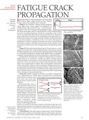
As shown <strong>in</strong> Fig. 3, a typical hip prosthesis consists<br />
<strong>of</strong> the femoral stem, a femoral ball, <strong>and</strong> a<br />
polymeric (ultrahigh molecular weight polyethylene,<br />
or UHMWPE) socket (cup) with or without<br />
a metallic back<strong>in</strong>g. Femoral components<br />
usually are manufactured from Co-Cr-Mo or<br />
Co-Ni-Cr-Mo alloys or titanium alloys (see<br />
Chapter 3, “Metallic Materials,” <strong>in</strong> this h<strong>and</strong>book<br />
for details). The ball (articulat<strong>in</strong>g portion<br />
<strong>of</strong> the femoral component) is made either <strong>of</strong><br />
highly polished Co-Cr alloys or <strong>of</strong> a ceramic<br />
(e.g., alum<strong>in</strong>a). Modular designs, where the<br />
stem <strong>and</strong> ball are <strong>of</strong> two different materials, are<br />
common. For example, hip replacement implants<br />
featur<strong>in</strong>g a titanium alloy femoral stem<br />
will have a Co-Cr femoral head. Similarly, the<br />
UHMWPE socket <strong>of</strong> the common acetabulum<br />
replacement can be implanted directly <strong>in</strong> the<br />
pelvis or be part <strong>of</strong> a modular arrangement<br />
where<strong>in</strong> the cup is placed <strong>in</strong>to a metallic shell<br />
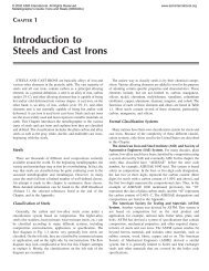
Fig. 3<br />
Typical components found <strong>in</strong> an unassembled total<br />
hip replacement (THR) implant. It should be noted<br />
that this is one <strong>of</strong> many artificial jo<strong>in</strong>t designs used <strong>in</strong> THR arthroplasty.<br />
For example, implants secured by bone cements would<br />
not be porous coated. Similarly, the ultrahigh molecular weight<br />
polyethylene (UHMWPE) acetabular cup is sometimes not<br />
capped by a metal (cobalt- or titanium-base alloys or unalloyed<br />
tantalum) shell.
© 2003 <strong>ASM</strong> <strong>International</strong>. All Rights Reserved.<br />
H<strong>and</strong>book <strong>of</strong> Materials for Medical Devices (#06974G)<br />
6 / H<strong>and</strong>book <strong>of</strong> Materials for Medical Devices<br />
(Fig. 4). Design variations <strong>in</strong>clude the modular<br />
approach, straight stems, curved stems, platforms<br />
<strong>and</strong> no platforms, holes <strong>and</strong> holes <strong>in</strong> the<br />
femoral stem, <strong>and</strong> so on.<br />
Table 3 lists some <strong>of</strong> the femoral head-tosocket<br />
comb<strong>in</strong>ations that have been used for<br />
total hip replacement arthroplasty. Cobalt-base<br />
alloys are the most commonly used metals for<br />
current metal-on-polymer implants. As <strong>in</strong>dicated<br />
<strong>in</strong> Table 3 <strong>and</strong> elaborated <strong>in</strong> Chapter 3,<br />
“Metallic Materials,” <strong>in</strong> this h<strong>and</strong>book, the oxide<br />
surface layer on titanium alloy femoral heads<br />
results <strong>in</strong> excessive wear to the UHMWPE acetabular<br />
cups. Figure 5 compares the wear behavior<br />
<strong>of</strong> various femoral head/cup comb<strong>in</strong>ations.<br />
Knee Implants<br />
In a total knee arthroplasty (TKA), the diseased<br />
cartilage surfaces <strong>of</strong> the lower femur<br />
Fig. 4<br />
Acetabular cup components, which are fitted over the<br />
the femoral head, featur<strong>in</strong>g plasma-sprayed shell with<br />
anatomic screw hole placement<br />
Table 3 Materials comb<strong>in</strong>ations <strong>in</strong> total hip replacement (THR) prostheses<br />
Femoral component Socket component Results<br />
Co-Cr-Mo Co-Cr-Mo Early high loosen<strong>in</strong>g rate <strong>and</strong> limited use; new developments show lowest wear rate<br />
(THR only—<strong>in</strong> cl<strong>in</strong>ical use <strong>in</strong> Europe)<br />
Co-Cr-Mo UHMWPE Widely employed; low wear<br />
Alum<strong>in</strong>a/zirconia UHMWPE Very low wear rate; zirconia more impact resistant<br />
Alum<strong>in</strong>a Alum<strong>in</strong>a M<strong>in</strong>imum wear rate (components matched); pa<strong>in</strong>—not <strong>in</strong> cl<strong>in</strong>ical use <strong>in</strong> the United States<br />
Ti-6Al-4V UHMWPE Reports <strong>of</strong> high UHMWPE wear due to breakdown <strong>of</strong> titanium surface<br />
Surface-coated Ti-6Al-4V UHMWPE Enhanced wear resistance to abrasion; only th<strong>in</strong> treated layer achieved<br />
UHMWPE, ultrahigh molecular weight polyethylene. Source: Ref 2<br />
www.asm<strong>in</strong>ternational.org<br />
(thighbone), the tibia (sh<strong>in</strong>bone), <strong>and</strong> the patella<br />
(kneecap) are replaced by a prosthesis made <strong>of</strong><br />
metal alloys <strong>and</strong> polymeric materials. Most <strong>of</strong><br />
the other structures <strong>of</strong> the knee, such as the connect<strong>in</strong>g<br />
ligaments, rema<strong>in</strong> <strong>in</strong>tact.<br />
Design. For simplicity, the knee is considered<br />
a h<strong>in</strong>ge jo<strong>in</strong>t because <strong>of</strong> its ability to bend<br />
<strong>and</strong> straighten like a h<strong>in</strong>ged door. In reality, the<br />
knee is much more complex, because the surfaces<br />
actually roll <strong>and</strong> glide, <strong>and</strong> the knee bends.<br />
The first implant designs used the h<strong>in</strong>ge concept<br />
<strong>and</strong> literally <strong>in</strong>cluded a connect<strong>in</strong>g h<strong>in</strong>ge between<br />
the components. Newer implant designs,<br />
recogniz<strong>in</strong>g the complexity <strong>of</strong> the jo<strong>in</strong>t, attempt<br />
to replicate the more complicated motions <strong>and</strong> to<br />
take advantage <strong>of</strong> the posterior cruciate ligament<br />
(PCL) <strong>and</strong> collateral ligaments for support.<br />
Up to three bone surfaces may be replaced<br />
dur<strong>in</strong>g a TKA: the lower ends (condyles) <strong>of</strong> the<br />
thighbone, the top surface <strong>of</strong> the sh<strong>in</strong>bone, <strong>and</strong><br />
the back surface <strong>of</strong> the kneecap. Components<br />
are designed so that metal always articulates<br />
aga<strong>in</strong>st a low-friction plastic, which provides<br />
smooth movement <strong>and</strong> results <strong>in</strong> m<strong>in</strong>imal wear.<br />
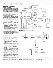
The metal femoral component curves<br />
around the end <strong>of</strong> the thighbone (Fig. 6) <strong>and</strong> has<br />
an <strong>in</strong>terior groove so the knee cap can move up<br />
<strong>and</strong> down smoothly aga<strong>in</strong>st the bone as the knee<br />
bends <strong>and</strong> straightens.<br />
The tibial component is a flat metal platform<br />
with a polymeric cushion (Fig. 6). The<br />
cushion may be part <strong>of</strong> the platform (fixed) or<br />
separate (mobile), with either a flat surface<br />
(PCL-reta<strong>in</strong><strong>in</strong>g) or a raised, slop<strong>in</strong>g surface<br />
(PCL-substitut<strong>in</strong>g).<br />
The patellar component is a dome-shaped<br />
piece <strong>of</strong> polyethylene that duplicates the shape<br />
<strong>of</strong> the kneecap, anchored to a flat metal plate<br />
(Fig. 6).<br />
Materials <strong>of</strong> Construction. The metal parts<br />
<strong>of</strong> the implant are made <strong>of</strong> titanium alloys (Ti-<br />
6Al-4V) or cobalt-chromium alloys. The plastic
© 2003 <strong>ASM</strong> <strong>International</strong>. All Rights Reserved.<br />
www.asm<strong>in</strong>ternational.org<br />
H<strong>and</strong>book <strong>of</strong> Materials for Medical Devices (#06974G)<br />
Chapter 1: <strong>Overview</strong> <strong>of</strong> <strong>Biomaterials</strong> <strong>and</strong> <strong>Their</strong> <strong>Use</strong> <strong>in</strong> Medical Devices / 7<br />
parts are made <strong>of</strong> UHMWPE. All together, the<br />
components weigh between 425 <strong>and</strong> 565 g (15<br />
<strong>and</strong> 20 oz), depend<strong>in</strong>g on the size selected.<br />
Fig. 5<br />
Wear behavior <strong>of</strong> various femoral head/cup comb<strong>in</strong>ations.<br />
Even higher ultrahigh molecular weight polyethylene<br />
(UHMWPE) wear rates are encountered with titaniumbase<br />
femoral heads. Source: Ref 2<br />
Fig. 6 Components <strong>of</strong> a total knee replacement arthroplasty. See text for details.<br />
Cardiac Pacemakers<br />
Function. Cardiac pacemakers are generally<br />
used to manage a slow or irregular heart<br />
rate. The pacemaker system applies precisely<br />
timed electrical signals to <strong>in</strong>duce heart muscle<br />
contraction <strong>and</strong> cause the heart to beat <strong>in</strong> a manner<br />
very similar to a naturally occurr<strong>in</strong>g heart<br />
rhythm. A pacemaker consists <strong>of</strong> a pulse generator,<br />
at least one electrode, <strong>and</strong> one or two pac<strong>in</strong>g<br />
leads connect<strong>in</strong>g the pacemaker to the heart.<br />
Figure 7 shows various types <strong>of</strong> pulse generators<br />
<strong>and</strong> pac<strong>in</strong>g leads.<br />
Components <strong>and</strong> Materials <strong>of</strong> Construction.<br />
The cas<strong>in</strong>g <strong>of</strong> the pulse generator functions<br />
as hous<strong>in</strong>g for the battery <strong>and</strong> circuits,<br />
which provide power. It is usually implanted<br />
between the sk<strong>in</strong> <strong>and</strong> pectoral muscle. The<br />
sealed lithium iod<strong>in</strong>e battery provides electrical<br />
energy to the pacemaker. This battery replaced<br />
the mercury-z<strong>in</strong>c battery <strong>in</strong> 1975, extend<strong>in</strong>g the<br />
life <strong>of</strong> some pacemaker models by over 10 yr.<br />
The circuitry converts the electrical energy to<br />
small electrical signals. The circuitry also con-
© 2003 <strong>ASM</strong> <strong>International</strong>. All Rights Reserved.<br />
H<strong>and</strong>book <strong>of</strong> Materials for Medical Devices (#06974G)<br />
8 / H<strong>and</strong>book <strong>of</strong> Materials for Medical Devices<br />
trols the tim<strong>in</strong>g <strong>of</strong> the electrical signals delivered<br />
to the heart. A connector block, made <strong>of</strong><br />
polyurethane, is located at the top <strong>of</strong> the pacemaker<br />
(Fig. 7). It serves to attach the pacemaker<br />
to the pacemaker lead. Formerly, glass materials<br />
were used to comprise the connector block.<br />
The pulse generator is encased <strong>in</strong> ASTM grade<br />
1 titanium. Titanium replaced ceramics <strong>and</strong><br />
epoxy res<strong>in</strong>, which were used for encapsulation<br />
<strong>of</strong> some pacemakers <strong>in</strong> the past, with silicone<br />
Fig. 7<br />
www.asm<strong>in</strong>ternational.org<br />
rubber. This upgrade to titanium allowed patients<br />
to safely use appliances such as microwave<br />
ovens, because titanium helps to shield<br />
the <strong>in</strong>ternal components <strong>and</strong> reduce the external<br />
electromagnetic <strong>in</strong>terference.<br />
A pac<strong>in</strong>g lead is vital to the pacemaker system,<br />
because it transmits the electrical signal<br />
from the pacemaker to the heart <strong>and</strong> <strong>in</strong>formation<br />
on the heart activity back to the pacemaker. One<br />
or two leads may be used, depend<strong>in</strong>g on the type<br />
Various pacemaker component designs. Top: Three examples <strong>of</strong> titanium-encased pulse generators. Connector blocks,<br />
which serve to attach the pacemaker to the pacemaker lead, are shown at the top <strong>of</strong> each pulse generator. Bottom: Various<br />
types <strong>of</strong> <strong>in</strong>sulated endocardial <strong>and</strong> myocardial leads. Note that the lead shown at the center <strong>of</strong> the figure has a silicone sew<strong>in</strong>g pad <strong>and</strong><br />
Dacram mesh disk for implant fixation. Source: Ref 5
© 2003 <strong>ASM</strong> <strong>International</strong>. All Rights Reserved.<br />
www.asm<strong>in</strong>ternational.org<br />
H<strong>and</strong>book <strong>of</strong> Materials for Medical Devices (#06974G)<br />
Chapter 1: <strong>Overview</strong> <strong>of</strong> <strong>Biomaterials</strong> <strong>and</strong> <strong>Their</strong> <strong>Use</strong> <strong>in</strong> Medical Devices / 9<br />
<strong>of</strong> pacemaker. One end <strong>of</strong> the lead is attached to<br />
the connector block <strong>of</strong> the pacemaker. The other<br />
end is <strong>in</strong>serted through a ve<strong>in</strong> <strong>and</strong> placed <strong>in</strong> the<br />
right ventricle or right atrium <strong>of</strong> the heart. The<br />
lead is an <strong>in</strong>sulated wire consist<strong>in</strong>g <strong>of</strong> a connector<br />
p<strong>in</strong>, lead body, fixation mechanism (Fig. 7),<br />
<strong>and</strong> at least one electrode. The connector p<strong>in</strong> is<br />
the portion <strong>of</strong> the lead that is <strong>in</strong>serted <strong>in</strong>to the<br />
connector block. The lead body is the <strong>in</strong>sulated<br />
metal wire that carries electrical energy from<br />
the pacemaker to the heart.<br />
The lead must be able to withst<strong>and</strong> the flex<strong>in</strong>g<br />
<strong>in</strong>duced by the cardiac contractions <strong>in</strong> the warm<br />
<strong>and</strong> corrosive environment <strong>in</strong> the body. Thus, the<br />
materials used must be <strong>in</strong>ert, nontoxic, <strong>and</strong><br />
durable. The lead body must be flexible, noncorrosive,<br />
<strong>and</strong> durable. It must also be a good electrical<br />
conductor. The early lead body was <strong>in</strong>sulated<br />
with polyethylene. Currently, the lead<br />
body is <strong>in</strong>sulated with a more resilient material<br />
such as silicone rubber tub<strong>in</strong>g or polyurethanes.<br />
Polyurethanes are generally stronger than silicone<br />
rubbers, which are easily damaged. The<br />
strength <strong>of</strong> polyurethanes enables a th<strong>in</strong>ner lead<br />
to be used <strong>in</strong> the pacemaker <strong>and</strong> <strong>of</strong>fers greater<br />
lead flexibility. Another advantage <strong>of</strong> polyurethanes<br />
is their very low coefficient <strong>of</strong> friction<br />
when wet. However, metal-ion-<strong>in</strong>duced oxidation<br />
may degrade polyurethanes, while silicones<br />
are not affected by this mechanism <strong>of</strong> degradation.<br />
The fixation mechanism serves to hold the<br />
tip <strong>of</strong> the lead <strong>in</strong> place <strong>in</strong> the heart. Currently,<br />
either a nickel-cobalt alloy with a silver core<br />
helix or an electrically active plat<strong>in</strong>um-iridium<br />
helix may be used to anchor the electrode <strong>of</strong> the<br />
lead to the surface <strong>of</strong> the heart. The electrode is<br />
located at the tip <strong>of</strong> the lead. It serves to deliver<br />
the electrical energy from the pacemaker to the<br />
heart <strong>and</strong> <strong>in</strong>formation about the natural activity<br />
<strong>of</strong> the heart back to the pacemaker. Electrodes<br />
may be composed <strong>of</strong> plat<strong>in</strong>um, titanium, sta<strong>in</strong>less<br />
steel, silver, or cobalt alloys. Titanium has<br />
been used because it forms a nonconduct<strong>in</strong>g<br />
oxide layer at the surface. This surface prevents<br />
the exchange <strong>of</strong> charge carriers across the<br />
boundary. Titanium also exhibits a high modulus<br />
<strong>of</strong> elasticity, high resistance to corrosion, <strong>and</strong><br />
high durability. Electrodes may be coated with<br />
iridium oxide to prevent nonconductive layers<br />
from form<strong>in</strong>g. The coated electrodes may also<br />
provide lower acute <strong>and</strong> chronic thresholds due<br />
to the reduced local <strong>in</strong>flammation.<br />
Drug-Elut<strong>in</strong>g Leads. Leads have developed<br />
immensely s<strong>in</strong>ce they were first <strong>in</strong>troduced. The<br />
earliest leads were attached to the outer surface<br />
<strong>of</strong> the heart. In the mid-1960s, transverse leads<br />
were <strong>in</strong>troduced. They could be <strong>in</strong>serted through<br />
a ve<strong>in</strong> lead<strong>in</strong>g to the heart, thus elim<strong>in</strong>at<strong>in</strong>g the<br />
need to open the chest cavity dur<strong>in</strong>g implantation.<br />
In the 1970s, t<strong>in</strong>ed <strong>and</strong> active fixation leads<br />
were developed to replace smooth tip leads. The<br />
prongs on the t<strong>in</strong>ed leads <strong>and</strong> the titanium alloy<br />
screws <strong>in</strong> the active fixation leads provide a more<br />
secure attachment to the heart <strong>and</strong> are still used<br />
today. In the early 1980s, steroid-elut<strong>in</strong>g leads<br />
were developed. These leads emit a steroid drug<br />
from the tip <strong>of</strong> the electrode on the lead to suppress<br />
<strong>in</strong>flammatory response <strong>of</strong> the heart wall,<br />
thus reduc<strong>in</strong>g the energy requirements <strong>of</strong> the<br />
pacemaker. The steroid also results <strong>in</strong> low<br />
chronic thresholds. Ceramic collars surround<strong>in</strong>g<br />
the electrode tip were first used to conta<strong>in</strong> <strong>and</strong><br />
emit the steroid. This technique is still used,<br />
where dexamethasone sodium phosphate is the<br />
eluted steroid. A silicone rubber matrix conta<strong>in</strong>s<br />
the steroid, <strong>and</strong> this matrix is conta<strong>in</strong>ed <strong>in</strong> a plat<strong>in</strong>um-iridium<br />
porous tip electrode. The comb<strong>in</strong>ation<br />
<strong>of</strong> plat<strong>in</strong>um <strong>and</strong> iridium results <strong>in</strong> a material<br />
stronger than most steels. The porous tip<br />
electrode provides an efficient pac<strong>in</strong>g <strong>and</strong> sens<strong>in</strong>g<br />
surface by promot<strong>in</strong>g fibrotic tissue growth<br />
<strong>and</strong> physically stabiliz<strong>in</strong>g the tissue <strong>in</strong>terface. In<br />
order to facilitate passage <strong>of</strong> the fixation mechanism<br />
to the heart, either a soluble polyethylene<br />
glycol capsule or a mannitol capsule is placed on<br />
the electrode tip. When the electrode tip is<br />
exposed to body fluids, the steroid is released.<br />
The polyethylene glycol capsule dissolves<br />
with<strong>in</strong> 2 to 4 m<strong>in</strong> after the electrode tip is <strong>in</strong>serted<br />
<strong>in</strong>to the ve<strong>in</strong>. The mannitol capsule dissolves<br />
with<strong>in</strong> 3 to 5 m<strong>in</strong> after the <strong>in</strong>sertion.<br />
ACKNOWLEDGMENTS<br />
The application examples describ<strong>in</strong>g knee<br />
implants <strong>and</strong> cardiac pacemakers were adapted<br />
from the follow<strong>in</strong>g web sites:<br />
• American Academy <strong>of</strong> Orthopaedic Surgeons,<br />
www.ortho<strong>in</strong>fo.aaos.org<br />
• T. Reilly, “Structure <strong>and</strong> Materials <strong>of</strong> Cardiac<br />
Pacemakers,” University <strong>of</strong> Wiscons<strong>in</strong>-<br />
Madison, www.pharmacy.wisc.edu/courses/<br />
718-430/2000presentation/Reilly.pdf<br />
REFERENCES<br />
1. C.M. Agrawal, Reconstruct<strong>in</strong>g the Human<br />
Body Us<strong>in</strong>g <strong>Biomaterials</strong>, JOM, Jan<br />
1998, p 31–35<br />
2. M. Long <strong>and</strong> H.J. Rack, Titanium Alloys
© 2003 <strong>ASM</strong> <strong>International</strong>. All Rights Reserved.<br />
H<strong>and</strong>book <strong>of</strong> Materials for Medical Devices (#06974G)<br />
10 / H<strong>and</strong>book <strong>of</strong> Materials for Medical Devices<br />
<strong>in</strong> Total Jo<strong>in</strong>t Replacement—A Materials<br />
Science Perspective, <strong>Biomaterials</strong>, Vol<br />
19, 1998, p 1621–1639<br />
3. D. Williams, An Introduction to Medical<br />
<strong>and</strong> Dental Materials, Concise Encyclopedia<br />
<strong>of</strong> Medical & Dental Materials, D.<br />
Williams, Ed., Pergamon Press <strong>and</strong> The<br />
MIT Press, 1990, p xvii–xx<br />
4. D. Dowson, Friction <strong>and</strong> Wear <strong>of</strong> Medical<br />
Implants <strong>and</strong> Prosthetic Devices, Friction,<br />
Lubrication, <strong>and</strong> Wear Technology,<br />
Vol 18, <strong>ASM</strong> H<strong>and</strong>book, <strong>ASM</strong> <strong>International</strong>,<br />
1992, p 656–664<br />
5. P. Didisheim <strong>and</strong> J.T. Watson, Cardiovascular<br />
Applications, <strong>Biomaterials</strong> Science:<br />
An Introduction to Materials <strong>in</strong> Medic<strong>in</strong>e,<br />
B.D. Ratner, A.S. H<strong>of</strong>fman, F.J. Schoen,<br />
<strong>and</strong> J.E. Lemons, Ed., Academic Press,<br />
1996, p 283–297<br />
SELECTED REFERENCES<br />
General<br />
• C.M. Agrawal, Reconstruct<strong>in</strong>g the Human<br />
Body Us<strong>in</strong>g <strong>Biomaterials</strong>. J. Met., Vol 50<br />
(No. 1). 1998, p 31–35<br />
• S.A. Barenberg, Abridged Report <strong>of</strong> the<br />
Committee to Survey the Needs <strong>and</strong> Opportunities<br />
for the <strong>Biomaterials</strong> Industry, J. Biomed.<br />
Mater. Res., Vol 22, 1988, p 1267–1291<br />
• J.S. Benson <strong>and</strong> J.W. Boretos, <strong>Biomaterials</strong><br />
<strong>and</strong> the Future <strong>of</strong> Medical Devices. Med. Device<br />
Diag. Ind., Vol 17 (No. 4). 1995, p 32–37<br />
• <strong>Biomaterials</strong> Science: An Introduction to<br />
Materials <strong>in</strong> Medic<strong>in</strong>e, B.D. Ratner, A.S.<br />
H<strong>of</strong>fman, F.J. Schoen, <strong>and</strong> J.E. Lemons, Ed.,<br />
Academic Press, 1996<br />
• M.M. Black et al., Medical Applications <strong>of</strong><br />
<strong>Biomaterials</strong>, Phys. Technol., Vol 13, 1982,<br />
p 50–65<br />
• Concise Encyclopedia <strong>of</strong> Medical Devices &<br />
Dental Materials, D. Williams, Ed., Pergamon<br />
Press <strong>and</strong> The MIT Press, 1990<br />
• Directory to Medical Materials, Med. Device<br />
Diag. Ind., published annually <strong>in</strong> the April<br />
issue<br />
• M. Donachie, <strong>Biomaterials</strong>, Metals H<strong>and</strong>book<br />
Desk Edition, 2nd ed., J.R. Davis, Ed.,<br />
<strong>ASM</strong> <strong>International</strong>, 1998, p 702–709<br />
• E. Duncan, <strong>Biomaterials</strong>: Look<strong>in</strong>g for Information,<br />
Med. Device Diag. Ind., Vol 13 (No.<br />
1), 1991, p 140–143<br />
• P.M. Galletti. Artificial Organs: Learn<strong>in</strong>g to<br />
Live with Risk, Tech. Rev., Nov/Dec 1988,<br />
p 34–40<br />
www.asm<strong>in</strong>ternational.org<br />
• P.M. Galletti, Organ Replacement by Man-<br />
Made Devices. J. Cardiothoracic Vascular<br />
Anesthesia, Vol 7 (No. 5), Oct 1993, p 624–<br />
628<br />
• J.S. Hanker <strong>and</strong> B.L. Giammara, <strong>Biomaterials</strong><br />
<strong>and</strong> Biomedical Devices, Science, Vol<br />
242, 11 Nov 1988, p 885–892<br />
• R.D. Lambert <strong>and</strong> M.E. Anthony, St<strong>and</strong>ardization<br />
<strong>in</strong> Orthopaedics, ASTM St<strong>and</strong>. News,<br />
Aug 1995, p 22–29<br />
• Medical Devices <strong>and</strong> Services, Section 13,<br />
Annual Book <strong>of</strong> ASTM St<strong>and</strong>ards, ASTM<br />
• J.B. Park <strong>and</strong> R.S. Lakes, <strong>Biomaterials</strong>: An<br />
Introduction, 2nd ed., Plenum Press, 1992<br />
<strong>Biomaterials</strong> for Nondental<br />
or General Application<br />
• J.M. Courtney <strong>and</strong> T. Gilchrist, Silicone Rubber<br />
<strong>and</strong> Natural Rubber as <strong>Biomaterials</strong>,<br />
Med. Biol. Eng. Comput., Vol 18, 1980, p<br />
538–540<br />
• R.H. Doremus, Bioceramics. J. Mater. Sci.,<br />
Vol 27, 1992, p 285–297<br />
• A.C. Fraker <strong>and</strong> A.W. Ruff, Metallic Surgical<br />
Implants: State <strong>of</strong> the Art. J. Met., Vol 29<br />
(No. 5), 1977, p 22–28<br />
• R.A. Fuller <strong>and</strong> J.J. Rosen, Materials for<br />
Medic<strong>in</strong>e, Sci. Am., Vol 255 (No. 4), Oct<br />
1986, p 119–125<br />
• B.S. Gupta, Medical Testile Structures: An<br />
<strong>Overview</strong>, Med. Plast. Biomater., Vol 5 (No.<br />
1), 1998, p 16, 19–21, 24, 26, 28, 30<br />
• G.H. Harth, “Metal Implants for Orthopedic<br />
<strong>and</strong> Dental Surgery,” MCIC-74-18, Metals<br />
<strong>and</strong> Ceramics Information Center Report.<br />
Feb 1974<br />
• G. Heimke <strong>and</strong> P. Griss, Ceramic Implant<br />
Materials, Med. Biol. Eng. Comput., Vol 18,<br />
1980, p 503–510<br />
• D.S. Hotter, B<strong>and</strong>-Aids for Broken Bones,<br />
Mach. Des., 4 April 1996, p 39–44<br />
• M. Hunt, Get Hip with Medical Metals,<br />
Mater. Eng., Vol 108 (No. 4), 1991, p 27–30<br />
• A.J. Kle<strong>in</strong>, <strong>Biomaterials</strong> Give New Life, Adv.<br />
Mater. Process., May 1986, p 18–22<br />
• F.G. Larson, Hydroxyapatite Coat<strong>in</strong>gs for<br />
Medical Implants. Med. Device Diag. Ind.,<br />
Vol 16 (No. 4). 1994, p 34–40<br />
• M. Long <strong>and</strong> H.J. Rack, Titanium Alloys <strong>in</strong><br />
Total Jo<strong>in</strong>t Replacement—A Materials Science<br />
Perspective, <strong>Biomaterials</strong>, Vol 19,<br />
1998, p 1621–1639<br />
• D.C. Mears, Metals <strong>in</strong> Medic<strong>in</strong>e <strong>and</strong> Surgery,<br />
Int. Met. Rev., Vol 22, June 1977, p 119–155
© 2003 <strong>ASM</strong> <strong>International</strong>. All Rights Reserved.<br />
www.asm<strong>in</strong>ternational.org<br />
H<strong>and</strong>book <strong>of</strong> Materials for Medical Devices (#06974G)<br />
Chapter 1: <strong>Overview</strong> <strong>of</strong> <strong>Biomaterials</strong> <strong>and</strong> <strong>Their</strong> <strong>Use</strong> <strong>in</strong> Medical Devices / 11<br />
• D.S. Metsger <strong>and</strong> S.F. Lebowitz, Medical<br />
Applications <strong>of</strong> Ceramics. Med. Device Diag.<br />
Ind., Vol 7, 1985, p 55–63<br />
• M. Moukwa, The Development <strong>of</strong> Polymer-<br />
Based <strong>Biomaterials</strong> S<strong>in</strong>ce the 1920s, J. Met.,<br />
Vol 49 (No. 2) 1997, p 46–50<br />
• S.J. Mraz, The Human Body Shop, Mach.<br />
Des., 7 Nov 1991, p 90–94<br />
• K. Neailey <strong>and</strong> R.C. Pond, Metal Implants,<br />
Mater. Eng., Vol 3, June 1982, p 470–478<br />
• D.E. Niesz <strong>and</strong> V.J. Tennery, “Ceramics for<br />
Prosthetic Applications—Orthopedic, Dental<br />
<strong>and</strong> Cardiovascular.” MCIC-74-21, Metals<br />
<strong>and</strong> Ceramics Information Center Report,<br />
July 1974<br />
• P.C. Noble, Special Materials for the<br />
Replacement <strong>of</strong> Human Jo<strong>in</strong>ts, Met. Forum,<br />
Vol 6 (No. 2), 1983, p 59–80<br />
• C.M. Rimnac et al., Failure <strong>of</strong> Orthopedic<br />
Implants: Three Case Histories, Mater.<br />
Char., Vol 26, 1991, p 201–209<br />
• W. Rostoker <strong>and</strong> J.O. Galante, Materials for<br />
Human Implantation, Trans. <strong>ASM</strong>E, Vol 101,<br />
Feb 1979, p 2–14<br />
• L.M. Sheppard, Build<strong>in</strong>g Teeth, Bones, <strong>and</strong><br />
Other Body Parts with Ceramics, Mater.<br />
Eng., April 1984, p 37–43<br />
• L.M. Sheppard, Cure It with Ceramics. Adv.<br />
Mater. Process., May 1986, p 26–31<br />
• H. Shimizu, Metal/Ceramic Implants, Med.<br />
Device Diag. Ind., Vol 8 (No. 7), 1986,<br />
p 30–35, 59–60<br />
• E. Smethurst <strong>and</strong> R.B. Waterhouse. Causes<br />
<strong>of</strong> Failure <strong>in</strong> Total Hip Prostheses, J. Mater.<br />
Sci., Vol 12, 1977, p 1781–1792<br />
• T. Stevens, Prescription: Plastics. Mater.<br />
Eng., Vol 108 (No. 4). 1991, p 23–26<br />
Dental Materials<br />
• S. B<strong>and</strong>yopadhyay, Dental Cements. Met.<br />
Forum, Vol 3 (No. 4), 1980, p 228–235<br />
• J.F. Bates <strong>and</strong> A.G. Knapton, Metals <strong>and</strong><br />
Alloys <strong>in</strong> Dentistry. Int. Met. Rev., Vol 22,<br />
March 1977, p 39–60<br />
• M.P. Dariel et al., New Technology for<br />
Mercury Free Metallic Dental Restorative<br />
Alloys, Powder Metall., Vol 37 (No. 2).<br />
1994, p 88<br />
• S. Espevik, Dental Amalgam, Ann Rev.<br />
Mater. Sci. Vol 7, 1977, p 55–72<br />
• R.M. German, Precious-Metal Dental Cast<strong>in</strong>g<br />
Alloys. Int. Met. Rev., Vol 27 (No. 5),<br />
1982, p 260–288<br />
• J.B. Moser, The <strong>Use</strong>s <strong>and</strong> Properties <strong>of</strong> Dental<br />
Materials, Int. Adv. Nondestruct. Test.,<br />
Vol 5, 1977, p 367–390<br />
• J.M. Powers <strong>and</strong> S.C. Bayne, Friction <strong>and</strong><br />
Wear <strong>of</strong> Dental Materials, Friction, Lubrication,<br />
<strong>and</strong> Wear Technology, Vol 18, <strong>ASM</strong><br />
H<strong>and</strong>book, <strong>ASM</strong> <strong>International</strong>, 1992, p<br />
665–681<br />
• Restorative Dental Materials, 11th ed., R.G.<br />
Craig <strong>and</strong> J.M. Powers, Ed., Mosby, Inc., An<br />
Affiliate <strong>of</strong> Elsevier Science, 2002<br />
• D.E. Southan, Dental Porcela<strong>in</strong>, Met. Forum,<br />
Vol 3 (No. 4), 1980, p 222–227<br />
• R.M. Waterstrat, Brush<strong>in</strong>g up on the History<br />
<strong>of</strong> Intermetallics <strong>in</strong> Dentistry, J. Met., Vol 42<br />
(No. 3), 1990, p 8–14<br />
Corrosion <strong>and</strong> Biocompatibility<br />
• J. Black, Biological Performance <strong>of</strong> Materials:<br />
Fundamentals <strong>of</strong> Biocompatibility, Marcel<br />
Dekker, 1981<br />
• A.C. Fraker, Corrosion <strong>of</strong> Metallic Implant<br />
<strong>and</strong> Prosthetic Devices. Corrosion, Vol 13,<br />
<strong>ASM</strong> H<strong>and</strong>book, 1987, p 1324–1335<br />
• K. Hayashi, Biodegradation <strong>of</strong> Implant Materials,<br />
JSME Int. J., Vol 30 (No. 268), 1987,<br />
p 1517–1525<br />
• H.J. Mueller, Tarnish <strong>and</strong> Corrosion <strong>of</strong> Dental<br />
Alloys. Corrosion, Vol 13, <strong>ASM</strong> H<strong>and</strong>book,<br />
1987, p 1336–1366<br />
• K.R. St. John, Biocompatibility Test<strong>in</strong>g for<br />
Medical Implant Materials, ASTM St<strong>and</strong>.<br />
News, March 1994, p 46–49<br />
• D.F. Williams, Corrosion <strong>of</strong> Implant Materials,<br />
Ann Rev. Mater. Sci., Vol 6, 1976, p 237–<br />
266<br />
• D.F. Williams, Tissue-Biomaterial Interactions,<br />
J. Mater. Sci., Vol 22, 1987, p 3421–<br />
3445
<strong>ASM</strong> <strong>International</strong> is the society for materials<br />
eng<strong>in</strong>eers <strong>and</strong> scientists, a worldwide network<br />
dedicated to advanc<strong>in</strong>g <strong>in</strong>dustry, technology, <strong>and</strong><br />
applications <strong>of</strong> metals <strong>and</strong> materials.<br />
<strong>ASM</strong> <strong>International</strong>, Materials Park, Ohio, USA<br />
www.asm<strong>in</strong>ternational.org<br />
This publication is copyright © <strong>ASM</strong> <strong>International</strong> ® . All rights reserved.<br />
Publication title Product code<br />
H<strong>and</strong>book <strong>of</strong> Materials for Medical Devices #06974G<br />
To order products from <strong>ASM</strong> <strong>International</strong>:<br />
Onl<strong>in</strong>e Visit www.asm<strong>in</strong>ternational.org/bookstore<br />
Telephone 1-800-336-5152 (US) or 1-440-338-5151 (Outside US)<br />
Fax 1-440-338-4634<br />
Mail<br />
Customer Service, <strong>ASM</strong> <strong>International</strong><br />
9639 K<strong>in</strong>sman Rd, Materials Park, Ohio 44073-0002, USA<br />
Email CustomerService@asm<strong>in</strong>ternational.org<br />
In Europe<br />
In Japan<br />
American Technical Publishers Ltd.<br />
27-29 Knowl Piece, Wilbury Way, Hitch<strong>in</strong> Hertfordshire SG4 0SX,<br />
United K<strong>in</strong>gdom<br />
Telephone: 01462 437933 (account holders), 01462 431525 (credit card)<br />
www.ameritech.co.uk<br />
Neutr<strong>in</strong>o Inc.<br />
Takahashi Bldg., 44-3 Fuda 1-chome, Ch<strong>of</strong>u-Shi, Tokyo 182 Japan<br />
Telephone: 81 (0) 424 84 5550<br />
Terms <strong>of</strong> <strong>Use</strong>. This publication is be<strong>in</strong>g made available <strong>in</strong> PDF format as a benefit to members <strong>and</strong><br />
customers <strong>of</strong> <strong>ASM</strong> <strong>International</strong>. You may download <strong>and</strong> pr<strong>in</strong>t a copy <strong>of</strong> this publication for your<br />
personal use only. Other use <strong>and</strong> distribution is prohibited without the express written permission <strong>of</strong><br />
<strong>ASM</strong> <strong>International</strong>.<br />
No warranties, express or implied, <strong>in</strong>clud<strong>in</strong>g, without limitation, warranties <strong>of</strong> merchantability or<br />
fitness for a particular purpose, are given <strong>in</strong> connection with this publication. Although this<br />
<strong>in</strong>formation is believed to be accurate by <strong>ASM</strong>, <strong>ASM</strong> cannot guarantee that favorable results will be<br />
obta<strong>in</strong>ed from the use <strong>of</strong> this publication alone. This publication is <strong>in</strong>tended for use by persons hav<strong>in</strong>g<br />
technical skill, at their sole discretion <strong>and</strong> risk. S<strong>in</strong>ce the conditions <strong>of</strong> product or material use are<br />
outside <strong>of</strong> <strong>ASM</strong>'s control, <strong>ASM</strong> assumes no liability or obligation <strong>in</strong> connection with any use <strong>of</strong> this<br />
<strong>in</strong>formation. As with any material, evaluation <strong>of</strong> the material under end-use conditions prior to<br />
specification is essential. Therefore, specific test<strong>in</strong>g under actual conditions is recommended.<br />
Noth<strong>in</strong>g conta<strong>in</strong>ed <strong>in</strong> this publication shall be construed as a grant <strong>of</strong> any right <strong>of</strong> manufacture, sale,<br />
use, or reproduction, <strong>in</strong> connection with any method, process, apparatus, product, composition, or<br />
system, whether or not covered by letters patent, copyright, or trademark, <strong>and</strong> noth<strong>in</strong>g conta<strong>in</strong>ed <strong>in</strong> this<br />
publication shall be construed as a defense aga<strong>in</strong>st any alleged <strong>in</strong>fr<strong>in</strong>gement <strong>of</strong> letters patent,<br />
copyright, or trademark, or as a defense aga<strong>in</strong>st liability for such <strong>in</strong>fr<strong>in</strong>gement.