- Page 4:
ContentsEditorial GroupAcknowledgem
- Page 8 and 9:
AppendicesAppendix 1 - Nicotine Rep
- Page 10 and 11:
AcknowledgementsA huge number of pe
- Page 12 and 13:
Summary of Major changes in Therape
- Page 14 and 15:
On discharge• As the discharge pr
- Page 16:
Other examples• Sevredol 10 mg ta
- Page 19 and 20:
Antiplatelets and anticoagulants:In
- Page 21 and 22:
non-Formulary medicines, a complete
- Page 23 and 24:
Quick guide to using the handbookWh
- Page 25 and 26:
Sections1. Resuscitation and Anaphy
- Page 27 and 28:
Section 1Resuscitation and Anaphyla
- Page 29 and 30:
Continued from previous page2. Adre
- Page 31 and 32:
Peri-arrest ArrhythmiasGeneral advi
- Page 33 and 34:
Broad QRS - continuedNarrow QRS - c
- Page 35 and 36:
Guidelines on Blood TransfusionThis
- Page 37 and 38:
Management of Major Haemorrhage(See
- Page 39 and 40:
Hospital Specific Information on Ma
- Page 41 and 42:
Table continued from previous pageG
- Page 43 and 44:
Assessment / MonitoringAKI stage I
- Page 45 and 46:
Section 2Drug Overdose and Toxicity
- Page 47 and 48:
Treatment of Paracetamol OverdoseTh
- Page 49:
Management of paracetamol overdose
- Page 52 and 53:
Lithium toxicityNormal range 0.4 -
- Page 54 and 55:
Management of DyspepsiaGastrointest
- Page 57 and 58:
Table continued from previous pageR
- Page 59 and 60:
Management of Gastroduodenal Ulcers
- Page 61 and 62:
Management of Upper Gastrointestina
- Page 63 and 64:
Post endoscopy continuedb. Arrange
- Page 65 and 66:
General management and treatment op
- Page 67 and 68:
- SAAG can differentiate ascites re
- Page 69 and 70:
General management and drug therapy
- Page 71 and 72:
Drug therapy• Unless contraindica
- Page 73 and 74:
Consider:• Serum caeruloplasmin,
- Page 75 and 76:
Section 4Cardiovascular SystemCardi
- Page 77 and 78:
Table 1 - Indicators of patients at
- Page 79 and 80:
Continued from previous pageIf the
- Page 81 and 82:
Diagnosis and Treatment of VenousTh
- Page 83 and 84:
Wells Clinical score should be util
- Page 85 and 86:
IV Drug Misusers• Given their cha
- Page 87 and 88:
Drug therapy / treatment options co
- Page 89 and 90:
RivaroxabanRivaroxaban is an oral d
- Page 91 and 92:
Diagnosis of VTE in pregnancy• If
- Page 93 and 94:
Referral of Patients to Anticoagula
- Page 95 and 96:
Reversal of Anticoagulant TherapyIn
- Page 97 and 98:
General management and drug therapy
- Page 99 and 100:
Elective admissions - Risk Stratifi
- Page 101:
Elective admissions - restarting an
- Page 104 and 105: Suspected Acute Coronary Syndrome (
- Page 106 and 107: If patient is for PPCI go to Box 1.
- Page 108 and 109: Drugs for acute coronary syndrome /
- Page 110 and 111: Secondary Prevention of Coronary He
- Page 112 and 113: Cardiovascular SystemTroponin posit
- Page 114 and 115: Atherosclerotic arterial disease is
- Page 116 and 117: Algorithm for Cardioversion of AFSt
- Page 118 and 119: Cardiovascular SystemVentricular ra
- Page 120 and 121: Prevention of stroke / thromboembol
- Page 122 and 123: Drugs for atrial fibrillation conti
- Page 124 and 125: CPAP in cardiogenic pulmonary oedem
- Page 126 and 127: Management of HypertensionCardiovas
- Page 128 and 129: General management and drug therapy
- Page 130 and 131: Cardiovascular SystemGeneral manage
- Page 132 and 133: Management of Acute Stroke 3Transie
- Page 134 and 135: Secondary prevention of stroke and
- Page 136 and 137: Cardiovascular SystemPage 126
- Page 138 and 139: Guidelines for Blood Gas AnalysisRe
- Page 140 and 141: Respiratory SystemNext consider aci
- Page 142 and 143: Management of Acute Severe Asthmain
- Page 144 and 145: Management of Acute Severe Asthma i
- Page 146 and 147: Management of Chronic ObstructivePu
- Page 148 and 149: Acute Exacerbation of Chronic Obstr
- Page 150 and 151: Treatment options continued• If p
- Page 153: Step 4 - Initiation of NIVAIM = To
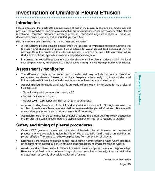
- Page 157 and 158: Investigation of unilateral pleural
- Page 159 and 160: General management• Oxygen as app
- Page 161 and 162: Management of PneumothoraxIntroduct
- Page 163 and 164: Management of StridorIntroductionSt
- Page 165 and 166: Initial Management of Superior Vena
- Page 167 and 168: Section 6Central Nervous SystemCent
- Page 169 and 170: Analgesics for acute painSTEP 3STEP
- Page 172 and 173: Analgesics for acute pain - prescri
- Page 174 and 175: Management of Postoperative Nausea
- Page 177 and 178: Management of Acutely Disturbed Pat
- Page 179 and 180: Pharmacological Management - Patien
- Page 181 and 182: Guidance on Night SedationPatients
- Page 183 and 184: Management of Alcohol Withdrawal Sy
- Page 185 and 186: Continued from previous pageUnable
- Page 187 and 188: Continued from previous pageImporta
- Page 189 and 190: Flow diagram for use with Hospital
- Page 191 and 192: Continued from previous pageMethado
- Page 193 and 194: Management of Suspected Subarachnoi
- Page 195 and 196: Sampling requirements for the Bioch
- Page 197 and 198: Referral to Neurosurgical Unit cont
- Page 199 and 200: Management of Status EpilepticusInt
- Page 201 and 202: OtherInitiating Long-term Anti-epil
- Page 203 and 204: Continued from previous page2. 'Top
- Page 205 and 206:
Decision making algorithm for the a
- Page 207 and 208:
Drug therapy / treatment optionsNil
- Page 209 and 210:
Section 7InfectionsInfectionsPage 1
- Page 211 and 212:
• In patients with sepsis -- Star
- Page 213 and 214:
Gentamicin and Vancomycin: Reducing
- Page 215 and 216:
IV-Oral Antibiotic Switch Therapy (
- Page 217 and 218:
Alert Antibiotic Policy(The full ve
- Page 219 and 220:
Table 1 - Alert Antibiotics continu
- Page 221 and 222:
Infection Management GuidelinesSeve
- Page 223 and 224:
Sepsis Source Unknown (not immunoco
- Page 225 and 226:
Table continued form previous pageI
- Page 227 and 228:
Table continued from previous pageE
- Page 229 and 230:
UTI in non-pregnant women Antibioti
- Page 231 and 232:
UTI in pregnant women Antibiotic Th
- Page 233 and 234:
Table continued from previous page
- Page 235 and 236:
Catheter-related UTI Antibiotic The
- Page 237 and 238:
Table continued from previous pageU
- Page 239 and 240:
Table continued from previous pageU
- Page 241 and 242:
Pneumonia(Also see page 148 for man
- Page 243 and 244:
Table continued from previous pageC
- Page 245:
Pneumonia (continued from previous
- Page 248 and 249:
InfectionsTable continued from prev
- Page 250 and 251:
Treatment of Clostridium difficile
- Page 252 and 253:
InfectionsSkin and soft tissue infe
- Page 254 and 255:
InfectionsSkin and soft tissue infe
- Page 256 and 257:
Central nervous system infectionsBa
- Page 258 and 259:
Meningitis Contacts• All suspecte
- Page 260 and 261:
Viral infectionsViral Encephalitis
- Page 262 and 263:
Guideline updated 11/10/13Genito-ur
- Page 264 and 265:
Genito-urinary infections (continue
- Page 266 and 267:
Patients with absent or non-functio
- Page 268 and 269:
MRSA Eradication Policy• In newly
- Page 270 and 271:
InfectionsDosage regimens for antib
- Page 272 and 273:
Gentamicin dosing guidelines (For p
- Page 274 and 275:
Step 2 continued from previous page
- Page 276 and 277:
Vancomycin dosing guidelines (For p
- Page 278 and 279:
Continued from previous page - If t
- Page 280 and 281:
B. Vancomycin continuous infusionSt
- Page 282 and 283:
Vancomycin - continuous infusion re
- Page 284 and 285:
InfectionsPage 274
- Page 286 and 287:
Management of Diabetic Ketoacidosis
- Page 288 and 289:
Endocrine System5. Signs of cerebra
- Page 290 and 291:
Assessment / monitoring• Glucose
- Page 292 and 293:
Important points1. This regimen is
- Page 294 and 295:
Diabetes therapy continued• Retro
- Page 296 and 297:
Insulin Sliding Scale (Not for use
- Page 298 and 299:
Management of HypoglycaemiaIntroduc
- Page 300 and 301:
Endocrine SystemContinued from prev
- Page 302 and 303:
Oral antidiabetic drugs continued3.
- Page 304 and 305:
Other InformationGeneral advice on
- Page 306:
Guideline updated 18/09/13Managemen
- Page 309 and 310:
Drug therapy / treatment options co
- Page 311 and 312:
Management of hypomagnesaemia in ad
- Page 313 and 314:
Drug therapy / treatment options co
- Page 315 and 316:
Drug therapy / treatment options co
- Page 317 and 318:
Management of Hyponatraemia• Earl
- Page 319 and 320:
Section 10Musculoskeletal and Joint
- Page 321 and 322:
Treatment optionsManagement of acut
- Page 323 and 324:
Treatment options continuedNSAIDs a
- Page 325 and 326:
Section 11Palliative CarePalliative
- Page 327 and 328:
Analgesic Step LadderStep 1: Mild P
- Page 329 and 330:
Opioid Conversion ChartThis is advi
- Page 331 and 332:
Palliative Care - SymptomsSee BNF,
- Page 333 and 334:
Table continued from previous pageP
- Page 335 and 336:
Management of symptoms present in l
- Page 337 and 338:
Section 12Oncological EmergenciesOn
- Page 339 and 340:
Malignant Spinal Cord Compression (
- Page 341 and 342:
Raised intracranial pressure in can
- Page 343 and 344:
Treatment /drug therapy continued5.
- Page 345 and 346:
N.B.• Laboratory TLS = > 2 labora
- Page 347 and 348:
• Avoid serum CA125 testing as it
- Page 349 and 350:
AppendicesAppendicesPage 339
- Page 351 and 352:
Appendix 2Preparation of Intravenou
- Page 353 and 354:
Glyceryl Trinitrate (GTN) infusionG
- Page 355 and 356:
Table 2 - Other drugsDrug Time to s
- Page 357 and 358:
Appendix 5Normal ImmunoglobulinsNor
- Page 359 and 360:
Drug MisuseHospital Liaison Addicti
- Page 361 and 362:
Outpatient Parenteral Antibiotic Th
- Page 363 and 364:
IndexPage 353
- Page 365 and 366:
AnticoagulantsIn treatment of VTE/P
- Page 367 and 368:
CiprofloxacinAlert Antibiotic Polic
- Page 369 and 370:
EEmergency sedationGeneral principl
- Page 371 and 372:
Hyoscine butylbromideIn palliative
- Page 373 and 374:
Mesalazine, oralIn inflammatory bow
- Page 375 and 376:
PhenytoinDosing and monitoring guid
- Page 377 and 378:
TeicoplaninDosing and monitoring...
- Page 379 and 380:
Notes
- Page 381 and 382:
Notes
- Page 384:
© NHS Greater Glasgow and ClydeAug


