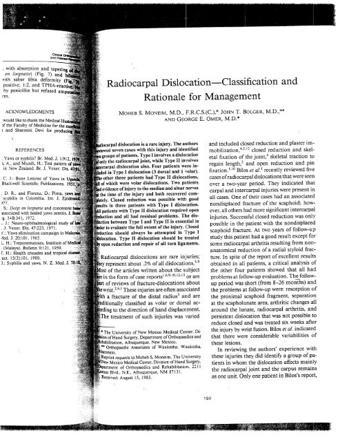
Radiocarpal Dislocation Classification Rationale for Management and
Radiocarpal Dislocation Classification Rationale for Management and
Radiocarpal Dislocation Classification Rationale for Management and

Create successful ePaper yourself
Turn your PDF publications into a flip-book with our unique Google optimized e-Paper software.
, with absorption <strong>and</strong> ~~:~<br />
en lorgnette) (Fig. 7) ~~~-.<br />
vith saber tibia defo~~<br />
i positive, 1:2, <strong>and</strong> TPHA~~:<br />
bv, penicillin but refused amp~<br />
i’m.<br />
ACKNOWLEDGMENTS<br />
would like to thank the Medical<br />
)f the Faculty of Medicine <strong>for</strong> the<br />
; <strong>and</strong> Sharmini Devi <strong>for</strong><br />
<strong>Radiocarpal</strong> <strong>Dislocation</strong> <strong>Classification</strong><br />
<strong>Rationale</strong> <strong>for</strong> <strong>Management</strong><br />
MOHEB S. MONEIM, M.D., F.R.C.S.(C.),* JOHN T. BOLGER, M.D.,**<br />
AND GEORGE E. OMER, M.D.*<br />
REFERENCES<br />
dislocation is a rare injury. The authors<br />
seven cases with this injury <strong>and</strong> identified<br />
, Yaws or syphilis? Br. Med. J. 1:912<br />
a, A., <strong>and</strong> Mundt, H.: Test pattern of<br />
in New Zeal<strong>and</strong>. Br. J. Vener. Dis.<br />
roups of patients. Type I involves a dislocation<br />
’only the radiocarpal joint, while Type II involves<br />
~al dislocation also. Four patients were inin<br />
Type I dislocation (3 dorsal <strong>and</strong> 1 volar).<br />
C. J.: Bone Lesions of Yaws in<br />
Blackwell Scientific Publications, 195<br />
other three patients had Type II disloc-qtions,<br />
of which were volar dislocations. Two patients<br />
[evidence of injury to the median <strong>and</strong> ulnar nerves<br />
, D. R.. <strong>and</strong> Florenz, D.: Pinta,<br />
:the time of the injury <strong>and</strong> both recoveredl corn-<br />
.syphilis in Colombia. Int. J.<br />
)77.<br />
S.: Doigt en lorgnette <strong>and</strong> concentric I<br />
~ssociated with healed yaws osteitis. J.<br />
-g. 54B:341, 1972.<br />
,. J.: Neuro-ophthalmological<br />
¯ J. Vener. Dis. 47:223, 1971.<br />
K: Yaws elimination campaign in<br />
4ed. J. 20:10t, 1965.<br />
Closed reduction was possible with good<br />
ults in three patients with Type I dislocation.<br />
patients with Type II dislocation required open<br />
<strong>and</strong> all had residual problems. The disbetween<br />
Type I <strong>and</strong> Type II is essential in<br />
r to evaluate the full extent of the injury. Closed<br />
should always be attempted in Type I<br />
Type II dislocation should be treated<br />
L. H.: Treponematoses. Institute<br />
. (Malaya). Bulletin 9:t21, 1959.<br />
’open reduction <strong>and</strong> repair of all torn liga:ments.<br />
[’. H.: Health crusades <strong>and</strong> tropical<br />
"act. 15(3):101. 1980.<br />
J.: Syphilis <strong>and</strong> yaws. N. Z. Med. J. 75:i<br />
)<strong>Radiocarpal</strong> dislocations are rare injuries;<br />
about 4"~ .2% of all dislocations.<br />
of the articles written about the subject<br />
the <strong>for</strong>m of case reports ~’6"9A°’~2"~3 or are<br />
of reviews of fracture-dislocations about<br />
~~e wrist. ~’4’5 These injuries are often associated<br />
~i?ii~~th a fracture of the distal radius 3 <strong>and</strong> are<br />
,’~ditionally classified as orvolar<br />
dorsal ac-<br />
-~rding~~ to the direction of h<strong>and</strong> displacement.<br />
¯~ ~.The treatment of such injuries was varied<br />
of New Mexico Medical Center, Di-<br />
,, Department of Orthopaedics <strong>and</strong><br />
Albuquerque, New Mexico.<br />
Orthopaedic Associates of Waukesha, Waukesha,<br />
Reprint requests to Moheb S. Moneim, The University<br />
~ Medical Center, Division of H<strong>and</strong> Surgery,<br />
of Orthopaedics <strong>and</strong> Rehabilitation, 2211<br />
Blvd.. N.E.. Albuquerque, NM 87131.<br />
Received: August 15, 1983.<br />
<strong>and</strong><br />
<strong>and</strong> included closed reduction <strong>and</strong> plaster im.mobilization,<br />
4’~-~2 closed reduction <strong>and</strong> skeletal<br />
fixation of the joint, ~ skeletal traction to<br />
regain length, 2 <strong>and</strong> open reduction <strong>and</strong> pin<br />
fixation, l’~° Bilos et al. ~ recently reviewed five<br />
cases ofradiocarpal dislocations that were seen<br />
over a two-year period. They indicated that<br />
carpal <strong>and</strong> intercarpal injuries were present in<br />
all cases. One of their cases had an associated<br />
nondisplaced fracture of the scaphoid; however,<br />
all others had more significant intercarpal<br />
injuries. Successful closed reduction was only<br />
possible in the patient with the nondisplaced<br />
scaphoid fracture. At two years of follow-up<br />
study this patient had a good result except <strong>for</strong><br />
some radiocarpal arthritis resulting from non.anatomical<br />
reduction of a radial styloid fracture.<br />
In spite of the report of excellent results<br />
obtained in all patients, a critical analysis of<br />
the other four patients showed that all had<br />
problems at follow-up evaluation. The__followup<br />
period was short (from 8-26 months) <strong>and</strong><br />
the problems at follow-up were: resorption of<br />
the proximal scaphoid fragment, separation<br />
at the scapholunatearea, arthritic changes all<br />
around the lunate, radiocarpal arthritis, <strong>and</strong><br />
persistent dislocation that was not possible to<br />
reduce closed <strong>and</strong> was treated six weeks after<br />
the injury by wrist fusion. Bilos et al. indicated<br />
that there were considerable<br />
these lesions.<br />
variabilities of<br />
In reviewing the authors’ experience with<br />
these injuries they did identify a group of patients<br />
in whom the dislocation affects mainly<br />
the radiocarpal joint <strong>and</strong> the carpus remains<br />
as one unit, Only one patient in Bilos’s repo~,<br />
199
200 Moneim et al.<br />
the patient with the nondisplaced scaphoid<br />
fracture, would probably fit in this group. It<br />
was evident that the prognosis is more favorable<br />
in this group of patients compared with<br />
the group with the associated intercarpal fractures<br />
or dislocations. The authors thought that<br />
the separation of these two groups was quite<br />
essential <strong>for</strong> management <strong>and</strong> prognosis.<br />
The purpose of this paper is to review sewm<br />
patients with such injuries <strong>and</strong> to introduce<br />
a new classification based on the extent of the<br />
injury.. Type I dislocation, where the carpus<br />
dislocates as one unit on the distal radius, <strong>and</strong><br />
Type II dislocation, where associated intercarpal<br />
dislocation is also present, will be discussed.<br />
MATERIAL (Table 1)<br />
Clinical<br />
<strong>and</strong> Related<br />
Another patient had successful closed red,<br />
tion of the dislocation, <strong>and</strong> open<br />
<strong>and</strong> internal fixation of the radial st<br />
ture, also with an excellent result (score of<br />
100). In the fourth patient the presence of~<br />
volar radius lip fracture prevented su~<br />
closed reduction. Open reduction <strong>and</strong> internal<br />
fixation was required. This patient scored 1<br />
lowest in the group (50) with marked sti:<br />
<strong>and</strong> ~ arthritic changes at one year of folh<br />
up evaluation.<br />
TYPE II DISLOCATION<br />
All patients in this group required open<br />
duction, two through both volar <strong>and</strong><br />
approaches <strong>and</strong> one through a dorsal approach<br />
only. Pin fixation was used in the first two<br />
patients. One patient had a bone graft to the<br />
radial styloid at the time of open reduction.<br />
One patient had a score of ?5 <strong>and</strong> resumed<br />
his work as a rancher in spite of the presence<br />
of arthritic changes. The patient with the lowest<br />
total score in the group (i 0) had an attempt<br />
at open reduction that was unsuccessful <strong>and</strong><br />
six months later required a proximal row carpectomy,<br />
which also failed to relieve his pain.<br />
Wrist fusion one year after the injury resulted<br />
in solid, painless fusion. The third patient in<br />
the group had an open reduction through a<br />
dorsal approach only, without pin fixation.<br />
This patient had a total score of 50 <strong>and</strong>__was<br />
unable to resume his occupation nine months<br />
after injury.<br />
Seven patients were treated between 1969 <strong>and</strong><br />
1982. This represents 20% of all cases of carpal<br />
dislocations seen in this period. This incidence is<br />
much higher than in previous reports. 4 Four patients<br />
had Type I dislocation, while three others had Type<br />
II dislocation, Of the Type I dislocation, three were<br />
dorsal <strong>and</strong> one was volar. All three patients with<br />
Type II dislocation were volar. The radial styloid<br />
was fractured in all but one patient with Type: I<br />
dislocation, while the ulnar styloid was fractured<br />
in four patients. The volar <strong>and</strong> the dorsal lips of<br />
the radius were fractured in two patients. One patient<br />
had evidence of inju~ to the median <strong>and</strong><br />
ulnar nerves at the time of the dislocation. Both<br />
nerves recovered completely, the median nerve<br />
shortly after closed reduction <strong>and</strong> the ulnar nerve<br />
seven weeks later. Another patient had paralysis of<br />
the ulnar nerve that recovered 16 months after injury,.<br />
Four patients had associated severe injuries<br />
to other parts of their bodies. Follow-up evaluation<br />
was available <strong>for</strong> all patients. The follow-up period<br />
CASE REPORTS<br />
ranged from nine months to five years <strong>and</strong> six<br />
months, with an average of 33 months.<br />
Clinical <strong>and</strong> radiologic evaluation was per<strong>for</strong>med<br />
with the system outlined by Green <strong>and</strong> O’Brien. 7<br />
This is a scoring system whereby pain, range of<br />
motion, grip strength, occupation change, <strong>and</strong> radiologic<br />
findings are given certain numbers. A score<br />
above 75 indicates a good result.<br />
Case 1. A 32-year-old housewife was involved<br />
in a motor vehicle accident in a head-on collision.<br />
Her examination revealed obvious de<strong>for</strong>mity <strong>and</strong><br />
volar displacement of the left h<strong>and</strong> in relation to<br />
the <strong>for</strong>earm. Radiographs of the h<strong>and</strong> showed volar<br />
radiocarpal dislocation, Type I dislocation. There<br />
was no fracture of the distal radius: however, the<br />
ulnar styloid was fractured (Fig. 1). The patient was<br />
given 15 mg of morphine <strong>and</strong> finger traps were<br />
RESULTS (Table 1)<br />
applied to the index, long, <strong>and</strong> ring fingers, with<br />
countertraction of ten pounds on the upper arm. ¯<br />
TYPE I DISLOCATION<br />
Closed reduction was successful in two patients<br />
with good results (scores of 90 <strong>and</strong> 85).<br />
After approximately 15 minutes of finger-trap trac-_<br />
tion, repeat radiographs showed good reduction<br />
the dislocation <strong>and</strong> a long-arm cast was applied<br />
¯ (Fig. 2). The cast was !eft on <strong>for</strong> five weeks <strong>and</strong>
TABLE 1. <strong>Radiocarpal</strong> <strong>Dislocation</strong>: Material <strong>and</strong> Results<br />
Patient Age Sex Mechanism qf lnjury Nerve Injury Associate Fractares Di.wlaeement Type Period Treatment Total Score<br />
H.M. 37 F MVA<br />
W.W. 32 M Fell off scaffold<br />
R.B. 22 M MVA--Head-on collision<br />
W.D. 30 M Motorcycle accident<br />
S.M. 51 M H<strong>and</strong> pinned between<br />
truck <strong>and</strong> wall<br />
D.V. 41 M Truck accident<br />
T.M. 29 M MVA, struck several cars,<br />
passenger died<br />
MVA = motor vehicle accident.<br />
Median <strong>and</strong> ulnar<br />
(resolved)<br />
Ulnar (resolved)<br />
Ulnar styloid<br />
Radial <strong>and</strong> ulnar<br />
styloids, volar<br />
<strong>and</strong> dorsal lips<br />
of radius<br />
Radial styloid<br />
Volar <strong>and</strong> dorsal<br />
lips of radius;<br />
radial <strong>and</strong> ulnar<br />
slyloids<br />
Radial styloid<br />
Radial styloid<br />
Radial <strong>and</strong> ulnar<br />
styloids<br />
Volar I 4 yrs & Closed reduction 90<br />
9 mos<br />
Dorsal I 2 yrs Closed reduction 85<br />
Dorsal I 16 mos<br />
Dorsal<br />
Volar<br />
Volar<br />
Volar<br />
1 yr<br />
II 5 yrs &<br />
6 mos<br />
11<br />
II<br />
3 yrs &<br />
8 mos<br />
9 mos<br />
Closed reduction<br />
of dislocation,<br />
open reduction<br />
<strong>and</strong> internal<br />
fixation of<br />
radial styloid<br />
Open reduction,<br />
volar <strong>and</strong><br />
dorsal<br />
Open reduction,<br />
volar <strong>and</strong><br />
dorsal<br />
Open reduction,<br />
volar <strong>and</strong><br />
dorsal; bone<br />
graft, radial<br />
styloid<br />
Open reduction,<br />
dorsal<br />
100<br />
5O<br />
75<br />
10 at 6 months of<br />
follow-up study.<br />
Successful wrist<br />
fusion 1 yr after<br />
injury<br />
50
202 Moneim et at.<br />
was then removed <strong>for</strong> active range of motion. At<br />
the time of follow-up study, which was four years<br />
<strong>and</strong> nine months later, she was asymptomatic with<br />
full function. Radiographs showed residual volar<br />
subluxation. However, there were no arthritic<br />
changes. She scored a 90 (Fig. 3).<br />
Case 2. A 22-year-old male miner was involved<br />
in a head-on collision automobile accident. His left<br />
wrist was de<strong>for</strong>med <strong>and</strong> radiographs revealed a<br />
dorsal radiocarpal fracture-dislocation (Type I dislocation)<br />
<strong>and</strong> a fracture of the radial styloid (Fig.<br />
4). He also had fib fractures <strong>and</strong> total paralysis of<br />
Clinical<br />
<strong>and</strong> Rela<br />
FIG. I.<br />
<strong>and</strong> lateral radi<br />
showi:<br />
of the entire carpus on<br />
distal radius. Note the<br />
sence of a fracture of~<br />
distal radius <strong>and</strong><br />
maintenance of<br />
intercarpal relation.<br />
is Type I dislocation.<br />
the ulnar nerve. Under general anesthesia close~<br />
reduction of the dislocation was conducted successfully.<br />
There was a stepoff of the radial styloid<br />
fracture. Open reduction <strong>and</strong> internal fixation of<br />
the radial styloid fracture was per<strong>for</strong>med (Fig. 5)<br />
<strong>and</strong> the patient was kept in a cast <strong>for</strong> six<br />
Sixteen months after the injury (Fig. 6) he had<br />
excellent result with a total score of 100. His ulnar<br />
nerve was explored nine months after injury <strong>and</strong><br />
was found to be in continuity. It was completely<br />
recovered at 16 months.<br />
Case 3. A 51-year-old male rancher injured his<br />
3. Fo<br />
of<br />
~lFig. 1 show<br />
axation e<br />
no arthri<br />
mainten<br />
bones<br />
reunited fro<br />
styloid<br />
wrist<br />
a lc<br />
! ag~ainst it by<br />
ir0om obvi~<br />
I ~.’:f0und. The<br />
Radiograph<br />
rarpal ffac~<br />
gap<br />
the patient<br />
reduc<br />
a vok<br />
long!<br />
FIG. 2. Anteroposterior FIG. 4. ~<br />
<strong>and</strong> lateral radiographs i i<strong>and</strong> latera<br />
of the patient in Fig- showing a<br />
1 showing satisfactory carpal fra<br />
closed reduction of.the<br />
tion. Not~<br />
dislocation. : .nance of t1<br />
relation. -<br />
i dislocatio~
FIG. 1.<br />
<strong>and</strong> lateral rad<br />
showin<br />
of the entire carpus or~<br />
distal radius. Note the<br />
sence of a fracture ofl<br />
distal radius <strong>and</strong><br />
maintenance of<br />
intercarpal relation.<br />
is Type I dislocation.<br />
er general anesthesia<br />
)cation was conducted<br />
, stepoff of the radial styloid i<br />
ion <strong>and</strong> internal fixation of<br />
ture was per<strong>for</strong>med (Fig. 5)<br />
:ept in a cast <strong>for</strong> six weeks.<br />
:he injury (Fig. 6) he had<br />
total score of 100. His ulnar<br />
ne months after injury <strong>and</strong> ~.<br />
~ntinuity. It was completely<br />
IS.<br />
~ld male rancher injured his<br />
FIG. 2. Anteroposterior<br />
<strong>and</strong> lateral radiographs<br />
of the patient in Fig.<br />
1 showing satisfactory<br />
closed reduction of the<br />
dislocation.<br />
1985<br />
i, FIG. 3. Follow-up raof<br />
the patient<br />
Fig. 1 showing residual<br />
ion of the carpus<br />
no arthritic changes<br />
maintenance of the<br />
bones relation. A<br />
mired fracture of the<br />
styloid is also evi-<br />
wrist when he attempted to brace himself<br />
nst a loading dock when he was pushed up<br />
against it by a truck. On his arrival at the emergency<br />
:room obvious de<strong>for</strong>mity of the right wrist was<br />
md. The h<strong>and</strong> was lying volar to the <strong>for</strong>earm.<br />
i~Radiographs of the h<strong>and</strong> revealed a volar radio-<br />
~¢arpal fracture-dislocation with severe volar dis-<br />
: placement of the lunate <strong>and</strong> the scaphoid (Type II<br />
) (Fig. 7). There was also a 7-mm scapholunate<br />
gap. Closed reduction was unsuccessful <strong>and</strong><br />
the patient was taken to the operating room <strong>for</strong><br />
open reduction under axillary block anesthesia.<br />
~Both a volar carpal tunnel release incision <strong>and</strong> a<br />
’IS!dorsal longitudinal incision were made. At the time<br />
FIG. 4. Anteroposterior<br />
<strong>and</strong> lateral radiographs<br />
showing a dorsal radiocarpal<br />
fracture dislocation.<br />
Note the mainte-<br />
dislocation.<br />
<strong>Radiocarpal</strong> <strong>Dislocation</strong><br />
203<br />
of ° surgery the lunate was found to be rotated 180<br />
on the distal radius <strong>and</strong> its capsular attachments<br />
to the radius were intact. There was a severe tear<br />
at the volar midcarpal level between the lunate <strong>and</strong><br />
the capitate that extended radially into the radiocapitate<br />
<strong>and</strong> radiolunate ligaments <strong>and</strong> ulnarly into<br />
the triquetrocapitate ligament. On the dorsal side<br />
the capsular ligaments between the radius <strong>and</strong> the<br />
scaphoid <strong>and</strong> lunate were torn. The scapholunate<br />
intercarpal ligament was completely torn <strong>and</strong> there<br />
was a fracture of the articular surface on the head<br />
of the capitate. The dislocation was reduced with<br />
some difficulty. Pins were used to fix the radial<br />
’styloid to the radius; two additional pins were used
204 Moneim et al.<br />
to fix the lunate to the ulna <strong>and</strong> one more pin was<br />
passed across the carpus. Intraoperative radiographs<br />
revealed reduction of the dislocation with persistence<br />
of widening of the scapholunate interval (Fig.<br />
8). Because of severe swelling, it was impossible to<br />
close the wound: however, that was successfully<br />
accomplished five days later by skin grafting to th~<br />
volar incision. The patient was maintained in a<br />
sugar-tong splint that was changed two days later<br />
to a long-arm cast. After six weeks both the cast<br />
<strong>and</strong> the pins were removed.<br />
<strong>and</strong>,<br />
FIG. 5. Same case as<br />
Fig. 4 showing the<br />
tomical reduction of [<br />
dislocation <strong>and</strong> the<br />
ning of the radial<br />
fracture.<br />
At the time of follow-up evaluation (Fig.<br />
:/ears <strong>and</strong> six months later, there was persistence<br />
of widening of the scapholunate interval with cystic<br />
changes in the lunate <strong>and</strong> arthritic changes affecting<br />
the wrist joint. This man’s total score was 75 <strong>and</strong><br />
he was per<strong>for</strong>ming his work as a rancher.<br />
Case 4. A 41-year-old male truck driver injured<br />
his right wrist after being involved in a truck<br />
cident. At the time he presented to the emergency.<br />
room there was obvious de<strong>for</strong>mity of the right wrist<br />
with volar displacement of the h<strong>and</strong> over the distal<br />
FIG. 6. ’Radiographs of<br />
the patient in Fig. 5 at follow-up<br />
evaluation showing<br />
the maintenance of<br />
the reduction <strong>and</strong> the ab-,<br />
sence of any arthritic:<br />
changes.<br />
192<br />
-February,<br />
FIG. 7. Ante.,"<br />
lateral rz.<br />
a vola<br />
fracture ci<br />
qote the sever<br />
of the 1<br />
scaphoid v,<br />
ius <strong>and</strong> t<br />
II disloc<br />
ntrast mater<br />
arthrogr:<br />
seen on the r:<br />
<strong>for</strong>earm. Rad<br />
radiocarpal "<br />
10). There ,x:<br />
Other systerv<br />
ation. Attem;<br />
room under<br />
was taken tc<br />
duction was<br />
dorsal incisic<br />
ing at surgerl<br />
which was r~<br />
FIG. 8. Rc<br />
;the patient<br />
i showing the :<br />
reduction ot<br />
tt-<br />
’ of the widen<br />
between the<br />
the lunate.
Clinical, 1985<br />
<strong>and</strong> Related<br />
7. Anteroposterior<br />
lateral radiographs<br />
a volar radiocar-<br />
:IG, 5. Same case as<br />
fracture dislocation.<br />
. 4 showing the<br />
the severe displace-<br />
~ical reduction of<br />
of the lunate <strong>and</strong><br />
ocation <strong>and</strong> the<br />
scaphoid volar to the<br />
g of the radial <strong>and</strong> the 7-mm<br />
:ture.<br />
~olunate gap. This is<br />
II dislocation. The<br />
~ material used <strong>for</strong><br />
arthrography is still<br />
~,en on the radiograph.<br />
:valuation (Fig. 9)five ,~! ’<br />
there was persistence i ~ it~i :. <strong>for</strong>earm. mdiocava Radiographs 1 fracture-dislocation of the h<strong>and</strong> revealed with severe a volar dis-<br />
~te inte~al with cystic ?~:~ placement of the lunate (Type II dislocation) (Fig.<br />
hfitic changes affe~ing .:~: 10). There was also a fracture of the radial styloid.<br />
:oral score was 75 <strong>and</strong> ~ Other system injuries revealed severe scalp lacer-<br />
: as a rancher.<br />
~ion. Attempt at closed reduction in the emergency<br />
le truck driver injured room under sedation was unsuccessful. The patient<br />
~volved in a truck ac- was taken to the operating room where open rented<br />
to the emergency duction was per<strong>for</strong>med through both volar <strong>and</strong><br />
,rmity of the right wrist .... dorsal incisions under general anesthesia. The find-<br />
he h<strong>and</strong> over the distal ing at surgery was volar dislocation of the lunate,<br />
:which was rotated 180 ° on the distal radius; the<br />
FIG. 6. Radiographs of<br />
; patient in Fig. 5 at folv-up<br />
evaluation show-<br />
~ the maintenance of<br />
; reduction <strong>and</strong> the ab-<br />
~ce of any arthritic<br />
tnges.<br />
FIG. 8. Radiographs of<br />
:~;the patient in Fig. 7<br />
showing the result of open<br />
of the dislocation.<br />
Note the persistence<br />
of the widening of the gap<br />
between the scaphoid <strong>and</strong><br />
the lunate.<br />
<strong>Radiocarpal</strong> <strong>Dislocation</strong> 205<br />
lur~ate was still attached to the distal radius with<br />
its capsular structures. There was, however, a severe<br />
midcarpal tear between the lunate <strong>and</strong> the capitate,<br />
extending both radialward into the radiocapitate<br />
<strong>and</strong> radiolunate ligaments <strong>and</strong> ulnarward into the<br />
triquetrocapitate ligament. On the dorsal side there<br />
was severe hemorrhage <strong>and</strong> avulsion of the capsular<br />
ligaments between the radius <strong>and</strong> the scaphoid <strong>and</strong><br />
lunate. The scapholunate intercarpal ligament was<br />
also completely disrupted. The radial styloid fracture<br />
was found to be comminuted. With difficulty,<br />
attempt at reduction was per<strong>for</strong>med <strong>and</strong> the radial
206 Moneim et al.<br />
styloid was fixed to the radius with bone grafting<br />
<strong>for</strong> comminution. Three pins were used to fix the<br />
radial styloid. Another pin was passed between the<br />
scaphoid <strong>and</strong> the lunate, <strong>and</strong> the torn ligaments<br />
were repaired. Postoperative radiographs showed<br />
<strong>for</strong>eshortening of the scaphoid <strong>and</strong> widening of the<br />
scapholunate interval, with residual volar subluxation<br />
of the lunate (Fig. 11). The patient was main-<br />
Clinical<br />
<strong>and</strong> Related<br />
FIG. 9. Patient in<br />
7 at follow-up<br />
Note the persistence<br />
the widening of<br />
scapholunate interval<br />
the arthritic changes.<br />
tained in a long-arm cast <strong>and</strong> after six weeks both<br />
the cast <strong>and</strong> the pins were removed.<br />
Six months after the injury, (Fig. 12) this patieni<br />
scored the poorest in the group. His total score was<br />
only 10. He had persistence of the gap between the.<br />
scaphoid <strong>and</strong> the lunate, <strong>and</strong> his range of motion<br />
wa.s restricted with severe pain. Follow-up evaluanon<br />
two months later involved a proximal row<br />
FIG. 10. Anteroposterior<br />
<strong>and</strong> lateral radiographs<br />
showing a<br />
radiocarpal fracture dislocation.<br />
Note the complete<br />
dissociation between :<br />
the scaphoid <strong>and</strong> the lunate<br />
<strong>and</strong> the severe<br />
placement of the lunate.<br />
This is Type II dislocation.<br />
:~,<br />
1985<br />
11. Same c:<br />
10 showing t.<br />
of open redu.<br />
the persister.,<br />
:widening betwe~<br />
<strong>and</strong> the 1<br />
the residual<br />
~n of the 1<br />
Sever:<br />
icarpectomy the p<br />
fusion was pc<br />
)injury. The fusior<br />
12:distal carpal row.<br />
iwrist fusion. He<br />
idriver.<br />
FIG. 12. Patier.<br />
) at tbllow-up<br />
Note the se<br />
thritic <strong>and</strong> cystic<br />
the<br />
<strong>and</strong> d<br />
!dius.
FIG. 9. Patient in<br />
7 at follow-~<br />
Note the persistence<br />
the widening of<br />
scapholunate "<br />
the arthritic chan<br />
1985<br />
qo. ll. Same case as<br />
10 showing the reof<br />
open reduction.<br />
the persistence of<br />
widening between the<br />
<strong>and</strong> the lunate<br />
the residual volar<br />
ion of the lunate.<br />
a cast <strong>and</strong> after six weeks both<br />
¢arpectomy. Several months after the proximal row<br />
Is were removed.. ....<br />
the injury (Fig. 12) ~nls panent<br />
: :<br />
’~,~wrisl<br />
~~;¢arpectomy the patient still had severe pain, <strong>and</strong><br />
a the group. His total score wa~ ~ i~ i inju~yf.Us~-~a~ fusion was per<strong>for</strong>med was between one year the after radius the <strong>and</strong> initial the<br />
sistence of the gap between the: "<br />
nate. <strong>and</strong> his range of motion :’i~ ~" distal carpal row. This resulted in solid, painless<br />
severe pain. Follow-up evalu.<br />
! wrist fusion. He is now back to work as a truck<br />
ater involved a proximal<br />
lriver.<br />
FIG. 10. Ante~oposterior<br />
<strong>and</strong> lateral radiographs<br />
showing a volar.<br />
radiocarpal fracture dislocation.<br />
Note the com,<br />
plete dissociation between<br />
the scaphoid <strong>and</strong> the lunate<br />
<strong>and</strong> the severe displacement<br />
of the lunate.<br />
This is Type II dislocation.<br />
FIG. 12. Patient in Fig.<br />
10 at follow-uP evaluation.<br />
Note the severe arthritic<br />
<strong>and</strong> cystic changes<br />
the lunate,<br />
<strong>and</strong> distal ra-<br />
<strong>Radiocarpal</strong> <strong>Dislocation</strong><br />
DISCUSSION<br />
207<br />
<strong>Radiocarpal</strong> dislocations are rare injuries<br />
<strong>and</strong> their management can be difficult. Four<br />
of seven patients in the authors’ series had<br />
severe injuries to other parts of their bodies.
208 Moneim et al.<br />
No uni<strong>for</strong>mity of opinion exists in the literature<br />
with regard to the indications <strong>for</strong> either<br />
closed or open reduction. Factors that affect<br />
the prognosis are not well outlined in the literature.<br />
Bohler in t9302 reported on a group of patients<br />
with radiocarpal dislocation <strong>and</strong> recommended<br />
skeletal traction to regain length<br />
with a nail through the metacarpals <strong>and</strong> another<br />
through the olecranon with the arm<br />
stretched in a screw-extension apparatus. He<br />
also used this method in conjunction with<br />
open reduction <strong>for</strong> lunate <strong>and</strong> scaphoid dislocations.<br />
One of his cases was a volar radiocarpal<br />
dislocation, similar to Case 1, that had<br />
a recurrence of the volar dislocation soon after<br />
a successful closed reduction. Dunn 4 reported<br />
on five cases with radiocarpal dislocation that<br />
were treated by closed reduction <strong>and</strong> additional<br />
skeletal fixation <strong>for</strong> unstable cases. On<br />
follow-up evaluation only one patient had recovered<br />
painless motion; however, the other<br />
four had some limitation <strong>and</strong> some pain at<br />
follow-up study. Fahey5 reported a single case<br />
of volar radiocarpal dislocation with a fracture<br />
of the radial styloid that was treated by closed<br />
reduction with a good result at two years of<br />
follow-up study. The carpus was dislocated as<br />
one 6 unit in this case. Freund <strong>and</strong> Ovesen<br />
reported on a case of dislocation of the radiocarpal<br />
joint. This dislocation was dorsal without<br />
an associated fracture of the radius <strong>and</strong><br />
was treated by closed reduction with good result<br />
at 12 months. In this case also, the carpus<br />
was dislocated as one unit without any intercarpal<br />
instability. Recently, Bilos et. al., ~ reported<br />
on five cases ofradiocarpal dislocation.<br />
Four were treated by open reduction <strong>and</strong><br />
closed reduction was per<strong>for</strong>med in one. They<br />
recommended early open reduction with internal<br />
fixation of the fracture <strong>and</strong> repair of all<br />
torn ligaments.<br />
The classification given here is based on the<br />
extent of the injury <strong>and</strong> can be used to predict<br />
the outcome in a given case. One can also<br />
<strong>for</strong>mulate a plan of management depending<br />
on that classification.<br />
Clinical<br />
<strong>and</strong> Related<br />
In Type I dislocation there is less<br />
<strong>and</strong> the ligamentous disruption is between 1<br />
distal radius <strong>and</strong> the carpus, as one unit.<br />
the one case of dorsal dislocation treated<br />
open reduction, the tear affected the volar ra.:<br />
diolunate ligament <strong>and</strong> extended<br />
to affect the radioulnar lunate capsular<br />
There was also a fracture of the volar<br />
the radius. Type I dislocation should be 1<br />
by closed reduction <strong>and</strong> plaster immoi<br />
tion <strong>for</strong> six weeks. During the first four<br />
a long-arm cast is applied <strong>and</strong> is then changN<br />
to a short-arm cast <strong>for</strong> two weeks. Open<br />
duction is reserved <strong>for</strong> cases where closed<br />
duction fails because of interposition in<br />
joint of bony fragments or <strong>for</strong> ’ "<br />
anatomical reduction of the radial styloid.<br />
can expect a good result in these cases. If the<br />
reduction is unstable, one can consider<br />
pinning of the radiocarpal joint to prevent<br />
future subluxation, as in Case 1.<br />
In Type II dislocation, the trauma is severe<br />
<strong>and</strong> the ligamentous disruption involves several<br />
areas. The authors found a dorsal tear<br />
affecting the dorsal capsular ligaments between<br />
the radius <strong>and</strong> the scaphoid <strong>and</strong> lunate, <strong>and</strong><br />
an extensive volar midcarpal tear between the<br />
lunate <strong>and</strong> the capitate that extended radially<br />
into the radiocapitate <strong>and</strong> radiolunate ligaments<br />
<strong>and</strong> ulnarly into the triquetrocapitate<br />
ligament. The scapholunate intercarpal ligament<br />
was also torn. In contrast to other published<br />
reports,1 all cases of Type II dislocation<br />
:in the authors’ series were volar dislocations.<br />
The lunate was severely displaced volarly in<br />
all cases. The authors agree with others I that<br />
the proper management of these severe dislocations<br />
is open reduction through both volar<br />
<strong>and</strong> dorsal approaches. The radial styloid<br />
should be pinned in anatomic position to<br />
achieve radiocarpal stability. Then the scaph-i<br />
old should be pinned to the lunate to achieve :<br />
midcarpal stability. Any other large fragments<br />
of bone are also pinned <strong>and</strong> all torn ligamentous<br />
structures repaired. Even then the<br />
nosis is guarded in these patients. If secondary<br />
procedures are deemed necessary because ....<br />
192<br />
19~<br />
results, tl<br />
~ion rather ttlatter<br />
pr(<br />
lction of t!<br />
~as a result oft<br />
ses had assc<br />
joi<br />
of fractu:<br />
carpal <strong>and</strong> di<br />
terposition ir<br />
i fra,,g, ment fro<br />
iclosed reduc:<br />
,’ur. I<br />
pai:<br />
The authors<br />
Thomas Flemim<br />
in this report.<br />
t. Bilos,<br />
Z, J..<br />
dislocation<br />
of five case
Clinical<br />
<strong>and</strong> Related<br />
,cation there is less traui ~ us disruption is between ~<br />
the carpus, as one unit<br />
orsal dislocation treated<br />
~e tear affected the volar<br />
It <strong>and</strong> extended<br />
alnar lunate capsular<br />
fracture of the volar<br />
or results, the author~ recommend wrist fu-<br />
~on rather than proximal row carpectomy.<br />
:The latter procedure will fail because of degruction<br />
of the distal radial articular surface<br />
a result of the injury) ~ None of the authors’<br />
had associated dislocation of the inferior<br />
nt. Weiss et al. ~3 reported on a<br />
lp lff~ of fracture-dislocation of both the radio-<br />
:tislocation should be |~:camal <strong>and</strong> distal radioulnar joints. Bony in-<br />
)n <strong>and</strong> plaster imnaobil~<br />
L~ terposition ~n the joint of an osteochondra<br />
During the first four ~fragment from the distal radius prevented<br />
applied <strong>and</strong> is then<br />
N~’:elosed reduction. Open reduction resulted in<br />
st <strong>for</strong> two weeks. Open good result at 14 months of follow-up study.<br />
d <strong>for</strong> cases where closed Residual pain at the distal ulna can, however,<br />
ruse of interposition in the<br />
~qents or <strong>for</strong> those<br />
ion of the radial<br />
ACKNOWLEDGMENTS<br />
! result in these cases. If the The authors wish to thank Drs. Robert Turner <strong>and</strong><br />
ble, one can consider cross- Thomas Fleming <strong>for</strong> contributing two of the cases included<br />
tdiocarpal joint to<br />
in this report.<br />
a, as in Case 1.<br />
REFERENCES<br />
,cation, the trauma is severe<br />
)us disruption involves sev- 1. Bilos, Z. J.. Pankovich, A. M.. <strong>and</strong> Yelda. S.: Fracturedislocation<br />
of the radiocarpal joint. A clinical study<br />
uthors found a dorsal tear of five cases. J. Bone Joint Surg. 59A:198, 1977.<br />
[ capsular ligaments between<br />
e scaphoid <strong>and</strong> lunate, <strong>and</strong><br />
’ midcarpal tear between the<br />
~itate that extended radially<br />
,kate <strong>and</strong> radiolunate ligay<br />
into the triquetrocapitate<br />
lpholunate intercarpal liga-,<br />
n. In contrast to other pubcases<br />
of Type II dislocation<br />
des were volar dislocations.<br />
,~verely displaced volarly in :<br />
hors ~ agree with others that<br />
;ement of these severe diseduction<br />
through both volar<br />
)aches. The radial<br />
1 in anatomic position to<br />
al stability. Then the scaphned<br />
to the lunate to<br />
¢. Any other large fragments i<br />
inned <strong>and</strong> all torn ligamen-<br />
~aired. Even then the prog-<br />
. these patients. If secondary<br />
:emed necessary because of<br />
1985<br />
<strong>Radiocarpal</strong> <strong>Dislocation</strong><br />
209<br />
2. Bohler, L.: Verrenkungen der H<strong>and</strong>gelenke. Acta Chit.<br />
Sc<strong>and</strong>. 67:154, 1930.<br />
3. Dobyns, J. D., <strong>and</strong> Linscheid, R. L.: Fractures <strong>and</strong><br />
dislocations of the wrist. In Rockwood, C. A. Jr., <strong>and</strong><br />
Green, D. P.(eds.): Fractures, vol. 1. Philadelphia,<br />
J. B. Lippincott, 1975, pp. 345-440.<br />
4. Dunn, A. W.: Fractures <strong>and</strong> dislocations of the carpus.<br />
Surg. Clin. North Am. 52:1513, 1972.<br />
5. Fahey, J. H.: Fractures <strong>and</strong> dislocations about the<br />
wrist. Surg. Clin. North Am. 37:19, 1957.<br />
6. Freund, L. G., <strong>and</strong> Ovesen, J.: Isolated dorsal dislocation<br />
of the radiocarpat joint: A case report. J.<br />
Bone Joint Surg. 59A:277, 1977.<br />
7. Green, D. P., <strong>and</strong> O’Brien, E. T.: Open reduction of<br />
carpal dislocations--lndications <strong>and</strong> operative techniques.<br />
J. H<strong>and</strong> Surg. 3:250, 1978.<br />
8. Gui, L.: Fratture e lussazioni. Firenze. Edizioni scientifiche<br />
Instituto ortopedico toscano, 1957, p. 510.<br />
9. Lourie, J. A.: An unusual dislocation of the lunate<br />
of the wrist. J. Trauma 22(I 1): 966, 1982.<br />
10. Mullan, G. B., <strong>and</strong> Lloyd, G. J.: Complete carpal<br />
disruption of the h<strong>and</strong>. H<strong>and</strong> 12:39, 1980.<br />
11. Neviaser, R. J.: Proximal row carpectomy <strong>for</strong> posttraumatic<br />
disorders of the carpus. J. H<strong>and</strong> Surg. 8:30 l,<br />
1983.<br />
12. Rosado, A. P.: A possible relationship of radiocarpal<br />
dislocation <strong>and</strong> dislocation of the lunate bone. J. Bone<br />
Joint Surg. 48B:504. 1966.<br />
13. Weiss. C., Laskin. R. S., <strong>and</strong> Spinner, M.: Irreducible<br />
radiocarpal dislocation: A case report. J. Bone Joint<br />
Surg. 52:562, 1970.