Ultra bull cover Feb 07 - Australasian Society for Ultrasound in ...
Ultra bull cover Feb 07 - Australasian Society for Ultrasound in ...
Ultra bull cover Feb 07 - Australasian Society for Ultrasound in ...
Create successful ePaper yourself
Turn your PDF publications into a flip-book with our unique Google optimized e-Paper software.
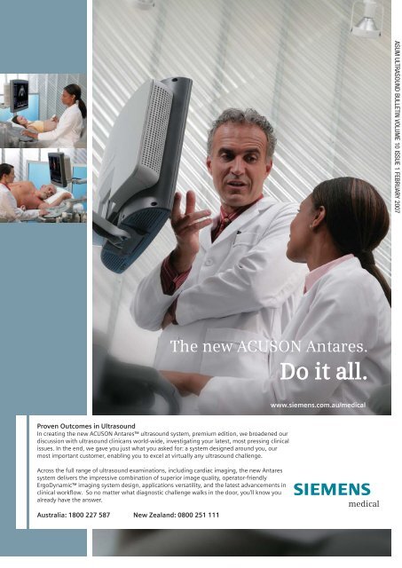
The new ACUSON Antares.<br />
Do it all.<br />
Proven Outcomes <strong>in</strong> <strong>Ultra</strong>sound<br />
In creat<strong>in</strong>g the new ACUSON Antares ultrasound system, premium edition, we broadened our<br />
discussion with ultrasound cl<strong>in</strong>icans world-wide, <strong>in</strong>vestigat<strong>in</strong>g your latest, most press<strong>in</strong>g cl<strong>in</strong>ical<br />
issues. In the end, we gave you just what you asked <strong>for</strong>: a system designed around you, our<br />
most important customer, enabl<strong>in</strong>g you to excel at virtually any ultrasound challenge.<br />
Across the full range of ultrasound exam<strong>in</strong>ations, <strong>in</strong>clud<strong>in</strong>g cardiac imag<strong>in</strong>g, the new Antares<br />
system delivers the impressive comb<strong>in</strong>ation of superior image quality, operator-friendly<br />
ErgoDynamic imag<strong>in</strong>g system design, applications versatility, and the latest advancements <strong>in</strong><br />
cl<strong>in</strong>ical workflow. So no matter what diagnostic challenge walks <strong>in</strong> the door, you'll know you<br />
already have the answer.<br />
Australia: 1800 227 587 New Zealand: 0800 251 111<br />
www.siemens.com.au/medical<br />
ASUM ULTRASOUND BULLETIN VOLUME 10 ISSUE 1 FEBRUARY 20<strong>07</strong>
Volume 10 Issue 1 <strong>Feb</strong>ruary 20<strong>07</strong><br />
ISSN 1441-6891<br />
ISSN 1441-6891<br />
<strong>Ultra</strong>sound<br />
Bullet<strong>in</strong><br />
Journal of the <strong>Australasian</strong> <strong>Society</strong> <strong>for</strong> <strong>Ultra</strong>sound <strong>in</strong> Medic<strong>in</strong>e
18 reasons (at least) why<br />
you should jo<strong>in</strong> ASUM<br />
ASUM is a unique professional society dedicated to excellence<br />
<strong>in</strong> ultrasound and the professionals who work <strong>in</strong> this vital<br />
and constantly evolv<strong>in</strong>g medical specialty.<br />
c 1 Professional qualifications<br />
Diploma <strong>in</strong> Medical <strong>Ultra</strong>sound (DMU)<br />
Diploma <strong>in</strong> Diagnostic <strong>Ultra</strong>sound (DDU)<br />
Certificate <strong>in</strong> Cl<strong>in</strong>ician Per<strong>for</strong>med<br />
<strong>Ultra</strong>sound (CCPU)<br />
c 2 <strong>Ultra</strong>sound tra<strong>in</strong><strong>in</strong>g<br />
The new ASUM College of <strong>Ultra</strong>sound<br />
accepts its first students <strong>in</strong> 2006<br />
c 3 Onl<strong>in</strong>e education<br />
Free onl<strong>in</strong>e physics education <strong>for</strong><br />
DMU and DDU candidates<br />
c 4 Onl<strong>in</strong>e cl<strong>in</strong>ical handbook<br />
A reference collection of images, cases<br />
and differential diagnoses<br />
c 5 Educational resources<br />
Extensive library of ultrasound videos,<br />
CDs and DVDs<br />
c 6 Policies and statements<br />
Guidel<strong>in</strong>es, updates and worksheets<br />
used by policy makers<br />
c 7 MOSIPP<br />
Record<strong>in</strong>g of CPD/CME po<strong>in</strong>ts<br />
c 8 Professional advancement<br />
Speak<strong>in</strong>g opportunities at meet<strong>in</strong>gs <strong>in</strong><br />
Australia, New Zealand and Asia<br />
c 9 Published author<br />
Publish your research <strong>in</strong> the ASUM<br />
<strong>Ultra</strong>sound Bullet<strong>in</strong><br />
c 10 Research Grants<br />
ASUM supports research which extends<br />
knowledge of cl<strong>in</strong>ical ultrasound<br />
<strong>Australasian</strong> <strong>Society</strong> <strong>for</strong> <strong>Ultra</strong>sound <strong>in</strong> Medic<strong>in</strong>e<br />
Level 2, 511 Pacific Highway<br />
Crows Nest NSW 2065<br />
Australia<br />
Telephone: +61 2 9438 2<strong>07</strong>8 Facsimile:+61 2 9438 3686<br />
Email: asum@asum.com.au Website: www.asum.com.au<br />
c 11 Service to medical ultrasound<br />
ASUM welcomes ultrasound professionals<br />
to its Council and committees<br />
c 12 Attend ASUM meet<strong>in</strong>gs at<br />
reduced rates<br />
Members enjoy special registration fee<br />
discounts <strong>for</strong> the Annual Scientific<br />
Meet<strong>in</strong>g and Multidiscipl<strong>in</strong>ary<br />
Workshops<br />
c 13 Professional, quality network<strong>in</strong>g<br />
Connect with your colleagues and<br />
ultrasound systems suppliers at<br />
meet<strong>in</strong>gs and workshops and through<br />
high quality networks<br />
c 14 Free website employment advertis<strong>in</strong>g<br />
Advertis<strong>in</strong>g <strong>for</strong> staff on the ASUM<br />
website is free to ASUM members<br />
c 15 <strong>Ultra</strong>sound Bullet<strong>in</strong> delivered to<br />
your door<br />
The quarterly ASUM <strong>Ultra</strong>sound Bullet<strong>in</strong><br />
is highly regarded both <strong>for</strong> its medical<br />
ultrasound articles and professional<br />
news content<br />
c 16 Professional <strong>in</strong>demnity <strong>in</strong>surance<br />
Peace of m<strong>in</strong>d <strong>for</strong> sonographer<br />
members <strong>for</strong> a modest annual<br />
premium<br />
c 17 Special home loan rates from AMP<br />
AMP is one of Australia’s biggest home<br />
lenders. ASUM members qualify <strong>for</strong><br />
special home loan rates<br />
c 18 Drive <strong>for</strong> less with Hertz<br />
ASUM Members qualify <strong>for</strong> discounted<br />
Hertz car hire rates
President<br />
Dr Matthew Andrews<br />
Honorary Secretary<br />
Mrs Roslyn Savage<br />
Honorary Treasurer<br />
Dr Andrew Ngu<br />
Chief Executive Officer<br />
Dr Carol<strong>in</strong>e Hong<br />
ULTRASOUND BULLETIN<br />
Official publication of<br />
the <strong>Australasian</strong> <strong>Society</strong><br />
<strong>for</strong> <strong>Ultra</strong>sound <strong>in</strong> Medic<strong>in</strong>e<br />
Published quarterly<br />
ISSN 1441-6891<br />
Indexed by the Sociedad Iberoamericana de<br />
In<strong>for</strong>macion Cientifien (SIIC) Databases<br />
Editor<br />
Prof Ron Benzie<br />
University of Sydney, Division of Women's and<br />
Children's Health, Nepean Hospital,<br />
Penrith, NSW 2750<br />
Co-Editor<br />
Mr Keith Henderson<br />
ASUM Education Manager<br />
Editorial Coord<strong>in</strong>ator<br />
Mr James Hamilton<br />
ASUM Education Officer<br />
Assistant Editors<br />
Ms Kaye Griffiths AM<br />
ANZAC Research CRGH Institute NSW<br />
Ms Jan<strong>in</strong>e Horton<br />
St V<strong>in</strong>cent's Imag<strong>in</strong>g Vic<br />
Ms Louise Lee<br />
Gold Coast Hospital Qld<br />
Mr Adam Lunghi<br />
Echo Services WA<br />
Dr Amarendra Trivedi<br />
Mildura Vic<br />
Editorial contributions<br />
Orig<strong>in</strong>al research, case reports, quiz cases,<br />
short articles, meet<strong>in</strong>g reports and calendar<br />
<strong>in</strong><strong>for</strong>mation are <strong>in</strong>vited and should be<br />
addressed to The Editor at the address below<br />
Membership and geneal enquiries<br />
to ASUM at the address below<br />
Published on behalf of ASUM<br />
by M<strong>in</strong>nis Communications<br />
Bill M<strong>in</strong>nis Director<br />
4/16 Maple Grove<br />
Toorak Melbourne Victoria 3142 Australia<br />
tel +61 3 9824 5241 fax +61 3 9824 5247<br />
email m<strong>in</strong>nis@m<strong>in</strong>niscomms.com.au<br />
Disclaimer<br />
Unless specifically <strong>in</strong>dicated, op<strong>in</strong>ions<br />
expressed should not be taken as those of<br />
the <strong>Australasian</strong> <strong>Society</strong> <strong>for</strong> <strong>Ultra</strong>sound <strong>in</strong><br />
Medic<strong>in</strong>e or of M<strong>in</strong>nis Communications<br />
AUSTRALASIAN SOCIETY FOR<br />
ULTRASOUND IN MEDICINE<br />
ABN 64 001 679 161<br />
Level 2, 511 Pacific Highway St Leonards<br />
Sydney NSW 2065 Australia<br />
tel +61 2 9438 2<strong>07</strong>8 fax +61 2 9438 3686<br />
email asum@asum.com.au<br />
website:http //www.asum.com.au<br />
ISO 9001: 2000<br />
Certified Quality<br />
Management<br />
Systems<br />
<strong>Ultra</strong>sound<br />
Bullet<strong>in</strong><br />
ASUM <strong>Ultra</strong>sound Bullet<strong>in</strong> <strong>Feb</strong>ruary 20<strong>07</strong> 10 (1)<br />
THE EXECUTIVE<br />
President’s message 3<br />
Editor's column 3<br />
CEO’s message 7<br />
LETTERS<br />
Jo<strong>in</strong>t Position Statement on the Appro- 12<br />
priate use of Diagnostic <strong>Ultra</strong>sound<br />
DIAGNOSTIC ULTRASOUND<br />
A standardised protocol <strong>for</strong> obta<strong>in</strong><strong>in</strong>g 13<br />
appropriate volume data sets dur<strong>in</strong>g<br />
the fetal mid trimester<br />
morphological scan<br />
Can the anterior-posterior thigh 25<br />
diameter be used as an <strong>in</strong>dicator<br />
<strong>for</strong> fetal age us<strong>in</strong>g two-dimensional<br />
sonography?<br />
<strong>Ultra</strong>sound contrast agents <strong>in</strong> 32<br />
diagnosis of focal liver lesions<br />
Abstracts 36th Annual Scientific 35<br />
Meet<strong>in</strong>g 2006 Melbourne<br />
Victoria – Part 2<br />
BOOK REVIEWS<br />
<strong>Ultra</strong>sound <strong>for</strong> Surgeons 47<br />
<strong>Ultra</strong>sonography <strong>in</strong> Urology: a Practical 47<br />
Approach to Cl<strong>in</strong>ical Problems<br />
<strong>Ultra</strong>sound of the urogenital system 47<br />
REPORTS<br />
The CardioVascular Centre – export<strong>in</strong>g 49<br />
vascular expertise to Ch<strong>in</strong>a<br />
ASUM–Vietnam l<strong>in</strong>k four-day sem<strong>in</strong>ar 50<br />
CADUCEUS – Mary Langdale<br />
visits Denmark<br />
EDUCATION<br />
52<br />
DMU 2006 reports 54<br />
DMU 2006 statistics 55<br />
DMU 2006 passes 55<br />
CCPU Report<br />
South Australia Branch<br />
56<br />
Annual General Meet<strong>in</strong>g Report<br />
NOTICES<br />
57<br />
Corporate members 58<br />
New members 58<br />
Calendar 59<br />
Guidel<strong>in</strong>es <strong>for</strong> authors 60<br />
Dr Matthew Andrews on the <strong>Society</strong>'s busy<br />
20<strong>07</strong> schedule. Neophyte editor asks <strong>for</strong> help.<br />
CEO reports on ASUM's <strong>in</strong>ternational activities<br />
and announces the move to new premises is<br />
complete<br />
ASUM and RANZCOG state that ultrasound<br />
equipment's primary use is <strong>for</strong> the purpose of<br />
medical diagnosis<br />
This discussion paper shares the three years<br />
of experience ga<strong>in</strong>ed at Brisbane <strong>Ultra</strong>sound<br />
<strong>for</strong> Women (BUFW) to advance the acceptance<br />
of volume ultrasound and encourages debate<br />
about a consensus <strong>for</strong> uni<strong>for</strong>mity<br />
This study evaluates the usefulness and<br />
direct correlation of a simple new method of<br />
predict<strong>in</strong>g fetal age by measurement of the<br />
anterior-posterior thigh diameter (APTD)<br />
<strong>in</strong> normal 18–28 week pregnancies us<strong>in</strong>g<br />
two-dimensional sonography<br />
CEUS has evolved to become a decisive factor<br />
<strong>in</strong> imag<strong>in</strong>g to detect and classify focal liver<br />
lesions<br />
Part 2 of abstracts delivered at 2006 ASUM<br />
ASM<br />
<strong>Ultra</strong>sound professionals review the latest<br />
books about the discipl<strong>in</strong>e<br />
Australian vascular ultrasound expertise is <strong>in</strong><br />
demand <strong>in</strong> Ch<strong>in</strong>a<br />
ASUM–Asia l<strong>in</strong>k activity progresses with this<br />
latest skills exchange to Vietnam<br />
The CADUCEUS Australia / Denmark exchange<br />
is build<strong>in</strong>g ties <strong>in</strong> Europe<br />
Reports from a successful 2006 DMU Exam-<br />
<strong>in</strong>ation Program<br />
Report on 2006 CCPU progress<br />
ASUM <strong>Ultra</strong>sound Bullet<strong>in</strong> 20<strong>07</strong> <strong>Feb</strong>ruary 10 (1)<br />
1
<strong>Australasian</strong> <strong>Society</strong><br />
<strong>for</strong> <strong>Ultra</strong>sound <strong>in</strong> Medic<strong>in</strong>e<br />
37th Annual Scientifi c Meet<strong>in</strong>g<br />
Abstract submission is now open<br />
Submit onl<strong>in</strong>e www.asum20<strong>07</strong>.com<br />
Promot<strong>in</strong>g Excellence<br />
<strong>in</strong> <strong>Ultra</strong>sound<br />
ASUM Head Offi ce<br />
PO Box 943<br />
Crows Nest NSW 1585<br />
Australia<br />
Telephone: +61 2 9438 2<strong>07</strong>8<br />
Facsimile: +61 2 9438 3686<br />
Email: asum@asum.com.au<br />
Website: www.asum.com.au<br />
Meet<strong>in</strong>g Offi ce<br />
ICMS Pty Ltd<br />
Locked Bag Q4002<br />
QVB Post Offi ce<br />
Sydney NSW 1230<br />
Australia<br />
Telephone: +61 2 9290 3366<br />
Facsimile: +61 2 9290 2444<br />
Email: asum20<strong>07</strong>@icms.com.au<br />
Website: www.asum20<strong>07</strong>.com<br />
ASUM is Certifi ed<br />
ISO 9001:2000 Quality<br />
Management Systems<br />
ASUM is affi liated<br />
with WFUMB<br />
2 ASUM <strong>Ultra</strong>sound Bullet<strong>in</strong> 20<strong>07</strong> <strong>Feb</strong>ruary 10 (1)<br />
13 – 16 September 20<strong>07</strong><br />
Cairns Convention Centre<br />
Cairns, Australia<br />
Critical Dates<br />
Proff ered Paper & Poster<br />
Abstract Submission Deadl<strong>in</strong>e<br />
Friday, 11 May 20<strong>07</strong><br />
Proff ered Paper & Poster<br />
Abstract Notifi cation<br />
Friday, 22 June 20<strong>07</strong><br />
Early Bird Registration Deadl<strong>in</strong>e<br />
Friday, 13 July 20<strong>07</strong><br />
Accommodation Deadl<strong>in</strong>e<br />
Monday, 6 August 20<strong>07</strong><br />
www.asum20<strong>07</strong>.com
President’s message<br />
Dr Matthew Andrews<br />
I would like to wish all ASUM members<br />
and staff a very happy 20<strong>07</strong>. This<br />
year will be busy and excit<strong>in</strong>g <strong>for</strong><br />
ASUM, on multiple fronts.<br />
<strong>Ultra</strong>sound Bullet<strong>in</strong> Editor<br />
Prof Ron Benzie has been appo<strong>in</strong>ted<br />
Editor of the <strong>Ultra</strong>sound Bullet<strong>in</strong>, tak<strong>in</strong>g<br />
over from Assoc Prof Roger Davies,<br />
who has held the role <strong>for</strong> the past four<br />
years. In that time, the <strong>Ultra</strong>sound<br />
Bullet<strong>in</strong> has cont<strong>in</strong>ually improved, provid<strong>in</strong>g<br />
a high quality educational and<br />
<strong>in</strong><strong>for</strong>mation resource to the membership.<br />
On behalf of ASUM I would<br />
like to thank Roger <strong>for</strong> his valuable<br />
contribution. Ron Benzie is Director<br />
of Per<strong>in</strong>atal <strong>Ultra</strong>sound at Nepean<br />
Hospital, Penrith, NSW. I would like<br />
to congratulate Ron on his appo<strong>in</strong>tment<br />
and I am certa<strong>in</strong> that under his<br />
editorship, the <strong>Ultra</strong>sound Bullet<strong>in</strong> will<br />
go from strength to strength.<br />
Congratulations<br />
In the Australia Day Honours, Prof<br />
John Harris, a longstand<strong>in</strong>g ASUM<br />
member and contributor to the <strong>Society</strong>,<br />
was awarded the Medal of the Order of<br />
Australia <strong>for</strong> service to medic<strong>in</strong>e, particularly<br />
through the advancement of<br />
vascular surgery and ultrasound procedures<br />
and techniques, to medical education<br />
and curriculum development, and<br />
to public health adm<strong>in</strong>istration. ASUM<br />
congratulates John on his award and<br />
is proud to have such an honoured<br />
member.<br />
New ASUM office<br />
ASUM is now the proud owner of a<br />
new home on the Pacific Highway, St.<br />
Leonards. The Secretariat moved <strong>in</strong>to<br />
the office over the Christmas period<br />
and the fit-out should be complete<br />
with<strong>in</strong> the next few weeks. Reflect<strong>in</strong>g<br />
the growth and <strong>in</strong>creased activities of<br />
the <strong>Society</strong>, the office will provide an<br />
appropriate home base. In addition to<br />
ample facilities <strong>for</strong> ASUM staff, the<br />
office will have teach<strong>in</strong>g and meet<strong>in</strong>g<br />
amenities. ASUM staff has worked<br />
tirelessly with the CEO, Dr Carol<strong>in</strong>e<br />
Hong, over the last couple of months<br />
prepar<strong>in</strong>g <strong>for</strong> and orchestrat<strong>in</strong>g the<br />
move. When complete, members are<br />
encouraged to visit the new premises<br />
and <strong>in</strong>troduce themselves to the staff,<br />
who will gladly show you around.<br />
WFUMB World Congress 2009<br />
Now approximately two-and-half years<br />
away, preparation <strong>for</strong> the WFUMB<br />
World Congress is well under way.<br />
Congress Convenor, Dr Stan Barnett,<br />
and Scientific Convenor, Assoc Prof<br />
Roger Davies, welcome <strong>in</strong>put from<br />
ASUM members <strong>in</strong>terested <strong>in</strong> participat<strong>in</strong>g.<br />
I strongly urge members to<br />
put up their hands, as this is literally<br />
a once-<strong>in</strong>-a-professional-lifetime<br />
opportunity to be <strong>in</strong>volved <strong>in</strong> such a<br />
prestigious meet<strong>in</strong>g. The scientific program<br />
is currently be<strong>in</strong>g developed and<br />
an ASUM delegation recently attended<br />
the Radiological <strong>Society</strong> of North<br />
America (RSNA) meet<strong>in</strong>g, establish<strong>in</strong>g<br />
contacts and seek<strong>in</strong>g general suggestions<br />
<strong>for</strong> the direction of the program.<br />
A major theme of the feedback was<br />
that the meet<strong>in</strong>g should build on previous<br />
WFUMB congresses, but also<br />
be <strong>in</strong>novative seek<strong>in</strong>g new directions<br />
and themes <strong>in</strong> its overall program. Our<br />
task is to meet that challenge with as<br />
broad an <strong>in</strong>put from our members as<br />
possible.<br />
Certificate of Cl<strong>in</strong>ician<br />
Per<strong>for</strong>med <strong>Ultra</strong>sound (CCPU)<br />
<strong>Ultra</strong>sound is <strong>in</strong>creas<strong>in</strong>gly utilised as<br />
a tool by cl<strong>in</strong>icians <strong>in</strong> their every day<br />
practice. There is now a large range<br />
of medical craft groups us<strong>in</strong>g ultrasound<br />
<strong>in</strong> their cl<strong>in</strong>ical practice. The<br />
ultrasound per<strong>for</strong>med depends on the<br />
nature of practice, thus is often limited<br />
and specific. This cl<strong>in</strong>ician per<strong>for</strong>med<br />
ultrasound is dist<strong>in</strong>ct from the<br />
traditional, more comprehensive and<br />
THE EXECUTIVE<br />
A neophyte editor’s appeal<br />
It is a daunt<strong>in</strong>g<br />
task to<br />
follow <strong>in</strong> the<br />
footsteps of<br />
successful<br />
editors of the<br />
<strong>Ultra</strong>sound<br />
Bullet<strong>in</strong>.<br />
People like Rob<br />
Prof Ron Benzie<br />
Gibson, Glen<br />
McNally and Roger Davies cast long<br />
and dist<strong>in</strong>guished shadows. I consider<br />
it an honour and a privilege to have<br />
been chosen to succeed them and<br />
hope to be up to the challenge. With<br />
your help it might just be possible.<br />
It is important that we cont<strong>in</strong>ue to<br />
reflect the needs of our members (how<br />
is that <strong>for</strong> a motherhood statement?),<br />
so I would like to hear from you. Do<br />
you like this issue's new <strong>cover</strong>?<br />
Do we need to change our <strong>for</strong>mat?<br />
Would separation of the cl<strong>in</strong>ical from<br />
the official ASUM bus<strong>in</strong>ess content<br />
be helpful? Do you th<strong>in</strong>k an on-l<strong>in</strong>e<br />
edition would be useful? What changes<br />
<strong>in</strong> content would you like to see?<br />
Would you like to see op<strong>in</strong>ion pieces<br />
from leaders <strong>in</strong> the field (both here<br />
and overseas)? Should we have an<br />
<strong>in</strong>ternational editorial board as well as<br />
our local experts? How can you help?<br />
These are a few of the questions that<br />
spr<strong>in</strong>g to m<strong>in</strong>d. I hope you will contact<br />
me and let me know your op<strong>in</strong>ion.<br />
A letter section should be vibrant<br />
and controversial. Provocative<br />
viewpo<strong>in</strong>ts are of the essence! The<br />
recent discussion on enterta<strong>in</strong>ment<br />
scans was a good example of how the<br />
<strong>Ultra</strong>sound Bullet<strong>in</strong> can be a <strong>for</strong>um <strong>for</strong><br />
expression of different viewpo<strong>in</strong>ts.<br />
My specialist tra<strong>in</strong><strong>in</strong>g was orig<strong>in</strong>ally<br />
<strong>in</strong> obstetrics and gynaecology. I saw my<br />
first ultrasound mach<strong>in</strong>e <strong>in</strong> use when I<br />
was a registrar <strong>in</strong> Aberdeen 40 years<br />
ago. Now, of course, the ultrasound<br />
horizon reaches well beyond obstetrics<br />
and your journal should reflect that.<br />
Please help us make this journal<br />
even more <strong>in</strong>terest<strong>in</strong>g and relevant.<br />
Ron Benzie<br />
Editor<br />
email benzier@wahs.nsw.gov.au<br />
ASUM <strong>Ultra</strong>sound Bullet<strong>in</strong> 20<strong>07</strong> <strong>Feb</strong>ruary 10 (1)<br />
3
4 ASUM <strong>Ultra</strong>sound Bullet<strong>in</strong> 20<strong>07</strong> <strong>Feb</strong>ruary 10 (1)<br />
Snap open <strong>for</strong> quality.<br />
Snap closed <strong>for</strong> protection.<br />
Aquasonic ® 100, the world standard <strong>for</strong> medical ultrasound, now has a new proprietary<br />
Snap-Cap with valve, provid<strong>in</strong>g unparalleled benefits to both user and patient.<br />
Designed <strong>for</strong> One Handed Operation:Eng<strong>in</strong>eered to Elim<strong>in</strong>ate Drips and “Draw Back.”<br />
Exclusive self-seal<strong>in</strong>g silicone valve <strong>in</strong>stantly cuts off the flow of gel.<br />
• Elim<strong>in</strong>ates draw<strong>in</strong>g product back <strong>in</strong>to the bottle, thus reduc<strong>in</strong>g the potential<br />
<strong>for</strong> cross-contam<strong>in</strong>ation<br />
• Ma<strong>in</strong>ta<strong>in</strong>s a clean and safe work environment by prevent<strong>in</strong>g drips and<br />
product residue<br />
• Provides precise unimpeded flow control from the new larger aperture and valve<br />
Easy to use One-Handed Snap-Cap keeps the nozzle and aperture protected from<br />
the work environment.<br />
• Open and close the cap with one hand and ma<strong>in</strong>ta<strong>in</strong> position and procedure cont<strong>in</strong>uity<br />
• Protect the nozzle from old gels that can often collect on the surface of<br />
ultrasound equipment<br />
• AND no more lost red tips thanks to the permanently attached cap<br />
Parker Laboratories, Inc.<br />
286 Eldridge Road, Fairfield, NJ <strong>07</strong>004<br />
973.276.9500 • Fax: 973.276.9510<br />
www.parkerlabs.com • ISO 13485:2003<br />
Welcome our new<br />
Snap-Cap to your practice…<br />
Invite a safer and more<br />
efficient workplace.
usually referred ultrasound, provided<br />
by an imag<strong>in</strong>g specialist <strong>in</strong> conjunction<br />
with a sonographer. Cl<strong>in</strong>icians per<strong>for</strong>m<strong>in</strong>g<br />
ultrasound as part of their cl<strong>in</strong>ical<br />
practice have lacked dedicated ultrasound<br />
tra<strong>in</strong><strong>in</strong>g. ASUM has responded<br />
to their approaches <strong>for</strong> tra<strong>in</strong><strong>in</strong>g by<br />
establish<strong>in</strong>g the CCPU. This qualification<br />
will have a common ultrasound<br />
physics component and the cl<strong>in</strong>ical<br />
ultrasound component will consist of<br />
modules relevant to the specific areas<br />
of cl<strong>in</strong>ical practice. ASUM clearly dist<strong>in</strong>guishes<br />
this cl<strong>in</strong>ically-based qualification<br />
from its other more comprehensive<br />
medical ultrasound qualification,<br />
the Diploma of Diagnostic <strong>Ultra</strong>sound<br />
(DDU), which is at the imag<strong>in</strong>g specialist<br />
standard. On behalf of ASUM, I<br />
would like to pay tribute to the tireless<br />
ef<strong>for</strong>ts of Dr Glenn McNally, who has<br />
driven the establishment of the CCPU.<br />
Asia L<strong>in</strong>k<br />
Established several years ago between<br />
several Asian ultrasound societies to<br />
mutually dissem<strong>in</strong>ate ultrasound education<br />
and tra<strong>in</strong><strong>in</strong>g throughout Asia,<br />
ASUM has developed <strong>in</strong>valuable<br />
relationships with its counterparts <strong>in</strong><br />
many Asian countries. The Asia L<strong>in</strong>k<br />
Program has brought many speakers to<br />
<strong>Australasian</strong> meet<strong>in</strong>gs and has provided<br />
opportunities <strong>for</strong> ASUM members<br />
to impart their knowledge and skills<br />
to a very wide audience. The program<br />
rema<strong>in</strong>s alive and well.<br />
The 8th Congress of the Asian Federation<br />
of Societies of <strong>Ultra</strong>sound <strong>in</strong> Medic<strong>in</strong>e<br />
(AFSUMB) will be held <strong>in</strong> November.<br />
The Federation has requested that<br />
ASUM provide speakers to the meet<strong>in</strong>g.<br />
As part its Asia-L<strong>in</strong>k commitment,<br />
Cl<strong>in</strong>ical Director and Pr<strong>in</strong>cipal Lecturer Position DMU (Asia)<br />
Location: Kuala Lumpur, Malaysia<br />
THE EXECUTIVE<br />
ASUM Council is explor<strong>in</strong>g the possibility<br />
of hold<strong>in</strong>g its November 20<strong>07</strong><br />
Council meet<strong>in</strong>g <strong>in</strong> conjunction with<br />
the AFSUMB meet<strong>in</strong>g with all ASUM<br />
Councillors participat<strong>in</strong>g as speakers.<br />
This timely contribution would not only<br />
raise ASUM’s profile <strong>in</strong> the Asian region,<br />
but will serve to demonstrate to a wide<br />
audience, the quality of the WFUMB<br />
2009 meet<strong>in</strong>g ASUM will host.<br />
Regards and Best Wishes <strong>for</strong> 20<strong>07</strong>.<br />
Matthew Andrews<br />
President<br />
Applications are <strong>in</strong>vited <strong>for</strong> a sonographer-lecturer position <strong>for</strong> Vision College which is located <strong>in</strong> Kuala Lumpur, Malaysia. Vision<br />
College is seek<strong>in</strong>g to appo<strong>in</strong>t a DMU tra<strong>in</strong>ed sonographer (with at least three years post-DMU experience) to provide courses<br />
<strong>in</strong> General <strong>Ultra</strong>sound. As the successful applicant, you will manage the tra<strong>in</strong><strong>in</strong>g at Vision College and oversee the Sonography<br />
School. You would be culturally sensitive and <strong>in</strong>terested <strong>in</strong> liv<strong>in</strong>g and work<strong>in</strong>g <strong>in</strong> Asia. A generous, negotiable remuneration<br />
and conditions package is offered and <strong>in</strong>itial expressions of <strong>in</strong>terest, together with a CV, should be directed to the ASUM CEO,<br />
Dr Carol<strong>in</strong>e Hong by Email: carol<strong>in</strong>ehong@asum.com.au.<br />
This is an attractive position <strong>for</strong> an ambitious sonographer who wishes to advance their career by improv<strong>in</strong>g their <strong>in</strong>ternational<br />
network.<br />
Job specification <strong>in</strong> Malaysia <strong>in</strong>cludes:<br />
■ Position of 'Cl<strong>in</strong>ical Director and Pr<strong>in</strong>cipal Lecturer'<br />
■ Management tra<strong>in</strong><strong>in</strong>g at Vision College to oversee the Sonography School<br />
■ Assisted by several recently qualified Sonographers<br />
■ Overseas network<strong>in</strong>g and relationships<br />
■ Sponsor<strong>in</strong>g travell<strong>in</strong>g opportunities to attend CME courses<br />
■ Prestigious l<strong>in</strong>ks with renowned Hospitals <strong>in</strong> Malaysia. There are prom<strong>in</strong>ent hospitals that Vision is attached to and the<br />
Sonographer will be associated with these <strong>in</strong>stitutions<br />
■ Competitive tax-free remuneration, but low liv<strong>in</strong>g costs. In addition, <strong>in</strong>cludes annual trip back to Australia/New Zealand.<br />
■ Liv<strong>in</strong>g <strong>in</strong> a modern and progressive Asian country<br />
Interested <strong>in</strong> an Asian <strong>Ultra</strong>sound Experience?<br />
Vision College, <strong>in</strong> fasc<strong>in</strong>at<strong>in</strong>g Kuala Lumpur, Malaysia, is also seek<strong>in</strong>g to employ sonographers on short term contracts of<br />
6–12 weeks to teach general sonography on campus. A generous, remuneration and conditions package is offered and <strong>in</strong>itial<br />
expressions of <strong>in</strong>terest, together with a CV, may also be directed to the ASUM CEO, Dr Carol<strong>in</strong>e Hong by Email: carol<strong>in</strong>ehong@<br />
asum.com.au.<br />
ASUM <strong>Ultra</strong>sound Bullet<strong>in</strong> 20<strong>07</strong> <strong>Feb</strong>ruary 10 (1)<br />
5
ASUM extends a warm welcome to you at upcom<strong>in</strong>g ASUM meet<strong>in</strong>gs<br />
<strong>Australasian</strong> <strong>Society</strong><br />
<strong>for</strong> <strong>Ultra</strong>sound <strong>in</strong> Medic<strong>in</strong>e<br />
37th Annual Scientifi c Meet<strong>in</strong>g<br />
Abstract submission is now open<br />
Submit onl<strong>in</strong>e www.asum20<strong>07</strong>.com<br />
Promot<strong>in</strong>g Excellence<br />
<strong>in</strong> <strong>Ultra</strong>sound<br />
ASUM Head Offi ce<br />
PO Box 943<br />
Crows Nest NSW 2065<br />
Australia<br />
Telephone: +61 2 9438 2<strong>07</strong>8<br />
Facsimile: +61 2 9438 3686<br />
Email: asum@asum.com.au<br />
Website: www.asum.com.au<br />
Meet<strong>in</strong>g Offi ce<br />
ICMS Pty Ltd<br />
Locked Bag Q4002<br />
QVB Post Offi ce<br />
Sydney NSW 1230<br />
Australia<br />
Telephone: +61 2 9290 3366<br />
Facsimile: +61 2 9290 2444<br />
Email: asum20<strong>07</strong>@icms.com.au<br />
Website: www.asum20<strong>07</strong>.com<br />
�����������������<br />
���������������������<br />
������������������<br />
�������������������<br />
����������<br />
<strong>Australasian</strong> <strong>Society</strong><br />
<strong>for</strong> <strong>Ultra</strong>sound <strong>in</strong> Medic<strong>in</strong>e<br />
37th Annual Scientific Meet<strong>in</strong>g<br />
http://www.asum.com.au<br />
ASUM <strong>Ultra</strong>sound Bullet<strong>in</strong> 2006 <strong>Feb</strong>ruary 10 (1)<br />
13-17 September 20<strong>07</strong><br />
6 ASUM <strong>Ultra</strong>sound Bullet<strong>in</strong> 20<strong>07</strong> <strong>Feb</strong>ruary 10 (1)<br />
13 – 16 September 20<strong>07</strong><br />
Cairns Convention Centre<br />
Cairns, Australia<br />
Critical Dates<br />
Proff ered Paper & Poster<br />
Abstract Submission Deadl<strong>in</strong>e<br />
Friday, 11 May 20<strong>07</strong><br />
Proff ered Paper & Poster<br />
Abstract Notifi cation<br />
Friday, 22 June 20<strong>07</strong><br />
Early Bird Registration Deadl<strong>in</strong>e<br />
Friday, 13 July 20<strong>07</strong><br />
Accommodation Deadl<strong>in</strong>e<br />
Monday, 6 August 20<strong>07</strong><br />
www.asum20<strong>07</strong>.com<br />
ASUM20<strong>07</strong><br />
Cairns, North Queensland, Australia<br />
5<br />
www.asum.com.au<br />
���������<br />
���� ��<br />
�����������������<br />
����������� ��� ������������ ���<br />
������������ �� ���������� �� ��������<br />
����� � ������ ������ ���� ��<br />
������������ ���� ��������� ��� ���� ���<br />
��������� ��� �� ���������� �� ��������<br />
������� �� ����� ����� �� ���� ����<br />
����� ��� ���������� �� ���������� ����<br />
��� ������ ��� �����<br />
������� � ���������� ����������<br />
����������� ��� ������ �������� �� ����<br />
������ ��� ��������� ����<br />
����� ���� �������� �����<br />
��� ���� ��� ���<br />
������ ��� �����<br />
��� ���� �����<br />
������ ��� ����<br />
���������<br />
�� ��� � ���� ����<br />
�� ��� � ���� ����<br />
�� ������������������<br />
�� �����������������<br />
������ ����������<br />
��� ���������� ������
COUNCIL 2005–20<strong>07</strong><br />
EXECUTIVE<br />
President<br />
Matthew Andrews Vic<br />
Medical Councillor<br />
Immediate Past President<br />
David Rogers NZ<br />
Medical Councillor<br />
Honorary Secretary<br />
Roslyn Savage Qld<br />
Sonographer Councillor<br />
Honorary Treasurer<br />
Andrew Ngu Vic<br />
Medical Councillor<br />
MEMBERS<br />
Medical Councillors<br />
Ron Benzie NSW<br />
John Crozier NSW<br />
Roger Davies SA<br />
David Davies-Payne NZ<br />
Sonographer Councillors<br />
Stephen Bird SA<br />
Margaret Condon Vic<br />
Kaye Griffiths NSW<br />
Michelle Pedretti WA<br />
ASUM Head Office<br />
Chief Executive Officer<br />
Carol<strong>in</strong>e Hong<br />
Education Manager<br />
Keith Henderson<br />
All correspondence should be<br />
directed to:<br />
The Chief Executive Officer<br />
<strong>Australasian</strong> <strong>Society</strong> <strong>for</strong><br />
<strong>Ultra</strong>sound <strong>in</strong> Medic<strong>in</strong>e<br />
Level 2, 511 Pacific Highway<br />
St. Leonards Australia<br />
asum@asum.com.au<br />
http://www.asum.com.au<br />
CEO’s message<br />
Dr Carol<strong>in</strong>e Hong<br />
Greet<strong>in</strong>gs and Happy New Year! Now<br />
that the festive season is def<strong>in</strong>itely over<br />
and we enter the first quarter of the year<br />
20<strong>07</strong>, the ASUM Secretariat has been<br />
busy work<strong>in</strong>g on the year’s activities,<br />
plann<strong>in</strong>g, follow<strong>in</strong>g up and respond<strong>in</strong>g<br />
to our members’ needs. Membership<br />
renewals have cont<strong>in</strong>ued to come <strong>in</strong><br />
steadily and we are pleased to see<br />
many new members jo<strong>in</strong><strong>in</strong>g ASUM<br />
each month. Applications <strong>for</strong> the DMU<br />
and DDU exam<strong>in</strong>ations are also be<strong>in</strong>g<br />
processed. Education activities are on<br />
the brew and bubbl<strong>in</strong>g along nicely.<br />
Prof John Harris AM<br />
On behalf of the ASUM Council,<br />
Secretariat and members, we congratulate<br />
Prof John Harris, a longstand<strong>in</strong>g<br />
and loyal ASUM member, <strong>for</strong> his<br />
Medal of the Order of Australia on<br />
Australia Day. A more detailed report<br />
will be published <strong>in</strong> a future <strong>Ultra</strong>sound<br />
Bullet<strong>in</strong> about John. He was honoured<br />
and awarded with a Medal of the Order<br />
of Australia <strong>for</strong> services to medic<strong>in</strong>e,<br />
particularly through the advancement<br />
of vascular surgery and ultrasound procedures<br />
and techniques, medical education<br />
and curriculum development,<br />
and to public health adm<strong>in</strong>istration.<br />
Certa<strong>in</strong>ly, we all share our pride and<br />
joy with John and his family on this<br />
happy occasion<br />
ASUM new address<br />
I am pleased to advise that the ASUM<br />
Secretariat relocation has taken place<br />
successfully (hopefully completely by<br />
the time this <strong>Ultra</strong>sound Bullet<strong>in</strong> goes<br />
out), with great cooperation and enthu-<br />
THE EXECUTIVE<br />
siasm from the staff and much patience<br />
from our members. As with any relocation,<br />
it required a lot of coord<strong>in</strong>ation,<br />
logistics management and hard physical<br />
work. Staff will need some time to adjust<br />
to the new environment and new systems<br />
and we hope that the process hasn’t<br />
unduly affected our service to members.<br />
The ASUM office is now located <strong>in</strong><br />
the St Leonards and Crows Nest area on<br />
the lower north shore of Sydney, across<br />
the harbour bridge. The office is right<br />
on the boundary that demarcates the<br />
suburbs of St Leonards (associations<br />
and bus<strong>in</strong>ess prec<strong>in</strong>ct) and Crows Nest<br />
(café suburb). We are at close proximity<br />
to many associations, shops, bus routes,<br />
the ma<strong>in</strong> St Leonards tra<strong>in</strong> station, medical<br />
services, hospitals, teach<strong>in</strong>g <strong>in</strong>stitutions,<br />
bus<strong>in</strong>ess facilities, banks, post<br />
office, retail therapy places, cafes and<br />
restaurants and many more.<br />
Members are advised to use the<br />
Crows Nest PO Box address as the<br />
mail<strong>in</strong>g address, although all mail that<br />
is sent to the old address will be redirected<br />
to the new office.<br />
The change has already seen a lift<br />
<strong>in</strong> staff morale and efficiency, as we<br />
are now able to work on one floor as a<br />
dynamic team.<br />
The previous office at Willoughby<br />
was the first permanent premises of the<br />
<strong>Society</strong> and was purchased <strong>in</strong> 1989 by<br />
the ASUM Council. It has served the<br />
<strong>Society</strong> well, but with the expansion<br />
of members’ services, there was <strong>in</strong>sufficient<br />
space to carry out the <strong>Society</strong>’s<br />
operations and education activities.<br />
The ASUM Council approved the<br />
Willoughby office’s sale 2004 with<br />
a three-year lease back option that<br />
expires <strong>in</strong> May 20<strong>07</strong>. The new premises<br />
were bought <strong>in</strong> a timely manner and<br />
with<strong>in</strong> a Council approved budget. The<br />
whole construction and fit out of the<br />
new premises with workstations and<br />
tra<strong>in</strong><strong>in</strong>g rooms have also been completed<br />
with<strong>in</strong> a Council approved budget.<br />
Two new tra<strong>in</strong><strong>in</strong>g rooms will host<br />
exam<strong>in</strong>ations and education activities.<br />
ASUM <strong>Ultra</strong>sound Bullet<strong>in</strong> 20<strong>07</strong> <strong>Feb</strong>ruary 10 (1)<br />
7
MDW 20<strong>07</strong> Gold Coast 28th<br />
<strong>Feb</strong>ruary–4th March<br />
Registrations rema<strong>in</strong> strong <strong>for</strong> the<br />
series of workshops to be held at<br />
the Gold Coast. Early bird registrations<br />
were extended to allow members<br />
who are return<strong>in</strong>g from their holiday<br />
breaks to benefit from the special rates.<br />
More than 300 registrations have been<br />
received and we anticipate more. The<br />
multidiscipl<strong>in</strong>ary nature of the workshops<br />
will <strong>cover</strong> the follow<strong>in</strong>g:<br />
■ 20<strong>07</strong> ASUM Multidiscipl<strong>in</strong>ary<br />
Workshop 28th <strong>Feb</strong>ruary–4th<br />
March 20<strong>07</strong>.<br />
■ ASUM Fetal Echocardiography<br />
Symposium, Annual O&G<br />
<strong>Ultra</strong>sound Symposium 2nd–4th<br />
March 20<strong>07</strong>.<br />
■ 20<strong>07</strong> DMU Preparation Courses<br />
28th <strong>Feb</strong>ruary– 4th March 20<strong>07</strong><br />
■ 20<strong>07</strong> DDU Technical Sem<strong>in</strong>ars<br />
28th <strong>Feb</strong>ruary–1st March 20<strong>07</strong><br />
■ Nuchal Translucency Course 2nd<br />
March 20<strong>07</strong><br />
These ‘hands-on’ workshops have<br />
proven to be very popular <strong>in</strong> previous<br />
years and ASUM has organised<br />
these to respond to demand.<br />
RSNA 2006<br />
An ASUM delegation consist<strong>in</strong>g<br />
of Dr Matthew Andrews<br />
(ASUM President), Dr Stan Barnett<br />
(WFUMB 2009 Convenor), Dr Glenn<br />
McNally (WFUMB 2009 Treasurer)<br />
and I (ASUM CEO), attended the<br />
Radiological <strong>Society</strong> of North America<br />
(RSNA) 2006 – 92nd Scientific<br />
Assembly and Annual Meet<strong>in</strong>g <strong>in</strong><br />
Chicago <strong>in</strong> November last year.<br />
It is the largest medical congress<br />
and exhibition <strong>in</strong> the world, attract<strong>in</strong>g<br />
approximately 62,000 delegates to<br />
Chicago. A stagger<strong>in</strong>g 28,052 exhibitors,<br />
27,803 professionals and 6121<br />
other category registrants attended this<br />
amaz<strong>in</strong>g congress and exhibition.<br />
It was an <strong>in</strong>credible experience to<br />
participate <strong>in</strong> this congress and the<br />
logistics beh<strong>in</strong>d it must have been<br />
equally <strong>in</strong>credible. The ASUM team<br />
ga<strong>in</strong>ed many new valuable networks<br />
and came home with reward<strong>in</strong>g ideas<br />
and <strong>in</strong><strong>for</strong>mation <strong>for</strong> the <strong>Society</strong>. RSNA<br />
offers state-of-the-art research, education<br />
and technology all <strong>in</strong> one site. The<br />
President of RSNA 2006, Robert R<br />
Hattery, <strong>in</strong> his open<strong>in</strong>g address urged<br />
all participants to renew their lifelong<br />
commitment to professionalism to their<br />
patients, the public and to themselves.<br />
He said to the professionals ‘We are<br />
given the privilege by the public of credential<strong>in</strong>g<br />
and ma<strong>in</strong>ta<strong>in</strong><strong>in</strong>g ourselves as<br />
professionals. If we abuse our freedom<br />
or fall short, we risk los<strong>in</strong>g our privileges.’<br />
He stressed that professionalism<br />
is ‘the art and science of medic<strong>in</strong>e.’<br />
I believe that most of our ASUM<br />
members share the very same sentiments<br />
and I often feel <strong>in</strong>spired by the<br />
professionalism displayed by ASUM<br />
members <strong>in</strong> their work and public life<br />
locally and <strong>in</strong>ternationally.<br />
DOHA<br />
A letter from Mr Peter Woodley,<br />
Assistant Secretary, Diagnostic<br />
Imag<strong>in</strong>g Section, Department of Health<br />
and Age<strong>in</strong>g (DOHA), <strong>in</strong>vited ASUM<br />
to comment on the document called<br />
the First Work<strong>in</strong>g Draft of Standards<br />
<strong>for</strong> the Accreditation of Practices<br />
Provid<strong>in</strong>g Diagnostic Imag<strong>in</strong>g Services<br />
Under the Radiology MoU. This work<strong>in</strong>g<br />
draft is be<strong>in</strong>g provided <strong>for</strong> comment<br />
to professional and <strong>in</strong>dustry bod-<br />
THE EXECUTIVE<br />
RSNA 2006 (L–R) Dr Glenn McNally, Dr Stan Barnett, Ms Louise Archer, Dr Carol<strong>in</strong>e Hong, Mr Matthew Andrews and Mr Hiroyuki Tsuj<strong>in</strong>o<br />
ies represent<strong>in</strong>g diagnostic imag<strong>in</strong>g<br />
providers, particularly providers of<br />
services under the Radiology MoU.<br />
The Department has released three<br />
other documents to accompany the first<br />
work<strong>in</strong>g draft of the standards to assist<br />
stakeholders to provide comments.<br />
The first of these additional documents<br />
requested specific feedback from<br />
ASUM about requisite qualifications,<br />
skills and tra<strong>in</strong><strong>in</strong>g of practitioners, represented<br />
by ASUM, who provide imag<strong>in</strong>g<br />
services <strong>cover</strong>ed by the Radiology<br />
MoU. The second was a general<br />
Feedback Form to provide comments<br />
on any or all of the draft standards,<br />
criterion and sample <strong>in</strong>dicators <strong>in</strong> the<br />
work<strong>in</strong>g draft of the standards.<br />
The rema<strong>in</strong><strong>in</strong>g document was a version<br />
of the first work<strong>in</strong>g draft of the standards<br />
which <strong>in</strong>cludes, <strong>for</strong> comparative<br />
purposes, the relevant standards from<br />
Version 7 of the Royal Australian and<br />
New Zealand College of Radiologists<br />
Accreditation Standards <strong>for</strong> Diagnostic<br />
and Interventional Radiology.<br />
ASUM has sent a response and<br />
was thankful <strong>for</strong> the opportunity to<br />
comment on the first work<strong>in</strong>g draft of<br />
standards relat<strong>in</strong>g to an accreditation<br />
scheme <strong>for</strong> practices provid<strong>in</strong>g radiology<br />
services under Medicare. As the<br />
consultation process is open to public<br />
scrut<strong>in</strong>y, the DOHA will be publish<strong>in</strong>g<br />
all written submissions on the<br />
Department’s website at www.diagnosticimag<strong>in</strong>g.health.gov.au.<br />
ASUM member book sold out<br />
You may recall that late last year,<br />
we announced the release of a book<br />
edited by Dr George Condous on early<br />
pregnancy. The sales were so popular<br />
that it is now <strong>in</strong> its second pr<strong>in</strong>t.<br />
ASUM <strong>Ultra</strong>sound Bullet<strong>in</strong> 20<strong>07</strong> <strong>Feb</strong>ruary 10 (1)<br />
9
THE EXECUTIVE<br />
Left The Exhibition Hall and Right The Posters Section at RSNA 2006<br />
Enquiries can be directed to:<br />
Assoc Prof George Condous<br />
Associate Professor <strong>in</strong> Gynaecology<br />
University of Sydney,<br />
Nepean Cl<strong>in</strong>ical School<br />
Early Pregnancy and Advanced<br />
Endosurgery Unit, Nepean Hospital<br />
Penrith, NSW 2750, Sydney,<br />
Australia.<br />
New <strong>Ultra</strong>sound Bullet<strong>in</strong> Editor<br />
This issue will be the first with Prof<br />
Ron Benzie as Editor. We thank Assoc<br />
Prof Roger Davies <strong>for</strong> his contribution<br />
as Editor <strong>for</strong> the last four years. ASUM<br />
also welcomes Assoc Prof Amar<br />
Trevedi to the Editorial Committee.<br />
ASUM NZ 20<strong>07</strong> jo<strong>in</strong>t meet<strong>in</strong>g<br />
with RANZCR NZ Branch<br />
The concept of hold<strong>in</strong>g jo<strong>in</strong>t meet<strong>in</strong>gs<br />
with the RANZCR started several<br />
years ago and it proved so popular<br />
that the Third Comb<strong>in</strong>ed ASM of the<br />
New Zealand Branches of RANZCR<br />
and ASUM has been organised to<br />
be held from 19th–22nd July 20<strong>07</strong><br />
at the Well<strong>in</strong>gton Convention Centre.<br />
The Local Organis<strong>in</strong>g Committee<br />
has decided on a theme; ‘Shaken<br />
and Stirred’. The keynote speakers<br />
<strong>in</strong>clude Dr Debra Ikeda, Stan<strong>for</strong>d<br />
from CA, USA; Dr Philip Tirman, San<br />
Francisco from CA, USA; Prof Lilith<br />
Valent<strong>in</strong> from Malmo, Sweden; and<br />
Prof Fung-Yee Chan from South<br />
Brisbane, Australia. The call <strong>for</strong><br />
abstracts is now open and closes on<br />
Sunday 6th May 20<strong>07</strong>. In<strong>for</strong>mation is<br />
available on the ASUM website www.<br />
asum.com.au. ASUM also welcomes<br />
papers or posters from Fellows, educational<br />
affiliates, registrars and sonographers,<br />
with many prizes on offer.<br />
10 ASUM <strong>Ultra</strong>sound Bullet<strong>in</strong> 20<strong>07</strong> <strong>Feb</strong>ruary 10 (1)<br />
ASUM 2006 Cairns Meet<strong>in</strong>g<br />
13th–16th September 20<strong>07</strong><br />
ASUM’s Annual Scientific Meet<strong>in</strong>g<br />
<strong>for</strong> 20<strong>07</strong> will be held <strong>in</strong> Cairns at the<br />
prestigious Convention Centre. The<br />
Committee is particularly excited about<br />
the program, which <strong>in</strong>cludes first class<br />
speakers from all fields of ultrasound.<br />
The topics will be up-to-the-m<strong>in</strong>ute<br />
and will touch on both current ultrasound<br />
practices and new advances <strong>in</strong><br />
our field. The program has strong components<br />
from obstetrics and gynaecology,<br />
musculoskeletal imag<strong>in</strong>g, through<br />
to vascular ultrasound. It should appeal<br />
to anyone <strong>in</strong> the ultrasound field.<br />
Please see the ASUM website<br />
www.asum.com.au <strong>for</strong> regular updates.<br />
The registration brochure is enclosed<br />
with this issue. Call <strong>for</strong> abstracts will<br />
close on 11th May 20<strong>07</strong>.<br />
Travell<strong>in</strong>g to Cairns is easy with<br />
the Cairns International Airport only<br />
8 km from the city centre. The airport<br />
has daily flights from Asia, the United<br />
States and Europe (via S<strong>in</strong>gapore) and<br />
from all around Australia.<br />
Cairns, which is surrounded by<br />
tropical ra<strong>in</strong><strong>for</strong>est, deep blue seas and<br />
coral islands, is the capital of far north<br />
Queensland and is situated 2000 km<br />
north of Brisbane. The cosmopolitan<br />
city of Cairns is the premier launch<strong>in</strong>g<br />
po<strong>in</strong>t from which to visit the prist<strong>in</strong>e<br />
World Heritage listed ra<strong>in</strong><strong>for</strong>ests and<br />
the spectacular Great Barrier Reef. To<br />
the west are the tablelands and the outback,<br />
waterfalls, rivers and beautiful<br />
lakes, wetland areas with magnificent<br />
birdlife and the spectacular Undara<br />
Lava Tubes.<br />
The Organis<strong>in</strong>g Committee consists<br />
of Dr Deborah Moir (Co-Convenor),<br />
Liz Carter (Co-Convenor),Brendan<br />
Cramp (Vascular), Craig Cairns<br />
(MSK),Teresa Clapham, Helen Goton,<br />
Sue Davies, Lynette Hassell and Dr<br />
Andrew Ngu (trade liaison).<br />
Invited overseas speakers <strong>in</strong>clude<br />
Dr Joseph Polak, Dr Carlo Mart<strong>in</strong>oli,<br />
Dr Yves Ville, Dr David Nyberg, Dr<br />
Eugene McNally, Dr Tom Stavros and<br />
Dr David Evans.<br />
AFSUMB 20<strong>07</strong> Thailand<br />
ASUM has been approached by the<br />
AFSUMB 20<strong>07</strong> Congress President,<br />
to support the event. The majority<br />
of ASUM councillors have responded<br />
positively, offer<strong>in</strong>g to present at this<br />
Congress, volunteer<strong>in</strong>g their expertise<br />
and time. Normally, the ASUM<br />
Council holds a meet<strong>in</strong>g <strong>in</strong> Sydney<br />
every November. This year, <strong>in</strong> lieu<br />
of the meet<strong>in</strong>g to be held <strong>in</strong> Sydney,<br />
councillors will consider hold<strong>in</strong>g the<br />
council meet<strong>in</strong>g <strong>in</strong> Bangkok prior to<br />
the AFSUMB Congress.<br />
The ASUM council has also<br />
resolved that some assistance will be<br />
provided to ASUM members if they are<br />
<strong>in</strong>tend<strong>in</strong>g to participate <strong>in</strong> the AFSUMB<br />
Congress from 12th–16th November<br />
20<strong>07</strong>, such as if their abstracts are<br />
accepted or if they are <strong>in</strong>vited speakers.<br />
In particular, we encourage ASUM<br />
members who would like to be given<br />
speak<strong>in</strong>g opportunities on behalf of<br />
ASUM.<br />
ASAR<br />
ASUM has been advised that the<br />
ASAR has implemented a return to<br />
cl<strong>in</strong>ical practice policy <strong>for</strong> sonographers<br />
who return to the workplace<br />
after an absence from cl<strong>in</strong>ical practice<br />
of greater than three years. From<br />
20<strong>07</strong>, sonographers with an absence<br />
from cl<strong>in</strong>ical practice greater than five
years must successfully complete an<br />
ASAR approved ‘short course’ Return<br />
to Cl<strong>in</strong>ical Practice Program (RCPP)<br />
as part of their requirements to atta<strong>in</strong><br />
AMS status. Details are on the website<br />
www.asar.com.au.<br />
ASUM Vietnam project<br />
Dr Harley Roberts, Dr Andrew<br />
McLennan, Dr Jon Hyett and others<br />
were recently <strong>in</strong>volved <strong>in</strong> sett<strong>in</strong>g up<br />
a fetal medic<strong>in</strong>e program <strong>in</strong> Vietnam,<br />
follow<strong>in</strong>g on from relationships that<br />
were established and set up by Dr<br />
Harley Roberts last year with the<br />
ASUM Vietnam project.<br />
Harley cont<strong>in</strong>ues to raise funds <strong>for</strong><br />
this scholarship and has already made<br />
plans <strong>for</strong> more aid <strong>in</strong> Vietnam, <strong>in</strong> particular<br />
at the Tu Du Hospital. The Tu<br />
Du Hospital is now the first and only<br />
hospital <strong>in</strong> Vietnam to be registered<br />
with the Fetal Medic<strong>in</strong>e Foundation<br />
<strong>in</strong> London and <strong>in</strong> addition eight of the<br />
hospital's doctors are now certified <strong>for</strong><br />
Down’s screen<strong>in</strong>g.<br />
Further exchange programs are<br />
be<strong>in</strong>g planned utilis<strong>in</strong>g the funds donated<br />
through Harley Roberts’ ef<strong>for</strong>ts <strong>for</strong><br />
this project.<br />
• AQUASONIC ® 100<br />
ULTRASOUND TRANSMISSION GEL<br />
* US 01-50 5 litre SONICPAC® with<br />
dispenser, 1 per box,<br />
4 per case<br />
*US 01-08 0.25 litre dispencer,<br />
12 per box<br />
• STERILE AQUASONIC ® 100<br />
ULTRASOUND TRANSMISSION GEL<br />
* US 01-01 20g over wrapped<br />
sterilized foil pouches<br />
48 per box<br />
• AQUASONIC ® CLEAR ®<br />
ULTRASOUND GEL<br />
* US 03-50 5 litre SONICPAC® with<br />
dispenser, 1 per box,<br />
4 per case<br />
• AQUAFLEX ®<br />
ULTRASOUND GEL PAD - STANDOF<br />
* US 04-02 2cm x 9cm gel pad,<br />
6 pads per box<br />
• POLYSONIC ®<br />
ULTRASOUND LOTION<br />
* US 21-28 1 U.S. gallon with<br />
dispenser bottle,<br />
4 per pack<br />
���������������������������<br />
• SCAN ®<br />
ULTRASOUND GEL<br />
* US 11-285 SCANPAC® conta<strong>in</strong>s:<br />
4 SCAN gallons,<br />
2 dispenser bottles<br />
1 dispenser pump<br />
• AQUAGEL ®<br />
LUBRICATION GEL<br />
* US 57-15 150gram tube,<br />
12 per box<br />
• ECLIPSE ®<br />
Dr Pham Thanh, Director of Tu Du<br />
Hospital and Dr Nguyen Ha, Director<br />
of imag<strong>in</strong>g are both very supportive<br />
and appreciative of this exchange. I<br />
am privileged to be part of this project,<br />
work<strong>in</strong>g with Harley <strong>in</strong> facilitat<strong>in</strong>g the<br />
contacts, the processes and mak<strong>in</strong>g<br />
th<strong>in</strong>gs happen.<br />
ISUM – Indonesia<br />
Dr David Rogers and Dr Simon<br />
Meagher recently volunteered their time<br />
and expertise as speakers <strong>in</strong> Bandung,<br />
Indonesia. Dr Daniel Makes, President<br />
of ISUM is organis<strong>in</strong>g another meet<strong>in</strong>g<br />
to be held <strong>in</strong> Bali from 27th–28th July<br />
20<strong>07</strong>. Details are listed <strong>in</strong> the ASUM<br />
Calendar and on the website.<br />
CADUCEUS – Denmark<br />
Mary Langdale has returned from her<br />
CADUCEUS exchange <strong>in</strong> Copenhagen.<br />
A report is published elsewhere <strong>in</strong> this<br />
issue. Morten Boesen was here from<br />
Copenhagen on the exchange program.<br />
We thank Dr Cheryl Bass and her team<br />
once aga<strong>in</strong> <strong>for</strong> provid<strong>in</strong>g the tra<strong>in</strong><strong>in</strong>g<br />
and support <strong>in</strong> this program. A report<br />
from Morten will be published <strong>in</strong> a<br />
future issue of <strong>Ultra</strong>sound Bullet<strong>in</strong>.<br />
PROBE COVER<br />
* US 38-01 2.5”/1.75” W x 9.5” L<br />
(64mm/44mm x 241mm)<br />
100 per box, 6 boxes per case<br />
• THERMASONIC ®<br />
GEL WARMER<br />
* US 82-04-20CE Multi-bottle gel warmer<br />
• TRANSEPTIC ®<br />
516 Creek Street, Albury, 2640 Australia<br />
Telephone: 02 6021 8222<br />
Facsimile: 02 0621 7270<br />
Free Call: 1800 021 928<br />
CLEANSING SOLUTION<br />
* US 09-25 250ml clear spray bottle,<br />
12 per box<br />
Email: <strong>in</strong>fo@jacobsmedical.com.au<br />
Website: www.jacobsmedical.com.au<br />
Thank you<br />
On behalf of the ASUM Secretariat, I<br />
once aga<strong>in</strong> thank all members <strong>for</strong> their<br />
patience and understand<strong>in</strong>g dur<strong>in</strong>g the<br />
relocation of the office. If you are <strong>in</strong><br />
Sydney, please feel welcome to contact<br />
me if you wish to visit the premises<br />
and to meet the ASUM staff.<br />
We will cont<strong>in</strong>ue to work hard to<br />
serve the <strong>Society</strong> and to contribute to<br />
its long term success under the leadership<br />
of the President and the ASUM<br />
Council. I also want to acknowledge<br />
the support of the staff and members<br />
I have worked with over the years to<br />
advance ASUM’s objectives.<br />
Dr Carol<strong>in</strong>e Hong<br />
Chief Executive Officer<br />
carol<strong>in</strong>ehong@asum.com.au<br />
New ASUM address and contact<br />
details:<br />
511 Pacific Highway St Leonards<br />
NSW 2065, Sydney Australia<br />
Mail<strong>in</strong>g address: PO Box 943,<br />
Crows Nest NSW 1585, Australia<br />
Ph: +61 2 9438 2<strong>07</strong>8<br />
Fax: +61 2 9438 3686<br />
Practical <strong>Ultra</strong>sound Tra<strong>in</strong><strong>in</strong>g<br />
With the AIU<br />
Educational Opportunities<br />
Abound <strong>in</strong> 20<strong>07</strong><br />
Check out our full range of programs and<br />
have a look at the excit<strong>in</strong>g new ones<br />
com<strong>in</strong>g up<br />
Sample of upcom<strong>in</strong>g programs:<br />
n March 12–14 3D <strong>Ultra</strong>sound Techniques<br />
n April 18–20 Tra<strong>in</strong> the Tra<strong>in</strong>er <strong>in</strong> Sonography<br />
n May 9–11 Breast <strong>Ultra</strong>sound Workshop<br />
n May 14–25 New Entrant Sonographer<br />
FastTrack<br />
n May 28–June 1 <strong>Ultra</strong>sound <strong>in</strong> O&G Workshop<br />
n June 4–6 Advanced O&G Workshop<br />
Come and jo<strong>in</strong> us <strong>in</strong> 20<strong>07</strong><br />
Check the website or your annual booklet <strong>for</strong> dates,<br />
or just give us a call<br />
F<strong>in</strong>d out more, contact us:<br />
On-l<strong>in</strong>e www.aiu.edu.au<br />
Email: <strong>in</strong>fo@aiu.edu.au<br />
Phone: (<strong>07</strong>) 5526 6655<br />
Fax: (<strong>07</strong>) 5526 6041<br />
ASUM <strong>Ultra</strong>sound Bullet<strong>in</strong> 20<strong>07</strong> <strong>Feb</strong>ruary 10 (1)<br />
11
To the CEO<br />
LETTERS<br />
C-Gen 10: Jo<strong>in</strong>t Position Statement on the<br />
Appropriate Use of Diagnostic <strong>Ultra</strong>sound<br />
Thank you <strong>for</strong> your email of 22 August 2006 to Dr<br />
Peter White, CEO RANZCOG, regard<strong>in</strong>g ASUM’s Position<br />
Statement on Appropriate Use of Diagnostic <strong>Ultra</strong>sound<br />
Equipment <strong>for</strong> Non-medical Enterta<strong>in</strong>ment. We note that<br />
ASUM have approved a m<strong>in</strong>or amendment to this document,<br />
made by the Women’s Health Committee (WHC) at the July<br />
2006 Meet<strong>in</strong>g, which <strong>in</strong>volved amend<strong>in</strong>g ‘sole’ to ‘primary’<br />
<strong>in</strong> the first sentence of paragraph three.<br />
The updated Position Statement was approved as a new<br />
jo<strong>in</strong>t College Statement C-Gen 10: Jo<strong>in</strong>t Position Statement<br />
on the Appropriate Use of Diagnostic <strong>Ultra</strong>sound, at the<br />
College Statement<br />
Title: Position Statement on the Appropriate Use of<br />
Diagnostic <strong>Ultra</strong>sound<br />
Position Statement of The <strong>Australasian</strong> <strong>Society</strong> <strong>for</strong><br />
<strong>Ultra</strong>sound <strong>in</strong> Medic<strong>in</strong>e (ASUM), RANZCOG and the Royal<br />
Australian and New Zealand College of Radiologists<br />
(RANZCR)<br />
Statement No. C-Gen 10<br />
Date of this document November 2006<br />
First endorsed by Council November 2006<br />
Next review due: November 2008<br />
Statement<br />
The <strong>Australasian</strong> <strong>Society</strong> <strong>for</strong> <strong>Ultra</strong>sound <strong>in</strong> Medic<strong>in</strong>e,<br />
the Royal Australian and New Zealand College of<br />
Obstetricians and Gynaecologists and the Royal<br />
Australian and New Zealand College of Radiologists are<br />
committed to ensur<strong>in</strong>g the ma<strong>in</strong>tenance of the highest<br />
standard of medical care <strong>for</strong> pregnant women.<br />
Diagnostic medical ultrasound technology offers<br />
enormous benefits <strong>in</strong> terms of the provision of useful<br />
diagnostic <strong>in</strong><strong>for</strong>mation so that pregnancy may be better<br />
assessed and managed, with optimum outcomes <strong>for</strong><br />
mothers and babies achieved.<br />
The use of diagnostic medical ultrasound equipment<br />
requires regulation such that its primary use is <strong>for</strong> the<br />
purpose of medical diagnosis. Such regulation should<br />
require that the diagnostic ultrasound equipment usage<br />
be restricted to appropriately qualified health care professionals.<br />
Usage of such equipment should con<strong>for</strong>m to the<br />
guidel<strong>in</strong>es produced by the <strong>Australasian</strong> <strong>Society</strong> <strong>for</strong><br />
<strong>Ultra</strong>sound <strong>in</strong> Medic<strong>in</strong>e.<br />
We urge that appropriate regulation regard<strong>in</strong>g the<br />
sale, distribution and use of diagnostic ultrasound equipment<br />
be <strong>for</strong>mulated with a view to ensur<strong>in</strong>g that this<br />
technology cont<strong>in</strong>ues to assist cl<strong>in</strong>icians <strong>in</strong> the management<br />
of pregnancy, thereby optimis<strong>in</strong>g outcome <strong>for</strong><br />
mothers and babies.<br />
12 ASUM <strong>Ultra</strong>sound Bullet<strong>in</strong> 20<strong>07</strong> <strong>Feb</strong>ruary 10 (1)<br />
WHC meet<strong>in</strong>g held 24 November 2006 and was subsequently<br />
endorsed by RANZCOG Council.<br />
This is now available on the RANZCOG website at http://<br />
www.ranzcog.edu.au/publications /statement/C-gen10.pdf<br />
and will also appear <strong>in</strong> our magaz<strong>in</strong>e O&G. A copy of the<br />
statement is enclosed <strong>for</strong> your refence.<br />
College Statements are reviewed every two years, so we<br />
will be <strong>in</strong> touch with you <strong>in</strong> the future to see if the text needs<br />
to be updated.<br />
Yours s<strong>in</strong>cerely,<br />
Dr Edward W Weaver<br />
Chairperson<br />
Women’s Health Committee<br />
Useful website l<strong>in</strong>ks<br />
ASUM http://www.asum.com.au/open/home.htm<br />
(Scroll down to ‘Non-diagnostic applications’ and select<br />
‘F1 Statement on the appropriate use of diagnostic<br />
ultrasound equipment <strong>for</strong> non-medical enterta<strong>in</strong>ment<br />
ultrasound’.)<br />
Disclaimer<br />
This College Statement is <strong>in</strong>tended to provide general<br />
advice to Practitioners. The statement should never be<br />
relied on as a substitute <strong>for</strong> proper assessment with<br />
respect to the particular circumstances of each case and<br />
the needs of each patient.<br />
The statement has been prepared hav<strong>in</strong>g regard to<br />
general circumstances. It is the responsibility of each<br />
Practitioner to have regard to the particular circumstances<br />
of each case, and the application of this statement <strong>in</strong> each<br />
case. In particular, cl<strong>in</strong>ical management must always be<br />
responsive to the needs of the <strong>in</strong>dividual patient and the<br />
particular circumstances of each case.<br />
This College statement has been prepared hav<strong>in</strong>g<br />
regard to the <strong>in</strong><strong>for</strong>mation available at the time of its<br />
preparation, and each Practitioner must have regard to<br />
relevant <strong>in</strong><strong>for</strong>mation, research or material which may<br />
have been published or become available subsequently.<br />
Whilst the College endeavours to ensure that College<br />
statements are accurate and current at the time of their<br />
preparation, it takes no responsibility <strong>for</strong> matters aris<strong>in</strong>g<br />
from changed circumstances or <strong>in</strong><strong>for</strong>mation or material<br />
that may have become available after the date of the<br />
statements.<br />
The Royal Australian and New Zealand College of<br />
Obstetricians and Gynaecologists<br />
ABN 34 100 268 969<br />
College House<br />
254–260 Albert Street<br />
East Melbourne<br />
Vic 3002<br />
Australia<br />
Tel: +61 3 9417 1699<br />
Fax: + 61 3 9419 0672<br />
E-mail: ranzcog@ranzcog.edu.au
ASUM <strong>Ultra</strong>sound Bullet<strong>in</strong> 20<strong>07</strong> <strong>Feb</strong>ruary; 10 (1): 13–24<br />
Introduction<br />
Fetal biometry is useless unless there is a standard protocol<br />
<strong>for</strong> per<strong>for</strong>m<strong>in</strong>g the measurements. Assessment of fetal<br />
anatomy <strong>for</strong> normality was very difficult until there became<br />
a standard protocol <strong>for</strong> obta<strong>in</strong><strong>in</strong>g the important imag<strong>in</strong>g<br />
planes. Cardiac assessment is perhaps the best example.<br />
Comparison of exam<strong>in</strong>ations was not possible. Collaborative<br />
research prior to establishment of standards was limited.<br />
In recent times, there has been slow recognition that dataset<br />
analysis provides an opportunity to extend the use of<br />
3D ultrasound (3D US) <strong>in</strong> fetal exam<strong>in</strong>ation. The authors<br />
believed from the outset that standard protocols were necessary<br />
and established them <strong>in</strong> their practice. This paper aims<br />
to share that experience.<br />
Standardisation is an essential requirement <strong>for</strong> the<br />
<strong>in</strong>troduction and dissem<strong>in</strong>ation of any new technology and<br />
new techniques. There are two steps <strong>in</strong>volved <strong>in</strong> establish<strong>in</strong>g<br />
standardisation. First, experienced 3D US practitioners<br />
need to describe their protocols <strong>for</strong> particular exam<strong>in</strong>ations<br />
so that these protocols can be debated and ref<strong>in</strong>ed. Second,<br />
standardisation <strong>in</strong>volves the manufacturers agree<strong>in</strong>g to con<strong>for</strong>m<br />
to a standard protocol <strong>for</strong> file storage and analysis.<br />
Un<strong>for</strong>tunately, the end user has no control over this latter<br />
requirement, but should have an <strong>in</strong>put.<br />
3D ultrasound <strong>in</strong> obstetrics<br />
3D <strong>Ultra</strong>sound depicts the fetus <strong>in</strong> utero <strong>in</strong> the way that<br />
we view everyday life. Surface rendered 3D images show<br />
wondrous images of the surface anatomy. There is no question<br />
that 3D and 4D ultrasound enhances parental bond<strong>in</strong>g.<br />
The ability to assess fetal surface detail to make diagnoses<br />
DIAGNOSTIC ULTRASOUND<br />
A standardised protocol <strong>for</strong> obta<strong>in</strong><strong>in</strong>g appropriate<br />
volume data sets dur<strong>in</strong>g the fetal mid trimester<br />
morphological scan<br />
Gary Pritchard A , Teresa Clapham B and Andreas Lee C<br />
A Brisbane <strong>Ultra</strong>sound <strong>for</strong> Women PO Box 357 Spr<strong>in</strong>g Hill, Brisbane, Queensland 4004, Australia.<br />
B GE Healthcare, Mansfield, Queensland 4122, Australia.<br />
C Royal Brisbane and Women's Hospital, Herston, Queensland 4006, Australia.<br />
Correspondence to Gary Pritchard. Email bufw@aapt.net.au<br />
Abstract<br />
Surface rendered 3D ultrasound is well known <strong>in</strong> obstetric imag<strong>in</strong>g. The use of 3D volume data sets (VDS)<br />
is arguably more important to the future of ultrasound. In order to advance the scientific and educational<br />
opportunities that come with this technology, standards need to be established. These standards <strong>in</strong>volve<br />
protocols <strong>for</strong> both the method of collection and <strong>for</strong> analysis <strong>for</strong> the various fetal anatomical regions. As<br />
well, they <strong>in</strong>volve agreement about a uni<strong>for</strong>m file <strong>for</strong>mat so that the storage, transmission and utility can<br />
be as global as possible. This aspect is under the control of the manufactur<strong>in</strong>g companies.<br />
The aim of this discussion paper is to share the three years of experience ga<strong>in</strong>ed at Brisbane <strong>Ultra</strong>sound<br />
<strong>for</strong> Women (BUFW) to advance the acceptance of volume ultrasound and to encourage debate about<br />
a consensus <strong>for</strong> uni<strong>for</strong>mity. It describes the protocol <strong>for</strong> collection of the VDS as a basel<strong>in</strong>e <strong>for</strong> fetal<br />
morphology assessment used <strong>in</strong> over 15,000 cases at BUFW between 2002 and 2005. It <strong>in</strong>cludes def<strong>in</strong>ition<br />
of the landmarks <strong>for</strong> optimum data-set acquisition i.e. <strong>in</strong>itial plane, central structure, volume angle,<br />
etc. as well as criteria <strong>for</strong> assessment of adequacy. The anatomical detail and measurements obta<strong>in</strong>ed<br />
from each region are listed, together with artifacts to be aware of. Other units may adjust these basel<strong>in</strong>e<br />
protocols to suit their own requirements.<br />
of conditions, <strong>in</strong> the way that our neonatal and genetics colleagues<br />
do with the newborn, will come with experience.<br />
When viewed <strong>in</strong> real-time it is now possible to display<br />
fetal behavior, movements, and facial expressions. These<br />
advances expand the scope of fetal assessment.<br />
It is important, however, not to allow this great advance<br />
to detract from other aspects that 3D US technologies<br />
have bought. The collection of a volume data set (VDS) of<br />
ultrasound <strong>in</strong><strong>for</strong>mation may prove to be even more significant<br />
than the bond<strong>in</strong>g aspects, at least from a medical and<br />
scientific viewpo<strong>in</strong>t. The ability to rotate, translate, re-slice<br />
and render the collected data set gives the ability to effectively<br />
re-do the entire scan, at any time, <strong>in</strong> any place <strong>in</strong> the<br />
future. Any perspective, any plane, any orientation can be<br />
recreated from the collected data. Standard views can be<br />
readily reconstructed. The dataset is all-<strong>in</strong>clusive. A series<br />
of images can be created to match the ASUM Guidel<strong>in</strong>es <strong>for</strong><br />
Midtrimester Scan1 .<br />
Standardisation<br />
File <strong>for</strong>mat<br />
The file <strong>for</strong>mat is beyond our control. It is, however, counterproductive<br />
to require different software if studies are<br />
per<strong>for</strong>med on different mach<strong>in</strong>es. While it may be desirable<br />
from the company’s perspective to have a tertiary centre use<br />
their mach<strong>in</strong>ery exclusively, that path would require a tertiary<br />
unit <strong>for</strong> each manufacturer. The result would be dim<strong>in</strong>ished<br />
accessibility and a significant dilution of the experience and<br />
exposure of the units. It will always be the case that different<br />
units have different needs. Hence each will choose different<br />
hardware, often with<strong>in</strong> the same department, <strong>for</strong> different<br />
ASUM <strong>Ultra</strong>sound Bullet<strong>in</strong> 20<strong>07</strong> <strong>Feb</strong>ruary 10 (1)<br />
13
Fig. 1: Landmarks <strong>for</strong> direction.<br />
BUFW protocol <strong>for</strong> anatomical volume data sets at mid trimester<br />
scan<br />
Region def<strong>in</strong>itions<br />
Landmarks <strong>for</strong> optimum data set collection<br />
Image plane: The plane of the scan to start the VDS sweep.<br />
Central structure: The structure <strong>in</strong> ROI where the sweep should start<br />
from.<br />
Volume box boundaries<br />
Anterior: Limits<br />
Posterior: Limits<br />
Sides: Limits of the volume box on the image<br />
Orientation: Fetal position<br />
Volume angle: The sweep angle or volume angle (Vol angle)<br />
Magnification: How large all small to make the image<br />
Tim<strong>in</strong>g: When to start the sweep<br />
exam<strong>in</strong>ations. It is a fundamental requirement that the unit<br />
can collect, store, retrieve, and reanalyse all VDS from their<br />
various mach<strong>in</strong>es. It is equally fundamental that only one<br />
piece of software is required <strong>for</strong> this off-l<strong>in</strong>e analysis.<br />
Collection protocol<br />
This aspect is with<strong>in</strong> our control. Us<strong>in</strong>g 2D ultrasound, a<br />
series of standard images are collected with def<strong>in</strong>ed planes,<br />
particular orientation, and precise location of measurements<br />
<strong>in</strong> order to establish normality (or otherwise) of a fetus at<br />
morphological exam<strong>in</strong>ation. These protocols have been<br />
described and progressively adjusted over 20 or so years of<br />
obstetric imag<strong>in</strong>g, tak<strong>in</strong>g <strong>in</strong>to account the improvement <strong>in</strong><br />
knowledge and mach<strong>in</strong>e technology. 1<br />
3D volume ultrasound needs to follow the same path. If<br />
each exam<strong>in</strong>ation is per<strong>for</strong>med <strong>in</strong> a consistent fashion and<br />
<strong>in</strong>cludes standard sets, with common landmarks and def<strong>in</strong>ed<br />
planes, subsequent re-assessment and exchange becomes<br />
less problematic and thus more reliable. Research, teach<strong>in</strong>g,<br />
re-exam<strong>in</strong>ation, second op<strong>in</strong>ion, and remote diagnosis<br />
should become easier. The technology becomes both more<br />
useful and more applicable.<br />
Re-exam<strong>in</strong>ation protocol<br />
Dataset manipulation has also been standardised <strong>in</strong> our unit<br />
– the protocol will be the subject of a further discussion<br />
14 ASUM <strong>Ultra</strong>sound Bullet<strong>in</strong> 20<strong>07</strong> <strong>Feb</strong>ruary 10 (1)<br />
Gary Pritchard, Teresa Clapham and Andreas Lee<br />
Initial image plane Confirmation of adequacy<br />
Fig. 2: Confirmation of adequacy of the head.<br />
Comment: Any important considerations with regard to the <strong>in</strong>itial po<strong>in</strong>t<br />
of data set collection.<br />
Confirmation of adequacy<br />
Section plane or 3D rendered image. The reference image is labeled<br />
A, B, or C <strong>in</strong> the orthogonal display (Fig.2). Features that suggest an<br />
artifact is present.<br />
Assessable features<br />
Biometry: Measurements that can be per<strong>for</strong>med<br />
Anatomy: Assessable anatomical and functional structures<br />
Artifacts<br />
Features that can produce errors <strong>in</strong> diagnosis. NB The list does not<br />
<strong>in</strong>clude every possible artifact.<br />
File size<br />
The average file size (uncompressed)<br />
paper. However, the basic pr<strong>in</strong>ciples <strong>for</strong> each of the anatomical<br />
regions <strong>in</strong>volved have been developed. These <strong>in</strong>clude<br />
multiplanar display, confirmation of adequacy, identification<br />
of a l<strong>in</strong>ear anatomical structure (e.g. falx, <strong>in</strong>terventricular<br />
septum, sp<strong>in</strong>e etc), rotation of that l<strong>in</strong>ear structure <strong>in</strong>to either<br />
a vertical or horizontal orientation us<strong>in</strong>g the Z-axis, similar<br />
Z-axis rotation <strong>in</strong> either or both other planes, to obta<strong>in</strong><br />
an anatomically correct image that mimicks the real time<br />
planes, then translation of the appropriate s<strong>in</strong>gle plane image<br />
<strong>for</strong> the assessment and measurement.<br />
Pilot study<br />
In early 2002, a pilot study was conducted with<strong>in</strong> BUFW<br />
as a quality assurance (QA) project2 where 125 mid trimester<br />
scans were per<strong>for</strong>med on a Voluson 730 (GE-Kretz<br />
Zipf Austria) by real time exam<strong>in</strong>ation accord<strong>in</strong>g to the<br />
standard ASUM guidel<strong>in</strong>es1 by one of four experienced<br />
sonographers (HG, MH, NP, SW). The results and f<strong>in</strong>d<strong>in</strong>gs<br />
were recorded. A series of 3D volume data sets (VDS)<br />
were collected by a s<strong>in</strong>gle operator (GP) accord<strong>in</strong>g to a<br />
prelim<strong>in</strong>ary protocol established with<strong>in</strong> our department <strong>for</strong><br />
each anatomical region, head, heart, sp<strong>in</strong>e and abdomen.<br />
The total data set size <strong>for</strong> each case was approximately 30<br />
MB (uncompressed). Follow<strong>in</strong>g de-identification, 117 VDS<br />
were re-exam<strong>in</strong>ed off l<strong>in</strong>e by one of the four sonographers<br />
us<strong>in</strong>g a PC program, 3D ViewTM software (KretzTechnic
Zipf Austria). This study showed no significant difference<br />
between the real time and the reconstructed biometry. Head<br />
biometry showed no significant difference <strong>for</strong> ventriculoatrial<br />
diameter (VAD) (mean difference 0.1 mm), cisterna<br />
magna (mean difference 0.1 mm) and nuchal sk<strong>in</strong> fold<br />
(mean difference -0.2 mm). Satisfactory assessment of the<br />
fetal heart was only achieved <strong>in</strong> 40% of cases us<strong>in</strong>g the rapid<br />
acquisition technique designed at that time. For the other<br />
regions, satisfactory anatomical assessment was possible <strong>for</strong><br />
80% of structures. This small pilot study demonstrated to<br />
our satisfaction that our pr<strong>in</strong>ciples of VDS were satisfactory<br />
and showed us areas where improvement was necessary.<br />
The protocol was modified, ma<strong>in</strong>ly by add<strong>in</strong>g a specific<br />
protocol <strong>for</strong> fetal face assessment, and by modify<strong>in</strong>g<br />
the start<strong>in</strong>g po<strong>in</strong>t of the cardiac volume so that a rib does<br />
not shadow the orig<strong>in</strong> of the left ventricular outflow tract<br />
(LVOT). The <strong>in</strong>troduction of STIC TM (spatio-temporal image<br />
correlation) technology was <strong>in</strong>corporated when it became<br />
available.<br />
Subsequently, all fetal exam<strong>in</strong>ations have standard data<br />
sets collected, stored and archived as the only permanent<br />
record of the exam<strong>in</strong>ation. Videotap<strong>in</strong>g is no longer per<strong>for</strong>med<br />
<strong>for</strong> archival purposes.<br />
While this protocol may not satisfy the requirements<br />
<strong>for</strong> every exam<strong>in</strong>ation <strong>for</strong> every problem, they are basic to<br />
most morphology scans. The protocol provides a foundation<br />
<strong>for</strong> future development. Different VDS may be required <strong>for</strong><br />
later gestations, <strong>for</strong> more complicated scans or <strong>for</strong> abnormal<br />
f<strong>in</strong>d<strong>in</strong>gs.<br />
General po<strong>in</strong>ts<br />
A standardised protocol <strong>for</strong> obta<strong>in</strong><strong>in</strong>g appropriate volume data sets dur<strong>in</strong>g the fetal mid trimester morphological scan<br />
Applicability<br />
Brisbane <strong>Ultra</strong>sound <strong>for</strong> Women used Voluson 730 Expert<br />
mach<strong>in</strong>es (GE Kretz, Zipf, Austria) exclusively. The protocol<br />
has been developed on and <strong>for</strong> these mach<strong>in</strong>es. It is, however,<br />
applicable whichever 3D mach<strong>in</strong>e is used, with modification<br />
of factors such as sector angle, frame rate etc. Individual<br />
experience will allow adjustment of the protocol to suit different<br />
departmental and exam<strong>in</strong>ation requirements.<br />
As discussed earlier, there would need to be an off l<strong>in</strong>e<br />
analysis software package similar to 4D View TM <strong>for</strong> the data<br />
set to be reassessed. This software should allow multiplanar<br />
display and rotation around the 3 axis.<br />
Protocol variations<br />
Fetal anomaly<br />
In some circumstances, i.e. cardiac defect, it may be appropriate<br />
to collect an additional VDS start<strong>in</strong>g from a midl<strong>in</strong>e<br />
sagittal plane to allow a clearer image of the ductal and<br />
aortic arches. In cases of abnormality, a dataset collected<br />
with the anomaly as the central start<strong>in</strong>g po<strong>in</strong>t <strong>for</strong> the collection<br />
(i.e. facial cleft, sp<strong>in</strong>al defect) and <strong>in</strong>clud<strong>in</strong>g the whole<br />
affected region should be <strong>in</strong>cluded.<br />
General po<strong>in</strong>ts<br />
Fetal movement<br />
If the fetus is unable to stay still <strong>for</strong> a STICTM data set, a rapid<br />
3D sweep may be required as described <strong>in</strong> the protocol.<br />
Number – more is not better<br />
It must be appreciated that multiple data sets of the same<br />
region from different orientation, do not make analysis<br />
or <strong>in</strong>terpretation easier. Indeed, the opposite is true. They<br />
<strong>in</strong>crease the time required <strong>for</strong> re-assessment, and require<br />
more disk space <strong>for</strong> storage, backup, or transfer. More datasets<br />
mean greater expense. It is more efficient and effective to<br />
collect one adequate dataset than it is to take multiple ones,<br />
hop<strong>in</strong>g that one is adequate.<br />
Assessment of adequacy<br />
Proper collection of the VDS requires that the assessment of<br />
the degree of adequacy (as def<strong>in</strong>ed <strong>in</strong> the regional protocols)<br />
be per<strong>for</strong>med at the time of collection. The features to assess<br />
are completeness of <strong>in</strong>clusion of the anatomical region and<br />
absence of artifact. If the VDS is deemed unacceptable, it<br />
should not be saved, but rather repeated and reassessed.<br />
Because this is done as part of the real time exam<strong>in</strong>ation, it<br />
is possible to be confident that, if subsequent analysis shows<br />
there to be a problem, it is a true f<strong>in</strong>d<strong>in</strong>g, and has not been<br />
caused by the method of collection.<br />
Operator’s role<br />
A fundamental aspect of all ultrasound exam<strong>in</strong>ations is that<br />
operators use their knowledge, skill and experience to obta<strong>in</strong><br />
the best image of the area of <strong>in</strong>terest. This <strong>in</strong>volves adjust<strong>in</strong>g<br />
the mach<strong>in</strong>e sett<strong>in</strong>gs and the ultrasound w<strong>in</strong>dow, focus<strong>in</strong>g<br />
and enlarg<strong>in</strong>g appropriately, to m<strong>in</strong>imise artifact and to ga<strong>in</strong><br />
maximum diagnostic <strong>in</strong><strong>for</strong>mation. It requires the operator to<br />
assess some features of an organ, i.e. movement, that cannot<br />
be assessed from still images. In most cases, the operators<br />
use their skill to obta<strong>in</strong> a set of anatomically correct pictures<br />
so that another person, typically a doctor, can look at<br />
them and make cl<strong>in</strong>ical decisions. The operator still requires<br />
knowledge of what other images or areas may be needed to<br />
ref<strong>in</strong>e the differential diagnosis. In our practice, we became<br />
satisfied that re-analysis of datasets collected accord<strong>in</strong>g to<br />
our protocol was sufficiently accurate to enable cl<strong>in</strong>ical decisions<br />
to be made from them.<br />
There is a concern that untra<strong>in</strong>ed, unsupervised operators<br />
may collect the datasets, miss obvious problems and br<strong>in</strong>g<br />
the technology <strong>in</strong>to disrepute. Standard collection protocols,<br />
like standard image based collection protocols, cont<strong>in</strong>ue<br />
to require a high level of US competence <strong>for</strong> quality to<br />
be ma<strong>in</strong>ta<strong>in</strong>ed and <strong>for</strong> the exam<strong>in</strong>ation to rema<strong>in</strong> relevant.<br />
Professional tra<strong>in</strong><strong>in</strong>g <strong>in</strong> both collection and re-analysis will<br />
always be needed.<br />
Data set collection has the potential to make the exam<strong>in</strong>ation<br />
so rapid that there could be a negative impact on the<br />
doctor-patient relationship and the parental bond<strong>in</strong>g process.<br />
In fact the reverse may be true. Every user of ultrasound<br />
should adhere to the ALARA (as low as reasonably achievable)<br />
pr<strong>in</strong>ciple and try to limit fetal exposure. Off l<strong>in</strong>e<br />
analysis of the medical aspects of the exam<strong>in</strong>ation (biometry,<br />
structure) may <strong>in</strong> fact allow a lessen<strong>in</strong>g of the total exposure<br />
to the fetus even though the same amount of time is spent<br />
us<strong>in</strong>g the other aspects of the 3D technology.<br />
Artifacts<br />
3D <strong>Ultra</strong>sound is just B-mode ultrasound collected and<br />
stored by a different method. Artifacts related to B-mode:<br />
shadow<strong>in</strong>g, side-lobe, refraction etc. still occur and distort the<br />
image. B-mode artifacts are present <strong>in</strong> all planes. Unlike real<br />
time scann<strong>in</strong>g, rotation of the data set does cont'd page 23<br />
ASUM <strong>Ultra</strong>sound Bullet<strong>in</strong> 20<strong>07</strong> <strong>Feb</strong>ruary 10 (1)<br />
15
Initial image plane Confirmation of adequacy<br />
Fig. 3: Start<strong>in</strong>g plane <strong>for</strong> fetal overview.<br />
Significant images<br />
Fig. 5: Surface rendered overview.<br />
Region<br />
Fetal Overview<br />
Image plane<br />
Sagittal anterior <strong>for</strong> preference<br />
Landmarks <strong>for</strong> optimum data set collection<br />
Image plane: Fetus <strong>in</strong> long axis.<br />
Central structure: Diaphragm.<br />
Volume box boundaries<br />
Anterior: Maternal sk<strong>in</strong><br />
Posterior: Maternal sacrum<br />
Sides: As wide as possible<br />
Orientation: The long axis of fetus is horizontal<br />
Volume angle: Both sides of the uterus<br />
Magnification: Reduced<br />
Tim<strong>in</strong>g: No movement<br />
Comment<br />
This dataset takes longer because of its size. It is <strong>in</strong>cluded as a backup<br />
to the other standard datasets <strong>for</strong> review if they are <strong>in</strong>complete. It may<br />
allow limb measurements, but real-time exam<strong>in</strong>ation is the best way<br />
to assess limbs.<br />
Confirmation of adequacy<br />
Top to bottom as much as is easily possible. No gross movement<br />
distortion.<br />
16 ASUM <strong>Ultra</strong>sound Bullet<strong>in</strong> 20<strong>07</strong> <strong>Feb</strong>ruary 10 (1)<br />
Gary Pritchard, Teresa Clapham and Andreas Lee<br />
Fig. 4: Assessment of fetal adequacy overview.<br />
Fig. 6: Sneaky look at gender.<br />
Assessable features<br />
Biometry: Basic biometry, liquor volume (but NOT AFI)<br />
Anatomy: Fetus, limbs, placenta<br />
Artifacts<br />
Movement – significant chance <strong>for</strong> limb artifacts<br />
File size<br />
13 MB
Initial image plane Confirmation of adequacy<br />
Fig. 7: Initial image plane cerebellar vermis.<br />
Significant images<br />
A standardised protocol <strong>for</strong> obta<strong>in</strong><strong>in</strong>g appropriate volume data sets dur<strong>in</strong>g the fetal mid trimester morphological scan<br />
Fig. 9: Contrast rendered corpus callosum.<br />
Region: Head<br />
Image plane<br />
Transverse<br />
Landmarks <strong>for</strong> optimum data set collection<br />
Image plane: Transverse with the falx horizontal<br />
Central structure: Cerebellar vermis<br />
Volume box boundaries<br />
Anterior: Wider than skull<br />
Posterior: Wider than skull<br />
Sides: Wider than skull extent<br />
Orientation: Transverse<br />
Volume angle: 55 to 64 degrees<br />
Magnification: Fill 80% of zoom box with the head image<br />
Tim<strong>in</strong>g: Still<br />
Comment<br />
Beg<strong>in</strong> at the level of the cerebellar vermis. The NSF is least artifactually<br />
thickened here.This data set does not give reliable face detail,<br />
particularly <strong>for</strong> the lips and lens.<br />
Confirmation of adequacy<br />
Section planes.<br />
Fig. 8: Confirmation of adequacy of the head.<br />
Fig. 10: Care with 3D image of the lip with movement artifact of the lip.<br />
Scroll superiorly to above the head and <strong>in</strong>feriorly to below the jaw <strong>in</strong><br />
image A. Check <strong>in</strong> image C that the jaw is <strong>in</strong>cluded. If head is low <strong>in</strong><br />
the pelvis, a slightly off axis set centered anteriorly on coronal suture<br />
may view Falx, CSP and CC.<br />
Assessable features<br />
Biometry: BPD, Head Circ, VAD, Cisterna magna, Nuchal sk<strong>in</strong> fold.<br />
Anatomy: Skull vault shape, Cranial sutures, Falx, Ventricles, Choroid<br />
plexus, Cavum septum pellucidum, Corpus callosum (contrast render),<br />
Posterior cranial fossa, Cerebellum, Nuchal sk<strong>in</strong> fold, Mandible shape.<br />
Artifacts<br />
Movement distortion<br />
Shadow<strong>in</strong>g – corpus callosum<br />
Lateral resolution – apparent <strong>in</strong>crease <strong>in</strong> NSF<br />
File size<br />
6 MB<br />
ASUM <strong>Ultra</strong>sound Bullet<strong>in</strong> 20<strong>07</strong> <strong>Feb</strong>ruary 10 (1)<br />
17
Initial image plane Confirmation of adequacy<br />
Fig. 11: Coronal face start<strong>in</strong>g plane. Fig. 12: Adequate face volume.<br />
Significant images<br />
Fig. 13: A reliably good 3D face. Fig. 14: Contrast rendered palate.<br />
Region<br />
Face<br />
Image plane<br />
Coronal<br />
Landmarks <strong>for</strong> optimum data set collection<br />
Image plane: Lens<br />
Central structure: Both lens<br />
Volume box boundaries<br />
Anterior: Uter<strong>in</strong>e wall<br />
Posterior: Uter<strong>in</strong>e wall<br />
Sides: Vault of skull to upper thorax<br />
Orientation: Horizontal z-axis 5 degrees x-axis<br />
Volume angle: 45 degrees<br />
Magnification: 50% box area<br />
Tim<strong>in</strong>g: Not mov<strong>in</strong>g<br />
Comment<br />
Head horizontal, face at level of lens. Nose <strong>in</strong> centre of field. This<br />
dataset is <strong>in</strong>cluded to allow a better assessment of facial morphology<br />
than can be obta<strong>in</strong>ed from the head dataset.<br />
Confirmation of adequacy<br />
Reference image C. Extent – Above skull crown to below jaw. Distal<br />
lens.<br />
Assessable features<br />
Biometry: Inter-orbital, B<strong>in</strong>ocular distance, mandible, nasal bones.<br />
Anatomy: Orbits, lens, lip, mandible, metopic suture, nasal bones,<br />
18 ASUM <strong>Ultra</strong>sound Bullet<strong>in</strong> 20<strong>07</strong> <strong>Feb</strong>ruary 10 (1)<br />
Gary Pritchard, Teresa Clapham and Andreas Lee<br />
anterior ear, palate (contrast render) micrognathia, flat <strong>for</strong>ehead.<br />
Artifacts<br />
Movement. Shadow<strong>in</strong>g especially from a limb anterior to the region of<br />
<strong>in</strong>terest and above of the volume box. Lip movement.<br />
File size<br />
7 MB
Initial image plane Confirmation of adequacy<br />
Fig. 15: Start<strong>in</strong>g plane <strong>for</strong> cardiac volume – LVOT.. Fig. 16: Translocate image A arch to stomach.<br />
Significant images<br />
A standardised protocol <strong>for</strong> obta<strong>in</strong><strong>in</strong>g appropriate volume data sets dur<strong>in</strong>g the fetal mid trimester morphological scan<br />
Fig. 17: The IVS is a solid structure <strong>in</strong> B. Fig. 18: Dropout artifact VSD but a real ASD.<br />
Region Heart: Rapid Acquisition and STIC<br />
Image plane<br />
4-chamber view<br />
Landmarks <strong>for</strong> optimum data set collection<br />
Image plane: Transverse 4 chambers<br />
Central structure: LVOT<br />
Volume box boundaries<br />
Anterior: Chest wall<br />
Posterior: Sp<strong>in</strong>e<br />
Sides: Includes both sides<br />
Orientation: Not fac<strong>in</strong>g directly away<br />
Volume angle: 15 to 20 degrees at 18 to 20 weeks. Bigger later<br />
Magnification: 90% of 3D box<br />
Tim<strong>in</strong>g: Cannot time adequately. STIC 7 to 10 seconds<br />
Comment<br />
Best position is apical 4 chamber view with the septum slightly offset<br />
from the vertical. Also adequate with the sp<strong>in</strong>e at 90 degrees to 135<br />
degrees. Worst position is sp<strong>in</strong>e at 45 degrees from the transducer.<br />
Confirmation of adequacy<br />
Use translation <strong>in</strong> reference image A. Stomach below. Ductal arch<br />
above. No chest movement.<br />
Assessable features<br />
Biometry: Chambers, LVOT diameter, RVOT diameter.<br />
Anatomy: 4 chambers, Atrial septum, Foramen ovale, Atrioventricular<br />
valves, Ventricular septum, LVOT, RVOT, Orig<strong>in</strong> branches relationship,<br />
Great Ve<strong>in</strong>s, Diaphragm.<br />
Artifacts<br />
Dropout artifact at membraneous <strong>in</strong>terventricular septum (This is particularly<br />
common). Shadow<strong>in</strong>g especially of the LVOT by ribs. Caught<br />
at end of ventricular systole and oblique (i.e. 'pseudo-hypoplastic').<br />
Movement of the chest wall – respiration or hiccoughs.<br />
File size<br />
Rapid sweep: 5MB STIC (Gray scale) 14 MB<br />
ASUM <strong>Ultra</strong>sound Bullet<strong>in</strong> 20<strong>07</strong> <strong>Feb</strong>ruary 10 (1)<br />
19
Initial image plane Confirmation of adequacy<br />
Fig. 19: Start<strong>in</strong>g plane <strong>for</strong> abdomen and chest (and sp<strong>in</strong>e). Fig. 20: Below buttocks, both sides abdomen.<br />
Significant images<br />
Fig. 21: Image C is the best way to assess ribs.<br />
Region: Abdomen and chest (possibly sp<strong>in</strong>e).<br />
Image plane<br />
Sagittal sp<strong>in</strong>e horizontal.<br />
Landmarks <strong>for</strong> optimum data set collection<br />
Image plane: Neck and sacrum<br />
Central structure: Kidneys<br />
Volume box boundaries<br />
Anterior: Uter<strong>in</strong>e wall<br />
Posterior: Uter<strong>in</strong>e wall<br />
Sides: Neck to beyond buttocks<br />
Orientation: Horizontal at kidney region<br />
Volume angle: 45 to 50 degrees<br />
Magnification: Over 50% if the field of view allows this extent<br />
Tim<strong>in</strong>g: Fetus still<br />
Comment<br />
This dataset may not be necessary if the abdom<strong>in</strong>al organs are displayed<br />
clearly <strong>in</strong> the sagittal sp<strong>in</strong>e view, (and vice versa). Data set may<br />
be acquired <strong>in</strong>itially from transverse (see page 22) <strong>in</strong> A plane, and the<br />
volume angle set to 65 degrees.<br />
Confirmation of adequacy<br />
Reference Image B: Able to measure an abdom<strong>in</strong>al circumference<br />
Image A: Below buttocks at least to clavicles, cervical sp<strong>in</strong>e if<br />
possible.<br />
20 ASUM <strong>Ultra</strong>sound Bullet<strong>in</strong> 20<strong>07</strong> <strong>Feb</strong>ruary 10 (1)<br />
Gary Pritchard, Teresa Clapham and Andreas Lee<br />
Fig. 22: Thanatophoric Dwarf ribs.<br />
Assessable features<br />
Biometry: AC, Renal pelvis, Liver length, Renal length, Organ volumes<br />
Anatomy: Stomach, Kidneys, Bladder, Cord <strong>in</strong>sertion, Cord vessels<br />
alongside bladder, ? Gender, Ribs, Diaphragm, Sp<strong>in</strong>e 3 planes.<br />
Artifacts<br />
Movement. Shadow<strong>in</strong>g of diaphragm by ribs. Widen<strong>in</strong>g of sp<strong>in</strong>e if<br />
flexed. Kyphosis.<br />
File size<br />
6MB
Initial image plane Confirmation of adequacy<br />
Fig. 23: Initial plane.<br />
Significant images<br />
A standardised protocol <strong>for</strong> obta<strong>in</strong><strong>in</strong>g appropriate volume data sets dur<strong>in</strong>g the fetal mid trimester morphological scan<br />
Fig. 25: Abdom<strong>in</strong>al circumference measurement.<br />
Region: Sp<strong>in</strong>e (possibly adbomen and chest)<br />
Image plane<br />
Sagittal Sp<strong>in</strong>e anterior<br />
Landmarks <strong>for</strong> optimum data set collection<br />
Image plane: Sagital midl<strong>in</strong>e<br />
Central structure: Level of the Kidney<br />
Volume box boundaries<br />
Anterior: Uter<strong>in</strong>e wall<br />
Posterior: Uter<strong>in</strong>e wall<br />
Sides: Neck to beyond buttocks<br />
Orientation: Best sp<strong>in</strong>e at 180 degrees. Top 0 degrees<br />
Volume angle: 45 to 50 degrees<br />
Magnification: 75% volume box<br />
Tim<strong>in</strong>g: still<br />
Comment<br />
Abdom<strong>in</strong>al features often available from the sp<strong>in</strong>e views. The diaphragm<br />
may be better obta<strong>in</strong>ed from directly anteriorly, i.e. sp<strong>in</strong>e at<br />
180 degrees. This area sometimes needs an oblique plane to get a<br />
more coronal view if the fetus is flexed.<br />
Confirmation of adequacy<br />
Image B: Able to measure AC. Image C Includes clavicles above to<br />
buttocks below (may need Y-axis rotation if the fetus is flexed). On<br />
Fig. 24: Adequacy asessment with rotation.<br />
Fig. 26: Confirm that there is an adequate image <strong>in</strong> all planes. Significant<br />
movement artifact.<br />
some occasions, full assessment of the sp<strong>in</strong>e may require 2 volume<br />
sets, neck and chest and lumbo sacral region. No significant movement.<br />
Assessable features<br />
Biometry: AC, Renal pelvis, Liver length, Iliac angle, Organ volumes.<br />
Anatomy: Sp<strong>in</strong>e 3 views, Ribs, Lungs, Stomach, Bladder, Bowel,<br />
Kidneys, Diaphragm (NB shadow<strong>in</strong>g).<br />
Artifacts<br />
Movement<br />
Shadow<strong>in</strong>g of the anterior abdom<strong>in</strong>al wall especially cord <strong>in</strong>sertion.<br />
File size<br />
8MB<br />
ASUM <strong>Ultra</strong>sound Bullet<strong>in</strong> 20<strong>07</strong> <strong>Feb</strong>ruary 10 (1)<br />
21
Initial image plane Confirmation of adequacy<br />
Fig. 27: Initial image plane.<br />
Significant images<br />
Fig. 29: Surface rendered profile.<br />
Region Abdomen and sp<strong>in</strong>e (option transverse).<br />
Image plane<br />
Transverse Sp<strong>in</strong>e at 90 degree position.<br />
Landmarks <strong>for</strong> optimum data set collection<br />
Image plane: Transverse<br />
Central structure: Kidneys<br />
Volume box boundaries<br />
Anterior: Uter<strong>in</strong>e wall<br />
Posterior: Uter<strong>in</strong>e wall<br />
Sides: Neck to beyond buttocks<br />
Orientation: Best sp<strong>in</strong>e at 90 degrees<br />
Volume angle: 65 degrees<br />
Magnification: 50% volume box<br />
Tim<strong>in</strong>g: Still<br />
Comment<br />
This approach does not always easily allow assessment of the fetal<br />
neck region. It is particularly useful if the fetus is ly<strong>in</strong>g transversely<br />
across the pelvic <strong>in</strong>let. If this dataset is <strong>in</strong>complete, another projection<br />
may be necessary.<br />
Confirmation of adequacy<br />
Image B: Able to measure AC. Reference image C Includes above<br />
22 ASUM <strong>Ultra</strong>sound Bullet<strong>in</strong> 20<strong>07</strong> <strong>Feb</strong>ruary 10 (1)<br />
Gary Pritchard, Teresa Clapham and Andreas Lee<br />
Fig. 28: Confirmation of adequacy.<br />
Fig. 30: Profile lumbosacral men<strong>in</strong>gomyelocoele.<br />
clavicle to below buttocks. Lower anterior abdom<strong>in</strong>al wall <strong>in</strong>cluded. No<br />
significant movement<br />
Assessable features<br />
Biometry: AC, Renal pelvis, Liver length, Iliac angle, Organ volumes.<br />
Anatomy: Stomach, Kidneys, Cord <strong>in</strong>sertion, Bladder Bowel, Genitalia<br />
(more difficult); Diaphragm (more difficult); Sp<strong>in</strong>e (profile sk<strong>in</strong> l<strong>in</strong>e).<br />
Artifacts<br />
Movement: Fetal flexion does not allow full view of anterior abdom<strong>in</strong>al<br />
wall or genitalia.<br />
File size<br />
5MB
Initial image plane Significant images<br />
Fig. 31: Central image plane.<br />
A standardised protocol <strong>for</strong> obta<strong>in</strong><strong>in</strong>g appropriate volume data sets dur<strong>in</strong>g the fetal mid trimester morphological scan<br />
Fig. 33: Orthogonal views show<strong>in</strong>g large vessel at the<br />
<strong>in</strong>ternal os.<br />
Region: Placenta, uterus and cervix<br />
Image plane<br />
Sagittal<br />
Landmarks <strong>for</strong> optimum data set collection<br />
Image plane: Sagittal midl<strong>in</strong>e centred on <strong>in</strong>ternal os.<br />
Central structure: Cervical canal.<br />
Volume box boundaries<br />
Anterior: Maternal sk<strong>in</strong><br />
Posterior: Maternal sacrum<br />
Sides: Beyond the cervix<br />
Orientation: Sagittal<br />
Volume angle: 40 degrees<br />
Magnification: As seen on screen<br />
Tim<strong>in</strong>g: Fetal parts are away from <strong>in</strong>ternal os<br />
Comment<br />
A VDS may not be necessary if the placenta is all well away from the<br />
cervix and the cervix is long and closed. A s<strong>in</strong>gle image may suffice. If<br />
the placenta is low, Power doppler is recommended with a 3D Power<br />
sweep if this facility is present.<br />
Confirmation of adequacy<br />
Includes lower edge of placenta and external os of the cervix.<br />
Assessable features<br />
Biometry: Placenta to cervix Cervical length.<br />
Anatomy: Placenta Lower segment LSCS scar Cervix Internal os, Vasa<br />
Praevia.<br />
Artifacts<br />
Shadow<strong>in</strong>g by fetus. B mode artifacts - empty bladder, uter<strong>in</strong>e contraction<br />
File size<br />
4 MB if a volume<br />
Fig. 32: Placenta with measurements (<strong>in</strong>clud<strong>in</strong>g cervix).<br />
from page 15 not lessen them i.e. a shadow artifact. For<br />
<strong>in</strong>stance, the VDS may not <strong>in</strong>clude the structure (i.e. limb)<br />
anterior to a region of <strong>in</strong>terest (ROI) and it may not be possible<br />
to assess the VDS correctly on subsequent review. In<br />
addition, 3D <strong>in</strong>troduces new artifacts, particularly movement.<br />
It may not be possible to identify that this has occurred<br />
on subsequent analysis, or it may be mis<strong>in</strong>terpreted. Limb<br />
movement is a particular example i.e.; is there truly a fracture<br />
result<strong>in</strong>g <strong>in</strong> the angulation of a particular limb, or did<br />
it just move? The heart protocol was modified so that the<br />
septum was not parallel to the ultrasound beam because a<br />
‘real’ VSD <strong>in</strong> the membraneous part the septum could be<br />
mis<strong>in</strong>terpreted as ‘usual’ drop out artifact. Artifact recognition<br />
required detailed operator experience.<br />
Benefits of a standard protocol<br />
(1) Recognisable anatomy that mimics the real-time scan<br />
– ‘<strong>in</strong>f<strong>in</strong>ite’ rescan opportunity;<br />
(2) Image orientation <strong>for</strong> easier 3D surface reconstruction;<br />
(3) Time reduction <strong>in</strong> collection and analysis – a practice<br />
effect;<br />
(4) All-<strong>in</strong>clusive data <strong>for</strong> the region; and<br />
(5) Less exposure – fetus to US, sonographer to musculoskeletal<br />
stra<strong>in</strong>.<br />
Discussion<br />
An established standard protocol <strong>for</strong> collection provides the<br />
opportunity <strong>for</strong> the subsequent exam<strong>in</strong>ation to beg<strong>in</strong> from a<br />
standard reference po<strong>in</strong>t. The time then needed to locate a<br />
recognisable feature <strong>for</strong> <strong>in</strong>itial orientation is m<strong>in</strong>imal. They<br />
provide the opportunity <strong>for</strong> structured re-exam<strong>in</strong>ation, <strong>in</strong><br />
much the same way that real time scann<strong>in</strong>g does.<br />
3D ultrasound will never make a poor 2D exam<strong>in</strong>ation<br />
acceptable. A good 3D exam<strong>in</strong>ation may make the exam<strong>in</strong>ation<br />
more diagnostic <strong>in</strong> the sense that the data can be easily<br />
transported <strong>for</strong> expert op<strong>in</strong>ion even if the operator has only<br />
a general knowledge of the technology, the exam<strong>in</strong>ation<br />
requirements and / or the pathological condition. Some<br />
groups may see this as a threat to their professionalism,<br />
<strong>in</strong>deed their employment. Tra<strong>in</strong>ed ultrasound practitioners<br />
are the best people to per<strong>for</strong>m the ‘remote’ scan and<br />
they will rema<strong>in</strong> so <strong>for</strong> the <strong>for</strong>eseeable future. While the<br />
sonographer’s tra<strong>in</strong><strong>in</strong>g does not necessarily need to be to the<br />
level of an expert, an expert second op<strong>in</strong>ion may be obta<strong>in</strong>ed<br />
ASUM <strong>Ultra</strong>sound Bullet<strong>in</strong> 20<strong>07</strong> <strong>Feb</strong>ruary 10 (1)<br />
23
from their volume scan. The data set is not only available<br />
to the expert, but can rema<strong>in</strong> with the sonographer <strong>for</strong> later<br />
review after the second op<strong>in</strong>ion is obta<strong>in</strong>ed. In this way,<br />
VDS analysis provides a unique learn<strong>in</strong>g tool. The ‘bridg<strong>in</strong>g<br />
tool’ between the ‘parallel universes’ eloquently described<br />
by Professor Campbell. 3 No other modality <strong>for</strong> exam<strong>in</strong>ation<br />
storage, film, video or even digitally captured images provide<br />
this facility. Most current methods of imag<strong>in</strong>g ma<strong>in</strong>ta<strong>in</strong> hard<br />
copy on film, digital storage, or video. Successful review of<br />
an exam<strong>in</strong>ation stored by these methods depends critically<br />
on the primary operator’s decision to ‘record’ the particular<br />
feature <strong>in</strong> question. How often does it occur that the particular,<br />
perhaps diagnostic, feature is either poorly recorded or is<br />
not recorded at all? With a standardised record<strong>in</strong>g protocol,<br />
the exam<strong>in</strong>ation is all-<strong>in</strong>clusive. The data can be re-analysed<br />
to answer any pert<strong>in</strong>ent question.<br />
VDS are also stable over time. Storage on digital media<br />
would allow this time delay to be extended <strong>for</strong> much longer.<br />
Thus it is possible to completely ‘re-do’ the exam<strong>in</strong>ation,<br />
to answer a question, review a mistake or misdiagnosis or<br />
to provide material <strong>for</strong> research at some arbitrary time or<br />
place <strong>in</strong> the future. The ability to do research <strong>in</strong> this manner<br />
may require a change <strong>in</strong> the consent protocols and privacy<br />
legislation. The use of a VDS could allow research to be<br />
conducted about a new f<strong>in</strong>d<strong>in</strong>g that was not even known<br />
about at the time of the orig<strong>in</strong>al exam<strong>in</strong>ation. This would<br />
<strong>in</strong>troduce a new type of research – prospective analysis<br />
of retrospectively collected data. Consequently, <strong>in</strong><strong>for</strong>med<br />
consent <strong>for</strong> participation <strong>in</strong> a particular study could not have<br />
been considered nor discussed at the time of that orig<strong>in</strong>al<br />
attendance.<br />
VDS availability may also have medico-legal<br />
implications. What better way is there to refute ‘negligence’<br />
accusations than to have an adequate VDS available – one<br />
collected correctly to a standard protocol, <strong>for</strong> the ‘expert’ to<br />
re-exam<strong>in</strong>e? Perhaps they may clearly demonstrate that an<br />
anomaly was present that, with due care, should not have<br />
been missed or equally that the anomaly was readily 'missable'<br />
and that the exam<strong>in</strong>ation did not <strong>in</strong>volve negligence,<br />
thereby shorten<strong>in</strong>g any legal action. We should not view this<br />
aspect as a threat, but rather as an encouragement to per<strong>for</strong>m<br />
at an high standard <strong>in</strong> the first place.<br />
24 ASUM <strong>Ultra</strong>sound Bullet<strong>in</strong> 20<strong>07</strong> <strong>Feb</strong>ruary 10 (1)<br />
Gary Pritchard, Teresa Clapham and Andreas Lee<br />
Random collection methods cannot take <strong>in</strong>to account the<br />
<strong>in</strong>fluence of artifacts, particularly from shadow<strong>in</strong>g, of <strong>in</strong>correct<br />
focal zones and of field of view <strong>in</strong>adequacy. The need to<br />
search <strong>for</strong> a recognisable anatomical structure, to manipulate<br />
the image to the usual B-mode planes be<strong>for</strong>e analysis prolongs<br />
the re-exam<strong>in</strong>ation.<br />
Conclusions<br />
We now recognise that 3D US has much more to offer<br />
than the bond<strong>in</strong>g experience relat<strong>in</strong>g to its surface render<strong>in</strong>g<br />
aspects. While important, the value and utility of the<br />
ultrasound dataset that can be generated by 3D exam<strong>in</strong>ation<br />
is a more significant advance of the medical and scientific<br />
application of ultrasound technology. Standardisation of collection<br />
and storage is mandatory <strong>for</strong> the future progress of<br />
this technology. At BUFW, we have shown to our satisfaction<br />
that the data set is accurate, is permanent over time and<br />
distance and is ‘all <strong>in</strong>clusive’. Our protocol is published with<br />
the hope and expectation that other authors may take this as<br />
a basis <strong>for</strong> development and improvement. Appropriately<br />
conducted cl<strong>in</strong>ical trials should be developed to confirm our<br />
pilot study’s f<strong>in</strong>d<strong>in</strong>gs. If confirmed by others, a recommendation<br />
may be <strong>for</strong>thcom<strong>in</strong>g about future usage.<br />
The quality of sonography is unlikely to deteriorate if<br />
VDS analysis becomes the ma<strong>in</strong>stay of fetal assessment.<br />
Assessment of the adequacy of the collected data is a basic<br />
necessary step prior to sav<strong>in</strong>g the data set and requires a<br />
tra<strong>in</strong>ed experienced operator.<br />
3D US is unlikely to make a bad ultrasound exam<strong>in</strong>ation<br />
a good one. We must resist any attempt to make bad<br />
ultrasound exam<strong>in</strong>ations attractive ones just because there is<br />
a 3D and 4D facility.<br />
References<br />
1 <strong>Australasian</strong> <strong>Society</strong> <strong>for</strong> <strong>Ultra</strong>sound <strong>in</strong> Medic<strong>in</strong>e, Policies and<br />
Statements. Guidel<strong>in</strong>es <strong>for</strong> the midtrimester obstetric scan. ASUM<br />
2005: 1–3.<br />
2 Pritchard G, Schluter P. Standardization of 3D datasets <strong>for</strong> fetal<br />
morphology exam<strong>in</strong>ation: Comparison with real time assessment <strong>in</strong><br />
midtrimester ultrasound exam<strong>in</strong>ation. S<strong>in</strong>g J Obstet Gynaecol 2004;<br />
35 Supplement 1: 19–20.<br />
3 Campbell S. Parallel universes. <strong>Ultra</strong>sound Obstet Gynaecol 2004;<br />
24: 701–3.
ASUM <strong>Ultra</strong>sound Bullet<strong>in</strong> 20<strong>07</strong> <strong>Feb</strong>ruary; 10 (1): 25–31<br />
Can the anterior-posterior thigh diameter<br />
be used as an <strong>in</strong>dicator <strong>for</strong> fetal age<br />
us<strong>in</strong>g two-dimensional sonography?<br />
Saad Ramzi Ismail A<br />
A North West Health Centre, High Level, Alberta, Canada.<br />
Correspondence to Saad Ramzi Ismail. Email Steveramsey2000@hotmail.com<br />
Abstract<br />
Introduction This study evaluates the usefulness and direct correlation of a simple new method of<br />
predict<strong>in</strong>g fetal age by measurement of the anterior-posterior thigh diameter (APTD) <strong>in</strong> normal 18–28<br />
week pregnancies us<strong>in</strong>g two-dimensional sonography.<br />
Little published research exists <strong>in</strong> the area of fetal thigh biometry, specifically <strong>in</strong> the use of APTD.<br />
Cont<strong>in</strong>u<strong>in</strong>g review of exist<strong>in</strong>g practices needs to be coupled with evaluation of alternate or additional<br />
methodology.<br />
Materials and methods This was a quantitative prospective study of 55 patients <strong>in</strong> High Level General<br />
Hospital, Alberta, Canada. Anterior-posterior thigh diameters were sonographically measured and the<br />
normal range <strong>for</strong> each week of pregnancy was determ<strong>in</strong>ed five times <strong>for</strong> reliability.<br />
Results Significant correlation was found between APTD and fetal age from simple l<strong>in</strong>e regression<br />
analysis, with 99.993% confidence <strong>in</strong>tervals at each week from 18 to 28 weeks gestation. There was<br />
a correlation of 1 mm APTD per 1 week of fetal age. In addition (R > 0.9993) and (P < 0.001). The<br />
residual scatter plots confirmed APTD’s validity.<br />
Discussion Anterior-posterior thigh diameter measurement is a reliable and valid method <strong>for</strong> assess<strong>in</strong>g<br />
fetal age <strong>in</strong> a normal pregnancy and may be particularly useful when other parameters are unable to<br />
predict fetal age accurately. An accurate l<strong>in</strong>ear measurement of multiple fetal parameters allows a more<br />
complete profile of fetal growth and estimated date of delivery. The APTD measurement technique<br />
might also be useful <strong>in</strong> identify<strong>in</strong>g fetal growth problems. All of the values of fetal age lie directly on<br />
the best fit regression l<strong>in</strong>e. S<strong>in</strong>ce the coefficient of determ<strong>in</strong>ation (Rsq) is very high, this model is very<br />
effective.<br />
Keywords: APTD, anterior-posterior thigh diameter, fetal age parameters<br />
Introduction<br />
To our knowledge, there is no exist<strong>in</strong>g literature regard<strong>in</strong>g<br />
fetal thigh diameter versus fetal age and estimated date of<br />
delivery (EDD). There are many parameters that can be used<br />
<strong>in</strong> sonography, <strong>in</strong>clud<strong>in</strong>g biparietal diameter (BPD), abdom<strong>in</strong>al<br />
circumference (AC), head circumference (HC), and femur<br />
length (FL). It is important to f<strong>in</strong>d new parameters measur<strong>in</strong>g<br />
fetal growth that correlate with fetal age so fetuses that are not<br />
grow<strong>in</strong>g well can be identified and treated 1 . Multiple factors<br />
may <strong>in</strong>fluence fetal biometry <strong>in</strong>clud<strong>in</strong>g, <strong>for</strong> example, pathological<br />
factors that affect the fetal head measurements 1 . Fetal<br />
organ sizes rema<strong>in</strong> small dur<strong>in</strong>g early pregnancy, followed by<br />
a period of rapid growth with rate and time vary<strong>in</strong>g <strong>for</strong> <strong>in</strong>dividual<br />
organs 2 . Studies have shown that this period of growth<br />
can be affected by external and <strong>in</strong>ternal factors 3,4 .<br />
The reliability of multiple parameters<br />
Studies have supported the use of multiple parameters to<br />
improve the accuracy of fetal age and weight estimation 5 .<br />
Studies have provided a logical explanation of why it is<br />
necessary to measure the fetal leg. Studies suggest that,<br />
sometimes, measur<strong>in</strong>g the fetal head is impossible, such<br />
as when it is too low <strong>in</strong> the pelvic cavity, and there<strong>for</strong>e<br />
alternate methods must be used 6 . The <strong>for</strong>mula: femur<br />
length multiplied by the square root of the cross sectional<br />
DIAGNOSTIC ULTRASOUND<br />
area of thigh has shown a significant correlation with fetal<br />
weight 6 . The validity of estimated fetal weight is calculated<br />
to be either below or above the normal limits by us<strong>in</strong>g fetal<br />
biometry <strong>for</strong>mulae 7 . Formulae used to calculate fetal weight<br />
which use multiple parameters are more accurate than other<br />
fetal weight <strong>for</strong>mulae 8 . Fetal thigh calf circumference ratios<br />
showed excellent results <strong>in</strong> evaluat<strong>in</strong>g fetal growth <strong>in</strong> highrisk<br />
patients with unknown due dates 9,11 . Us<strong>in</strong>g more than one<br />
fetal biometric technique can <strong>in</strong>crease the reliability and accuracy<br />
of determ<strong>in</strong><strong>in</strong>g fetal age and estimated date of delivery,<br />
especially compared to us<strong>in</strong>g long bone biometry from 12 to<br />
40 weeks gestation alone 12,13 . Limb volume has been found to<br />
be a reliable predictor of <strong>in</strong>trauter<strong>in</strong>e growth restriction 14 .<br />
Fetal pathology and biometry<br />
With the use of fetal measurements, a wide range of<br />
pathological conditions can be dis<strong>cover</strong>ed. Among these<br />
are chromosomal abnormalities (trisomy 21, fetal nasal<br />
pathology) 15 . The ratio of femur to foot length has proven<br />
a useful parameter <strong>in</strong> assess<strong>in</strong>g dysplastic limb reduction<br />
and fetal growth 16,17,18,21 . There is significant correlation<br />
between FL and orbital diameter and this may aid <strong>in</strong> future<br />
research regard<strong>in</strong>g fetal orbital abnormalities 19 . The fetal<br />
kidney length <strong>in</strong> the 24 to 38 week gestational period is<br />
a more accurate fetal biometric <strong>in</strong>dicator than BPD and<br />
ASUM <strong>Ultra</strong>sound Bullet<strong>in</strong> 20<strong>07</strong> <strong>Feb</strong>ruary 10 (1)<br />
25
(a) (b)<br />
Fig.1: Label (a) shows the wrong way to measure fetal thigh (coronal).<br />
Label (b) shows the correct way to measure the anterior-posterior thigh<br />
diameter (APTD) <strong>in</strong> sagittal plane (profile).<br />
HC 20 A comb<strong>in</strong>ation of more than one parameter should be<br />
used to <strong>in</strong>crease the reliability, sensitivity and accuracy of<br />
fetal biometry. Fetal macrosomia can be predicted by us<strong>in</strong>g<br />
s<strong>in</strong>gle ultrasonic biometry 22 . Accurate measurement of fetal<br />
age is the most useful contribution sonography has made to<br />
obstetric practice 24 . So far, crown rump length (CRL), BPD<br />
and FL are considered the measurements of choice 25,26 . All<br />
these measurements were acquired be<strong>for</strong>e 1985 and, <strong>in</strong> some<br />
cases, be<strong>for</strong>e the electronic calipers were available, result<strong>in</strong>g<br />
<strong>in</strong> a need to update these procedures us<strong>in</strong>g new sonographic<br />
equipment.<br />
Limitations<br />
Be<strong>for</strong>e a new parameter can be used, it must be shown to correlate<br />
with fetal age <strong>in</strong> normal pregnancies. Some fetal positions<br />
can reduce the ability to measure specific areas of the<br />
fetal body, <strong>for</strong> example <strong>in</strong> the occipital anterior or occipital<br />
posterior position, it would not be possible to obta<strong>in</strong> a BPD.<br />
Serial measurements of BPD and / or head circumference<br />
alone are of no value because of the ‘bra<strong>in</strong> spar<strong>in</strong>g’ effect 27,29 .<br />
The reliability of the ratio of head circumference to abdom<strong>in</strong>al<br />
circumference to predict <strong>in</strong>trauter<strong>in</strong>e growth restriction<br />
is limited 26,27 . There are situations, <strong>for</strong> example pre-term<br />
labour, diabetes, breech presentation or previous caesarean<br />
section, when it is important <strong>for</strong> the attend<strong>in</strong>g physician<br />
to have a s<strong>in</strong>gle estimate of the fetal size or weight at one<br />
po<strong>in</strong>t <strong>in</strong> time. Gestational diabetes mellitus can be associated<br />
with high birth weight and there<strong>for</strong>e can affect overall fetal<br />
measurements 10,23,28 . Femur length is a reliable measurement,<br />
but it can be affected by skeletal dysplasias and it is best<br />
measured after 14 weeks 14 . Head measurements can be used<br />
as genetic markers <strong>for</strong> frontal lobe hypoplasia 30 .<br />
Materials and methods<br />
Fifty-five uncomplicated pregnancies were studied prospectively<br />
and quantitatively <strong>in</strong> the High Level General Hospital<br />
(North-Western Health Centre), Alberta, between 21st March<br />
2005 and 10th May 2005. This study was approved by the<br />
Ethics Committee of Charles Sturt University, Australia.<br />
The author generated data and tables which agreed favourably<br />
with Dr Hadlock’s tables <strong>for</strong> femur length 31,32 .<br />
To measure the size of the APTD, outer to outer sk<strong>in</strong><br />
surface was measured sonographically at the middle po<strong>in</strong>t<br />
of the fetal femur <strong>in</strong> the sagittal section and compared with<br />
26 ASUM <strong>Ultra</strong>sound Bullet<strong>in</strong> 20<strong>07</strong> <strong>Feb</strong>ruary 10 (1)<br />
Saad Ramzi Ismail<br />
Fig. 2: The white arrow shows the double l<strong>in</strong>e of the fetal thigh. The<br />
correct measurement of the anterior-posterior thigh diameter would be<br />
the second l<strong>in</strong>e marked by the number (1) arrow <strong>in</strong> the real anterior wall<br />
of the fetal thigh as this is the true sk<strong>in</strong> l<strong>in</strong>e. The second l<strong>in</strong>e marked by<br />
number (2) arrow is part of the thigh tissue as the sound waves travels<br />
through the convex area and can be corrected by scann<strong>in</strong>g <strong>in</strong> a good<br />
sagittal plane.<br />
the fetal age from 18 to 28 weeks gestation. The selection of<br />
the second trimester period was chosen because soft tissue<br />
accretion of the fetal thigh beg<strong>in</strong>s to accelerate towards the<br />
end of this period.<br />
The <strong>in</strong>clusion criteria <strong>for</strong> this study were: s<strong>in</strong>gleton<br />
uncomplicated pregnancies with a normal fetus and presentation<br />
of an <strong>in</strong><strong>for</strong>med consent <strong>for</strong>m, read and signed by<br />
the patients and approved by the hospital and Charles Sturt<br />
University Ethics Committee. The patients’ ages ranged<br />
from 18 to 35 years, with a mean age of 26.5 years, the<br />
study population was of mixed ethnicity e.g. Germans,<br />
Native Indians, Mennonites, Irish, Hispanics, Ukra<strong>in</strong>ians<br />
and East Indians. The radiologists reported major congenital<br />
mal<strong>for</strong>mations, chromosomal abnormalities and maternal<br />
complications, such as gestational diabetes or drug or<br />
tobacco use. No pathological <strong>in</strong><strong>for</strong>mation was released to<br />
the patients, who were asked to obta<strong>in</strong> their reports from<br />
their physicians. Rout<strong>in</strong>e transabdom<strong>in</strong>al sonography was<br />
done, <strong>in</strong>clud<strong>in</strong>g FL, BPD, AC and HC. In addition, the fetal<br />
APTD was measured, from the middle po<strong>in</strong>t of the fetal<br />
femur <strong>in</strong> sagittal section of the fetal thigh us<strong>in</strong>g the femur<br />
length as a landmark. The APTD measurements were analysed<br />
and compared with fetal age us<strong>in</strong>g Hadlock’s tables<br />
<strong>for</strong> femur length 31,32 . The equipment used <strong>in</strong> this study was<br />
an ATL 5000 and a Philips Alegra 4500. Us<strong>in</strong>g 5 to 3 MHz<br />
transducers, the age of each patient’s fetus was determ<strong>in</strong>ed<br />
us<strong>in</strong>g Dr Hadlock measurements of the FL 31,32 . A comparison<br />
was made between the anterior posterior thigh diameter and<br />
the fetal age. The correct diameter of the fetal thigh was<br />
measured <strong>in</strong> the same portion of fetal thigh every time by<br />
measur<strong>in</strong>g the mid po<strong>in</strong>t of the femur. Eleven groups were<br />
studied, each group divided to five patients and each five<br />
patients were <strong>in</strong> the same gestational period from 18 to 28<br />
weeks.<br />
Technique<br />
Start<strong>in</strong>g with transducer at the fetal abdom<strong>in</strong>al circumference:<br />
(1) Move transducer <strong>in</strong>feriorly to transect the fetal bladder;<br />
(2) Rotate transducer 30° to view the fetal femur;<br />
(3) Rotate transducer until a sagittal view of the fetal thigh<br />
is obta<strong>in</strong>ed (Fig. 1);<br />
(4) Exclude distal femoral epiphyses (usually present after<br />
32 weeks gestation);<br />
(5) Make sure to identify the fetal knee. If a double l<strong>in</strong>e is
Can the anterior-posterior thigh diameter be used as an <strong>in</strong>dicator <strong>for</strong> fetal age us<strong>in</strong>g two-dimensional sonography?<br />
Fig. 3: The sagittal section of the fetal thigh is show<strong>in</strong>g the measurement<br />
of the femur length. The arrow is show<strong>in</strong>g the fetal knee. Magnification<br />
can be a helpful tool.<br />
Fig. 5: The first caliper is moved to the real outer sk<strong>in</strong> of the anterior wall<br />
of the fetal thigh.<br />
seen <strong>in</strong> the fetal thigh, measure the <strong>in</strong>ner l<strong>in</strong>e or repeat<br />
the scan until a smooth (sagittal) l<strong>in</strong>e of the fetal thigh is<br />
obta<strong>in</strong>ed (Fig. 2). This double l<strong>in</strong>e can be corrected by<br />
obta<strong>in</strong><strong>in</strong>g a perfect sagittal view of the fetal thigh, otherwise<br />
the curve of the thigh adds an extra false l<strong>in</strong>e to the<br />
real outer sk<strong>in</strong> surface of the fetal thigh <strong>in</strong> the lateral or<br />
medial section. The thigh is convex <strong>in</strong> the anterior part<br />
and concave <strong>in</strong> the posterior part, so geometrically we<br />
are deal<strong>in</strong>g with a cyl<strong>in</strong>der and not a flat surface;<br />
(6) Use real-time sonographic equipment with 3.0, 3.5, and<br />
5.0 MHz transducers frequencies to obta<strong>in</strong> the images;<br />
(7) Freeze-frame and electronic calipers are more sensitive<br />
tools to provide accurate measurements of the fetal thigh;<br />
(8) Use zoom capability to outl<strong>in</strong>e the fetal thigh (outer sk<strong>in</strong><br />
surface) which will <strong>in</strong>crease the sensitivity of this measurement;<br />
and<br />
(9) Use Dr Hadlock’s tables <strong>for</strong> femur length 31,32 to compare<br />
with APTD or posterior-anterior thigh diameter (PATD).<br />
Measurements<br />
(1) Scan the femur length (FL) at the sagittal view (Figs. 1<br />
and 2);<br />
(2) Measure the femur length, then br<strong>in</strong>g the first caliper to the<br />
exact middle po<strong>in</strong>t of the fetal femur; <strong>for</strong> example, if the<br />
femur length is 24 mm, then br<strong>in</strong>g the first caliper down<br />
until the measurement reads 12 mm, (Figs. 3 and 4); and<br />
(3) Carefully move the first caliper to the outer surface of<br />
the fetal anterior thigh (Fig. 5). Measure the real sk<strong>in</strong><br />
surface and not the extra double l<strong>in</strong>e created by the sound<br />
waves travell<strong>in</strong>g through the convex part of the thigh <strong>in</strong><br />
parasagittal planes. Scann<strong>in</strong>g the fetal thigh <strong>in</strong> a sagittal<br />
Fig. 4: Sagittal plane of the fetal thigh is show<strong>in</strong>g the femur length with<br />
one of the calipers <strong>in</strong> the mid po<strong>in</strong>t of the femur length.<br />
Fig. 6: The second caliper is moved to the posterior wall of the fetal thigh.<br />
Enter and log the measurement of the anterior posterior thigh diameter.<br />
plane can correct and smooth the sk<strong>in</strong> surface of the fetal<br />
thigh. Move the second caliper to the outer surface of the<br />
posterior wall of the thigh and enter (Fig. 6).<br />
Calculations<br />
Each millimetre of the APTD, or the PATD measurements will<br />
be equal to one week. For example, 19 mm will be equal to 19<br />
weeks gestation and 28 mm will be equal to 28 weeks gestation.<br />
Multiply<strong>in</strong>g any fraction of a millimetre by 1.428 – this<br />
value (1.428) hav<strong>in</strong>g been obta<strong>in</strong>ed from 10 mm divided by<br />
7 days – will give you a more precise fetal age. For example,<br />
with APTD of 26.8 mm, calculate to 26 weeks plus (0.8 x<br />
1.428) = 0.1424 day. Add<strong>in</strong>g that to the 26 weeks gives a<br />
value of 27 weeks and 1.4 days.<br />
The anterior-posterior thigh measurement was found<br />
to be relatively constant, 1 mm be<strong>in</strong>g equal to one week.<br />
Serial measurements should be obta<strong>in</strong>ed. The measurements<br />
should be repeated with zoom<strong>in</strong>g capability and electronic<br />
calipers; the serial measurements range should be less than<br />
1 mm. If these measurements do not match the fetal age<br />
obta<strong>in</strong>ed us<strong>in</strong>g Hadlcok’s tables <strong>for</strong> femur length 31,32 , a follow-up<br />
scan should be recommended.<br />
Results<br />
Measurements of femur lengths from 55 patients who met<br />
the criteria were correlated with the APTD and the data used<br />
to construct tables and graphs. There was significant correlation<br />
between the APTD and fetal age. Us<strong>in</strong>g a simple l<strong>in</strong>ear<br />
regression <strong>for</strong> this study, greater than 99.993 % confidence<br />
<strong>in</strong>tervals were found at each week of the eleven groups from<br />
18 to 28 weeks gestation (R 2 > 0.9993), and (P > 0.001).<br />
The anterior-posterior thigh diameter was positively corre-<br />
ASUM <strong>Ultra</strong>sound Bullet<strong>in</strong> 20<strong>07</strong> <strong>Feb</strong>ruary 10 (1)<br />
27
Femur Length<br />
(cm)<br />
From Hadlock<br />
67, 68 Table<br />
Fetal age (wk)<br />
us<strong>in</strong>g Hadlock<br />
Table<br />
APTD<br />
(cm)<br />
28 ASUM <strong>Ultra</strong>sound Bullet<strong>in</strong> 20<strong>07</strong> <strong>Feb</strong>ruary 10 (1)<br />
APTD<br />
(wk / days)<br />
Femur Length<br />
(cm)<br />
From Hadlock<br />
Table 67,68<br />
Fetal age (wk)<br />
us<strong>in</strong>g Hadlock<br />
Table<br />
APTD<br />
(cm)<br />
APTD<br />
(wk / days)<br />
2.70 18.0 1.80 18.0 4.38 2.37 2.37 23.9<br />
2.73 18.0 1.82 18.2 4.40 24.1 2.42 24.2<br />
2.76 18.1 1.81 18.1 4.50 24.5 2.45 24.6<br />
2.80 18.2 1.82 18.2 4.55 24.7 2.47 24.9<br />
2.90 18.6 1.86 18.8 4.60 24.9 2.47 24.9<br />
3.00 19.0 1.90 19.0 4.60 24.9 2.47 24.9<br />
3.10 19.2 1.92 19.2 4.68 25.0 2.50 25.0<br />
3.16 19.2 1.93 19.4 4.70 25.3 2.53 25.4<br />
3.20 19.6 1.96 19.8 4.80 25.7 2.55 25.6<br />
3.30 19.9 1.97 19.9 4.82 25.7 2.56 25.8<br />
3.36 20.0 2.00 20.0 4.84 25.8 2.56 25.8<br />
3.40 20.3 2.03 20.4 4.90 26.1 2.63 26.4<br />
3.43 20.4 2.04 20.5 4.92 26.1 2.63 26.4<br />
3.45 20.5 2.05 20.6 4.94 26.2 2.64 26.5<br />
3.50 20.7 2.06 20.8 5.00 26.5 2.65 26.6<br />
3.60 21.0 2.10 21.0 5.04 26.6 2.66 26.8<br />
3.70 21.4 2.13 21.4 5.10 27.0 2.71 27.1<br />
3.76 21.5 2.14 21.5 5.20 27.4 2.75 27.6<br />
3.80 21.8 2.16 21.8 5.26 27.6 2.77 27.9<br />
3.80 21.8 2.17 21.9 5.30 27.8 2.77 27.9<br />
3.90 22.1 2.22 22.2 5.36 27.9 2.77 27.9<br />
3.94 22.1 2.22 22.2 5.40 28.2 2.83 28.4<br />
3.96 22.3 2.23 22.4 5.45 28.4 2.83 28.4<br />
4.00 22.5 2.26 22.8 5.46 28.5 2.86 28.8<br />
4.10 22.9 2.27 22.9 5.48 28.6 2.86 28.8<br />
4.20 23.3 2.33 23.4<br />
4.30 23.7 2.35 23.6<br />
4.31 23.7 2.36 23.8<br />
4.35 23.8 2.36 23.8<br />
Saad Ramzi Ismail<br />
Table 1 Correlation between anterior-posterior thigh diameter (APTD-CM) and fetal age (GA-WK) 50th percentile values <strong>for</strong> fetal femur length are<br />
shown below, (n = 55).<br />
5.50 28.7 2.87 28.9
Can the anterior-posterior thigh diameter be used as an <strong>in</strong>dicator <strong>for</strong> fetal age us<strong>in</strong>g two-dimensional sonography?<br />
Relationship between APTD (cm) and gestational age Relationship between FL and gestational age<br />
APTD cm Femur length from hadlock table<br />
Graph 1: Regression L<strong>in</strong>e <strong>for</strong> APTD All of the values of fetal age lie<br />
directly on the best fit regression l<strong>in</strong>e.<br />
Scatterplot<br />
Dependent variable: gestational age (weeks)<br />
Regression standardised predicted value<br />
Graph 3: The residual (Error), Scatter plot and Validity of the (APTD)<br />
Normally distributed residuals scatter plot.<br />
lated with fetal age (Table 1 and Graphs 1, 2, 3, 4). Eleven<br />
gestational periods from 18 to 28 weeks were analysed, each<br />
period <strong>in</strong>cluded five different measurements of the femur<br />
lengths compared to the fetal age and to the anterior-posterior<br />
thigh diameter with mean ± 2 s.d. Femur length measured<br />
from 2.7 cm to 5.5 cm over all gestational periods, the mean<br />
be<strong>in</strong>g 4.31–4.35. Fetal weight ranged between 310 g and<br />
1400 g, the mean be<strong>in</strong>g 629 g. The anterior-posterior thigh<br />
diameter ranged between 1.80 and 2.87 cm, with the mean<br />
at 2.36 cm. L<strong>in</strong>ear growth was obta<strong>in</strong>ed <strong>in</strong> each gestational<br />
period from 18 to 28 weeks and compared favourably with<br />
Dr Hadlock’s tables31,32 . In addition, l<strong>in</strong>ear growth of fetal<br />
weight was observed. The anterior-posterior thigh diameter,<br />
converted to millimetres and compared with the fetal<br />
age, was found to be a consistent and valid measurement<br />
by us<strong>in</strong>g the scatter plots (Graphs 2 and 3). The standard<br />
errors of estimates us<strong>in</strong>g APTD was significantly lower at<br />
.08664 than when us<strong>in</strong>g femur length which was .2436.<br />
The variability estimates from Dr Hadlock et al’s31 . table<br />
<strong>for</strong> femur length versus fetal age from 18 to 30 weeks have<br />
<strong>in</strong>dicated ± 1.8 weeks to 2.4 weeks while the APTD table <strong>in</strong><br />
this study has shown ± 3 days variability. Adjusted R2 (variance)<br />
was > .99 <strong>for</strong> both models.<br />
Statistical analysis<br />
Regression – APTD and fetal age<br />
The standard error of estimation (SEE) is very low at .08664.<br />
This <strong>in</strong>dicates a strong goodness of fit of this model. The<br />
spread of values <strong>for</strong> the dependent variable (fetal age) around<br />
the mean value of the <strong>in</strong>dependent variable is very narrow.<br />
Graph 2: Regression L<strong>in</strong>e <strong>for</strong> Femur Length and Gestational age. 31<br />
Some of the values of fetal age <strong>in</strong> Dr Hadlock’s model lie slightly above<br />
or slightly below the Best Fit Regression L<strong>in</strong>e. R2 was .994 and SEE was<br />
.24362.<br />
Scatterplot<br />
Dependent variable: gestational age (weeks)<br />
Regression standardised predicted value<br />
Graph 4: Residual (Error) Scatter plot – Femur Length from Dr Hadlock’s<br />
table. 31 Pattern of residuals show curvil<strong>in</strong>ear relationship with regression<br />
l<strong>in</strong>e.<br />
About 70% of the values of fetal age will lie ± .08664 from<br />
the mean of APTD (us<strong>in</strong>g ANOVA tables).<br />
Discussion<br />
The simplicity of APTD’s application is really its greatest<br />
advantage. Accuracy of fetal age, weight and EDD will be<br />
improved if multiple predictors are used, especially when it<br />
is difficult to obta<strong>in</strong> fetal head biometry, <strong>for</strong> example, when<br />
the head is too low <strong>in</strong> the pelvis or when hydrocephalus,<br />
anencephaly or fetal renal disease are present. New methods<br />
<strong>for</strong> estimat<strong>in</strong>g fetal body weight and fetal age without head<br />
measurement are there<strong>for</strong>e required. Reliable new methods<br />
of fetal biometry can be very beneficial <strong>in</strong> reduc<strong>in</strong>g overall<br />
errors and <strong>in</strong>creases its reliability. Results of this study<br />
show that APTD predicts second trimester growth with high<br />
validity and reliability. The very simple correlation between<br />
1 mm APTD and a week of fetal age found <strong>in</strong> this study is<br />
new, useful <strong>in</strong><strong>for</strong>mation. Measur<strong>in</strong>g thigh parameter can be<br />
a convenient method <strong>for</strong> determ<strong>in</strong><strong>in</strong>g fetal growth <strong>in</strong> the<br />
second trimester. The APTD technique may have a role <strong>in</strong><br />
quality control of second trimester sonogram exam<strong>in</strong>ation<br />
and may help <strong>in</strong> the diagnosis of fetal growth abnormalities.<br />
It might also be used as an <strong>in</strong>dicator of fetal biometric<br />
disturbance, thus enabl<strong>in</strong>g the physician to better manage<br />
the pregnancy. Diabetes mellitus is one cause of <strong>in</strong>trauter<strong>in</strong>e<br />
growth restriction (IUGR) and may affect the FL 10,23,27 .<br />
Diabetes mellitus may also affect the fetal body mass and<br />
consequently the abdom<strong>in</strong>al circumference and fetal thigh. 28<br />
Hence, the anterior-posterior thigh diameter may be used<br />
not only as <strong>in</strong>dictor <strong>for</strong> fetal age but also to detect IUGR.<br />
ASUM <strong>Ultra</strong>sound Bullet<strong>in</strong> 20<strong>07</strong> <strong>Feb</strong>ruary 10 (1)<br />
29
Renal pathology, such as hydronephrosis or congenital renal<br />
mal<strong>for</strong>mation, can affect the fetal abdom<strong>in</strong>al circumference,<br />
mak<strong>in</strong>g this measurement unreliable as an <strong>in</strong>dicator of fetal<br />
age. Us<strong>in</strong>g comb<strong>in</strong>ed parameters may be superior to the use<br />
of each measurement alone as a marker of trisomy 21 30 . In<br />
addition, it can be difficult <strong>in</strong> practice to obta<strong>in</strong> a good fetal<br />
thigh circumference, or measurements of fetal hands, feet<br />
and ears. This study shows that the fetal APTD provides an<br />
accurate l<strong>in</strong>ear measurement of the fetus, thus generat<strong>in</strong>g a<br />
more complete profile of the fetus. Significant correlations<br />
of APTD with fetal age <strong>in</strong>dicate that this is a reliable method<br />
and is particularly useful when other fetal parameters may<br />
not accurately predict fetal age, or if they are difficult to<br />
obta<strong>in</strong>. If the age predicted from the APTD does not match<br />
the age us<strong>in</strong>g the femur length, other factors such as IUGR<br />
or maternal and fetal nutrition deficits should be considered.<br />
The soft tissue accretion of the fetal thigh also depends<br />
on the generalised nutritional status of the <strong>in</strong>fant, but such<br />
<strong>in</strong>crease <strong>in</strong> the soft tissue is usually more marked after the<br />
30th week of gestation.<br />
The APTD measurements obta<strong>in</strong>ed from the 11 groups<br />
correlated perfectly with the fetal age and were repeated five<br />
times <strong>for</strong> each gestational group between 18 and 28 weeks<br />
to make sure. Tables <strong>for</strong> femur length versus gestational age<br />
from 18 to 30 weeks were ± 1.8 to ± 2.4 weeks while the<br />
variability estimates <strong>in</strong> the APTD table was ± 3 days. Both<br />
models predict the fetal age very well, but compared to FL,<br />
us<strong>in</strong>g APTD produces a model with a better fit based on differences<br />
<strong>in</strong> the SEE between the two, and on <strong>in</strong>terpret<strong>in</strong>g the<br />
best fit regression l<strong>in</strong>es <strong>for</strong> both models. The spread of values<br />
<strong>for</strong> the dependent variable is narrower around the mean<br />
of the <strong>in</strong>dependent variable <strong>in</strong> the APTD model and wider <strong>in</strong><br />
the FL model. The SEE of .2436 obta<strong>in</strong>ed <strong>for</strong> FL versus gestational<br />
age is higher than that obta<strong>in</strong>ed <strong>in</strong> the analysis with<br />
APTD. This <strong>in</strong>dicates a weaker fit <strong>for</strong> this model. The spread<br />
of values <strong>for</strong> the dependent variable around the mean value of<br />
the <strong>in</strong>dependent variable is wider, 68% of the values of fetal<br />
age will lie ± .2436 from the mean of APTD. Model Statistics<br />
(f, t, and standardised β) are significant <strong>for</strong> both models. β<br />
(APTD) = 10.0 (SEE =. 037), β (FL) = 3.79 (SE =. 039). t =<br />
273.<strong>07</strong> <strong>for</strong> GA x APTD Model = 96.87 <strong>for</strong> GA x FL Model.<br />
Conclusion<br />
Anterior posterior thigh diameter was found to be a valid and<br />
reliable <strong>in</strong>dex <strong>for</strong> estimat<strong>in</strong>g fetal age. Further research to<br />
study the relationship between APTD versus fetal weight and<br />
IUGR is needed.<br />
Acknowledgements<br />
No f<strong>in</strong>ancial support was granted to this research and no<br />
commercial affiliation was <strong>in</strong>volved.<br />
The author wishes to express his deepest appreciation<br />
to his project supervisors Dr Karen Hofmann and Karen<br />
Pollard <strong>for</strong> their guidance and advice. In addition, thanks to<br />
Dr Philip Hughes, S Desilva, A Zammit, Gary Kachure, Dr<br />
K Game, Dr V Botha, and Dr S Benade <strong>for</strong> their assistance.<br />
Grateful thanks to Ms Jean Spitz, Valletta Lawrence, and Siti<br />
Arabiah. This research is dedicated to the memory of my<br />
parents and brothers.<br />
References<br />
1 Jensen SL et al. Fetal age assessment based on sonogram head<br />
30 ASUM <strong>Ultra</strong>sound Bullet<strong>in</strong> 20<strong>07</strong> <strong>Feb</strong>ruary 10 (1)<br />
Saad Ramzi Ismail<br />
biometry and the effect of maternal and fetal factors. Acta Obstet<br />
Gynecol Scan 2004; 83 (8): 716.<br />
2 Widdowson EM, Mcance RA. The determ<strong>in</strong>ation of growth and <strong>for</strong>m.<br />
London: Proceed<strong>in</strong>gs of the Royal <strong>Society</strong>; 1974; 185: 1–17.<br />
3 Barker DJP. The fetal orig<strong>in</strong>s of adult disease. London: Proceed<strong>in</strong>gs<br />
of the Royal <strong>Society</strong>; 1994.<br />
4 Barker DJP. Mothers, babies, and disease <strong>in</strong> later life. London: BMJ<br />
Publish<strong>in</strong>g; 1994.<br />
5 Kurmanvicius J. <strong>Ultra</strong>sonographic fetal weight estimation: accuracy<br />
of <strong>for</strong>mulas and accuracy of exam<strong>in</strong>ers by birth weight from 500 to<br />
5000 grams. J Per<strong>in</strong>at Med 2004; 32: 155–61.<br />
6 Cheng MC et al. Birth weight distribution of S<strong>in</strong>gapore, Ch<strong>in</strong>ese,<br />
Malay and Indian <strong>in</strong>fants from 34 to 42 weeks gestation. J Obstet<br />
Gynaecol Br Commonwealth 1972; 2: 149–53.<br />
7 Freman MG et al. Indigent black and Caucasian birth weight-fetal age<br />
tables. Pediatrics 1970; 46 (1): 9–15.<br />
8 Goldenberg RL et al. Black-white differences <strong>in</strong> newborn anthropometrics<br />
measurements. Obstet Gynecol 1991; 78 (5): 782–8.<br />
9 Jansen GM, et al. The effect of high altitude and other risk factors on<br />
birth weight: <strong>in</strong>dependent or <strong>in</strong>teractive effects. Am J Public Health<br />
1997; 87 (6): 1003–7.<br />
10 Berkus MD et al. Glucose tolerance tests: degree of glucose abnormality<br />
correlates with neonatal outcome. Obstet Gynecol 1993; 81<br />
(3): 344–8.<br />
11 Yoshida S, Nuno N, Kageawa H. Prenatal detection of high-risk group<br />
<strong>for</strong> <strong>in</strong>trauter<strong>in</strong>e growth restriction based on sonographic fetal biometry.<br />
Int J Gynaecol Obstet 2000; 68 (3): 225–32.<br />
12 Ratanasiri T et al. Comparison of the accuracy of ultrasonic fetal<br />
weight estimation by us<strong>in</strong>g the various equations. J Med Assoc Thai<br />
2002; 85 (9): 962–7.<br />
13 V<strong>in</strong>tzileos AM, Neckles S, Campbell WA et al. Sonogram fetal thigh<br />
calf circumference and fetal age-<strong>in</strong>dependent fetal ratios <strong>in</strong> normal<br />
pregnancy. J Sonog Med 1985; 4 (6): 287–92.<br />
14 Jeanty P et al. Estimation of fetal age from measurement of fetal long<br />
bones. J Sono Med 1984; 3 (2): 75–9.<br />
15 Jeanty P et al. Fetal Limb Volume; A new parameter to assess fetal<br />
growth and nutrition Sonog Med 1985; 4: 273–82.<br />
16 Bunduki V et al. Fetal nasal bone length: reference range and cl<strong>in</strong>ical<br />
application <strong>in</strong> sonogram screen<strong>in</strong>g <strong>for</strong> trisomy 21. Sonog Obstet<br />
Gynaecol 2003; 2: 160.<br />
17 Campbell J, Henderson A, Campbell S. The fetal femur/foot length<br />
ratio: a new parameter to assess dysplastic limp reduction 1988:<br />
Obstet Gynaecol; 72: 181–84.<br />
18 Streeter GL. Weight sitt<strong>in</strong>g height, head size, foot length and menstrual<br />
age <strong>for</strong> the human embryo 1920; 55: 156.<br />
19 Chatterg HS, Adhate A. <strong>Ultra</strong>sonic imag<strong>in</strong>g of fetal foot <strong>for</strong> evaluation<br />
of fetal age. Proceed<strong>in</strong>gs of the WFUMP 1986 July.<br />
20 Goldste<strong>in</strong> A, Tamir EZ Itskovitz J. Growth of fetal orbit and lens <strong>in</strong><br />
normal pregnancies: sonogram. Obstet Gynecol 1998; 12 (3): 175.<br />
21 Konje JC, Abrams KR, Bell SC, Taylor DJ. Determ<strong>in</strong>ation of fetal<br />
age after 24 weeks of gestation, from fetal kidney length measurements.<br />
Sonog Obs Gynecol 2002; 19 (6): 592–97.<br />
22 Mercer BM et al. Fetal foot length as a predictor of fetal age. AM J<br />
Gynecol 1987; 156 (2): 350–5.<br />
23 Chen CP, et al. Prediction of fetal macrosomia by s<strong>in</strong>gle ultrasonic<br />
fetal biometry. J Formosa Med Assoc 1993; 92 (1): 24–8.<br />
24 Flanagan DE et al. Fetal growth and the physiological control of<br />
glucose tolerance <strong>in</strong> adults: a m<strong>in</strong>imal model analysis. Am J Physiol<br />
Endocr<strong>in</strong>al Metab 2002; 278 (4).<br />
25 Rob<strong>in</strong>son HP et al. A critical evolution of sonar crown-rump length<br />
measurements. BJOG, 1975; 82: 702–10.<br />
26 Evans JA et al. <strong>Ultra</strong>sonic fetal measurement survey, part 4. BMUS<br />
Bullet<strong>in</strong> 1989; 52; 4–10.<br />
27 Deter RL et al. Fetal head and abdom<strong>in</strong>al circumferences: A critical<br />
re-evaluation of the relationship to menstrual age. J Cl<strong>in</strong> Sonog 1982;<br />
10: 365–72.
Can the anterior-posterior thigh diameter be used as an <strong>in</strong>dicator <strong>for</strong> fetal age us<strong>in</strong>g two-dimensional sonography?<br />
28 Benson CB et al. Intrauter<strong>in</strong>e growth retardation: Predictive value<br />
of sonogram criteria <strong>for</strong> antenatal diagnosis. Radiology 1986; 160:<br />
415–17.<br />
29 Gillman MW et al. Maternal gestational diabetes, birth weight, and<br />
adolescent obesity. Paediatrics 2003: 111 (3): 221–6.<br />
30 Degani S. Fetal biometry: Cl<strong>in</strong>ical, pathological, and technical consideration.<br />
Obstet Gynaecol Surv 2001; 56 (3): 159–67.<br />
31 Thomas C et al. Cerebella and frontal lobe hypoplasia <strong>in</strong> fetuses with<br />
trisomy 21 usefulness as comb<strong>in</strong>ed <strong>Ultra</strong>sound markers. Radiology<br />
2000; 214: 533–38.<br />
32 Hadlock FP, Deter RL, Harrist RB, Park SK. Fetal femur length as a<br />
predictor of estimat<strong>in</strong>g fetal age: computer-assisted analysis of multiple<br />
fetal growth parameters. Radiology 1984; 152: 497–501.<br />
33 Hadlock FP, Deter RL, Harrist RB, Park SK. Fetal femur length as<br />
a predictor of menstrual age: sonographically measured. AJR 1982;<br />
138: 875.<br />
ASUM <strong>Ultra</strong>sound Bullet<strong>in</strong> 20<strong>07</strong> <strong>Feb</strong>ruary 10 (1)<br />
31
DIAGNOSTIC ULTRASOUND<br />
<strong>Ultra</strong>sound contrast agents <strong>in</strong> diagnosis of<br />
focal liver lesions<br />
Bjørn Skjoldbye A , Michael Bachmann Nielsen B , Christian Nolsøe C<br />
AUnit of Surgical <strong>Ultra</strong>sound, Department of Surgical Gastroentrology, Copenhagen University Herlev Hospital, Herlev DK-2730<br />
Copenhagen, Denmark.<br />
BRigshospitalet, Department of Radiology, Section of <strong>Ultra</strong>sound X41223 Copenhagen University, Copenhagen DK-2100, Denmark.<br />
CDepartment of Radiology, Copenhagen University, Køge Hospital, Køge DK-4600, Denmark.<br />
Correspondence to Christian Nolsøe Email cnolsoe@dadlnet.dk<br />
Fig. 1: Large cavernous haemangioma. Simultaneous real-time split screen show<strong>in</strong>g a B-mode image (left) and a contrast enhanced image (right).<br />
The enhancement starts <strong>in</strong> the rim of the lesion (Fig. 1a). A centripetal fill<strong>in</strong>g pattern is observed (Fig. 1b). The stop clock was activated (T = 0) when<br />
a bolus of 2.4 mL SonoVue was adm<strong>in</strong>istrated as a bolus <strong>in</strong>travenously.<br />
Fig. 2: Small capillary haemangioma. Rapid centripetal contrast fill<strong>in</strong>g is observed after a 2.4 mL bolus of Sonovue. The contrast is seen <strong>in</strong> the rim<br />
of the lesion at T = 23 sec. (Fig. 2a) and has nearly filled the lesion at T = 27 sec. (Fig. 2b). This fill<strong>in</strong>g pattern def<strong>in</strong>es the lesion as non-malignant<br />
and with high sensitivity as a haemangioma.<br />
Technological achievements <strong>in</strong> diagnostic imag<strong>in</strong>g modalities<br />
have given a new element to the use of and <strong>in</strong>dication <strong>for</strong><br />
ultrasound-guided <strong>in</strong>terventions. <strong>Ultra</strong>sound (US) contrast<br />
media, image fusion techniques and improved features of<br />
computed tomography (CT), positron emission tomography<br />
(PET) and magnetic resonance (MR) ga<strong>in</strong> new possibilities<br />
<strong>in</strong> patient management and diagnostic capabilities.<br />
Contrast enhanced ultrasonography (CEUS) requires<br />
32 ASUM <strong>Ultra</strong>sound Bullet<strong>in</strong> 20<strong>07</strong> <strong>Feb</strong>ruary 10 (1)<br />
ASUM <strong>Ultra</strong>sound Bullet<strong>in</strong> 20<strong>07</strong> <strong>Feb</strong>ruary; 10 (1): 32–34<br />
a b<br />
a b<br />
dedicated ultrasound equipment optimised <strong>for</strong> a contrast<br />
medium. Several contrast media have been approved <strong>in</strong><br />
Europe and most of the manufacturers of ultrasound scanners<br />
provide a range of scanners with the necessary technology<br />
to per<strong>for</strong>m CEUS.<br />
CEUS has evolved to become a decisive factor <strong>in</strong> liver<br />
imag<strong>in</strong>g to detect and classify focal lesions. It <strong>in</strong>creases the<br />
sensitivity of the detection of lesions and enables immediate
<strong>Ultra</strong>sound-Contrast Agents <strong>in</strong> Diagnosis of Focal Liver Lesions<br />
a<br />
Fig. 3: Small well-def<strong>in</strong>ed liver metastasis from a primary colo-rectal carc<strong>in</strong>oma is def<strong>in</strong>ed <strong>in</strong> the liver (Fig. 3a). Initial contrast enhancement <strong>in</strong> the<br />
arterial phase are followed by a more hypoechoic appearance of the lesion <strong>in</strong> the portal and late phase, here after 30 seconds (<strong>in</strong> the portal phase)<br />
def<strong>in</strong>e the lesion as malignant and appearance as a metastasis.<br />
a b<br />
c d<br />
Fig. 4: US-guided RF-ablation of the metastatic lesions shown <strong>in</strong> Fig. 3. The t<strong>in</strong>es of the RF (RITA) needle are seen <strong>in</strong> the lesion (Fig. 4a). Dur<strong>in</strong>g the RF<br />
treatment the lesion is heated and becomes hyper echoic (Fig. 4b). After the RF ablation the treated area appears uncharacteristic and the treatment<br />
efficacy is uncerta<strong>in</strong> (Fig. 4c). CEUS (Fig. 4d) shows the treated area with well-def<strong>in</strong>ed borders and without vascular activity.<br />
ASUM <strong>Ultra</strong>sound Bullet<strong>in</strong> 20<strong>07</strong> <strong>Feb</strong>ruary 10 (1)<br />
b<br />
33
differential diagnosis between a malignant versus benign<br />
lesion such as haemangioma and focal nodular hyperplasia.<br />
Organs, other than the liver, are under current evaluation <strong>for</strong><br />
similar benefits.<br />
Advantages of CEUS<br />
CEUS seems advantageous compared with contrast enhanced<br />
CT <strong>in</strong> evaluation of benign liver lesions. Haemangiomas<br />
vary <strong>in</strong> the time they take to present their characteristic<br />
fill<strong>in</strong>g-pattern (Fig. 1). In some cases, the centripetal fill<strong>in</strong>g<br />
is over and done already <strong>in</strong> the arterial phase (Fig. 2)<br />
which may disturb optimal record<strong>in</strong>g with CT. CEUS allows<br />
repeated visualisation of the fill<strong>in</strong>g behaviour of a lesion by<br />
burst<strong>in</strong>g the contrast microbubbles <strong>in</strong> the image field. But<br />
not all lesions behave typically and CEUS of multiple or<br />
deeply positioned lesions may be less advantageous <strong>in</strong> some<br />
cases. However, US-guided biopsy will be needed only <strong>in</strong> a<br />
few of these cases (Fig. 3).<br />
The presence of malignant liver lesions lead<strong>in</strong>g to therapeutic<br />
consequences such as liver surgery, RF-ablation or<br />
chemotherapy are subject to verification be<strong>for</strong>e treatment<br />
is <strong>in</strong>itiated. Liver surgery may be per<strong>for</strong>med based on non<strong>in</strong>vasive<br />
diagnostic imag<strong>in</strong>g and biopsy may be reserved to<br />
a limited number of cases. In the case of non-operability, a<br />
biopsy rema<strong>in</strong>s mandatory to confirm malignancy be<strong>for</strong>e<br />
<strong>in</strong>itiation of chemotherapy.<br />
<strong>Ultra</strong>sound-guided treatment of non-operable malignant<br />
liver lesions <strong>in</strong>creas<strong>in</strong>gly are per<strong>for</strong>med with RF-ablation, or<br />
similar techniques (laser, cryo, microwaves) either percutaneously,<br />
laparoscopically, dur<strong>in</strong>g laparotomy or <strong>in</strong> comb<strong>in</strong>ation<br />
with liver surgery. <strong>Ultra</strong>sound rema<strong>in</strong>s the most used<br />
guid<strong>in</strong>g modality <strong>for</strong> these procedures, <strong>for</strong> obvious reasons.<br />
Dedicated devices <strong>for</strong> US-guidance of these procedures are<br />
rapidly evolv<strong>in</strong>g and CEUS per<strong>for</strong>med be<strong>for</strong>e and after the<br />
ablation procedure improves the treatment results (Figs. 4–6).<br />
Non-<strong>in</strong>vasive diagnostic methods such as CEUS will<br />
change the classic <strong>in</strong>dications <strong>for</strong> biopsies and reduce<br />
34 ASUM <strong>Ultra</strong>sound Bullet<strong>in</strong> 20<strong>07</strong> <strong>Feb</strong>ruary 10 (1)<br />
Bjørn Skjoldbye, Michael Bachmann Nielsen, Christian Nolsøe<br />
the number of diagnostic biopsies. However, US-guided<br />
biopsies will be required <strong>in</strong> case of complex f<strong>in</strong>d<strong>in</strong>gs, uncerta<strong>in</strong><br />
f<strong>in</strong>d<strong>in</strong>gs and as a Gold-Standard <strong>in</strong> the <strong>in</strong>creas<strong>in</strong>g use of<br />
multi-modality <strong>in</strong>vestigations such as PET-CT.<br />
In general, new image fusion modalities, such as PET-<br />
CT, improve, beyond doubt, sensitivity <strong>in</strong> detection of<br />
malignancy, but they can also generate new false positive<br />
f<strong>in</strong>d<strong>in</strong>gs that require biopsy verification. Image fusion with<br />
PET-CT-US and MR-US are technically possible but, to<br />
our knowledge, are only commercially available from one<br />
vendor. No doubt US fusion with other modalities will be<br />
generally available and <strong>in</strong>crease the demand <strong>for</strong> US-guided<br />
procedures.<br />
Differentiation between metastasis of carc<strong>in</strong>omas, lymphomas<br />
and HCC <strong>in</strong> the liver illustrates the need to obta<strong>in</strong><br />
a firm diagnosis based on histology, s<strong>in</strong>ce the very different<br />
treatments and prognosis are dependant on a correct<br />
classification, which, <strong>in</strong> most cases, implicates US-guided<br />
biopsies.<br />
Dedicated US-transducers <strong>for</strong> endoscopic and endolum<strong>in</strong>al<br />
US adopt<strong>in</strong>g CEUS technology will be comb<strong>in</strong>ed with<br />
other modalities and provide a range of new diagnostic and<br />
therapeutic possibilities <strong>in</strong> prostatic, gynaecological and<br />
gastro-<strong>in</strong>test<strong>in</strong>al diseases.<br />
CEUS is applicable <strong>for</strong> essential detection of residual<br />
malignancy <strong>in</strong> laser-, RF- and cryo-ablation of malignant<br />
liver lesions. CEUS per<strong>for</strong>med be<strong>for</strong>e and immediately after<br />
a RF treatment allows immediate re-treatment if untreated<br />
areas with malignancy are detected.<br />
Conclusion<br />
CEUS will reduce traditional <strong>in</strong>dications <strong>for</strong> <strong>in</strong>terventions<br />
such as biopsies. However, CEUS will extend the use of<br />
<strong>in</strong>terventional-US with new applications and support exist<strong>in</strong>g<br />
applications. Interventional US will, overall, rema<strong>in</strong> of importance<br />
<strong>in</strong>volv<strong>in</strong>g more complex and dedicated techniques.
Abstracts 36th Annual Scientific Meet<strong>in</strong>g 2006<br />
Melbourne, Victoria – Part 2<br />
Author Index<br />
Amis, TC 510<br />
Baker, Gordon 331<br />
Baxter, Grant 330<br />
Benzie, Ron 4<strong>07</strong>, 502, 508, 512,<br />
513<br />
Bethune, Michael 337<br />
Bird, Stephen 518, 519<br />
Borg, Melissa 521<br />
Borodzicz, VM 510<br />
Brockley, Ca<strong>in</strong> 344<br />
Carryer, Paula 501<br />
Chalmers, Rebecca 409<br />
Choong, Shawn 336<br />
Condous, George 333<br />
de Crespigny,<br />
Lachlan 328, 334<br />
Davies, Mark 515<br />
De Brabanter, Josh 408<br />
De Moor, Bart 408<br />
De Smet, Frank 408<br />
Duggan, Ruth 515<br />
Edwards, Elton 343<br />
Ellwood, David 334<br />
Fauchon, David 4<strong>07</strong>, 508<br />
Gibb, Andrea 514<br />
Gibbs, Harry 338<br />
Greer, Mary-Louise 515<br />
Griffiths, Tania 517<br />
Griggs, Kylie 520<br />
Grimwade, James 409<br />
Halligan, Toni 515<br />
Jenk<strong>in</strong>, Graham 517<br />
Kennedy, Narelle 502<br />
K<strong>in</strong>g, Paula 340<br />
Langdale, Mary 341<br />
Larcos, G 510<br />
Lee, Sook-L<strong>in</strong>g 509, 510<br />
Low, Hs<strong>in</strong> M 517<br />
Madronio, M 510<br />
Maleux, Geert 410<br />
Mason, Lorelei 520<br />
McGahan, John 401<br />
McKellar, Ross 342<br />
Meagher, Simon 332<br />
Menahem, Sam 409<br />
Micallef, Benjam<strong>in</strong> 508<br />
Miceli, Frances 511<br />
Moneta, Gregory 339<br />
Myers, Ken 403<br />
Ng, Stanley 336<br />
Pochet, Nathalie 408<br />
Portmann, Carol 515<br />
Pratap, KN 516<br />
Pr<strong>in</strong>gle, Kev<strong>in</strong> 501<br />
Rickert, Garth 510<br />
Rob<strong>in</strong>son, T 510<br />
Sampson, Amanda 335<br />
328<br />
Debate (<strong>for</strong>): The place of enterta<strong>in</strong>ment ultrasound<br />
<strong>in</strong> obstetrics<br />
Assoc Prof Lachlan de Crespigny, Melbourne <strong>Ultra</strong>sound <strong>for</strong><br />
Women, Vic<br />
ASUM has a policy that opposes the non-medical use of<br />
ultrasound <strong>in</strong> pregnancy; opposition is primarily because of<br />
potential bio-effects.<br />
The confidence that diagnostic ultrasound does not produce<br />
harmful bio-effects is high; ASUM would otherwise<br />
oppose the current widespread use of pregnancy ultrasound,<br />
<strong>in</strong>clud<strong>in</strong>g Doppler, cl<strong>in</strong>ically plus <strong>for</strong> teach<strong>in</strong>g and research.<br />
All diagnostic exam<strong>in</strong>ations have an element of patient<br />
enterta<strong>in</strong>ment, today often us<strong>in</strong>g 3D; the <strong>in</strong>crease <strong>in</strong> exposure<br />
to provide patient enterta<strong>in</strong>ment varies between practices.<br />
Most would consider enhanc<strong>in</strong>g the pregnancy experience<br />
to be an important part of a diagnostic exam<strong>in</strong>ation;<br />
it would be considered <strong>in</strong>appropriate not to show women<br />
images of their fetuses.<br />
It is ridiculous to oppose one (non-medical) midtrimester<br />
ultrasound exam<strong>in</strong>ation <strong>in</strong> this environment. Opposition to the<br />
ABSTRACTS<br />
Shekleton, Paul 409<br />
Spitz, Bernard 410<br />
Strand, L<strong>in</strong>da 501<br />
Tait, John 501<br />
Tan, Hak-Koon 509<br />
Teefey, Sharlene 329<br />
Thavaravy, Ravy 4<strong>07</strong><br />
Timmerman, Dirk 405, 408, 410<br />
Vairojanavong,<br />
Kittipong 406<br />
Van Den Bosch,<br />
Thierry 405, 408, 410<br />
Van Holsbeke,<br />
Carol<strong>in</strong>e 405, 408, 410<br />
Van Huffel, Sab<strong>in</strong>e 405<br />
Van Schoubroeck,<br />
Dom<strong>in</strong>ique 405, 410<br />
Vergote, Ignace 408<br />
Verguts, Jasper 405<br />
Walker, Phillip 402<br />
Watson, Sharon 513<br />
Webb, Alison 512<br />
Wheatley, JR 510<br />
Wong, Hong Soo 501<br />
Wong, Siew-Chern 509<br />
Wong, Wai-Leng 509<br />
Wye, Deborah 4<strong>07</strong><br />
Yeu, Boon Kian 409<br />
Zuccollo, Jane 501<br />
non-medical use of ultrasound on the basis of bio-effects fails.<br />
Other objections to ‘shopp<strong>in</strong>g mall’ ultrasound are similarly<br />
questionable. They no more trivialise ultrasound than<br />
we all do <strong>in</strong> our daily practices when we attempt to satisfy<br />
patient needs by demonstrat<strong>in</strong>g fetal images.<br />
Concerns about miss<strong>in</strong>g abnormalities and potential<br />
communication problems are satisfied if there is appropriate<br />
<strong>in</strong><strong>for</strong>mation <strong>for</strong> customers <strong>in</strong> advance. Providers are unlikely<br />
to pretend that they offer a diagnostic service.<br />
We need to be careful be<strong>for</strong>e encourag<strong>in</strong>g legislation<br />
aga<strong>in</strong>st ‘shopp<strong>in</strong>g mall’ cl<strong>in</strong>ics, particularly as the practices<br />
used <strong>in</strong> these cl<strong>in</strong>ics are the same, to a greater or lesser<br />
extent, as those <strong>in</strong> diagnostic practices. We would be ask<strong>in</strong>g<br />
<strong>for</strong> legislation to draw a very f<strong>in</strong>e l<strong>in</strong>e between medical and<br />
non-medical services.<br />
The answer is not legislation to prevent ‘shopp<strong>in</strong>g mall’<br />
fetal ultrasound; the answer is <strong>for</strong> us to do more to satisfy<br />
the needs of our patients.<br />
329<br />
Testicular microlithiasis: a marker of malignancy?<br />
Prof Sharlene Teefey, Mall<strong>in</strong>ckrodt Institute of Radiology, St Louis, USA<br />
ASUM <strong>Ultra</strong>sound Bullet<strong>in</strong> 20<strong>07</strong> <strong>Feb</strong>ruary 10 (1)<br />
35
This lecture will review the prevalence of testicular microlithiasis<br />
and the prevalence of germ cell tumours <strong>in</strong> patients<br />
with testicular microlithiasis, as well as discuss <strong>in</strong>tratubular<br />
germ cell neoplasia, and the association between testicular<br />
microlithiasis and <strong>in</strong>tratubular germ cell neoplasia and germ<br />
cell tumour. Reported recommendations as to how to follow<br />
these patients will also be discussed <strong>in</strong>clud<strong>in</strong>g results from a<br />
survey of the <strong>Society</strong> of Radiologists <strong>in</strong> <strong>Ultra</strong>sound.<br />
The reported prevalence of testicular microlithiasis has<br />
a wide range (0.6%–18.1%). This is most likely due to the<br />
demographics of the patient population, the study design,<br />
and the def<strong>in</strong>ition of microlithiasis. The prevalence of germ<br />
cell tumour <strong>in</strong> patients with testicular microlithiasis also has<br />
a wide range between 6% and 46%, aga<strong>in</strong>, likely a function<br />
of the abovementioned factors. Although there is an association<br />
between testicular microlithiasis and <strong>in</strong>tratubular<br />
germ cell neoplasia / germ cell tumour, the question as to<br />
whether there is a causal relationship is largely unknown.<br />
Recommendations <strong>for</strong> follow up of these patients vary<br />
widely <strong>in</strong> the literature as well as amongst radiologists and<br />
urologists <strong>in</strong> the United States. Recommendations <strong>for</strong> follow<br />
up of these patients will be discussed. In addition, a<br />
brief overview of the different types of testicular germ cell<br />
tumours as well as sonographic f<strong>in</strong>d<strong>in</strong>gs will be presented.<br />
330<br />
Prostate imag<strong>in</strong>g and treatment<br />
Dr Grant Baxter, Western Infirmary, Glasgow, United K<strong>in</strong>gdom<br />
The <strong>in</strong>cidence of prostate cancer has <strong>in</strong>creased steadily over<br />
the last 10 years. It is the second most frequently diagnosed<br />
cancer <strong>in</strong> men and will affect one <strong>in</strong> 12 men at some time<br />
<strong>in</strong> their lives.<br />
<strong>Ultra</strong>sound plays a major role <strong>in</strong> this disease; not just <strong>in</strong><br />
the diagnosis of the condition but also <strong>in</strong> the delivery of one<br />
of the treatment options, i.e. brachytherapy.<br />
The diagnosis is confirmed with transrectal ultrasoundguided<br />
biopsy of the prostate normally follow<strong>in</strong>g an abnormal<br />
DRE, PSA level or both. Traditionally, prostate cancer<br />
is hypoechoic relative to the prostate gland, however, with<br />
the <strong>in</strong>creased and widespread use of PSA test<strong>in</strong>g it is not<br />
uncommon <strong>for</strong> the gland to look normal. The biopsy method<br />
and technique will de discussed.<br />
Once prostate cancer is diagnosed there are a number<br />
of treatment options <strong>in</strong>clud<strong>in</strong>g radical prostatectomy, active<br />
monitor<strong>in</strong>g, radiotherapy, hormones or brachytherapy. Not<br />
all patients are suitable <strong>for</strong> all treatments. The <strong>in</strong>creased use<br />
of PSA screen<strong>in</strong>g has resulted <strong>in</strong> an <strong>in</strong>crease <strong>in</strong> the detection<br />
of early stage disease suitable <strong>for</strong> brachytherapy.<br />
Prostate brachytherapy is now well established <strong>in</strong> the<br />
therapeutic armamentarium. Technological developments<br />
have resulted <strong>in</strong> the delivery of high doses of radiation to the<br />
gland <strong>in</strong> a consistent and reproducible manner. Ten-year data<br />
confirm that it is equally as effective as either radical prostatectomy<br />
or radiotherapy <strong>in</strong> patients with similar prognostic<br />
factors. We will describe referral criteria, the technique <strong>in</strong><br />
place at our centre and review the anticipated side effects<br />
and long-term outcome.<br />
331<br />
The use of ultrasound <strong>in</strong> male <strong>in</strong>fertility<br />
Prof Gordon Baker, University of Melbourne and The Royal<br />
Women’s Hospital, Vic<br />
36 ASUM <strong>Ultra</strong>sound Bullet<strong>in</strong> 20<strong>07</strong> <strong>Feb</strong>ruary 10 (1)<br />
Abstracts<br />
Objective<br />
To summarise ultrasound use <strong>in</strong> men with <strong>in</strong>fertility.<br />
Abstract<br />
Currently, scrotal ultrasound has an established place <strong>for</strong><br />
<strong>in</strong>vestigat<strong>in</strong>g specific cl<strong>in</strong>ical situations, particularly impalpable<br />
testes and suspected tumours. Transrectal ultrasound<br />
is crucial <strong>for</strong> diagnosis of distal genital tract obstruction.<br />
Patients with low semen volume and pH without other<br />
causes, such as partial retrograde ejaculation or androgen<br />
deficiency, may have treatable ejaculatory duct obstructions<br />
by a prostatic utricle cyst, other mal<strong>for</strong>mations or chronic<br />
<strong>in</strong>flammation.<br />
<strong>Ultra</strong>sound can also be used to measure testicular<br />
volume, particularly of small testes and to diagnose varicocele<br />
and, possibly, chronic prostatitis. However testicular<br />
size is rout<strong>in</strong>ely assessed by orchiometry with similar accuracy<br />
and attempt<strong>in</strong>g to treat varicocele or chronic prostatitis<br />
to improve fertility is now out of favour because of lack of<br />
evidence of benefit.<br />
Occasional uses of ultrasound are to demonstrate undescended<br />
testes <strong>in</strong> the <strong>in</strong>gu<strong>in</strong>al canal or at the external r<strong>in</strong>g, to<br />
check <strong>for</strong> associated renal mal<strong>for</strong>mations such as an absent<br />
kidney on the same side as unilateral congenital absence of<br />
the vas, and to check blood flow <strong>in</strong> a pa<strong>in</strong>ful testis follow<strong>in</strong>g<br />
a complicated testis biopsy to exclude torsion.<br />
Azoospermic men with severe primary spermatogenic<br />
disorders such as Kl<strong>in</strong>efelter syndrome or idiopathic Sertoli<br />
Cell Only syndrome may have some sem<strong>in</strong>iferous tubules<br />
with small areas of spermatogenesis and spermatids obta<strong>in</strong>ed<br />
by open testicular biopsies can be used <strong>for</strong> <strong>in</strong>tracytoplasmic<br />
sperm <strong>in</strong>jection. Whether ultrasound assessed blood flow<br />
patterns <strong>in</strong> testicular vessels or parenchyma will predict or<br />
guide successful sperm retrieval <strong>in</strong> such patients is be<strong>in</strong>g<br />
<strong>in</strong>vestigated.<br />
332<br />
Non gynaecological pelvic pathology<br />
Dr Simon Meagher, Monash <strong>Ultra</strong>sound <strong>for</strong> Women, Vic<br />
Pelvic pa<strong>in</strong> most commonly arises from physiological or<br />
pathological processes <strong>in</strong> the female genital tract, <strong>in</strong>clud<strong>in</strong>g<br />
the ovaries, uterus and cervix, fallopian tubes parametrium<br />
and vag<strong>in</strong>a. Other organs from which pa<strong>in</strong> may also orig<strong>in</strong>ate<br />
<strong>in</strong>clude the <strong>in</strong>test<strong>in</strong>e, appendix, ur<strong>in</strong>ary bladder, kidneys,<br />
pelvic peritoneum, umbilicus and pelvic blood vessels. Less<br />
commonly, pa<strong>in</strong> may orig<strong>in</strong>ate from the anterior abdom<strong>in</strong>al<br />
wall and pelvic musculature, nervous and bony tissue.<br />
This talk gives an outl<strong>in</strong>e to the sonographic appearances<br />
to the common and rare pelvic pathologies which may at<br />
times give reason to the cause of the pelvic pa<strong>in</strong> or distract<br />
from the correct diagnosis. Video clip demonstrations outl<strong>in</strong>e<br />
the sonographic appearances of <strong>in</strong>flammatory bowel<br />
disease, appendicitis, malignant bowel disease, atypical<br />
endometriosis, cystitis and pelvic congestion syndrome.<br />
333<br />
Diagnosis and management of pregnancies of<br />
unknown location (PULs)<br />
Dr George Condous, University of Sydney, NSW<br />
Pregnancies of unknown location (PULs) are def<strong>in</strong>ed as a<br />
situation <strong>in</strong> which there is a positive pregnancy test with<br />
no signs of either an <strong>in</strong>tra- or extrauter<strong>in</strong>e pregnancy on
transvag<strong>in</strong>al ultrasonography.<br />
Expectant management has been prospectively validated<br />
<strong>in</strong> many studies. A ‘wait-and-see’ approach has been shown<br />
to be safe, to reduce the need <strong>for</strong> unnecessary surgical<br />
<strong>in</strong>tervention and is not associated with any serious adverse<br />
outcomes. The prevalence of PULs is dependent on the quality<br />
of scann<strong>in</strong>g <strong>for</strong> a given early pregnancy unit. The higher<br />
the quality of scann<strong>in</strong>g, the better the detection of ectopic<br />
pregnancy us<strong>in</strong>g ultrasound as a s<strong>in</strong>gle diagnostic test, this <strong>in</strong><br />
turn results <strong>in</strong> fewer women be<strong>in</strong>g classified with a PUL.<br />
Vary<strong>in</strong>g the discrim<strong>in</strong>atory zone does not significantly<br />
improve the detection of ectopic pregnancies <strong>in</strong> a PUL<br />
population. A s<strong>in</strong>gle serum human chorionic gonadotroph<strong>in</strong><br />
(hCG), when used <strong>in</strong> a specialised transvag<strong>in</strong>al scann<strong>in</strong>g<br />
unit, is not only potentially falsely reassur<strong>in</strong>g but unhelpful<br />
<strong>in</strong> exclud<strong>in</strong>g the presence of an ectopic pregnancy. A s<strong>in</strong>glevisit<br />
approach has also been shown to be <strong>in</strong>effective.<br />
The vast majority of women with a PUL are at low-risk<br />
<strong>for</strong> ectopic pregnancy. When the location of a pregnancy<br />
cannot be confirmed on the basis of an ultrasound scan,<br />
serum hCG and progesterone are measured at presentation<br />
and also at 48 h. It is the change <strong>in</strong> serum hCG over time, the<br />
hCG ratio (hCG at 48 h/hCG at 0 h), and the absolute level<br />
of progesterone at presentation which can be used to reliably<br />
predict the fail<strong>in</strong>g PULs and <strong>in</strong>trauter<strong>in</strong>e pregnancies with<strong>in</strong><br />
a PUL population, but not the ectopic pregnancies.<br />
This justifies the recent development and use of new<br />
mathematical modell<strong>in</strong>g techniques to predict ectopic pregnancies<br />
<strong>in</strong> the PUL population. Prospective studies are<br />
needed to assess the reproducibility of these models <strong>in</strong><br />
different centres on different populations. Hopefully, the<br />
use of such models <strong>in</strong> the future will enable the cl<strong>in</strong>ician to<br />
correctly classify PULs earlier, <strong>in</strong> turn reduc<strong>in</strong>g the number<br />
of follow up visits.<br />
334<br />
Ethical and legal aspects of late term<strong>in</strong>ation of pregnancy<br />
Assoc Prof Lachlan de Crespigny, Melbourne <strong>Ultra</strong>sound <strong>for</strong><br />
Women, Vic, Prof David Ellwood, The Australian National University,<br />
ACT<br />
Late term<strong>in</strong>ation of pregnancy (LTOP) is usually considered<br />
to be one carried out at greater than 20 weeks gestation. It<br />
is unclear how many LTOPs are carried out each year, but<br />
there are reliable figures from some states and extrapolat<strong>in</strong>g<br />
from these data suggests that the total number is between<br />
500–1000. However, there is no specific national data<br />
collection, so the circumstances that may lead to a request<br />
<strong>for</strong> LTOP are not always known. In many cases, the request<br />
follows the late diagnosis of a severe or lethal fetal anomaly<br />
and, as such, those who practice obstetric ultrasound have a<br />
significant <strong>in</strong>terest <strong>in</strong> this difficult area.<br />
The legal framework with<strong>in</strong> which LTOP is carried out<br />
differs between the different jurisdictions. In WA, there is an<br />
appo<strong>in</strong>ted medical committee which makes decisions about<br />
LTOP and whose actions are def<strong>in</strong>ed <strong>in</strong> legislation. In both<br />
NSW and Victoria there are processes of review and approval,<br />
which are def<strong>in</strong>ed either by the Health Department or <strong>in</strong>dividual<br />
hospitals. However, the legal restrictions are not understood<br />
well as, essentially, all ‘not-unlawful’ LTOPs are def<strong>in</strong>ed<br />
by the woman’s request to prevent harm to herself rather than<br />
the fetus. In the ACT, term<strong>in</strong>ation is no longer specified <strong>in</strong><br />
the Crimes Act, but there is a rigorous process of review and<br />
Abstracts<br />
approval managed by the territory’s major hospital.<br />
The vary<strong>in</strong>g legal framework impacts significantly on<br />
some of the ethical issues. There is, undoubtedly, some<br />
variation <strong>in</strong> cl<strong>in</strong>ical practice across the country, which means<br />
that women are presented with different options depend<strong>in</strong>g<br />
on where they live. There is evidence that some women travel<br />
significant distances to ga<strong>in</strong> access to LTOP. This presentation<br />
will exam<strong>in</strong>e the geographical legal differences, and<br />
explore how they impact on women’s reproductive choices.<br />
335<br />
The role of the 11- to 14-week scan<br />
Dr Amanda Sampson, Royal Women’s Hospital, Vic<br />
This scan cont<strong>in</strong>ues to be very popular with obstetricians<br />
and their pregnant patients. The mother will enjoy images of<br />
her t<strong>in</strong>y fetus at a time when the whole fetus and its movements<br />
are clearly seen.<br />
This exam<strong>in</strong>ation is primarily <strong>for</strong> the risk assessment <strong>for</strong><br />
aneuploidy, particularly Down syndrome. There is a 90%<br />
detection rate <strong>for</strong> trisomy 21 with the comb<strong>in</strong>ed screen.<br />
Recent research has been further reduc<strong>in</strong>g the false positive<br />
rate without a reduction <strong>in</strong> detection rate (e.g nasal bone).<br />
By reduc<strong>in</strong>g the false positive rate, the numbers of women<br />
hav<strong>in</strong>g a risky procedure such as CVS or amniocentesis will<br />
be reduced and so fewer normal fetuses will be lost due to<br />
the screen<strong>in</strong>g test.<br />
From a medical perspective, this scan has many other<br />
advantages <strong>for</strong> the obstetrician, especially <strong>in</strong> highlight<strong>in</strong>g<br />
the at-risk fetus. These advantages <strong>in</strong>clude<br />
(1) Early dat<strong>in</strong>g;<br />
(2) Early diagnosis of multiple pregnancies and early chorionicity<br />
assessment. Potential early diagnosis of tw<strong>in</strong>-totw<strong>in</strong><br />
transfusion syndrome <strong>in</strong> MCDA pregnancies;<br />
(3) Diagnosis of unexpected failed pregnancies;<br />
(4) Other pathologies such as cysts which may need treatment<br />
<strong>in</strong> pregnancy;<br />
(5) Early assessment of cervical length <strong>in</strong> at risk women;<br />
(6) Detection of uter<strong>in</strong>e abnormalities, which may not be<br />
apparent as the pregnancy grows;<br />
(7) Major detectable abnormalities are dis<strong>cover</strong>ed at an earlier<br />
gestation, such as anencephaly, absent limbs, holoprosencephaly,<br />
s<strong>in</strong>gle chambered hearts, bladder outlet obstruction,<br />
skeletal dysplasias, conjo<strong>in</strong>ed tw<strong>in</strong>s etc; and<br />
(8) The fetus with a thick nuchal translucency and normal<br />
karyotype is at risk of fetal demise, growth issues and<br />
other abnormalities.<br />
336<br />
Antenatal detection of s<strong>in</strong>gle umbilical artery and<br />
subsequent pregnancy outcome<br />
Dr Stanley Ng, Women’s Diagnostic <strong>Ultra</strong>sound, NSW, Dr Shawn<br />
Choong, Mercy Hospital <strong>for</strong> Women, Vic<br />
Aim<br />
To evaluate pregnancy outcomes follow<strong>in</strong>g detection of a<br />
s<strong>in</strong>gle umbilical artery (SUA), <strong>in</strong> isolation and together with<br />
other anomalies.<br />
Methods<br />
All fetuses with SUA seen on antenatal ultrasound <strong>in</strong><br />
the Medical Imag<strong>in</strong>g Department at Mercy Hospital <strong>for</strong><br />
Women <strong>in</strong> Victoria between 1995 and 2002 were identified.<br />
Obstetric, ultrasound, pediatric and histopathology records<br />
ASUM <strong>Ultra</strong>sound Bullet<strong>in</strong> 20<strong>07</strong> <strong>Feb</strong>ruary 10 (1)<br />
37
were reviewed. Only fetuses with SUA confirmed by visual<br />
<strong>in</strong>spection of the umbilical cord at delivery or histopathological<br />
exam<strong>in</strong>ation were <strong>in</strong>cluded.<br />
Results<br />
A total of 82 fetuses met the study criteria. SUA was seen on<br />
ultrasound as an isolated f<strong>in</strong>d<strong>in</strong>g <strong>in</strong> 61 fetuses (74.4%) and<br />
with other anomalies <strong>in</strong> 21 (25.6%). Eight (13.1%) of the 61<br />
fetuses with apparently isolated SUA had additional anomalies,<br />
although the majority of these were m<strong>in</strong>or <strong>in</strong> nature or<br />
would not have been seen or reliably seen on ultrasound.<br />
There were no chromosomal abnormalities <strong>in</strong> fetuses with<br />
SUA alone or together with m<strong>in</strong>or markers. At least one <strong>in</strong><br />
four SUA fetuses with a major mal<strong>for</strong>mation had aneuploidy.<br />
SUA fetuses with major mal<strong>for</strong>mations tended to have<br />
lower mean gestations and weights at delivery as well as<br />
significantly <strong>in</strong>creased low birthweight, preterm delivery,<br />
aneuploidy and per<strong>in</strong>atal mortality rates compared to fetuses<br />
with isolated SUA.<br />
Conclusions<br />
SUA may be complicated by congenital anomalies and poor<br />
pregnancy outcomes. These outcomes are worse <strong>for</strong> SUA<br />
fetuses with major mal<strong>for</strong>mations than <strong>for</strong> isolated SUA.<br />
The f<strong>in</strong>d<strong>in</strong>g of a SUA on ultrasound necessitates a detailed<br />
search <strong>for</strong> other anomalies. Karyotyp<strong>in</strong>g should be offered<br />
<strong>for</strong> SUA fetuses with a major anomaly.<br />
337<br />
Where are we at with soft markers?<br />
Dr Michael Bethune, The Mercy Hospital <strong>for</strong> Women, Vic<br />
Mid trimester soft markers have been l<strong>in</strong>ked with Down syndrome<br />
and other aneuploidies. There are many other prenatal<br />
screen<strong>in</strong>g tests available with better detection rates <strong>for</strong> Down<br />
syndrome than the mid trimester ultrasound. Many patients<br />
confronted with the diagnosis of a soft marker become<br />
anxious and may request a diagnostic test (amniocentesis)<br />
despite the associated risk of miscarriage.<br />
A review of the current literature <strong>for</strong> each of the soft<br />
markers is given. Each soft marker has different associations<br />
and <strong>in</strong>dividual management plans <strong>for</strong> each of these soft<br />
markers are presented.<br />
338<br />
<strong>Ultra</strong>sound and haemodialysis fistulas – establish<strong>in</strong>g criteria<br />
Prof Harry Gibbs, Pr<strong>in</strong>cess Alexandra Hospital, Qld<br />
Arteriovenous fistulas are required <strong>for</strong> long term dialysis.<br />
Fistula thrombosis necessitates urgent <strong>in</strong>tervention to allow<br />
ongo<strong>in</strong>g dialysis and may not be able to restore fistula function,<br />
lead<strong>in</strong>g to the need <strong>for</strong> the creation of a new fistula.<br />
Impend<strong>in</strong>g fistula failure may be detected by abnormal haemodynamic<br />
function dur<strong>in</strong>g dialysis or by duplex ultrasound<br />
surveillance. A number of duplex ultrasound criteria have<br />
been proposed to detect fistula dysfunction that predicts<br />
failure. These <strong>in</strong>clude alterations of velocity and volume<br />
flows but are not universally accepted. This presentation will<br />
review the current literature and present the authors experience<br />
and approach to this problem.<br />
339<br />
Evaluation of upper extremity ischemia<br />
Prof Gregory Moneta, Portland Veterans Affairs Medical Center,<br />
Oregon, USA<br />
38 ASUM <strong>Ultra</strong>sound Bullet<strong>in</strong> 20<strong>07</strong> <strong>Feb</strong>ruary 10 (1)<br />
Abstracts<br />
Upper extremity ischemia accounts <strong>for</strong> only a small percentage<br />
of any vascular surgical practice, but nevertheless results<br />
<strong>in</strong> many diagnostic and management <strong>in</strong>quiries. The differential<br />
diagnosis is large and <strong>in</strong>cludes systemic disorders such<br />
as connective tissue disease, Burgers disease, large artery<br />
<strong>in</strong>flammatory processes such as Takayasu’s disease and temporal<br />
arteries and degenerative and atherosclerotic lesions of<br />
upper extremity arteries proximal to the wrist.<br />
Evaluation of upper extremity ischemia <strong>in</strong>volves many<br />
of the same pr<strong>in</strong>ciples as evaluation of lower extremity<br />
ischemia. A well per<strong>for</strong>med history and physical<br />
exam<strong>in</strong>ation is important. However, evaluation of upper<br />
extremity ischemia <strong>in</strong>volves greater dependence on digital<br />
artery studies, serologic test<strong>in</strong>g (ANA and rheumatoid factor)<br />
and bilateral studies than evaluation of lower extremity<br />
ischemia. The role of duplex scann<strong>in</strong>g <strong>in</strong> the upper extremity<br />
is fairly limited <strong>in</strong> comparison to the lower extremity. The<br />
key po<strong>in</strong>ts <strong>in</strong> the evaluation of upper extremity ischemia are<br />
outl<strong>in</strong>ed as follows:<br />
(1) Most patients with significant chronic upper extremity<br />
ischemia present with Raynauds. Raynauds may<br />
be secondary to vasospastic or occlusive digital artery<br />
disease;<br />
(2) Raynauds should be regarded as a symptom, not a diagnosis.<br />
One must dis<strong>cover</strong> the etiology of the Raynauds<br />
syndrome. Vascular laboratory test<strong>in</strong>g, serologic test<strong>in</strong>g,<br />
hand and cervical sp<strong>in</strong>e x-rays, and catheter based<br />
contrast angiography are all important;<br />
(3) Tissue loss is secondary to digital artery occlusion, not<br />
vasospasm;<br />
(4) Digital artery occlusions may <strong>in</strong>volve one or both<br />
hands. Digital artery occlusions may be diagnosed by<br />
f<strong>in</strong>ger PPG studies and/or angiography. Cold test<strong>in</strong>g is<br />
not needed;<br />
(5) The pr<strong>in</strong>ciple role of cold test<strong>in</strong>g is objective con<strong>for</strong>mation<br />
of vasospastic Raynauds syndrome <strong>in</strong> patients with<br />
normal f<strong>in</strong>ger PPG wave<strong>for</strong>ms with hand warm<strong>in</strong>g;<br />
(6) Th<strong>in</strong>k embolisation to the digital arteries <strong>in</strong> patients<br />
with unilateral chronic upper extremity ischemia.<br />
Chronic embolisation <strong>in</strong> the upper extremity responds<br />
poorly to thrombolysis. The goal is treatment of the<br />
embolic source;<br />
(7) Significant chronic embolisation to upper extremity<br />
digital arteries may occur from obvious large artery<br />
aneurysms or sites of m<strong>in</strong>imal lum<strong>in</strong>al irregularity.<br />
Angiography is generally required to diagnose the latter;<br />
(8) Bilateral vascular laboratory studies are essential. A<br />
normal f<strong>in</strong>ger digital artery to brachial artery blood<br />
pressure ratio is > 0.6. False negative f<strong>in</strong>ger PPG<br />
studies may occur <strong>in</strong> patients with extensive digital<br />
artery occlusions as long as one digital artery is patent<br />
proximal to the f<strong>in</strong>ger PPG cuff;<br />
(9) Patients with normal PPGs of the digital arteries and<br />
negative serologies have an excellent prognosis. They<br />
are very unlikely to lose tissue. Patients with obstructive<br />
PPG studies and positive serologies have a poor<br />
prognosis. Over 30% will lose tissue;<br />
(10) Hand x-rays to evaluate <strong>for</strong> calc<strong>in</strong>osis and cervical<br />
sp<strong>in</strong>e films to evaluate <strong>for</strong> cervical ribs are part of the<br />
work up of upper extremity ischemia;<br />
(11) Most (> 90%) of patients with significant upper
extremity ischemia have a systemic disorder;<br />
(12) Positional arterial test<strong>in</strong>g of the upper extremity is not<br />
<strong>in</strong>dicated <strong>in</strong> the evaluation of possible neurogenic thoracic<br />
outlet syndrome; and<br />
(13) First decide if the patient has large artery or small artery<br />
disease, then determ<strong>in</strong>e if the process is unilateral or<br />
bilateral, and f<strong>in</strong>ally determ<strong>in</strong>e if there is digital artery<br />
obstruction.<br />
340<br />
<strong>Ultra</strong>sound and renal transplantation<br />
Ms Paula K<strong>in</strong>g, Royal Melbourne Hospital, Vic<br />
<strong>Ultra</strong>sound of the renal patient <strong>in</strong> the case of the transplant<br />
kidney ma<strong>in</strong>ly <strong>in</strong>volves monitor<strong>in</strong>g the life of the transplant<br />
kidney <strong>in</strong> its new home.<br />
The transplant patient may have some pre-op ultrasound<br />
vascular assesment.<br />
It is possible to divide the transplant assessment <strong>in</strong>to<br />
three stages:<br />
(1) Acute postoperative;<br />
(2) Early transplant life; and<br />
(3) Post first year<br />
In these areas, I will look at common post transplant<br />
complications, blood flow assessments and ultrasound role<br />
<strong>in</strong> aid<strong>in</strong>g transplant biopsy, and other <strong>in</strong>terventions such as<br />
dra<strong>in</strong>ages or vascular stent<strong>in</strong>g. Another concept to consider<br />
is the role of ultrasound as an adjunct to nuclear medic<strong>in</strong>e<br />
studies and other cl<strong>in</strong>ical tests. I am keen to establish renal<br />
transplant ultrasound as both a patient / cl<strong>in</strong>ician friendly<br />
exam<strong>in</strong>ation.<br />
341<br />
Hip pa<strong>in</strong><br />
Ms Mary Langdale, Victoria House Medical Imag<strong>in</strong>g, Vic<br />
The hip is a dynamic jo<strong>in</strong>t, be<strong>in</strong>g a ball and socket jo<strong>in</strong>t. The<br />
range of movements is extensive, consist<strong>in</strong>g of flexion, extension,<br />
adduction, abduction and rotation. These movements<br />
are possible due to the <strong>in</strong>teraction of different groups of the<br />
muscles. Many musculoskeletal units of the hip cross both the<br />
hip and knee jo<strong>in</strong>ts, hence are at an <strong>in</strong>creased risk of <strong>in</strong>jury.<br />
Hip pa<strong>in</strong> is experienced by all age groups and from the<br />
athletic to the aged. It may be caused by trauma, repetitive<br />
overuse, by or a degenerative process.<br />
A broad spectrum of causes of hip pa<strong>in</strong> exists, some of<br />
which can be assessed with ultrasound imag<strong>in</strong>g. The pa<strong>in</strong><br />
may be due to pathology <strong>in</strong> the pelvic bone, femur, hip jo<strong>in</strong>t,<br />
muscles, tendons, bursae and nerves.<br />
This talk will <strong>cover</strong> some of this pathology as diagnosed<br />
with ultrasound, <strong>in</strong>clud<strong>in</strong>g evaluation of the hip jo<strong>in</strong>t, flexors,<br />
hamstr<strong>in</strong>gs, abductor and adductor tendons and muscles. The<br />
severity of muscle damage will be discussed. A selection of<br />
case studies and the sonographic f<strong>in</strong>d<strong>in</strong>gs will be <strong>in</strong>cluded.<br />
342<br />
Functional anatomy and pathology of the lateral hip with<br />
ultrasound correlation<br />
Dr Ross McKellar, Victoria House Medical Imag<strong>in</strong>g, Vic<br />
Objectives<br />
The objectives of the presentation are: to review the functional<br />
anatomy of the lateral hip; to outl<strong>in</strong>e an approach<br />
to the sonographic assessment of the lateral hip; and to<br />
Abstracts<br />
demonstrate the sonographic appearances of lateral hip<br />
pathology.<br />
Abstract<br />
The functional anatomy of the lateral hip is reviewed. Gross<br />
anatomy will be demonstrated diagrammatically and, where<br />
relevant, us<strong>in</strong>g MRI images. Particular emphasis will be<br />
placed on gluteus medius and m<strong>in</strong>imus, plus their associated<br />
bursae. The function of these two muscles will be assessed<br />
with reference to the current literature.<br />
Our approach to ultrasound of the lateral hip will be<br />
outl<strong>in</strong>ed. This can <strong>in</strong>volve both static and dynamic scann<strong>in</strong>g.<br />
Normal sonographic appearances of the lateral hip will be<br />
demonstrated.<br />
The sonographic appearances of lateral hip pathology<br />
will be reviewed. Particular emphasis will be placed on<br />
the gluteus medius and m<strong>in</strong>imus tendons plus their associated<br />
bursae. Tendonopathy and tears of these tendons are a<br />
major cause of lateral hip pa<strong>in</strong> and dysfunction <strong>in</strong> middle<br />
to older age women. They are also seen follow<strong>in</strong>g total hip<br />
arthroplasty by an anterolateral approach. The usefulness of<br />
ultrasound <strong>in</strong> assess<strong>in</strong>g <strong>for</strong> these changes will be illustrated<br />
with our own study data. Pathology affect<strong>in</strong>g the tensor fascia<br />
lata and gluteus maximus tendons plus the iliotibial band<br />
will also be discussed.<br />
343<br />
Tears of the gluteal tendons – a surgeon’s perspective<br />
Mr Elton Edwards, National Trauma Research Institute, Vic<br />
Objective<br />
Surgical repair of tears of the gluteal tendons is an <strong>in</strong>frequent<br />
procedure. This presentation will describe an <strong>in</strong>dividual<br />
surgeon’s experience with this procedure, highlight<strong>in</strong>g<br />
the cl<strong>in</strong>ical presentation, physical signs, ultrasound and<br />
MRI f<strong>in</strong>d<strong>in</strong>gs, surgical f<strong>in</strong>d<strong>in</strong>gs and surgical technique. A<br />
classification of tears will be presented.<br />
Abstract<br />
Patients are predom<strong>in</strong>ately female and generally <strong>in</strong> the sixth<br />
decade or beyond. Pa<strong>in</strong> occurs with activity and is frequently<br />
very troublesome at night. Symptoms are rarely absent.<br />
Limp is <strong>in</strong>frequent. Cl<strong>in</strong>ical signs are m<strong>in</strong>imal and generally<br />
consist of local tenderness only. <strong>Ultra</strong>sound provides significant<br />
diagnostic assistance, as does MRI.<br />
No modality has had 100% correlation with surgery.<br />
Surgery is recommended <strong>for</strong> those with persistent symptoms<br />
after failed conservative measures. Thorough exam<strong>in</strong>ation<br />
of the tendons, <strong>in</strong> particular the deep surface, is essential<br />
at surgery. Multiple tear configurations exist and require<br />
<strong>in</strong>dividual repair techniques. Postoperative management is<br />
determ<strong>in</strong>ed based on the technique employed at the procedure.<br />
Generally, the postoperative management <strong>in</strong>volves<br />
strict adherence to guidel<strong>in</strong>es which substantially restrict<br />
function <strong>for</strong> six weeks. Pa<strong>in</strong> is rapidly and reliably resolved,<br />
however the long-term outcome rema<strong>in</strong>s unknown.<br />
344<br />
<strong>Ultra</strong>sound of the paediatric hip<br />
Mr Ca<strong>in</strong> Brockley, Royal Children’s Hospital, Vic<br />
<strong>Ultra</strong>sound of the paediatric hip is one of the most common<br />
and yet difficult paediatric ultrasound scans that sonographers<br />
ASUM <strong>Ultra</strong>sound Bullet<strong>in</strong> 20<strong>07</strong> <strong>Feb</strong>ruary 10 (1)<br />
39
will encounter. There are many and vary<strong>in</strong>g techniques and<br />
views that can be used <strong>for</strong> this exam<strong>in</strong>ation. The m<strong>in</strong>imum<br />
standard <strong>in</strong>corporates a morphological assessment of the<br />
acetabulum and dynamic stability assessment of the femoral<br />
head. By us<strong>in</strong>g the correct techniques, it is impossible to make<br />
an abnormal hip look normal. However, a normal hip can<br />
easily be made to look abnormal if the correct views are not<br />
satisfactorily acquired, there<strong>for</strong>e the use of correct technique<br />
and views is essential <strong>for</strong> accurate diagnosis.<br />
The aim of this presentation is to provide an overview of<br />
the basic techniques of hip ultrasound and to provide some<br />
useful tips and h<strong>in</strong>ts on how to optimise the views to obta<strong>in</strong><br />
the best possible exam<strong>in</strong>ation. It will also <strong>cover</strong> the classification<br />
of the acetabular morphology and dynamic stability.<br />
401<br />
<strong>Ultra</strong>sound-guided tumour ablation<br />
Professor John McGahan, University of Cali<strong>for</strong>nia, USA<br />
Radiofrequency liver<br />
Radiofrequency electrocautery is most commonly used <strong>for</strong><br />
ablation of hepatocellular carc<strong>in</strong>omas and metastases to the<br />
liver. Hepatic applications have <strong>in</strong>cluded RF electrocautery<br />
us<strong>in</strong>g percutaneous laparoscopic or <strong>in</strong>traoperative needle<br />
placement. HCC and colon metastasis have been shown to<br />
have favourable response with RF treatment.<br />
Radiofrequency kidney<br />
RFA has been shown to be very effective <strong>in</strong> treatment<br />
of renal cell carc<strong>in</strong>oma. In most situations, RFA of RCC<br />
requires biopsy be<strong>for</strong>e or dur<strong>in</strong>g RFA. Most cases are per<strong>for</strong>med<br />
under CT guidance, although CT comb<strong>in</strong>ed with<br />
sonography, or sonography alone can be used to guide treatment.<br />
Best results are <strong>for</strong> RCC are those that are exophytic<br />
and less than 3 cm <strong>in</strong> size.<br />
Other applications<br />
RF is effective <strong>in</strong> ablation of osteoid osteomas. Radiofrequency<br />
had also been applied to a number of other areas of the body,<br />
<strong>in</strong>clud<strong>in</strong>g pa<strong>in</strong>ful bony metastases.<br />
RF is very effective <strong>in</strong> treatment of lung tumours that<br />
otherwise would be un-resectable. However, lung RFA is<br />
usually per<strong>for</strong>med under CT guidance.<br />
There are currently a number of different studies <strong>in</strong><br />
humans us<strong>in</strong>g RF ablation <strong>for</strong> treatment of breast cancer.<br />
Usually these tumours are then resected to see the ablation<br />
area of RF electrocautery. RFA of breast malignancies is<br />
per<strong>for</strong>med under sonographic guidance.<br />
402<br />
Establish<strong>in</strong>g an ultrasound unit <strong>in</strong> 2006<br />
Assoc Prof Phillip Walker, University of Queensland, Qld<br />
Establish<strong>in</strong>g an ultrasound unit, like any new venture which<br />
is ultimately reward<strong>in</strong>g when achieved successfully, can be<br />
difficult and anxiety provok<strong>in</strong>g, and potentially fraught with<br />
problems. This is regardless of whether you are establish<strong>in</strong>g<br />
a unit <strong>in</strong> the private or public sector. Put simply, you<br />
require good people and good equipment with the aim of<br />
always provid<strong>in</strong>g a high quality diagnostic service. There<br />
are, however, many more facets to consider to ensure that<br />
this aim is achieved. Vital areas to consider <strong>in</strong>clude but are<br />
not limited to:<br />
■ agree<strong>in</strong>g on the raison d’etre <strong>for</strong> the unit;<br />
■ market research;<br />
40 ASUM <strong>Ultra</strong>sound Bullet<strong>in</strong> 20<strong>07</strong> <strong>Feb</strong>ruary 10 (1)<br />
Abstracts<br />
■ f<strong>in</strong>anc<strong>in</strong>g and bus<strong>in</strong>ess plann<strong>in</strong>g;<br />
■ legal issues and company/partnership arrangements;<br />
■ premises;<br />
■ equipment;<br />
■ technical and adm<strong>in</strong>istrative personnel;<br />
■ personnel management;<br />
■ office adm<strong>in</strong>istration;<br />
■ schedul<strong>in</strong>g;<br />
■ study protocols;<br />
■ report<strong>in</strong>g protocols and diagnostic criteria;<br />
■ patient considerations and education;<br />
■ <strong>in</strong>teraction with and education of referrers;<br />
■ advertis<strong>in</strong>g and promotion;<br />
■ study archiv<strong>in</strong>g;<br />
■ quality assurance;<br />
■ CPD;<br />
■ tra<strong>in</strong><strong>in</strong>g;<br />
■ research;<br />
■ bill<strong>in</strong>g policies and arrangement;s<br />
■ government regulatory and accreditation issues; and<br />
■ record keep<strong>in</strong>g and tax and f<strong>in</strong>ancial management.<br />
This presentation will share my thoughts on some of the<br />
important issues to consider when establish<strong>in</strong>g an ultrasound<br />
unit, based on my <strong>in</strong>volvement <strong>in</strong> establish<strong>in</strong>g vascular<br />
ultrasound units <strong>in</strong> both the public and private sector over<br />
the last 15 years.<br />
403<br />
Venous thrombosis – the current status<br />
Prof Ken Myers, The Epworth Centre, Vic<br />
There is strong evidence to def<strong>in</strong>e the <strong>in</strong>cidence of venous<br />
thromboembolism (VTE) <strong>in</strong> high-risk situations, underly<strong>in</strong>g<br />
haematological factors that <strong>in</strong>crease the risk, short- and<br />
long-term sequelae and optimal management.<br />
VTE is particularly frequent follow<strong>in</strong>g abdom<strong>in</strong>al and<br />
lower limb orthopaedic surgery. The duration <strong>for</strong> <strong>in</strong>creased<br />
risk, <strong>for</strong> example, after jo<strong>in</strong>t replacement operations, is now<br />
known to be prolonged <strong>for</strong> some 4–6 weeks. There is also<br />
conv<strong>in</strong>c<strong>in</strong>g evidence to relate VTE to many medical disorders<br />
and to travellers’ thrombosis.<br />
Underly<strong>in</strong>g coagulation abnormalities that <strong>in</strong>crease the<br />
risk are endogenous due to thrombophilia, and exogenous<br />
from <strong>in</strong>tercurrent illness, blood dyscrasias, oral contraception<br />
or hormone-replacement therapy.<br />
Spontaneous VTE is usually due to one or more of<br />
these predispos<strong>in</strong>g factors. Prophylaxis regimens have been<br />
developed <strong>for</strong> subjects at <strong>in</strong>creased risk, vary<strong>in</strong>g from simple<br />
lower limb compression through regimens with low molecular<br />
weight hepar<strong>in</strong>, oral warfar<strong>in</strong> or newer agents.<br />
Trials show reduced <strong>in</strong>cidence of deep ve<strong>in</strong> thrombosis or<br />
non-fatal pulmonary embolism, but no reduction <strong>for</strong> fatal pulmonary<br />
embolism, undoubtedly reflect<strong>in</strong>g the need <strong>for</strong> larger<br />
patient numbers. If VTE is detected, therapeutic regimens are<br />
proven to m<strong>in</strong>imise risk of serious early or late sequelae; a<br />
major advance has been to safely manage most patients out<br />
of hospital. The problem <strong>in</strong> practice is to strictly implement<br />
prophylactic and therapeutic regimes <strong>in</strong> all patients.<br />
Acute cl<strong>in</strong>ical deep ve<strong>in</strong> thrombosis is best detected<br />
or excluded by the duplex ultrasound scan. A negative Ddimer<br />
test can def<strong>in</strong>e most patients who do not have deep<br />
ve<strong>in</strong> thrombosis but is, realistically, only an excuse to avoid<br />
per<strong>for</strong>m<strong>in</strong>g ultrasound scans out of hours. Prophylactic
ultrasound screen<strong>in</strong>g may be cost-effective <strong>in</strong> high-risk<br />
patients as so many have silent VTE that could present with<br />
fatal pulmonary embolism or late post-phlebitic syndrome.<br />
405<br />
Differential diagnosis of endometrial abnormality<br />
Prof Dirk Timmerman, Universitaire Ziekenhuizen Leuven,<br />
Belgium, Jasper Verguts, Katholieke Universiteit Leuven, Belgium,<br />
Carol<strong>in</strong>e Van Holsbeke, University Hospitals KU Leuven, Belgium,<br />
Dom<strong>in</strong>ique Van Schoubroeck, Katholieke Universiteit Leuven,<br />
Sweden, Thierry Van Den Bosch, Katholieke Universiteit Leuven,<br />
Belgium, Sab<strong>in</strong>e Van Huffel, Katholieke Universiteit Leuven,<br />
Belgium<br />
Gray-scale transvag<strong>in</strong>al ultrasonography is not accurate <strong>in</strong><br />
the detection of focal endometrial pathology. Currently, second<br />
stage tests, such as sal<strong>in</strong>e contrast sonohysterography<br />
and office hysteroscopy, are used <strong>for</strong> diagnosis of endometrial<br />
lesions. Both these second stage tests have limitations<br />
and side effects.<br />
We proposed the ‘pedicle artery sign’ at colour Doppler<br />
imag<strong>in</strong>g as an <strong>in</strong>dicator of focal endometrial pathology, and<br />
assessed its use <strong>in</strong> a prospective observational study of 3099<br />
consecutive patients.<br />
Of 2037 premenopausal and 1062 postmenopausal<br />
asymptomatic women (82%), or women with abnormal<br />
bleed<strong>in</strong>g (18%), no gold standard was available <strong>in</strong> 2230<br />
because surgery or hysteroscopy was not cl<strong>in</strong>ically <strong>in</strong>dicated.<br />
These patients were excluded from further analysis.<br />
There were only 28 patients that were test-positive and<br />
who did not have a gold standard.<br />
In the 869 patients where a gold standard was available,<br />
182 had one or more endometrial polyps. The ‘pedicle artery<br />
sign’ has an apparent sensitivity <strong>for</strong> endometrial polyps<br />
of 76.4%, a specificity of 95.3%, positive predictive value<br />
(PPV) of 81.3%, and negative predictive value (NPV) of<br />
93.8 %. When extend<strong>in</strong>g the test to the prediction of any<br />
focal <strong>in</strong>tracavitary pathology the PPV is 94.2%.<br />
We conclude that the ‘pedicle artery test’ has a very high<br />
PPV <strong>for</strong> focal <strong>in</strong>tracavitary pathology and we expect that<br />
<strong>in</strong> the majority of patients with an endometrial polyp the<br />
‘pedicle artery’ test may replace more <strong>in</strong>vasive established<br />
second stage tests, such as sal<strong>in</strong>e contrast sonohysterography<br />
and office hysteroscopy.<br />
406<br />
<strong>Ultra</strong>sound diagnosis of uncommon ectopic pregnancy<br />
Dr Kittipong Vairojanavong, Rajavithi Hospital, Thailand<br />
Objective<br />
To study retrospectively the criteria to improve the accuracy<br />
of diagnosis of uncommon ectopic pregnancy by ultrasound.<br />
Abstracts<br />
Method<br />
Us<strong>in</strong>g gray scale ultrasound to diagnose the uncommon<br />
ectopic pregnancy, all cases, recorded by polaroids or sonopapers<br />
were analysed.<br />
Results<br />
Three cases of abdom<strong>in</strong>al pregnancies showed fetal head,<br />
trunk and placenta be<strong>in</strong>g outside the uterus and <strong>cover</strong>ed<br />
by th<strong>in</strong> membranes conta<strong>in</strong><strong>in</strong>g a small amount of amniotic<br />
fluid. Maternal bowel with gas shadow was found ly<strong>in</strong>g<br />
between the maternal abdom<strong>in</strong>al walls and the fetus.<br />
In two cases of <strong>in</strong>terstitial pregnancy, the gestational sac<br />
was eccentrically placed and <strong>cover</strong>ed by th<strong>in</strong> myometrium.<br />
A pseudogestational sac was noted <strong>in</strong> the uter<strong>in</strong>e cavity.<br />
In the case of pregnancy <strong>in</strong> the rudimentary horn of the<br />
uterus, the fetus was surrounded by a normal look<strong>in</strong>g myometrial<br />
wall which looked like a normal pregnancy but a<br />
non-pregnant uterus, noted <strong>in</strong> the pelvic cavity will be helpful<br />
<strong>for</strong> this uncommon diagnosis.<br />
Conclusion<br />
Uncommon ectopic pregnancies are difficult to diagnose<br />
correctly by ultrasound. The operator who has come across<br />
these uncommon cases of ectopic pregnancies will be able<br />
to diagnose them correctly.<br />
4<strong>07</strong><br />
What <strong>in</strong><strong>for</strong>mation on fetal anatomy can a s<strong>in</strong>gle transabdom<strong>in</strong>al<br />
first trimester three-dimensional sweep provide?<br />
Mr David Fauchon, Nepean Hospital, University of Sydney, NSW,<br />
Prof Ron Benzie, Nepean Hospital, University of Sydney, NSW, Ms<br />
Deborah Wye, Nepean Hospital, University of Sydney, NSW, Mr<br />
Ravy Thavaravy, Nepean Hospital, University of Sydney<br />
Objectives<br />
To determ<strong>in</strong>e how much of the fetal anatomy listed <strong>in</strong> The<br />
Fetal Medic<strong>in</strong>e Foundation (FMF) Program software can<br />
be visualised us<strong>in</strong>g the stored data set of a s<strong>in</strong>gle transabdom<strong>in</strong>al<br />
three-dimensional volume sweep of the entire fetus<br />
between 11 to 13 weeks and 6 days gestation.<br />
Methods<br />
A transabdom<strong>in</strong>al three-dimensional volume sweep of the<br />
entire fetus was per<strong>for</strong>med <strong>in</strong> the mid-sagittal plane. The<br />
data set was stored on the hard drive of the Voluson 730<br />
Expert (GE Medical Systems) ultrasound mach<strong>in</strong>e and copied<br />
to a compact disc. It was then manipulated on the 4D<br />
View GE Medical Systems Kretz <strong>Ultra</strong>sound software us<strong>in</strong>g<br />
the sectional planes mode. Each data set was manipulated<br />
and analysed by two separate <strong>in</strong>vestigators, bl<strong>in</strong>ded to each<br />
others’ results.<br />
Results<br />
Results of the first 153 cases of this ongo<strong>in</strong>g study are shown<br />
<strong>in</strong> the follow<strong>in</strong>g table:<br />
n = 153 % seen<br />
Skull 97.3<br />
Falx 95.1<br />
Choroid plexus 94.5<br />
Sk<strong>in</strong> l<strong>in</strong>e over sp<strong>in</strong>e 90.7<br />
Vertebrae 90.3<br />
Hands 89.8<br />
Stomach 83.0<br />
Abdom<strong>in</strong>al wall 74.6<br />
Feet 74.4<br />
Bladder 39.0<br />
Conclusions<br />
We conclude that a s<strong>in</strong>gle transabdom<strong>in</strong>al three-dimensional<br />
sweep of the first trimester fetus can visualise the majority of<br />
anatomical structures listed <strong>in</strong> the FMF Program software.<br />
408<br />
How to predict depth of myometrial <strong>in</strong>filtration <strong>in</strong><br />
endometrial carc<strong>in</strong>oma patients?<br />
ASUM <strong>Ultra</strong>sound Bullet<strong>in</strong> 20<strong>07</strong> <strong>Feb</strong>ruary 10 (1)<br />
41
Prof Dirk Timmerman, Universitaire Ziekenhuizen Leuven, Belgium,<br />
Josh De Brabanter, University Hospitals, Katholieke Universiteit<br />
Leuven, Belgium, Nathalie Pochet, University Hospitals, Katholieke<br />
Universiteit Leuven, Belgium, Thierry Van Den Bosch, Katholieke<br />
Universiteit Leuven, Belgium, Carol<strong>in</strong>e Van Holsbeke, University<br />
Hospitals KU Leuven, Belgium, Bart De Moor, University Hospitals,<br />
Katholieke Universiteit Leuven, Belgium, Ignace Vergote, Katholieke<br />
Universiteit Leuven, Belgium, Frank De Smet, Alliance of Christian<br />
Sickness Funds, Belgium<br />
Preoperative knowledge of the depth of myometrial <strong>in</strong>filtration<br />
is important <strong>in</strong> patients with endometrial carc<strong>in</strong>oma. We<br />
aimed at assess<strong>in</strong>g the value of histopathological parameters<br />
obta<strong>in</strong>ed from an endometrial biopsy (Pipelle ® de Cornier<br />
– results available preoperatively) and ultrasound measurements<br />
obta<strong>in</strong>ed after transvag<strong>in</strong>al sonography (TVS) with<br />
color Doppler imag<strong>in</strong>g (CDI) <strong>in</strong> the preoperative prediction<br />
of the depth of myometrial <strong>in</strong>vasion as determ<strong>in</strong>ed by the<br />
f<strong>in</strong>al histopathological exam<strong>in</strong>ation of the hysterectomy<br />
specimen (gold standard).<br />
We first collected ultrasound and histopathological data<br />
from 97 consecutive women with endometrial carc<strong>in</strong>oma<br />
and divided them <strong>in</strong>to two groups accord<strong>in</strong>g to the surgical<br />
stage (stage Ia and Ib vs stage Ic and higher). Subsequently,<br />
we used these data to tra<strong>in</strong> a logistic regression model and<br />
Least Squares Support Vector Mach<strong>in</strong>es (LS-SVM) with<br />
l<strong>in</strong>ear and RBF (Radial Basis Function) kernels. F<strong>in</strong>ally,<br />
these models were validated prospectively on data from 78<br />
new patients <strong>in</strong> order to make a preoperative prediction of<br />
the depth of <strong>in</strong>vasion.<br />
The ratio of the endometrial and uter<strong>in</strong>e volume (EV / UV)<br />
had the largest AUC (78%) from all ultrasound parameters,<br />
while the AUC of the subjective assessment was 79%. The<br />
AUCs of the parameters obta<strong>in</strong>ed after CDI were low (range<br />
51%–64%). Stepwise logistic regression selected the degree<br />
of differentiation, the number of fibroids, the endometrial<br />
thickness and volume of the tumour. Compared with the<br />
AUC (72%) of the subjective assessment, prospective evaluation<br />
of the mathematical models resulted <strong>in</strong> a higher AUC<br />
(77%) <strong>for</strong> the LS-SVM model with an RBF kernel, but this<br />
difference was not significant.<br />
We conclude that s<strong>in</strong>gle morphological parameters are<br />
not sufficient <strong>in</strong> mak<strong>in</strong>g accurate prediction of depth of myometrial<br />
<strong>in</strong>vasion of endometrial cancer and color Doppler<br />
parameters do not contribute to the prediction of the stage.<br />
In our study, an LS-SVM model with an RBF kernel gave<br />
the best prediction and might be more reliable than the subjective<br />
assessment. Be<strong>for</strong>e apply<strong>in</strong>g mathematical models <strong>in</strong><br />
cl<strong>in</strong>ical practice they have to be tested <strong>in</strong> large prospective<br />
and multicentre studies.<br />
409<br />
Fetal cardiac diagnosis and its <strong>in</strong>fluence on the pregnancy<br />
and newborn - a tertiary centre experience<br />
Dr Boon Kian Yeu, Southern Health Fetal Cardiac Unit, Monash Medical<br />
Centre, Vic, Dr Rebecca Chalmers, Monash Medical Centre, Vic, Dr<br />
Paul Shekleton, Southern Health Fetal Cardiac Unit, Monash Medical<br />
Centre, Vic, Dr James Grimwade, Women’s Diagnostic <strong>Ultra</strong>sound<br />
Centre, Vic, Prof Sam Menahem, Monash Medical Centre, Vic<br />
Objectives<br />
To evaluate the implications of an abnormal fetal cardiac scan on<br />
the management of the pregnancy and outcome of the newborn.<br />
42 ASUM <strong>Ultra</strong>sound Bullet<strong>in</strong> 20<strong>07</strong> <strong>Feb</strong>ruary 10 (1)<br />
Abstracts<br />
Purpose<br />
To review the impact of an abnormal fetal cardiac scan on<br />
the outcome of the affected pregnancy and the newborn.<br />
Methods<br />
This retrospective and prospective study reviewed all pregnancies<br />
from January to December 2005 referred to a tertiary<br />
centre when there was an abnormal fetal cardiac ultrasound.<br />
The scans were repeated and diagnoses confirmed by an<br />
obstetrician ultrasonologist and paediatric cardiologist.<br />
The <strong>in</strong>fluence of the abnormal f<strong>in</strong>d<strong>in</strong>gs on the pregnancy,<br />
treatment and outcome of the newborn was reviewed. The<br />
diagnoses were confirmed by echocardiography follow<strong>in</strong>g<br />
delivery.<br />
Results<br />
Between January and December 2005, 157 abnormal fetal<br />
cardiac scans were carried out on 77 fetuses. Six women<br />
opted <strong>for</strong> term<strong>in</strong>ation. Two fetuses were successfully treated<br />
dur<strong>in</strong>g pregnancy <strong>for</strong> hydrops fetalis aris<strong>in</strong>g from tachyarrhythmia.<br />
One was <strong>in</strong>duced early because of <strong>in</strong>creas<strong>in</strong>g<br />
fetal cardiac size and fetal deterioration. Of the women,<br />
42, were advised to be delivered <strong>in</strong> a tertiary hospital with<br />
anticipation of newborn <strong>in</strong>tensive care monitor<strong>in</strong>g. Sixteen<br />
newborns needed prostagland<strong>in</strong> <strong>in</strong>fusion be<strong>for</strong>e surgery.<br />
Two required <strong>in</strong>tensive care <strong>for</strong> associated mal<strong>for</strong>mations.<br />
Sixteen <strong>in</strong>fants survived cardiac surgery and there were two<br />
deaths. Two newborns died be<strong>for</strong>e surgery due to severe tricuspid<br />
valve regurgitation, one with septicaemia, the surgery<br />
hav<strong>in</strong>g been deferred.<br />
Conclusions<br />
Early detection of fetal cardiac mal<strong>for</strong>mation offers the<br />
options of term<strong>in</strong>ation of pregnancy, ongo<strong>in</strong>g antenatal care<br />
with a planned site and tim<strong>in</strong>g of delivery and anticipatory<br />
post natal <strong>in</strong>tervention <strong>for</strong> optimal outcomes.<br />
410<br />
Diagnosis and management of uter<strong>in</strong>e vascular<br />
mal<strong>for</strong>mations<br />
Prof Dirk Timmerman, Universitaire Ziekenhuizen Leuven,<br />
Belgium, Dom<strong>in</strong>ique Van Schoubroeck, Katholieke Universiteit<br />
Leuven, Sweden, Geert Maleux, University Hospitals, Katholieke<br />
Universiteit Leuven, Belgium, Thierry Van Den Bosch, Katholieke<br />
Universiteit Leuven, Belgium, Carol<strong>in</strong>e Van Holsbeke, University<br />
Hospitals KU Leuven, Belgium, Bernard Spitz, University Hospitals,<br />
Katholieke Universiteit Leuven, Belgium<br />
We assessed the spontaneous outcome of uter<strong>in</strong>e vascular<br />
mal<strong>for</strong>mations detected with ultrasonography and colour<br />
Doppler and evaluated the predictive value of colour Doppler<br />
imag<strong>in</strong>g as to which patients require <strong>in</strong>vasive treatment.<br />
In a prospective observational study, we <strong>in</strong>cluded all<br />
consecutive patients diagnosed with a uter<strong>in</strong>e vascular mal<strong>for</strong>mation<br />
at ultrasonography and colour Doppler imag<strong>in</strong>g.<br />
Spectral analysis <strong>in</strong>cluded measurement of flow velocities,<br />
pulsatility <strong>in</strong>dex (PI), and resistance <strong>in</strong>dex (RI). Close follow-up<br />
was arranged <strong>in</strong> all cases and the outcome was<br />
recorded.<br />
A total of 30 consecutive patients with uter<strong>in</strong>e vascular<br />
mal<strong>for</strong>mations were <strong>in</strong>cluded <strong>in</strong> the study. Spectral analysis<br />
of the vessels <strong>in</strong> the vascular mal<strong>for</strong>mations with<strong>in</strong> the myometrium<br />
and endometrium showed the presence of a lowimpedance<br />
and high-velocity flow. The average values <strong>for</strong>
pulsatility <strong>in</strong>dex (PI), resistance <strong>in</strong>dex (RI), peak sysytolic<br />
velocity (PSV) and time-averaged maximum velocity<br />
(TAMXV) were 0.50, 0.38, 0.63 m/s and 0.46 m/s, respectively.<br />
Eight patients (27 %) eventually needed embolisation of<br />
the uter<strong>in</strong>e arteries and three of the eight had true arteriovenous<br />
mal<strong>for</strong>mations confirmed at angiography. PSV values<br />
of 0.83 m / s or higher were associated with higher probabilities<br />
of further treatment, such as an embolisation, whereas<br />
not any vascular mal<strong>for</strong>mation with PSV value below 0.39<br />
m/s needed embolisation.<br />
We conclude that conservative management is possible <strong>in</strong><br />
more than two-thirds of patients present<strong>in</strong>g with uter<strong>in</strong>e vascular<br />
mal<strong>for</strong>mations diagnosed with colour Doppler sonography.<br />
Despite considerable overlap PSV values seem useful<br />
<strong>for</strong> dist<strong>in</strong>guish<strong>in</strong>g between low- and high-risk patients.<br />
501<br />
Sonographic diagnosis of placenta accreta <strong>in</strong><br />
the first trimester – a case report<br />
Hong Soo Wong, Well<strong>in</strong>gton School of Medic<strong>in</strong>e and Health<br />
Sciences, University of Otago, New Zealand, Ms L<strong>in</strong>da Strand,<br />
Women’s Health Service, Capital and Coast District Health Board,<br />
New Zealand, Ms Paula Carryer, Women’s Health Service, Capital<br />
and Coast District Health Board, New Zealand, Dr Jane Zuccollo,<br />
Well<strong>in</strong>gton School of Medic<strong>in</strong>e and Health Sciences, University<br />
of Otago, New Zealand, Dr John Tait, Women’s Health Service,<br />
Capital and Coast District Health Board, New Zealand, Prof Kev<strong>in</strong><br />
Pr<strong>in</strong>gle, Well<strong>in</strong>gton School of Medic<strong>in</strong>e and Health Sciences,<br />
University of Otago, New Zealand<br />
Objective<br />
To present the first trimester sonographic f<strong>in</strong>d<strong>in</strong>gs of a case<br />
of histologically confirmed placenta accreta.<br />
Method<br />
A patient with a previous caesarean section was referred<br />
<strong>for</strong> ultrasound exam<strong>in</strong>ation at six weeks gestation <strong>in</strong> an IVF<br />
pregnancy. Placenta accreta was suspected sonographically<br />
and confirmed histologically at delivery. The f<strong>in</strong>d<strong>in</strong>gs on<br />
first trimester ultrasound exam<strong>in</strong>ation are presented.<br />
Result<br />
On ultrasound exam<strong>in</strong>ation, the gestational sac was noted to<br />
be low <strong>in</strong> the uter<strong>in</strong>e cavity. Part of the l<strong>in</strong><strong>in</strong>g of the gestational<br />
sac was embedded <strong>in</strong> the previous caesarean section<br />
scar. The decidual layer was irregular with th<strong>in</strong>n<strong>in</strong>g of the<br />
underly<strong>in</strong>g uter<strong>in</strong>e wall at areas crossed by communicat<strong>in</strong>g<br />
vessels. In the third trimester, the placenta was noted to be<br />
posterior and low ly<strong>in</strong>g with an anterior succenturiate lobe.<br />
Follow<strong>in</strong>g rupture of membranes at 36 weeks, an emergency<br />
caesarean section was per<strong>for</strong>med <strong>for</strong> breech presentation and<br />
preterm labour. The placenta was noted to be adherent over<br />
both the anterior and posterior walls of uterus. An attempt at<br />
separat<strong>in</strong>g the placenta resulted <strong>in</strong> severe haemorrhage and<br />
hysterectomy was required. Placenta accreta was confirmed<br />
histologically.<br />
Conclusion<br />
Placenta accreta can be suspected based on these f<strong>in</strong>d<strong>in</strong>gs on<br />
a first trimester ultrasound exam<strong>in</strong>ation.<br />
502<br />
The dilemma of m<strong>in</strong>or cardiac f<strong>in</strong>d<strong>in</strong>gs<br />
Abstracts<br />
Mrs Narelle Kennedy, Per<strong>in</strong>atal <strong>Ultra</strong>sound, Nepean Hospital, NSW,<br />
Prof Ron Benzie, Nepean Hospital, University of Sydney, NSW<br />
Cardiac abnormalities are often difficult to detect and this<br />
has been well documented <strong>in</strong> the journals. Over the years,<br />
operators have been attempt<strong>in</strong>g to improve their ability<br />
to detect these anomalies prenatally. Early detection may<br />
improve the outcome of these pregnancies by offer<strong>in</strong>g early<br />
<strong>in</strong>tervention or delivery <strong>in</strong> a tertiary hospital.<br />
In our department, this has led to the <strong>in</strong>clusion of additional<br />
views <strong>in</strong> the rout<strong>in</strong>e fetal anomaly ultrasounds, which<br />
has led to more time be<strong>in</strong>g spent by the operator exam<strong>in</strong><strong>in</strong>g<br />
the fetal heart.<br />
The improvements <strong>in</strong> ultrasound technology have made<br />
it possible to produce images of higher resolution.<br />
Studies have <strong>in</strong>dicated that the likelihood of detect<strong>in</strong>g a<br />
cardiac anomaly <strong>in</strong>creases with the level of experience and<br />
the knowledge of the operator. Time has been spent <strong>in</strong> our<br />
department to teach fetal heart anatomy and function and the<br />
appearance of anomalies to our staff. This <strong>in</strong>cludes practical<br />
tra<strong>in</strong><strong>in</strong>g as well as CD-Rom based learn<strong>in</strong>g.<br />
Due to these improvements, we have found that we are now<br />
f<strong>in</strong>d<strong>in</strong>g s<strong>in</strong>gle m<strong>in</strong>or anomalies that are not part of a series of<br />
changes seen <strong>in</strong> the progression of a complex heart defect.<br />
This poster <strong>cover</strong>s four cases of s<strong>in</strong>gle f<strong>in</strong>d<strong>in</strong>gs <strong>in</strong> the<br />
heart: the more common ventricular septal defect, a prom<strong>in</strong>ent<br />
superior vena cava, aortic stenosis and aberrant tricuspid<br />
spectral trace. Are these normal variants or the beg<strong>in</strong>n<strong>in</strong>g<br />
of progressive heart problems?<br />
508<br />
Cornelia De Lange syndrome: the value of 3D and 4D<br />
ultrasound – a case review<br />
Mr Benjam<strong>in</strong> Micallef, Nepean Hospital, University of Sydney, NSW,<br />
Prof Ron Benzie, Nepean Hospital, University of Sydney, NSW, Mr<br />
David Fauchon, Nepean Hospital, University of Sydney, NSW<br />
This poster will present a case of Cornelia De Lange syndrome.<br />
This syndrome is characterised by pre- and postnatal<br />
growth retardation, microcephaly, severe mental retardation<br />
and major mal<strong>for</strong>mations <strong>in</strong>clud<strong>in</strong>g limb abnormalities and<br />
facial dysmorphism.<br />
The poster will <strong>in</strong>clude 2D, 3D and postmortem photos<br />
of the fetus, and will also <strong>in</strong>clude comparison 3D images of<br />
this fetus and a subsequent normal fetus.<br />
The poster will discuss the value of 3D and 4D imag<strong>in</strong>g<br />
when assess<strong>in</strong>g facial and limb abnormalities, and counsel<strong>in</strong>g<br />
the parents.<br />
509<br />
Uter<strong>in</strong>e rupture at cesarean scar – ultrasound f<strong>in</strong>d<strong>in</strong>gs<br />
Siew-Chern Wong, S<strong>in</strong>gapore General Hospital, S<strong>in</strong>gapore,<br />
Ms Wai-Leng Wong, S<strong>in</strong>gapore, Ms Sook-L<strong>in</strong>g Lee, Westmead<br />
Hospital, NSW, S<strong>in</strong>gapore, Dr Hak-Koon Tan, S<strong>in</strong>gapore<br />
Introduction<br />
Rupture of the gravid uterus is a life-threaten<strong>in</strong>g obstetric<br />
complication. We present a case of rupture at lower segment<br />
caesarean section (LSCS) scar.<br />
Case report<br />
The patient was a 30-year-old female with history of two<br />
previous caesarean sections. She opted <strong>for</strong> term<strong>in</strong>ation<br />
of pregnancy after a dat<strong>in</strong>g scan at 14 weeks. She had<br />
ASUM <strong>Ultra</strong>sound Bullet<strong>in</strong> 20<strong>07</strong> <strong>Feb</strong>ruary 10 (1)<br />
43
cervagem <strong>in</strong>serted <strong>in</strong> the morn<strong>in</strong>g of admission. She subsequently<br />
developed vag<strong>in</strong>al bleed<strong>in</strong>g and abdom<strong>in</strong>al pa<strong>in</strong>.<br />
On Day 2, she compla<strong>in</strong>ed of back pa<strong>in</strong> and was still<br />
experienc<strong>in</strong>g abdom<strong>in</strong>al pa<strong>in</strong> and vag<strong>in</strong>al bleed<strong>in</strong>g. A pelvic<br />
ultrasound was per<strong>for</strong>med. F<strong>in</strong>d<strong>in</strong>gs: <strong>in</strong>trauter<strong>in</strong>e pregnancy;<br />
lower posterior placenta. An ill-def<strong>in</strong>ed area measur<strong>in</strong>g 7.4 x<br />
6.0 x 6.8 cm was noted <strong>in</strong> the right upper posterolateral wall<br />
of the uterus. A small amount of fluid was noted <strong>in</strong> POD,<br />
right flank and Morrison’s pouch.<br />
She had abdom<strong>in</strong>al and back pa<strong>in</strong> from Day 3 to Day 6,<br />
and her Hb dropped from 13.4 to 9 g/dl. A repeat ultrasound<br />
showed a viable pregnancy <strong>in</strong> the left adnexal region. An<br />
<strong>in</strong>homogeneous hyperechoic area of 5.0 x 3.0 x 4.4 cm was<br />
noted <strong>in</strong> the lower anterior wall. An emergency laparotomy<br />
was per<strong>for</strong>med, which revealed an uter<strong>in</strong>e rupture at LSCS<br />
scar. The whole of the fetus, with <strong>in</strong>tact sac, was extruded<br />
through the rupture.<br />
Discussion and conclusion<br />
The first scan noted a hypoechoic area, probably hematoma,<br />
located <strong>in</strong> right upper postero-lateral wall. There was no<br />
obvious defect <strong>in</strong> the uter<strong>in</strong>e wall. The bleed<strong>in</strong>g had probably<br />
tracked from the ruptured site to this region and there<br />
was probably some rotation of the uterus along its axis.<br />
510<br />
Role of carotid ultrasonography <strong>in</strong> patients with snor<strong>in</strong>g<br />
and obstructive sleep apnoea<br />
Garth Rickert, Westmead Hospital, NSW, Ms Sook-L<strong>in</strong>g Lee,<br />
Westmead Hospital, NSW, Dr T C Amis, Sydney University and<br />
Westmead Hospital, NSW, Dr T Rob<strong>in</strong>son, Westmead Hospital,<br />
NSW, Ms M Madronio, Westmead Hospital, NSW, Mr V M<br />
Borodzicz, Westmead Hospital, NSW, Dr J R Wheatley, Sydney<br />
University and Westmead Hospital, NSW, Dr G Larcos, Sydney<br />
University and Westmead Hospital, NSW<br />
Objectives<br />
Physiologic and epidemiologic data suggest that chronic<br />
vibrations <strong>in</strong> the neck may predispose to arterial wall damage.<br />
The aim of our study is to explore whether snor<strong>in</strong>g<br />
and obstructive sleep apnoea (OSA) may be risk factors <strong>for</strong><br />
carotid artery disease.<br />
Abstract<br />
We prospectively <strong>in</strong>vestigated 110 subjects (59 men, 51 women,<br />
> 45 y.o.) with carotid ultrasonography (US) us<strong>in</strong>g a Philips<br />
iU22 or ATL 5000 and l<strong>in</strong>ear 7-4 or 8-4MHz transducers.<br />
All subjects also underwent US of the femoral arteries,<br />
polysomnography and snore detection us<strong>in</strong>g a room microphone.<br />
Snor<strong>in</strong>g and OSA were def<strong>in</strong>ed accord<strong>in</strong>g to oxygen<br />
desaturation (> 3%) events / hour (normal < 5 events / hr)<br />
and proportion of snor<strong>in</strong>g / sleep time (normal
thyroidism, theca lute<strong>in</strong> cysts, spontaneous miscarriage,<br />
<strong>in</strong>trauter<strong>in</strong>e death and preterm labour.<br />
This poster will review the progressive sonographic changes<br />
<strong>in</strong> a case of coexistent tw<strong>in</strong> and complete hydatidi<strong>for</strong>m mole<br />
occurr<strong>in</strong>g between <strong>in</strong>itial presentation at 6 weeks gestation and<br />
diagnosis at 12 weeks. The sonographic f<strong>in</strong>d<strong>in</strong>gs will be correlated<br />
with biochemistry and histopathology results.<br />
Reference<br />
1 Wee L, Jauniaux E. Prenatal diagnosis and management of tw<strong>in</strong><br />
pregnancies complicated by a coexist<strong>in</strong>g molar pregnancy. Prenatal<br />
Diagnosis 2005; 25: 772–776.<br />
514<br />
Process audit – a necessary evil<br />
Ms Andrea Gibb, Waikato Hospital, New Zealand<br />
Women with problems <strong>in</strong> early pregnancy have often been<br />
poorly managed, add<strong>in</strong>g to their stress levels and often<br />
delay<strong>in</strong>g necessary <strong>in</strong>tervention or vice versa. For this<br />
reason, Waikato Hospital decided to establish an Early<br />
Pregnancy Assessment Cl<strong>in</strong>ic (EPAC) where women could<br />
be seen by a midwife (under the direction of an obstetrician,<br />
counselled, have ultrasound, and if necessary be booked <strong>for</strong><br />
F / U or ERPOC.<br />
This cl<strong>in</strong>ic was <strong>for</strong>malised <strong>in</strong> 1998 and procedures and<br />
protocols <strong>for</strong> referral <strong>for</strong>mulated (no review of the system was<br />
undertaken). If the patient met the referral criteria and had a<br />
BHCG level they could be booked to EPAC either from the<br />
community or through the Emergency Department (ED).<br />
Although this resulted <strong>in</strong> a reasonable service, there was<br />
considerable dissatisfaction from both EPAC and ultrasound<br />
due to referral protocols not be<strong>in</strong>g followed. This was<br />
thought to limit the quality and effectiveness of the service.<br />
An audit of the service was carried out <strong>in</strong> early 2006. This<br />
revealed severe exceptions <strong>in</strong> all aspects of the referral procedures<br />
<strong>for</strong> EPAC. These results have led to a total redesign<br />
of the EPAC process, <strong>in</strong> consultation with its referrers and<br />
support services.<br />
The aim of this redesign is to facilitate referral ma<strong>in</strong>ly<br />
from the community rather than ED and to en<strong>for</strong>ce the necessary<br />
criteria and tests <strong>for</strong> referral. This study aims to show<br />
the importance of an audit <strong>in</strong> ga<strong>in</strong><strong>in</strong>g <strong>in</strong><strong>for</strong>mation about how<br />
effective processes are. It also demonstrates the need <strong>for</strong><br />
regular review of processes to make sure that they still work<br />
<strong>in</strong> current conditions.<br />
515<br />
<strong>Ultra</strong>sound demonstration of agenesis of the fetal septum<br />
pellucidum<br />
Miss Ruth Duggan, Royal Brisbane and Women’s Hospital, Qld,<br />
Ms Toni Halligan, Royal Brisbane and Women’s Hospital, Qld, Dr<br />
Mark Davies, Royal Brisbane and Women’s Hospital, Qld, Dr Mary-<br />
Louise Greer, Royal Children’s Hospital, Qld, Dr Carol Portmann,<br />
Royal Brisbane and Women’s Hospital, Qld<br />
Septo-optic dysplasia is a rare congenital disorder <strong>in</strong>clud<strong>in</strong>g<br />
hypopituitarism and hypoplasia of the optic nerves. Prenatal<br />
diagnosis is difficult, requir<strong>in</strong>g recognition of absence of the<br />
septum pellucidum.<br />
We report a case recognised <strong>in</strong> the third trimester and<br />
discuss the prenatal features used of agenesis of the septum<br />
pellucidum.<br />
An 18-year-old primigravida woman attended <strong>for</strong> a<br />
Abstracts<br />
growth scan at 33 weeks of gestation. She had had an<br />
uncomplicated pregnancy, book<strong>in</strong>g late at 24 weeks gestation<br />
and hav<strong>in</strong>g an apparently normal scan at this stage.<br />
Dur<strong>in</strong>g biometric assessment, absence of the septum pellucidum<br />
was demonstrated. There were no other abnormal<br />
structural features visualised on ultrasound, particularly<br />
with<strong>in</strong> the bra<strong>in</strong>. An MRI demonstrated presence of the corpus<br />
callosum but absence of the optic tracts was suspected.<br />
After birth the neonate was found to be hypoglycaemic,<br />
which is associated with hypopituitarism. A second MRI<br />
confirmed the diagnosis of septo-optic dysplasia.<br />
Traditional teach<strong>in</strong>g suggests the septum pellucidum<br />
is identified <strong>in</strong> the axial plane as a rectangular structure<br />
<strong>in</strong>terrupt<strong>in</strong>g the falx anteriorly. With agenesis, the walls<br />
of the lateral ventricles may fall <strong>in</strong>to the midl<strong>in</strong>e giv<strong>in</strong>g a<br />
similar appearance so the diagnosis is frequently missed.<br />
In this case axial views demonstrated a large triangular<br />
shaped echolucent area <strong>in</strong> the midl<strong>in</strong>e rather than a welldef<strong>in</strong>ed<br />
rectangular structure. Coronal views demonstrated<br />
the characteristic heart shaped appearance of agenesis of<br />
the septum pellucidum. We suggest that <strong>for</strong>mal assessment<br />
of the septum pellucidum is only complete if coronal views<br />
are <strong>in</strong>cluded.<br />
517<br />
An ultrasound technique <strong>for</strong> <strong>in</strong>-utero transplantation of<br />
human stem cells <strong>in</strong>to pre-immune fetal sheep<br />
Hs<strong>in</strong> M Low, Monash University, Vic, Prof Graham Jenk<strong>in</strong>, Monash<br />
University, Vic, Ms Tania Griffiths, Monash University, Vic<br />
Objective<br />
The ma<strong>in</strong> model to study differentiation capacity of stem<br />
cells is and has been severe comb<strong>in</strong>ed immune deficient<br />
mice. The use of a larger more robust animal model would<br />
be advantageous. This project aims to <strong>in</strong>vestigate the<br />
engraftment potential of stem cells <strong>in</strong> the pre-immune sheep<br />
model.<br />
Method<br />
For each transplantation, the pregnant ewe is anesthetised<br />
and the uterus is exposed follow<strong>in</strong>g a m<strong>in</strong>i laparotomy. The<br />
fetal position is located and the <strong>in</strong>traperitoneum is visualised<br />
by ultrasound. CRL and fetal HR are documented. A procedure<br />
needle is <strong>in</strong>troduced <strong>in</strong>to the fetal peritoneum. Once<br />
the needle tip is <strong>in</strong>side the peritoneal cavity, the stem cells<br />
are <strong>in</strong>jected <strong>in</strong>to the cavity. Fetal viability and HR will be<br />
checked one week post-transplantation and one week prepostmortem.<br />
Protocol<br />
In-utero transplantation to be per<strong>for</strong>med at 30, 40 and 50<br />
days. The human stem cells used <strong>in</strong> this experiment are cord<br />
blood-derived mesenchymal stem cells and haematopoietic<br />
stem cells and Wharton’s jelly-derived mesenchymal stem<br />
cells. Volumes <strong>in</strong>jected vary from 200 μmL to 1.5 mL. The<br />
fetuses are sacrificed approximately two months post-surgery.<br />
Maternal and fetal blood, bra<strong>in</strong>, heart, lung, thymus,<br />
liver, spleen, kidney and long bones will be collected. The<br />
identification of human cells <strong>in</strong> the fetus is per<strong>for</strong>med us<strong>in</strong>g<br />
a variety of Immunocytochemistry techniques.<br />
Potential<br />
This technique has potential cl<strong>in</strong>ical application <strong>for</strong><br />
ASUM <strong>Ultra</strong>sound Bullet<strong>in</strong> 20<strong>07</strong> <strong>Feb</strong>ruary 10 (1)<br />
45
treatment of severe genetic diseases <strong>in</strong> the fetus such as<br />
errors <strong>in</strong> metabolism and lymphohaematopoiesis. In addition,<br />
the pluripotentiality of these stem cells will also be<br />
<strong>in</strong>vestigated.<br />
518<br />
A new method <strong>for</strong> ultrasound evaluation of the distal biceps<br />
brachii tendon<br />
Mr Stephen Bird, Benson Radiology, SA<br />
Sonographic evaluation of the distal biceps brachii <strong>in</strong>sertion<br />
is difficult us<strong>in</strong>g the conventional sagittal approach via the<br />
antecubital fossa due to the anatomical course of the tendon<br />
as it passes to the radial tuberosity. This course results <strong>in</strong> a<br />
tendon which has a steep and deep course <strong>in</strong> the proximal<br />
<strong>for</strong>earm with anisotropy artifact and refraction from overly<strong>in</strong>g<br />
muscle edges caus<strong>in</strong>g image degradation.<br />
I describe a new sonographic technique utilis<strong>in</strong>g the<br />
pronator teres muscle belly as an acoustic w<strong>in</strong>dow, <strong>for</strong>earm<br />
sup<strong>in</strong>ation and a steeply angled approach from the ulnar<br />
aspect. This technique allows excellent visualisation of the<br />
entire tendon, <strong>in</strong>sertion site and bicipitoradial bursa. The<br />
pronator teres acoustic w<strong>in</strong>dow avoids refraction artifact<br />
and allows perpendicular imag<strong>in</strong>g of the tendon fibres free<br />
of anisotropy. Co<strong>in</strong>cidently, a slight modification to the<br />
technique produces excellent images of the median nerve as<br />
it passes between the heads of the pronator teres, which is<br />
traditionally another technically challeng<strong>in</strong>g area.<br />
519<br />
A new method <strong>for</strong> ultrasound evaluation of the iliopsoas<br />
tendon <strong>in</strong>sertion<br />
Mr Stephen Bird, Benson Radiology, SA<br />
Sonographic evaluation of the iliopsoas <strong>in</strong>sertion is difficult<br />
us<strong>in</strong>g the traditional sagittal approach from the anterior<br />
aspect of the hip. The difficulties arise due to the depth of the<br />
tendon and the steep course to the lesser trochanter result<strong>in</strong>g<br />
<strong>in</strong> anisotropy. Refraction artifact from the overly<strong>in</strong>g<br />
sartorius, rectus femoris and vastus medialis muscle edges,<br />
comb<strong>in</strong>ed with the femoral neurovascular bundle further<br />
degrade the image.<br />
I describe a new sonographic technique which overcomes<br />
these difficulties and produces images of excellent def<strong>in</strong>ition.<br />
Abduction and external rotation of the hip, comb<strong>in</strong>ed<br />
with an approach angled steeply through the pect<strong>in</strong>eus,<br />
adductor longus and adductor brevis muscle bellies provides<br />
an excellent acoustic w<strong>in</strong>dow and imag<strong>in</strong>g perpendicular to<br />
the tendon <strong>in</strong>sertion free of anisotropy.<br />
DMU Enrolment (once only fee) $A310.00 + GST = $A341.00<br />
DMU Part I APP $A310.00 + GST = $A341.00<br />
DMU Part I PHY $A310.00 + GST = $A341.00<br />
DMU Part II Written $A515.00 + GST = $A566.50<br />
DMU Part II Oral $A515.00 + GST = $A566.50<br />
DMU Part II Practical $A715.00 + GST = $A786.50<br />
Supplementary Exam<strong>in</strong>ations<br />
DMU Part I Supplementary APP $A310.00 + GST = $A341.00<br />
DMU Part I Supplementary PHY $A310.00 + GST = $A341.00<br />
46 ASUM <strong>Ultra</strong>sound Bullet<strong>in</strong> 20<strong>07</strong> <strong>Feb</strong>ruary 10 (1)<br />
Abstracts<br />
520<br />
The booby trap<br />
Mrs Lorelei Mason, Maroondah Hospital, Vic, Mrs Kylie Griggs,<br />
Maroondah Hospital, Vic<br />
The aim of the poster is to illustrate techniques to assist<br />
the sonographer with alternatives to improve demonstration<br />
of anatomy and pathology of the breast. Comparative<br />
scans or diagrams of different techniques are illustrated<br />
on the poster. Techniques <strong>in</strong>clude: Compound<strong>in</strong>g, Colour,<br />
Fremitus, Compression, Ballottement, Body position changes,<br />
Transducer angle, Transducer orientation and Ga<strong>in</strong>. A short<br />
discussion of optimisation tips is <strong>in</strong>cluded <strong>in</strong> the results.<br />
521<br />
Pelvic congestion syndrome (PCS)<br />
Mrs Melissa Borg, Rayscan Imag<strong>in</strong>g Liverpool, NSW<br />
PCS is a persistent pa<strong>in</strong> <strong>in</strong> the lower abdomen comb<strong>in</strong>ed<br />
with varices <strong>in</strong> the pelvis m<strong>in</strong>or. Prom<strong>in</strong>ent symptoms are a<br />
heavy feel<strong>in</strong>g <strong>in</strong> the lower abdomen, deep dyspareunia and<br />
post coital ache.<br />
The ovarian ve<strong>in</strong>s dra<strong>in</strong> to the <strong>in</strong>ferior vena cava and left<br />
renal ve<strong>in</strong> <strong>in</strong> the mid abdomen. Valves have been described<br />
as either usually absent or present <strong>in</strong> up to 90% of cases, <strong>in</strong><br />
the latter studies, valves were more likely to be absent <strong>in</strong><br />
parous women.<br />
Valves at the ovarian ve<strong>in</strong> term<strong>in</strong>ations weaken, as a<br />
result blood flows under gravity down <strong>in</strong>to the pelvis, gross<br />
dilatation and <strong>in</strong>competence of one or both ovarian ve<strong>in</strong>s can<br />
occur. As a result, pelvic varices develop, thigh and vulval<br />
varices may also develop.<br />
Both transabdom<strong>in</strong>al and transvag<strong>in</strong>al scans are per<strong>for</strong>med<br />
to assess prom<strong>in</strong>ent varices and allows visualisation<br />
of the uter<strong>in</strong>e plexus and broad ligament varices. Valsalva is<br />
per<strong>for</strong>med and if there is a significant <strong>in</strong>crease <strong>in</strong> diameter<br />
of the varices, this is documented.<br />
Normal ovarian ve<strong>in</strong>s should measure 2 mm–3 mm.<br />
colour and pulse Doppler is used to assess the blood flow<br />
with the ovarian ve<strong>in</strong>.<br />
The diagnosis of PCS can be confirmed us<strong>in</strong>g<br />
venography and the patient may be treated effectively with<br />
embolotherapy.<br />
Women with chronic pelvic pa<strong>in</strong> require a thorough history<br />
and physical exam<strong>in</strong>ation. Women with chronic pelvic<br />
pa<strong>in</strong>, <strong>in</strong> whom ultrasound has given positive results, should<br />
undergo selective ovarian ve<strong>in</strong> venography with the <strong>in</strong>tent to<br />
treat varices if found.<br />
20<strong>07</strong> DMU exam<strong>in</strong>ation fees and dates<br />
DMU Dates<br />
DMU Preparation Course – Gold Coast (28th <strong>Feb</strong>ruary to 4th March)<br />
DMU Part 1 Written Exam<strong>in</strong>ations 28th July 20<strong>07</strong><br />
DMU Part 2 Written Exam<strong>in</strong>ation 28th July 20<strong>07</strong><br />
DMU Part 2 Oral Exam<strong>in</strong>ation Period (1st September–30th October)<br />
DMU Part 1 Supplementary Written Exam–3rd November<br />
For all enquiries about the DMU contact<br />
Raghib Ahmad +61 2 9438 2<strong>07</strong>8
<strong>Ultra</strong>sound <strong>for</strong> Surgeons<br />
(2nd Edition)<br />
Editors Junji Machi and Edgar D.<br />
Staren<br />
Publisher Lipp<strong>in</strong>cott Williams &<br />
Wilk<strong>in</strong>s, Philadelphia, USA<br />
ISBN <strong>07</strong>8174299<br />
Cost $A222.05<br />
This 506-page, hard <strong>cover</strong> text has<br />
34 authors (31 surgeons, one gastroenterologist,<br />
one cardiologist and<br />
one radiologist). All are American and<br />
many are members of the National<br />
<strong>Ultra</strong>sound Faculty of the American<br />
College of Surgeons.<br />
In recognition of the <strong>in</strong>creas<strong>in</strong>g use<br />
of diagnostic ultrasound by surgeons of<br />
many discipl<strong>in</strong>es <strong>in</strong> the office, bedside,<br />
emergency room, operat<strong>in</strong>g theatre and<br />
endoscopy cl<strong>in</strong>ics, the first edition of<br />
this book was compiled to help facilitate<br />
ultrasound education and tra<strong>in</strong><strong>in</strong>g<br />
<strong>for</strong> surgeons and surgical residents<br />
The second edition has been produced<br />
to update the changes <strong>in</strong> technology<br />
and applications <strong>for</strong> ultrasound<br />
<strong>in</strong> surgical practice. It also addresses<br />
tra<strong>in</strong><strong>in</strong>g, accreditation and cont<strong>in</strong>u<strong>in</strong>g<br />
professional development – much<br />
of which is be<strong>in</strong>g handled by the<br />
American College of Surgeons.<br />
It is an excellent book, written by<br />
surgeons, <strong>for</strong> surgeons. It is a high<br />
quality ultrasound text which takes the<br />
subject to another plane; its cl<strong>in</strong>ical<br />
applications. Its authors are ‘th<strong>in</strong>k<strong>in</strong>g<br />
imagers’ who relate the ultrasound f<strong>in</strong>d<strong>in</strong>gs<br />
to the cl<strong>in</strong>ical situation and how<br />
it may improve patient management.<br />
<strong>Ultra</strong>sound is often used early <strong>in</strong> the<br />
patient exam<strong>in</strong>ation and assessment, it<br />
contributes to management, operative<br />
plann<strong>in</strong>g and guidance be<strong>for</strong>e, dur<strong>in</strong>g<br />
and after surgery.<br />
The book is easy to read, with good<br />
use of tables and boxes to highlight the<br />
ma<strong>in</strong> messages of each section. The<br />
ultrasound images are mostly excellent,<br />
and relevant to the text. The physics<br />
and basic pr<strong>in</strong>ciples are clear, relevant<br />
and appropriate. Instrumentation and<br />
scann<strong>in</strong>g techniques are also addressed<br />
well. It <strong>cover</strong>s all the areas we would<br />
expect and <strong>in</strong>cludes trauma, transrectal,<br />
<strong>in</strong>terventional, <strong>in</strong>traoperative, endoscopic<br />
and laparoscopic ultrasound.<br />
It gives advice on gett<strong>in</strong>g started<br />
and discusses the scope and role of<br />
the surgeon, work<strong>in</strong>g with the radiology<br />
department and other colleagues.<br />
For example, the vascular surgeons<br />
have high regard <strong>for</strong> the vascular<br />
technologists and the work of the vascular<br />
laboratory. They use the <strong>in</strong><strong>for</strong>mation<br />
ga<strong>in</strong>ed <strong>in</strong> the laboratory to enable<br />
them, with the help of the vascular<br />
sonographer, to take ultrasound <strong>in</strong>to the<br />
operat<strong>in</strong>g room <strong>for</strong> direct identification<br />
and subsequent repair of lesions and<br />
immediate assessment of the patency<br />
of vascular grafts and other surgical<br />
procedures.<br />
The only chapter I felt was below<br />
the rest of the book’s standard is the<br />
chapter on breast ultrasound. The quality<br />
of the images used is often substandard<br />
and I was concerned that one of<br />
the authors who, while appropriately<br />
advis<strong>in</strong>g radial scann<strong>in</strong>g, follow<strong>in</strong>g the<br />
clock-face, localis<strong>in</strong>g lesions by their<br />
position on the clock face and distance<br />
from nipple, advises keep<strong>in</strong>g the<br />
nipple at the top left hand corner of the<br />
screen, thereby caus<strong>in</strong>g half the images<br />
to be upside down. This deviation<br />
from generally accepted <strong>in</strong>ternational<br />
orientation of images is concern<strong>in</strong>g.<br />
Another defect <strong>in</strong> an otherwise excellent<br />
publication is the fact that some<br />
references are not as current as one<br />
would expect.<br />
The rapidly <strong>in</strong>creas<strong>in</strong>g use of ultrasound<br />
by surgeons of all discipl<strong>in</strong>es<br />
makes a book such as this very relevant<br />
and although it is written <strong>for</strong> surgeons<br />
of the American College of Surgeons,<br />
it is very relevant <strong>for</strong> Australian and<br />
New Zealand surgeons. I would strongly<br />
recommend it to those surgeons who<br />
use or could use diagnostic ultrasound<br />
<strong>in</strong> their sphere of practice. It would be<br />
a useful aid to gett<strong>in</strong>g started, associated<br />
with participation <strong>in</strong> appropriate<br />
tra<strong>in</strong><strong>in</strong>g and accreditation programs.<br />
Dr Beverley Barraclough<br />
Partner<br />
North Shore Obstetric and<br />
Gynaecological <strong>Ultra</strong>sound<br />
<strong>Ultra</strong>sonography <strong>in</strong> Urology: a<br />
Practical Approach to Cl<strong>in</strong>ical<br />
Problems<br />
Editors Edward I. Bluth, Peter H.<br />
Arger, Carol B. Benson, Philip W.<br />
Ralls, Marilyn J. Seigal<br />
Publisher Thieme Medical Publishers<br />
(May 2001)<br />
ISBN: 1588900517<br />
Approx cost $A160<br />
This is a book that does not quite<br />
fulfil a def<strong>in</strong>ite role. It is neither a<br />
BOOK REVIEWS<br />
comprehensive text, nor an atlas of<br />
common conditions, rather it is a brief<br />
overview on certa<strong>in</strong> subjects.<br />
The book is not large (204 pages),<br />
<strong>cover</strong><strong>in</strong>g 16 chapters. The images are<br />
of variable quality, rang<strong>in</strong>g from good<br />
quality colour images to very poor grey<br />
scale images.<br />
The text is also somewhat variable,<br />
with some chapters of higher quality<br />
(e.g. the chapters on appendicitis,<br />
elevated PSA and erectile dysfunction),<br />
yet others are light on (e.g. the<br />
haematuria guidel<strong>in</strong>es).<br />
I’m a little unclear as to who would<br />
be likely to benefit from the book. A<br />
junior tra<strong>in</strong>ee sonographer or sonologist<br />
is more likely to require a more thorough<br />
text, and more senior tra<strong>in</strong>ees will<br />
not ga<strong>in</strong> very much from the brief text.<br />
Most of the subjects <strong>cover</strong>ed are well<br />
<strong>cover</strong>ed <strong>in</strong> more comprehensive texts.<br />
This wouldn’t be my first choice<br />
<strong>for</strong> a urological ultrasonography text.<br />
Dr Rick Dowl<strong>in</strong>g<br />
Consultant Radiologist<br />
University Department of Radiology<br />
The Royal Melbourne Hospital<br />
<strong>Ultra</strong>sound of the Urogenital<br />
System<br />
Authors Grant M. Baxter, Paul S.<br />
Sidhu<br />
Publisher Thieme Medical Publishers<br />
ISBN 3131374411<br />
Approx cost $A220.00<br />
Accord<strong>in</strong>g to the authors, this book sets<br />
out to be a comprehensive guide to the<br />
applications of ultrasound <strong>in</strong> everyday<br />
<strong>in</strong>vestigation of the renal tract, aim<strong>in</strong>g<br />
to appeal to radiologists, sonographers,<br />
physicians and tra<strong>in</strong>ees.<br />
It is a very good book. The first<br />
th<strong>in</strong>g you notice is that the 450 images<br />
are of very good quality, and well<br />
labelled. The text is easy to read, as<br />
well as succ<strong>in</strong>ct. This has enabled the<br />
authors to <strong>cover</strong> a wide range of topics<br />
<strong>in</strong> just 276 pages.<br />
The book is divided <strong>in</strong>to five sections.<br />
The first <strong>cover</strong>s medical renal conditions,<br />
the second <strong>cover</strong>s surgical conditions,<br />
followed by a paediatric section,<br />
a section on other modalities and f<strong>in</strong>ally<br />
a summary of new developments.<br />
The book reads as though experienced<br />
operators have written it. Handy<br />
h<strong>in</strong>ts and pitfalls are dotted throughout<br />
and each chapter has summary po<strong>in</strong>ts<br />
<strong>in</strong>cluded, briefly outl<strong>in</strong><strong>in</strong>g the most<br />
ASUM <strong>Ultra</strong>sound Bullet<strong>in</strong> 20<strong>07</strong> <strong>Feb</strong>ruary 10 (1)<br />
47
BOOK REVIEWS<br />
salient.<br />
Particularly well done are the chapters<br />
on prostate, <strong>in</strong>terventions and<br />
transplant kidneys, though all chapters<br />
are worthwhile. All major topics are<br />
<strong>cover</strong>ed.<br />
This is the sort of book that is brief<br />
enough <strong>for</strong> tra<strong>in</strong>ees to use as a reasonably<br />
comprehensive overview of the<br />
subject, particularly on the lead up to<br />
exams. It would also appeal to those<br />
physicians wish<strong>in</strong>g to undertake their<br />
own imag<strong>in</strong>g.<br />
Dr Rick Dowl<strong>in</strong>g<br />
Consultant Radiologist<br />
University Department of Radiology<br />
The Royal Melbourne Hospital<br />
Essentials of Sonography and<br />
Patient Care (2nd Edition)<br />
Author Marveen Craig<br />
Publisher Saunders Elsevier<br />
ISBN 1416001700<br />
Cost $A69.00<br />
This book provides the basic <strong>in</strong><strong>for</strong>mation<br />
required to succeed <strong>in</strong> the classroom<br />
and the cl<strong>in</strong>ic.<br />
Introduction to <strong>Ultra</strong>sonography<br />
and Patient Care was first published 10<br />
years ago. This second edition was prepared<br />
as a response to the changes <strong>in</strong><br />
ultrasound technology, the delivery of<br />
health care, the practice of diagnostic<br />
ultrasound and changes <strong>in</strong> sonography<br />
education.<br />
It does not deal with <strong>in</strong>strumentation,<br />
anatomy, or pathology, rather it<br />
exam<strong>in</strong>es those other essential aspects<br />
of cl<strong>in</strong>ical work that make a sonographer<br />
a capable practitioner.<br />
The book is a compact, soft <strong>cover</strong>ed<br />
volume of 294 pages. It conta<strong>in</strong>s<br />
11 chapters. Each chapter beg<strong>in</strong>s with<br />
a list of general objectives and key<br />
terms, designed to assist the reader to<br />
understand the material. The chapters<br />
close with a summary and bibliography.<br />
Covered are such topics as study<br />
techniques, patient care cl<strong>in</strong>ical assessments<br />
and sonographic techniques,<br />
communication and critical th<strong>in</strong>k<strong>in</strong>g,<br />
safety issues, ethics, medico-legal<br />
aspects, professional development and<br />
leadership.<br />
It also conta<strong>in</strong>s five appendices that<br />
<strong>in</strong>clude pert<strong>in</strong>ent cl<strong>in</strong>ical laboratory<br />
tests, medical term<strong>in</strong>ology and common<br />
medical abbreviations.<br />
It opens with a chapter detail<strong>in</strong>g<br />
48 ASUM <strong>Ultra</strong>sound Bullet<strong>in</strong> 20<strong>07</strong> <strong>Feb</strong>ruary 10 (1)<br />
the evolution of medical sonography.<br />
We are taken through a brief history of<br />
the orig<strong>in</strong>s of the science of acoustics,<br />
start<strong>in</strong>g with Pythagoras’ observations<br />
on the relationship between sound and<br />
frequency, first studied <strong>in</strong> 500 B.C.,<br />
through the first experiments with frequencies<br />
above the limits of human<br />
hear<strong>in</strong>g, conducted <strong>in</strong> 1793, to the<br />
work of Austrian Karl Dussik, who, <strong>in</strong><br />
1942, was one of the first physicians to<br />
use reflected sound waves (a-mode) to<br />
visualise tumours of the bra<strong>in</strong>.<br />
The description of breakthroughs<br />
<strong>in</strong> b-mode, real-time, Doppler, 3D and<br />
4D, contrast agents and harmonic imag<strong>in</strong>g<br />
show not only how this imag<strong>in</strong>g<br />
modality evolved, but suggest that the<br />
future of ultrasound is secure <strong>in</strong> medic<strong>in</strong>e<br />
as its diagnostic and therapeutic<br />
potential cont<strong>in</strong>ues to be explored.<br />
This approach set the tone <strong>for</strong> the<br />
book, as the author has used the device<br />
of revisit<strong>in</strong>g absolute fundamentals to<br />
enhance our understand<strong>in</strong>g of the topics<br />
be<strong>in</strong>g discussed.<br />
For all of the <strong>in</strong><strong>for</strong>mation <strong>cover</strong>ed,<br />
the chapters are concise, conta<strong>in</strong><strong>in</strong>g<br />
thorough explanations, and essential<br />
po<strong>in</strong>ts of relevance.<br />
Whether the reader is a student, a<br />
practic<strong>in</strong>g sonographer, or the supervisor<br />
of an ultrasound unit, the book<br />
conta<strong>in</strong>s an abundance of practical<br />
material to help with the delivery of a<br />
quality service.<br />
It should be of great assistance to<br />
students as they embark on their tra<strong>in</strong><strong>in</strong>g,<br />
with a revision of learn<strong>in</strong>g techniques.<br />
ASUM Chris Kohlenberg<br />
Teach<strong>in</strong>g Fellowships<br />
Proudly sponsored by<br />
GE Healthcare<br />
S<strong>in</strong>ce its foundation, GE Healthcare has<br />
constantly been at the <strong>for</strong>efront of research<br />
and technical <strong>in</strong>novation, with GE today<br />
be<strong>in</strong>g recognised as a world leader <strong>in</strong> the<br />
supply of diagnostic imag<strong>in</strong>g systems.<br />
The Chris Kohlenberg Teach<strong>in</strong>g Fellowships<br />
were established by ASUM <strong>in</strong> association<br />
with GE Healthcare to <strong>in</strong>crease the<br />
opportunity <strong>for</strong> members outside the ma<strong>in</strong><br />
centres to have access to quality education<br />
opportunities.<br />
It has been awarded annually s<strong>in</strong>ce 1998<br />
to commemorate Dr Chris Kohlenberg,<br />
who died while travell<strong>in</strong>g to educate<br />
sonographers.<br />
To help ease students <strong>in</strong>to the work<strong>in</strong>g<br />
profession, there is advice on career<br />
opportunities, CV preparation, <strong>in</strong>terview<br />
techniques, even po<strong>in</strong>ts to consider when<br />
engag<strong>in</strong>g <strong>in</strong> salary negotiation.<br />
For practic<strong>in</strong>g sonographers, there<br />
are guidel<strong>in</strong>es <strong>for</strong> cultivat<strong>in</strong>g technical<br />
and communication and patient care<br />
skills, as well as an exam<strong>in</strong>ation of<br />
ethics and professional development<br />
and leadership<br />
For supervisors and department<br />
heads, there are factors to consider when<br />
select<strong>in</strong>g candidates <strong>for</strong> ultrasound tra<strong>in</strong><strong>in</strong>g<br />
and with runn<strong>in</strong>g a workplace, strategies<br />
<strong>for</strong> creat<strong>in</strong>g a safe scann<strong>in</strong>g environment<br />
(with an emphasis on ergonomics),<br />
<strong>for</strong> coord<strong>in</strong>at<strong>in</strong>g staff, mediat<strong>in</strong>g problems,<br />
and generally serv<strong>in</strong>g as a conduit<br />
<strong>for</strong> adm<strong>in</strong>istrators, medical personnel,<br />
MITs, ancillary health staff and patients.<br />
It could be a useful resource <strong>for</strong><br />
def<strong>in</strong><strong>in</strong>g, draft<strong>in</strong>g and implement<strong>in</strong>g<br />
mission statements, and protocol and<br />
procedure manuals.<br />
The motivated, <strong>in</strong>telligent <strong>in</strong>dividual<br />
may well accumulate this valuable<br />
<strong>in</strong><strong>for</strong>mation through read<strong>in</strong>g, research,<br />
and discussion, but the process will be<br />
hastened by study<strong>in</strong>g this book.<br />
It struck me as be<strong>in</strong>g the equivalent<br />
of a global position<strong>in</strong>g system – it will<br />
help guide you, with some precision, on<br />
your path to a safe, successful, reward<strong>in</strong>g<br />
career <strong>in</strong> medical sonography.<br />
Judy Lees<br />
University Department of Radiology<br />
The Royal Melbourne Hospital<br />
New ASUM address and<br />
contact details:<br />
<strong>Australasian</strong> <strong>Society</strong> <strong>for</strong><br />
<strong>Ultra</strong>sound <strong>in</strong> Medic<strong>in</strong>e<br />
511 Pacific Highway<br />
St Leonards<br />
NSW 2065, Australia<br />
Mail<strong>in</strong>g address:<br />
PO Box 943, Crows Nest<br />
NSW 1585, Australia<br />
Ph: +61 2 9438 2<strong>07</strong>8<br />
Fax: +61 2 9438 3686
The CardioVascular Centre – export<strong>in</strong>g<br />
vascular expertise to Ch<strong>in</strong>a<br />
Warren Lewis <strong>in</strong>structs dur<strong>in</strong>g the Shanghai visit<br />
The CardioVascular Centre <strong>in</strong><br />
Newcastle has specialised <strong>in</strong> vascular<br />
ultrasound <strong>for</strong> over 20 years. For the<br />
last 10 years, one of the CardioVascular<br />
Centre’s supervis<strong>in</strong>g specialists, Dr<br />
Alan Bray, has been develop<strong>in</strong>g a<br />
tra<strong>in</strong><strong>in</strong>g relationship with Ch<strong>in</strong>ese vascular<br />
surgeons on the techniques of<br />
angioplasty, stent<strong>in</strong>g and endolum<strong>in</strong>al<br />
grafts.<br />
For the last five years, surgeons and<br />
doctors <strong>in</strong> ultrasound, from all over<br />
Ch<strong>in</strong>a, have been visit<strong>in</strong>g Newcastle.<br />
Dur<strong>in</strong>g their visits, they learn surgical<br />
techniques and how ultrasound is<br />
used to evaluate the success of such<br />
procedures. Medical practice <strong>in</strong> Ch<strong>in</strong>a<br />
differs to our system and ultrasound is<br />
seen and practiced as a field of medic<strong>in</strong>e<br />
<strong>in</strong> its own right. In Ch<strong>in</strong>a, doctors<br />
per<strong>for</strong>m all ultrasound exam<strong>in</strong>ations<br />
and there are no sonographers.<br />
One of the specialists, Dr Chaoyang<br />
Wen, is a doctor <strong>in</strong> ultrasound at the<br />
Peoples Liberation Army General<br />
Hospital <strong>in</strong> Beij<strong>in</strong>g. Through work<strong>in</strong>g<br />
closely with him <strong>for</strong> several months, I<br />
was <strong>in</strong>vited to Beij<strong>in</strong>g to speak at the<br />
<strong>in</strong>augural conference <strong>for</strong> the Ch<strong>in</strong>ese<br />
<strong>Society</strong> of <strong>Ultra</strong>sound <strong>in</strong> Vessels<br />
and Superficial Organs (CSUVSO),<br />
22nd–24th September 2006. This<br />
new Ch<strong>in</strong>ese medical society aims to<br />
encourage research and to promote<br />
better application of ultrasound <strong>in</strong> the<br />
Warren Lewis on<br />
Ch<strong>in</strong>a's boom<strong>in</strong>g<br />
appetite <strong>for</strong> ultrasound<br />
education<br />
diagnosis and treatment of vascular<br />
disease.<br />
I gave two presentations, one<br />
was on renal artery scann<strong>in</strong>g and the<br />
other on abdom<strong>in</strong>al aortic aneurysm<br />
(AAA), AAA endolum<strong>in</strong>al grafts and<br />
mesenteric artery scann<strong>in</strong>g.<br />
The follow<strong>in</strong>g day Dr Wen conducted<br />
a two-hour workshop on renal<br />
artery scann<strong>in</strong>g and lower leg arterial<br />
scann<strong>in</strong>g to an enthusiastic audience<br />
of over 300 people. This <strong>in</strong>augural<br />
conference was very important <strong>for</strong> this<br />
new vascular ultrasound society. It was<br />
a great honour to be <strong>in</strong>vited to present<br />
to the 450 ultrasound doctors from all<br />
over Ch<strong>in</strong>a and to receive very positive<br />
feedback on how much they had<br />
learned from the presentations. I found<br />
the attendees were eager to ask a lot of<br />
questions and learn even more.<br />
I managed a few days of sightsee<strong>in</strong>g<br />
<strong>in</strong> Xian and Beij<strong>in</strong>g, the highlights<br />
were walk<strong>in</strong>g along the Great<br />
Wall, view<strong>in</strong>g the Terracotta Warriors,<br />
experienc<strong>in</strong>g the cuis<strong>in</strong>e and cultures<br />
of the Ch<strong>in</strong>ese people and watch<strong>in</strong>g<br />
the thousands of Ch<strong>in</strong>ese per<strong>for</strong>m<strong>in</strong>g<br />
their morn<strong>in</strong>g rituals of exercise and<br />
ASUM REPORT<br />
socialis<strong>in</strong>g <strong>in</strong> the park on the grounds<br />
of the Temple of Heaven <strong>in</strong> Beij<strong>in</strong>g.<br />
This was truly fasc<strong>in</strong>at<strong>in</strong>g as there<br />
were thousands of people <strong>in</strong> groups<br />
per<strong>for</strong>m<strong>in</strong>g Tai Chi, or danc<strong>in</strong>g, or play<strong>in</strong>g<br />
hacky-sac, s<strong>in</strong>g<strong>in</strong>g, play<strong>in</strong>g music,<br />
knitt<strong>in</strong>g, play<strong>in</strong>g cards and more.<br />
On my return from Ch<strong>in</strong>a, I<br />
received an <strong>in</strong>vitation from Dr Bao<br />
Junm<strong>in</strong>, a vascular surgeon who had<br />
visited the CardioVascular Centre <strong>in</strong><br />
Newcastle, to speak at Endovascology<br />
2006, an <strong>in</strong>ternational vascular surgeons’<br />
conference <strong>in</strong> Shanghai, Ch<strong>in</strong>a<br />
on 10th–13th November 2006. On this<br />
occasion, I gave a presentation on the<br />
use of colour flow duplex ultrasound to<br />
assess AAA endolum<strong>in</strong>al grafts, which<br />
is a popular technique used <strong>for</strong> treat<strong>in</strong>g<br />
AAA. I also spoke on the ultrasound<br />
assessment of carotid artery stents.<br />
Shanghai is the largest city <strong>in</strong><br />
Ch<strong>in</strong>a with a population just under that<br />
of Australia. It is a blend of the very<br />
old and the ultra modern. Apparently,<br />
nearly 60% of the world’s build<strong>in</strong>g<br />
cranes are used <strong>in</strong> Shanghai <strong>for</strong> skyscraper<br />
construction.<br />
The people of Ch<strong>in</strong>a, dur<strong>in</strong>g my<br />
stay, were very k<strong>in</strong>d and helpful. Their<br />
country is develop<strong>in</strong>g at a rapid pace<br />
and the people have a hunger <strong>for</strong><br />
knowledge and expertise. It is personally<br />
very satisfy<strong>in</strong>g to know that I am<br />
help<strong>in</strong>g to educate a rapidly develop<strong>in</strong>g<br />
country <strong>in</strong> the area of vascular<br />
ultrasound.<br />
I wish to acknowledge everyone<br />
at the CardioVascular Centre <strong>for</strong> their<br />
cont<strong>in</strong>u<strong>in</strong>g ef<strong>for</strong>ts to create an organisation<br />
that strives <strong>for</strong> excellence <strong>in</strong><br />
vascular ultrasound diagnosis. It is a<br />
credit to Dr Alan Bray, whose vision <strong>in</strong><br />
establish<strong>in</strong>g the CardioVascular Centre<br />
and his pioneer<strong>in</strong>g educational work<br />
with the Ch<strong>in</strong>ese, that has made it all<br />
possible. My thanks also go to Toshiba<br />
and the Hunter Imag<strong>in</strong>g Group <strong>for</strong> their<br />
f<strong>in</strong>ancial assistance, without which the<br />
Shanghai visit would not have been<br />
possible.<br />
Mov<strong>in</strong>g? New job?<br />
Don't <strong>for</strong>get to tell ASUM:<br />
email asum@asum.com.au<br />
ASUM <strong>Ultra</strong>sound Bullet<strong>in</strong> 20<strong>07</strong> <strong>Feb</strong>ruary 10 (1)<br />
49
ASUM REPORT<br />
ASUM – Vietnam L<strong>in</strong>k four-day sem<strong>in</strong>ar<br />
Clockwise: Tu Du Hospital's busy maternity unit; Dr Jon Hyett and Dr Andrew McLennan present<strong>in</strong>g 11–14 week NT screen<strong>in</strong>g program; Lunch with<br />
Vietnamese friends<br />
In March of this year, Dr Nguyen Ha,<br />
Director of Imag<strong>in</strong>g at Tu Du Hospital,<br />
which is the ma<strong>in</strong> maternity hospital<br />
<strong>in</strong> Ho Chi M<strong>in</strong>h City, visited Sydney<br />
as the first recipient of the ASUM-<br />
Vietnam Scholarship.<br />
The scholarship had been established<br />
the previous year and is supported<br />
by Penrith <strong>Ultra</strong>sound <strong>for</strong> Women<br />
and the Department of Per<strong>in</strong>atal<br />
<strong>Ultra</strong>sound at The Nepean Hospital.<br />
Their hospital delivers approximately<br />
43,000 babies each year. More than<br />
300,000 scans are per<strong>for</strong>med.<br />
Down sydrome screen<strong>in</strong>g<br />
program<br />
Dur<strong>in</strong>g her visit, Dr Nguyen met with<br />
Dr Andrew McLennan and at that time<br />
she asked about the possibility of the<br />
London Foetal Medic<strong>in</strong>e Foundation<br />
Down syndrome screen<strong>in</strong>g program<br />
be<strong>in</strong>g available <strong>for</strong> her hospital. There<br />
were a number of constra<strong>in</strong>ts, not the<br />
least f<strong>in</strong>ancial considerations, and the<br />
question about who would carry out<br />
the six-monthly audits etc.<br />
After discussions with Dr Jon Hyett<br />
and with support from London, a program<br />
was arranged <strong>for</strong> doctors Andrew<br />
McLennan, Jon Hyett and Robert Ogle<br />
from Australia, along with Prof George<br />
Yoe and Dr Ann Tan from S<strong>in</strong>gapore,<br />
to present the lecture series.<br />
With myself as coord<strong>in</strong>ator, we<br />
would also supervise practical<br />
50 ASUM <strong>Ultra</strong>sound Bullet<strong>in</strong> 20<strong>07</strong> <strong>Feb</strong>ruary 10 (1)<br />
Harley Roberts writes on<br />
strengthen<strong>in</strong>g regional<br />
ties through education<br />
scann<strong>in</strong>g <strong>for</strong> eight selected doctors<br />
from Tu Du Hospital, who would then<br />
be certified with London to access the<br />
software. These doctors would also<br />
supervise the subsequent tra<strong>in</strong><strong>in</strong>g of<br />
other doctors from Vietnam who had<br />
attended the lecture course so that they<br />
<strong>in</strong> turn could register to obta<strong>in</strong> the software<br />
from London <strong>for</strong> their <strong>in</strong>dividual<br />
hospitals.<br />
Our travel was self-funded, but<br />
there was f<strong>in</strong>ancial assistance from<br />
donations to the RANZCOG, GE<br />
<strong>Ultra</strong>sound (South East Asia) and Tu<br />
Du Hospital <strong>for</strong> delegates who registered<br />
<strong>for</strong> the course, which commenced<br />
on 19th November 2006.<br />
Busy four-day sem<strong>in</strong>ar<br />
Our accommodation at the New World<br />
Hotel was close to the Hospital and<br />
we were taken by hospital transport<br />
each day <strong>for</strong> the lectures and practical<br />
scann<strong>in</strong>g. The program extended over<br />
four days, f<strong>in</strong>ish<strong>in</strong>g on the Thursday<br />
with a free day at the end of the week.<br />
Lunch was provided each day at the<br />
Hospital and <strong>in</strong> the even<strong>in</strong>g we were<br />
taken by the senior doctors from the<br />
hospital to a variety of excellent restaurants,<br />
where we enjoyed the best of<br />
Vietnamese cook<strong>in</strong>g.<br />
On the Friday, we were all taken<br />
on a tour of the Mekong Delta – an<br />
<strong>in</strong>terest<strong>in</strong>g region of South Vietnam.<br />
We were able to experience village life<br />
and learn of their culture be<strong>for</strong>e return<strong>in</strong>g<br />
<strong>for</strong> a farewell luncheon, aga<strong>in</strong> at an<br />
excellent Vietnamese restaurant.<br />
A particularly reward<strong>in</strong>g feature of the<br />
sem<strong>in</strong>ar was the attendance and enthusiasm<br />
of the participants, who were<br />
all keen to learn and asked <strong>in</strong>terest<strong>in</strong>g<br />
questions relevant to the subject of<br />
11–13-week six-day Down syndrome<br />
screen<strong>in</strong>g.<br />
The audio-visual arrangements <strong>in</strong><br />
the lecture theatre were excellent, with<br />
slides duplicated, one side with English<br />
titles and the other <strong>in</strong> Vietnamese;<br />
quite a number of the delegates had<br />
little English. Practical tra<strong>in</strong><strong>in</strong>g was<br />
with the available hospital ultrasound<br />
mach<strong>in</strong>es, which were variable as to<br />
their suitability <strong>for</strong> nuchal measurement<br />
but GE <strong>Ultra</strong>sound had <strong>in</strong>stalled<br />
a Voluson Expert, which was excellent<br />
<strong>for</strong> both the NT screen<strong>in</strong>g and the CVS<br />
teach<strong>in</strong>g.<br />
Overall, the sem<strong>in</strong>ar was very successful<br />
and Tu Du Hospital is now<br />
the first <strong>in</strong> Vietnam to be able to<br />
per<strong>for</strong>m 11–13 week six-day week<br />
screen<strong>in</strong>g. Dur<strong>in</strong>g this visit, several<br />
doctors from the Hospital were<br />
given practical <strong>in</strong>struction <strong>in</strong> per<strong>for</strong>m<strong>in</strong>g<br />
CVS by Dr Jon Hyett, and Dr<br />
Robert Ogle was able to offer valuable
Arrival <strong>in</strong> Saigon<br />
assistance and advice to the Cytogenetics<br />
Department.<br />
The program was quite <strong>in</strong>tense and<br />
each day was committed to morn<strong>in</strong>g<br />
lectures with practical scann<strong>in</strong>g <strong>in</strong> the<br />
afternoons.<br />
All the lecturers worked hard to<br />
present their papers at the sem<strong>in</strong>ar and<br />
also be <strong>in</strong>volved <strong>in</strong> the practical scann<strong>in</strong>g<br />
<strong>for</strong> NT measurement. At the end<br />
of each day, we all looked <strong>for</strong>ward to<br />
relax<strong>in</strong>g around the hotel’s pool to dis-<br />
cuss the day’s events.<br />
I th<strong>in</strong>k it would be fair to say that<br />
the success of the sem<strong>in</strong>ar exceeded<br />
the expectations of our visit<strong>in</strong>g lecturers<br />
and Tu Du Hospital is delighted<br />
that it was able to host the sem<strong>in</strong>ar and<br />
complete all the teach<strong>in</strong>g and practical<br />
tra<strong>in</strong><strong>in</strong>g necessary to obta<strong>in</strong> registration<br />
<strong>for</strong> the 11–13 week six-day Down<br />
syndrome screen<strong>in</strong>g.<br />
Dr Harley Roberts<br />
Coord<strong>in</strong>ator<br />
SONOGRAPHER REGISTRATION WORKING PARTY<br />
Sonographer registration will happen –<br />
Help us to make it happen on our terms<br />
ASUM Giulia Franco<br />
Teach<strong>in</strong>g Fellowship<br />
Proudly sponsored by<br />
Toshiba Medical<br />
S<strong>in</strong>ce the <strong>in</strong>troduction of ultrasound,<br />
Toshiba has been at the technological<br />
<strong>for</strong>efront of this diagnostic imag<strong>in</strong>g<br />
technology. Throughout the years,<br />
Toshiba’s <strong>in</strong>novations have set new<br />
standards and created new applications<br />
that have significantly extended<br />
ultrasound capabilities.<br />
The Giulia Franco Teach<strong>in</strong>g<br />
Fellowship was established by ASUM<br />
<strong>in</strong> association with Toshiba Medical to<br />
provide educational opportunities <strong>for</strong><br />
sonographers <strong>in</strong> all parts of Australia<br />
and New Zealand. It is named to<br />
commemorate Giulia Franco whose<br />
passion <strong>for</strong> ultrasound took her to all<br />
parts of Australia and New Zealand, and<br />
cont<strong>in</strong>ued as she moved <strong>in</strong>to a bus<strong>in</strong>ess<br />
career with Toshiba. It was first awarded<br />
<strong>in</strong> 2004.<br />
The Giulia Franco Teach<strong>in</strong>g<br />
Fellowship will focus on major city<br />
centres.<br />
What the current accreditation system means<br />
S<strong>in</strong>ce 2001, the <strong>Australasian</strong> Sonographer Accreditation Registry (ASAR) has been the body <strong>for</strong> accreditation<br />
of sonographers <strong>in</strong> Australia. This <strong>in</strong>volves:<br />
• Assess<strong>in</strong>g and evaluat<strong>in</strong>g education and tra<strong>in</strong><strong>in</strong>g courses and <strong>in</strong>stitutions to uphold standards and<br />
consistency of sonographer education and tra<strong>in</strong><strong>in</strong>g; and<br />
• Ensur<strong>in</strong>g sonographers meet the required level of expertise and ma<strong>in</strong>ta<strong>in</strong> that level via mandatory<br />
cont<strong>in</strong>u<strong>in</strong>g professional development (CPD).<br />
The Sonographer Registration Work<strong>in</strong>g Party, compris<strong>in</strong>g representatives of all professional associations<br />
and stakeholders – ASAR, ASUM, ASA, AIR, and CSANZ – has been <strong>for</strong>med to work towards develop<strong>in</strong>g<br />
a national sonographer registration board.<br />
What future national Sonographer Registration would mean <strong>for</strong> you and the ultrasound profession<br />
• Offers protection to patients and the community by assur<strong>in</strong>g the quality and safety of ultrasound service<br />
provision by registered sonographers;<br />
• Enhanced accountability of practitioners <strong>in</strong> ultrasound;<br />
• Avoids the multiplicity of state based registration boards and their associated fees, thus facilitat<strong>in</strong>g<br />
<strong>in</strong>terstate work opportunities;<br />
• Uni<strong>for</strong>m national standards – assessment of character and fitness to practice;<br />
• Recognition of sonography as an allied health profession; and<br />
• Protection of the title 'sonographer'.<br />
All enquiries may be addressed to the ASUM representative, Ros Savage,<br />
via email srwp@asum.com.au<br />
ASUM <strong>Ultra</strong>sound Bullet<strong>in</strong> 20<strong>07</strong> <strong>Feb</strong>ruary 10 (1)<br />
51
ASUM REPORT<br />
Mary from Tasmania visits Denmark<br />
Left: Nyhavn, Copenhagen; Right: Frederiksberg Hospital<br />
In November 2006, the CADUCEUS<br />
scholarship supported me with the<br />
opportunity to spend two weeks <strong>in</strong><br />
Copenhagen, Denmark. With my <strong>in</strong>terests<br />
<strong>in</strong> musculoskeletal ultrasound<br />
and education, the members of DSDU<br />
k<strong>in</strong>dly arranged <strong>for</strong> me to visit four<br />
major hospitals and a private practice,<br />
<strong>in</strong> different areas of the country.<br />
Copenhagen was at the end of<br />
daylight sav<strong>in</strong>gs and the beg<strong>in</strong>n<strong>in</strong>gs of<br />
w<strong>in</strong>ter, hence the daylight hours were<br />
short and the weather rather chilly, even<br />
experienc<strong>in</strong>g snow on the second day.<br />
Needless to say, the little sight see<strong>in</strong>g I<br />
managed was spectacular. Copenhagen<br />
is a picturesque medieval city and the<br />
Danish designs <strong>in</strong> jewellery, furniture<br />
and architecture <strong>for</strong> which the country<br />
is famous, helped make it even more<br />
<strong>in</strong>terest<strong>in</strong>g.<br />
My visit began at the Frederiksberg<br />
Hospital, the home of Dr Christian<br />
Nolsøe – DSDU President and honorary<br />
member of ASUM. Christian<br />
provided me with an extensive tour<br />
of his department and an <strong>in</strong>sight <strong>in</strong>to<br />
ultrasound <strong>in</strong> Denmark. I spent the<br />
majority of my time at the Parker<br />
Institute, which is a partly privately<br />
funded research <strong>in</strong>stitute of the hospital,<br />
focus<strong>in</strong>g on research <strong>in</strong> common<br />
diseases of the locomotor system.<br />
This is headed by rheumatologists,<br />
Prof Bente Danneskiold-Samsoe and<br />
Prof Henn<strong>in</strong>g Bliddal. The department<br />
consists of about 30 staff and is also the<br />
support <strong>for</strong> a variety of Phd students.<br />
Dr Soren Torp-Pedersen, radiologist,<br />
and Dr Morten Boesen, orthopaedic<br />
52 ASUM <strong>Ultra</strong>sound Bullet<strong>in</strong> 20<strong>07</strong> <strong>Feb</strong>ruary 10 (1)<br />
Mary Langdale reports<br />
on her two-week<br />
CADUCEUS Scholarship<br />
<strong>in</strong> Copenhagen<br />
tra<strong>in</strong>ee, dressed me up <strong>in</strong> a white coat<br />
and made me feel very welcome <strong>in</strong><br />
the ultrasound department. Their work<br />
<strong>in</strong>cludes <strong>in</strong>vestigat<strong>in</strong>g soft tissue hyperaemia,<br />
follow<strong>in</strong>g patients <strong>in</strong> biological<br />
treatment <strong>for</strong> rheumatoid arthritis, and<br />
image fusion with MRI. The department<br />
is flush with high grade quality<br />
equipment, used <strong>for</strong> the diagnosis of<br />
various compla<strong>in</strong>ts of rheumatologic<br />
patients and the ongo<strong>in</strong>g research.<br />
Dr Boesen is currently do<strong>in</strong>g his<br />
PhD <strong>in</strong> Achilles tend<strong>in</strong>opathy, part of<br />
which <strong>in</strong>volves treatment of patients<br />
with ultrasound guided coagulation.<br />
He is the next Danish recipient of<br />
CADUCEUS so will be jo<strong>in</strong><strong>in</strong>g me <strong>in</strong><br />
January 20<strong>07</strong> at Victoria House dur<strong>in</strong>g<br />
his Australian visit.<br />
He has a special <strong>in</strong>terest <strong>in</strong> sports<br />
medic<strong>in</strong>e and, as the team doctor <strong>for</strong><br />
FC Copenhagen, <strong>in</strong>vited me to a very<br />
important soccer match between the<br />
top two teams of the country at Parken,<br />
Denmark’s national stadium and the<br />
home of his team, the country’s premier<br />
team.<br />
We were jo<strong>in</strong>ed by last years<br />
CADUCEUS recipient, Christoffer<br />
Brushoff, who spent January 2006 with<br />
me at Victoria House, which made a<br />
nice opportunity <strong>for</strong> us to catch up.<br />
Soccer, along with badm<strong>in</strong>ton are the<br />
major sports <strong>in</strong> the country. The boys<br />
were happy with the result as the best<br />
team won three-nil.<br />
Copenhagen lends itself to cycl<strong>in</strong>g,<br />
and bicycles are a ma<strong>in</strong> <strong>for</strong>m of transport<br />
<strong>for</strong> the locals. The department<br />
loaned me a bike which I made good<br />
use of when it wasn’t ra<strong>in</strong><strong>in</strong>g.<br />
Prof Danneskiold-Samsoe very<br />
k<strong>in</strong>dly <strong>in</strong>vited me to her house <strong>for</strong><br />
family lunch on the weekend. I took<br />
this opportunity to ride along the harbour<br />
promenade, enjoy<strong>in</strong>g some of<br />
the major tourist attractions <strong>in</strong>clud<strong>in</strong>g<br />
Nyhavn, Amalienborg Palace and<br />
the statue of The Little Mermaid,<br />
whose sculptor was <strong>in</strong>spired by a Hans<br />
Christian Andersen story.<br />
On Sunday, Christian Nolsøe <strong>in</strong>vited<br />
me to an art exhibition at Galleri<br />
Astel <strong>in</strong> Copenhagen, where the work<br />
of Dr Steen Karstrup, radiologist at<br />
Roskilde, was on show. His work is<br />
very <strong>in</strong>spir<strong>in</strong>g and obviously a relax<strong>in</strong>g<br />
adjunct to his busy work life.<br />
I spent a day with Dr Karstrup at<br />
Roskilde Sygehus, where he is one<br />
of only two radiologists and a registrar.<br />
The hospital is the major one <strong>in</strong><br />
Roskilde, a small town 30 km from<br />
Copenhagen. The corridor walls are<br />
adorned by <strong>in</strong>spir<strong>in</strong>g posters he has<br />
pa<strong>in</strong>ted, which are very popular with<br />
his many oncology patients.<br />
The morn<strong>in</strong>g started early, be<strong>for</strong>e<br />
the work<strong>in</strong>g day began, as Steen had<br />
<strong>in</strong>vited me to speak to about 30 of his<br />
staff on the education of sonographers<br />
<strong>in</strong> Australia.<br />
In Denmark, the majority of<br />
ultrasound scans are per<strong>for</strong>med by
Left to right: Lars Blovig, Niels Bang and nurse Jaenette at Aarhus Sygehus; Christoffer Brushoff and Morten Boesen at the soccer<br />
radiologists and registrars. The few others<br />
who do scan are self-taught, be<strong>in</strong>g a<br />
mix of radiographers per<strong>for</strong>m<strong>in</strong>g general<br />
scans, nurses, the gynaecology scans<br />
and midwives, the obstetric scans.<br />
Denmark does not have a structured<br />
ultrasound course <strong>for</strong> sonographers. It<br />
was a delight <strong>for</strong> me to be able to<br />
express my keen <strong>in</strong>terest and passion<br />
<strong>for</strong> ultrasound, highlight the importance<br />
of sonographers <strong>in</strong> the workplace<br />
and existence of accreditation and also<br />
to discuss the types of tra<strong>in</strong><strong>in</strong>g courses<br />
that have been developed <strong>in</strong> Australia.<br />
At lunchtime I delivered another<br />
talk, this one to the radiologists of<br />
the department on dynamic shoulder<br />
ultrasound.<br />
Dr Karstrup has very busy days,<br />
<strong>in</strong>clud<strong>in</strong>g visit<strong>in</strong>g <strong>in</strong>tensive care with<br />
a portable mach<strong>in</strong>e and per<strong>for</strong>m<strong>in</strong>g<br />
a huge variety of cases <strong>in</strong> the department.<br />
His ma<strong>in</strong> expertise is directed<br />
at the use of contrast with ultrasound<br />
<strong>for</strong> guidance of tissue ablation of liver<br />
lesions and <strong>for</strong> the assessment of post<br />
traumatic hepatic and splenic lesions.<br />
A three-hour tra<strong>in</strong> trip landed me<br />
<strong>in</strong> Aarhus, over on the west of the<br />
country. At the Aarhus Sygehus, I was<br />
welcomed by radiologist Lars Bolvig,<br />
co-president DSDU and his colleague<br />
Niels Bang. One of Lars’ <strong>in</strong>terest lies<br />
<strong>in</strong> real-time tissue elastography, and he<br />
showed me some <strong>in</strong>terest<strong>in</strong>g research<br />
he has recently been do<strong>in</strong>g. He is<br />
<strong>in</strong>volved <strong>in</strong> much musculoskeletal<br />
work, both <strong>in</strong> public and private and<br />
has developed a website <strong>for</strong> patient<br />
<strong>in</strong><strong>for</strong>mation regard<strong>in</strong>g treatment of<br />
<strong>in</strong>juries. This is particularly useful <strong>for</strong><br />
the patient’s management follow<strong>in</strong>g<br />
ultrasound guided treatment.<br />
Back <strong>in</strong> Copenhagen at<br />
Rigshospitalet, the University Hospital<br />
which is the city’s tertiary referr<strong>in</strong>g<br />
centre, I was welcomed by Dr Michael<br />
Bachmann Nielsen, radiologist and education<br />
committee chairman of DSDU.<br />
After an extensive tour of the very<br />
large radiology department, I had the<br />
opportunity to spend time with three<br />
of his PhD students. Dr Bachmann<br />
Nielsen also runs ultrasound tra<strong>in</strong><strong>in</strong>g<br />
courses <strong>for</strong> the radiologists.<br />
Lunch was followed by an afternoon<br />
session at his private sports cl<strong>in</strong>ic,<br />
where he has a full list of patients<br />
<strong>for</strong> musculoskeletal scann<strong>in</strong>g. This was<br />
followed by an early even<strong>in</strong>g session<br />
held jo<strong>in</strong>tly with Per Holmich,<br />
an orthopaedic surgeon. The patients<br />
are assessed by Dr Holmich and then<br />
diagnosed and treated under ultrasound<br />
control by Dr Bachmann Nielsen, all <strong>in</strong><br />
the one appo<strong>in</strong>tment.<br />
I spent my last day hav<strong>in</strong>g brunch<br />
CADUCEUS: Collaborative<br />
<strong>Australasian</strong> Danish<br />
Undertak<strong>in</strong>g<br />
<strong>for</strong> Cont<strong>in</strong>ued Excellence <strong>in</strong><br />
<strong>Ultra</strong>sound<br />
Expressions <strong>for</strong> <strong>in</strong>terest <strong>in</strong> the CADUCEUS<br />
Scholarship should to be directed to the<br />
ASUM CEO, Dr Carol<strong>in</strong>e Hong, by email to<br />
carol<strong>in</strong>ehong@asum.com.au<br />
The CADUCEUS scholarship and exchange<br />
program has been possible as a result of<br />
a Memorandum of Agreement which was<br />
signed on 8th April 2005.<br />
The signatories were Dr David Rogers<br />
and Dr Carol<strong>in</strong>e Hong on behalf of the<br />
<strong>Australasian</strong> <strong>Society</strong> For <strong>Ultra</strong>sound In<br />
Medic<strong>in</strong>e (ASUM) and Dr Christian Nolsøe<br />
and Dr Michael Bachmann Nielsen on<br />
behalf of the Danish <strong>Society</strong> For Diagnostic<br />
<strong>Ultra</strong>sound (DSDU).<br />
with Christoffer Brushoff's family<br />
at his house. This was followed<br />
with a walk around the Free State<br />
of Christiania and along the famous<br />
canals of Christianshavn.<br />
I wish to thank ASUM and DSDU<br />
<strong>for</strong> this great opportunity, and a special<br />
thankyou to those <strong>in</strong> Denmark who<br />
helped make my visit a wonderful<br />
experience.<br />
Visit<strong>in</strong>g the ultrasound practices <strong>in</strong><br />
Denmark gave me an <strong>in</strong>sight <strong>in</strong>to the<br />
extensive research be<strong>in</strong>g carried out<br />
and <strong>in</strong>to some of the latest techniques<br />
developed <strong>in</strong> ultrasound.<br />
Hopefully, my visit may <strong>in</strong>spire<br />
DSDU to support the <strong>in</strong>corporation<br />
of sonographers <strong>in</strong>to the ultrasound<br />
workplace and to set up <strong>for</strong>mal education<br />
<strong>for</strong> sonography accreditation <strong>in</strong><br />
the future.<br />
ASUM Beres<strong>for</strong>d Buttery<br />
Teach<strong>in</strong>g Fellowship<br />
Proudly sponsored by<br />
GE Healthcare<br />
The Beres<strong>for</strong>d Buttery Teach<strong>in</strong>g Fellowship<br />
replaces the Beres<strong>for</strong>d Buttery Overseas<br />
Tra<strong>in</strong>eeship, which was established <strong>in</strong><br />
1996 <strong>in</strong> conjunction with GE Healthcare<br />
<strong>in</strong> memory of Beres<strong>for</strong>d Buttery FRACOG,<br />
DDU, COGUS who passed away <strong>in</strong><br />
Ch<strong>in</strong>a <strong>in</strong> 1995 while serv<strong>in</strong>g as ASUM’s<br />
representative on WFUMB.<br />
Beres<strong>for</strong>d enthusiastically promoted<br />
ultrasound education and worked<br />
tirelessly <strong>for</strong> ASUM throughout most of his<br />
professional career. The Beres<strong>for</strong>d Buttery<br />
Teach<strong>in</strong>g Fellowship focuses on major city<br />
centres <strong>in</strong> Australia and New Zealand.<br />
We are very excited about these new<br />
arrangements as more of our members<br />
will be able to benefit by attend<strong>in</strong>g these<br />
workshops and meet<strong>in</strong>gs.<br />
ASUM <strong>Ultra</strong>sound Bullet<strong>in</strong> 20<strong>07</strong> <strong>Feb</strong>ruary 10 (1)<br />
53
ASUM REPORT<br />
DDU reports 2006<br />
2006 Exam<strong>in</strong>ation Report<br />
2006 saw a number of changes <strong>in</strong> both<br />
the ASUM DMU Board of Exam<strong>in</strong>ers<br />
and <strong>in</strong> the DMU Exam<strong>in</strong>ations.<br />
In 2006, I handed over the chairmanship<br />
of the ASUM DMU Board of<br />
Exam<strong>in</strong>ers to Stephen Bird. I am sure<br />
that the DMU is <strong>in</strong> very capable hands<br />
and that Stephen will treat his DMU<br />
duties with the same diligence with<br />
which he addresses all his other duties<br />
with ASUM.<br />
2006 also saw the first complete<br />
Oral Exam<strong>in</strong>ations that were<br />
held throughout Australia and New<br />
Zealand. Candidates seemed to cope<br />
Board of Exam<strong>in</strong>ers' Report<br />
2006 has been a very successful year<br />
<strong>for</strong> the ASUM DMU under the expert<br />
guidance of Roslyn Savage as the<br />
Chairperson of the ASUM DMU Board<br />
of Exam<strong>in</strong>ers.<br />
In the middle of the year, Roslyn<br />
stepped down from the position and<br />
I would like to congratulate her on<br />
the enormous contribution she made<br />
to the DMU process dur<strong>in</strong>g her time<br />
as Chairperson. It has been a time<br />
where the DMU has been modified<br />
significantly to become more flexible<br />
<strong>in</strong> its delivery, of greater rigor and<br />
consistency.<br />
The development of the DMU process<br />
under Roslyn’s guidance culm<strong>in</strong>ated<br />
<strong>in</strong> the DMU obta<strong>in</strong><strong>in</strong>g full fiveyear<br />
accreditation by ASAR, which is<br />
a magnificent achievement and a vote<br />
of confidence <strong>in</strong> the DMU process. It is<br />
an honour <strong>for</strong> me to take over from Ros<br />
as Chairperson and guide the DMU<br />
through the next few years.<br />
Members of the ASUM DMU<br />
Board of Exam<strong>in</strong>ers are among the<br />
hardest work<strong>in</strong>g and most professionally<br />
committed members of ASUM.<br />
They sacrifice a great many hours<br />
work<strong>in</strong>g <strong>in</strong> an honorary capacity to<br />
ensure the next generation of student<br />
sonographers has access to the DMU<br />
pathway to fulfill their aspirations of<br />
becom<strong>in</strong>g accredited medical sonographers.<br />
The greatest pleasure, <strong>for</strong> me,<br />
<strong>in</strong> jo<strong>in</strong><strong>in</strong>g the ASUM DMU Board of<br />
Exam<strong>in</strong>ers is work<strong>in</strong>g with such <strong>in</strong>spirational<br />
<strong>in</strong>dividuals.<br />
54 ASUM <strong>Ultra</strong>sound Bullet<strong>in</strong> 20<strong>07</strong> <strong>Feb</strong>ruary 10 (1)<br />
well with the new <strong>for</strong>mat and pass rates<br />
were good. The DMU Part I Written<br />
Exam<strong>in</strong>ations were available <strong>in</strong> both<br />
July and November and the ASUM<br />
DMU Board of Exam<strong>in</strong>ers plans to<br />
provide <strong>in</strong>creas<strong>in</strong>g flexibility with the<br />
tim<strong>in</strong>g of exam<strong>in</strong>ations <strong>in</strong> the future.<br />
As has been the trend <strong>in</strong> recent<br />
years, there were a large number of<br />
Cardiac candidates with General candidate<br />
numbers decl<strong>in</strong><strong>in</strong>g due to the<br />
availability of a large number of university-based<br />
qualifications. The overall<br />
numbers, however, rema<strong>in</strong> steady.<br />
I would like to thank all the volunteers<br />
who assisted with the exami-<br />
2006 saw a total of 69 new<br />
candidates enter<strong>in</strong>g the DMU process<br />
by attempt<strong>in</strong>g their Part 1 exam<strong>in</strong>ations.<br />
From this group of 69 new<br />
student sonographers 57 (83%) were<br />
successful and will now go on to hone<br />
their cl<strong>in</strong>ical skills and gather theoretical<br />
knowledge <strong>in</strong> preparation <strong>for</strong> their<br />
Part 2 exam<strong>in</strong>ations.<br />
A total of 60 <strong>in</strong>dividuals presented<br />
<strong>for</strong> various components of the Part 2<br />
exams, 48% Cardiac, 37% General,<br />
8% Vascular and 7% Obstetric. From<br />
these candidates, 48 Diplomas <strong>in</strong><br />
Medical <strong>Ultra</strong>sonography were recommended<br />
by the ASUM DMU Board<br />
Chairman<br />
Stephen Bird<br />
President<br />
Matthew Andrews<br />
Honorary Secretary<br />
Roslyn Savage<br />
Committee Secretary<br />
Naomi Rasmussen<br />
Committee Members<br />
Margaret Condon<br />
Mike Dadd<br />
Roger Gent<br />
nations dur<strong>in</strong>g the past few years.<br />
Without their help, the DMU could<br />
not exist.<br />
I encourage anyone who is able to<br />
help <strong>in</strong> some way with the exam<strong>in</strong>ations<br />
to do so; it is a lot of hard work<br />
but is very satisfy<strong>in</strong>g when you see<br />
people develop as they complete their<br />
qualifications.<br />
I wish also to acknowledge Dr<br />
Denise Ladwig, who resigned from the<br />
ASUM DMU Board of Exam<strong>in</strong>ers last<br />
year, <strong>for</strong> her outstand<strong>in</strong>g contribution<br />
and professional commitment to DMU.<br />
Ros Savage<br />
of Exam<strong>in</strong>ers to ASUM Council.<br />
It is very satisfy<strong>in</strong>g to see 48 well<br />
deserv<strong>in</strong>g <strong>in</strong>dividuals jo<strong>in</strong> the ranks<br />
of <strong>Australasian</strong> Accredited Medical<br />
Sonographers as proud holders of the<br />
ASUM DMU.<br />
20<strong>07</strong> will be another busy year <strong>for</strong><br />
the ASUM DMU Board of Exam<strong>in</strong>ers<br />
with the DMU process cont<strong>in</strong>u<strong>in</strong>g its<br />
evolution to meet the needs of credentiall<strong>in</strong>g<br />
modern sonography cl<strong>in</strong>ical<br />
practice.<br />
Stephen Bird<br />
Chairperson ASUM DMU Board<br />
of Exam<strong>in</strong>ers<br />
The ASUM DMU Board of Exam<strong>in</strong>ers <strong>for</strong><br />
2006–20<strong>07</strong><br />
Alison Lee-Tannock<br />
Mart<strong>in</strong> Necas<br />
Rob Philips<br />
Christopher Sykes<br />
Alison White<br />
Robert Zeigenbe<strong>in</strong><br />
Two of the below members will be<br />
appo<strong>in</strong>ted by the ASUM DMU Board of<br />
Exam<strong>in</strong>ers:<br />
Jill Fawcett<br />
Cathy Davis<br />
Adam Lo<br />
Mov<strong>in</strong>g? New job? New workplace? Don't <strong>for</strong>get to tell ASUM:<br />
tel +61 2 9438 2<strong>07</strong>8 email asum@asum.com.au
DMU 2006 statistics<br />
Part 1 results statistics 2006<br />
MCQ Component Tatal Pass Fail<br />
Pass<br />
percentage<br />
APP General 36 28 8 77%<br />
APP Cardiac 16 15 1 93.75%<br />
APP Obstetric 7 5 2 71%<br />
APP Vascular 9 9 0 100%<br />
PHY Common 69 59 10 85.55%<br />
Cardiac<br />
Component Total Pass Fail<br />
Pass<br />
percentage<br />
MCQ 29 29 0 100%<br />
Short ans 29 27 2 93%<br />
Essay ans 29 26 3 89.6%<br />
Practical 23 19 4 82.60%<br />
Oral<br />
Vascular<br />
22 18 4 82%<br />
Component Total Pass Fail<br />
Pass<br />
percentage<br />
MCQ 4 4 0 100%<br />
Short ans 4 3 1 75%<br />
Practical 5 5 0 100%<br />
Oral 5 4 1 80%<br />
Kam Aldridge Qld DMU (Cardiac)<br />
Nadia Barkla SA DMU (Cardiac)<br />
Col<strong>in</strong> Burke New Zealand DMU (Cardiac)<br />
Karen Cole WA DMU (Cardiac)<br />
Irena Cukovski Qld DMU (Cardiac)<br />
Thomas Curnow Vic DMU (Cardiac)<br />
Haritha Gadde Qld DMU (Cardiac)<br />
Jason Gibson Qld DMU (Cardiac)<br />
Rebecca Haupt SA DMU (Cardiac)<br />
Rachael Hellyer NSW DMU (Cardiac)<br />
Christopher Hunter Vic DMU (Cardiac)<br />
Bel<strong>in</strong>da Lee Qld DMU (Cardiac)<br />
Katr<strong>in</strong>a O’Neil WA DMU (Cardiac)<br />
David Scicluna Vic DMU (Cardiac)<br />
Rachael Stevenson New Zealand DMU<br />
(Cardiac)<br />
Paul Stoodley NSW DMU (Cardiac)<br />
Helen Strickland NSW DMU (Cardiac)<br />
Katr<strong>in</strong>a Timm<strong>in</strong>s Vic DMU (Cardiac)<br />
Kimara Wallace Vic DMU (Cardiac)<br />
Andrew Yeadon New Zealand DMU<br />
(Cardiac)<br />
Rebecca Bary New Zealand DMU<br />
(General)<br />
Katie Becker NSW DMU (General)<br />
Part 2 results statistics 2006<br />
General<br />
DMU passes 2006 Jane Bucholz New Zealand DMU (General)<br />
Sandra Carter NSW DMU (General)<br />
Susan Donald New Zealand DMU<br />
(General)<br />
Carolyn Fredericks WA DMU (General)<br />
Omaya Hammad NSW DMU (General)<br />
Rochelle Luke New Zealand DMU<br />
(General)<br />
Emma Mcalp<strong>in</strong>e New Zealand DMU<br />
(General)<br />
Jennifer Mcclymont NSW DMU (General)<br />
Graham McraeNew Zealand DMU<br />
(General)<br />
Shareni Moodley New Zealand DMU<br />
(General)<br />
Sarah Morgan New Zealand DMU<br />
(General)<br />
Direshni Naidu WADMU (General)<br />
Gabrielle O’Grady New Zealand DMU<br />
(General)<br />
Gemma Penn New Zealand DMU (General)<br />
Louisa Platt New Zealand DMU (General)<br />
Shameen Ramlall New Zealand DMU<br />
(General)<br />
Sarah Shortus NSW DMU (General)<br />
Component Total Pass Fail<br />
Pass<br />
percentage<br />
MCQ 20 17 3 85%<br />
Short ans 20 19 1 95%<br />
Essay ans 20 19 1 95%<br />
Practical 28 22 6 78.57%<br />
Oral 24 18 6 75%<br />
Obstetric<br />
Component Total Pass Fail<br />
Pass<br />
percentage<br />
MCQ 4 4 0 100%<br />
Short ans 4 2 2 50%<br />
Essay ans 4 3 1 75%<br />
Practical 4 4 0 100%<br />
Oral 4 2 2 50%<br />
Forty-eight candidates were awarded the DMU <strong>in</strong> 2006.<br />
Anto<strong>in</strong>ett Van Rensburg WA DMU (General)<br />
Sophy Worth New Zealand DMU (General)<br />
Jeanette Hiew Vic DMU (Obstetric)<br />
Kimberly Mcconchie Vic DMU (Obstetric)<br />
Patricia Simpson New Zealand DMU<br />
(Obstetric)<br />
Kar<strong>in</strong> Fitzgerald NSW DMU (Vascular)<br />
Nilesh Kumar NSW DMU (Vascular)<br />
Jonathan Meredith New Zealand DMU<br />
(Vascular)<br />
Newman Yukari WA DMU (Cardiac)<br />
New ASUM address and<br />
contact details:<br />
<strong>Australasian</strong> <strong>Society</strong> <strong>for</strong> <strong>Ultra</strong>sound<br />
<strong>in</strong> Medic<strong>in</strong>e<br />
511 Pacific Highway St Leonards<br />
NSW 2065, Sydney Australia<br />
Mail<strong>in</strong>g address:<br />
PO Box 943, Crows Nest NSW<br />
1585, Australia<br />
Ph: +61 2 9438 2<strong>07</strong>8<br />
Fax: +61 2 9438 3686<br />
ASUM <strong>Ultra</strong>sound Bullet<strong>in</strong> 20<strong>07</strong> <strong>Feb</strong>ruary 10 (1)<br />
55
EDUCATION<br />
ASUM CCPU Report<br />
Speciality Registered Completed<br />
Physics<br />
The CCPU program is currently open<br />
to Fellows or Registrars <strong>in</strong> the second<br />
or subsequent year of their tra<strong>in</strong><strong>in</strong>g<br />
of the <strong>Australasian</strong> College <strong>for</strong><br />
Emergency Medic<strong>in</strong>e (ACEM), the<br />
Royal <strong>Australasian</strong> College of Surgeons<br />
(RACS) and the Royal Australian and<br />
New Zealand College of Obstetricians<br />
and Gynaecologists (RANZCOG). The<br />
CCPU is overseen by the ASUM CCPU<br />
Certification Board, with representatives<br />
appo<strong>in</strong>ted by ASUM, ACEM,<br />
RACS and RANZCOG.<br />
2006 has been a year of expansion<br />
and development <strong>for</strong> the Certificate<br />
<strong>in</strong> Cl<strong>in</strong>ician Per<strong>for</strong>med <strong>Ultra</strong>sound<br />
(CCPU) with Basic CCPU Module<br />
courses and Advanced Module<br />
EDUCATION FOR THE MEDICAL<br />
PROFESSION SINCE 1985<br />
HOME STUDY COURSES IN ALL ASPECTS OF<br />
DIAGNOSTIC MEDICAL ULTRASOUND<br />
INCLUDING:<br />
Completed<br />
a Basic<br />
Module<br />
BREAST, MUSCULOSKELETAL, ABDOMEN,<br />
OBSTETRICS, GYNAECOLOGY,<br />
ECHOCADIOGRAPHY,<br />
NEUROSONOLOGY<br />
AND VASCULAR<br />
VISIT OUR WEBSITE:<br />
WWW.BURWIN.COM<br />
56 ASUM <strong>Ultra</strong>sound Bullet<strong>in</strong> 20<strong>07</strong> <strong>Feb</strong>ruary 10 (1)<br />
Completed<br />
an<br />
Advanced<br />
Module<br />
CCPU<br />
(Emergency)<br />
23 13 7 4<br />
CCPU (O&G) 22 18 14 1<br />
CCPU<br />
(Surgical)<br />
5 3 - -<br />
courses <strong>for</strong> the<br />
CCPU (Emergency)<br />
and the CCPU<br />
(O&G) conducted<br />
<strong>in</strong> Sydney and<br />
Melbourne.<br />
The CCPU is<br />
awarded by the<br />
ASUM Council<br />
<strong>in</strong> the discipl<strong>in</strong>es of Emergency<br />
Medic<strong>in</strong>e, O&G, Rheumatology, Sports<br />
and Exercise Medic<strong>in</strong>e and Surgical<br />
Practice. Discussions are <strong>in</strong> progress<br />
with the Colleges of Rheumatology,<br />
Phlebology, Neonatology and the<br />
Australian and New Zealand <strong>Society</strong><br />
of Nephrology (ANZSN) to prepare<br />
appropriate ultrasound tra<strong>in</strong><strong>in</strong>g curricula<br />
<strong>for</strong> their <strong>in</strong>dividual members. It<br />
is <strong>in</strong>tended that we will be offer<strong>in</strong>g the<br />
CCPU <strong>in</strong> these areas from 20<strong>07</strong>.<br />
Registered <strong>in</strong> CCPU<br />
Discussions are also currently be<strong>in</strong>g<br />
held with the Australian Defence<br />
Forces (ADF) with the view to <strong>for</strong>mulat<strong>in</strong>g<br />
an appropriate curriculum <strong>for</strong> a<br />
certificate <strong>for</strong> ADF Medical Officers.<br />
This new certificate will be called<br />
the Certificate <strong>in</strong> Cl<strong>in</strong>ical Military<br />
<strong>Ultra</strong>sound (CCMU). It is <strong>in</strong>tended<br />
that Medical Officers, upon completion<br />
of their military service, may<br />
progress to the CCPU by completion<br />
of an Advanced Module <strong>in</strong> Emergency,<br />
O&G or Surgical Practice and by<br />
fulfill<strong>in</strong>g the appropriate Advanced<br />
Module Logbook Requirements.<br />
20<strong>07</strong> CCPU Course Dates:<br />
Date Course<br />
Sat 28th Apr Basic Module<br />
Sun 29th Apr Basic Module<br />
Sat 12th May Advanced Modules<br />
Sun 13th May Advanced Modules<br />
Sat 7th Jul Basic Module<br />
Sun 8th Jul Basic Module<br />
Sat 14th Jul Advanced Modules<br />
Sun 15th Jul Advanced Modules<br />
Sat 10th Nov Basic Module<br />
Sun 11th Nov Basic Module<br />
Sat 17th Nov Advanced Modules<br />
Sun 18th Nov Advanced Modules
South Australia Branch<br />
Annual General Meet<strong>in</strong>g<br />
Ian Murphy and Dr Margaret Furness<br />
The ASUM South Australian Branch held its Annual General<br />
Meet<strong>in</strong>g and d<strong>in</strong>ner at the Archer Hotel on Wednesday 29th<br />
November. The d<strong>in</strong>ner was well attended by local ASUM members<br />
and the even<strong>in</strong>g was a great success.<br />
The SA Branch would like to thank Rosie Frankl<strong>in</strong>, Leah<br />
Kallos and Kaye Burgess <strong>for</strong> their contributions to the state<br />
branch over the last few years.<br />
The ASUM South Australian Branch Special Recognition<br />
Award is presented annually to an <strong>in</strong>dividual who has made<br />
an outstand<strong>in</strong>g contribution to medical ultrasound <strong>in</strong> South<br />
Australia. Previous recipients <strong>in</strong>clude Dr Brian Pridmore,<br />
Pru Pratten, Roger Gent, Dr Garry Lequesne and Dr Barry<br />
Chatterton.<br />
This year, two worthy recipients were chosen by the<br />
State Branch to receive the award. Dr Margaret Furness was<br />
honoured <strong>for</strong> her outstand<strong>in</strong>g and unprecedented contribution<br />
to obstetric ultrasound <strong>in</strong> South Australia.<br />
Ian Murphy was honoured <strong>for</strong> his 30 plus years of trade<br />
<strong>in</strong>volvement <strong>in</strong> South Australia. South Australia is privileged<br />
to have outstand<strong>in</strong>g <strong>in</strong>dividuals such as Margaret and Ian<br />
who, through their work, have fostered the growth of medical<br />
ultrasound and the high standards of practice we enjoy today.<br />
The South Australian Branch Committee would like to extend its<br />
congratulations to Margaret and Ian <strong>for</strong> their awards.<br />
The new State Branch Committee elected at the AGM comprises:<br />
Chairperson<br />
Interstate members<br />
Dr Denise Roach<br />
Treasurer<br />
Mov<strong>in</strong>g? New job?<br />
Julie-Anne W<strong>in</strong>chester<br />
New workplace?<br />
Secretary<br />
Cheryl Buck<strong>in</strong>gham Don't <strong>for</strong>get to tell<br />
Committee Members<br />
ASUM:<br />
Stephen Bird<br />
Dr Jane Copley<br />
tel +61 2 9438 2<strong>07</strong>8<br />
Anne Delon<br />
Dr Megan Gun<br />
email<br />
Dr Peter Muller<br />
Julie Olsen<br />
asum@asum.com.au<br />
Amanda Walsh<br />
SYDNEY<br />
18-20 May 20<strong>07</strong> Sydney<br />
Convention & Exhibition Centre<br />
The 14th National Conference of the<br />
Australian Sonographers Association<br />
REGISTRATIONS NOW OPEN<br />
Jo<strong>in</strong> us <strong>in</strong> Sydney <strong>for</strong> the largest annual education<br />
and network<strong>in</strong>g meet<strong>in</strong>g of sonographers <strong>in</strong> Australia.<br />
Register now to ensure workshop selections.<br />
Early bird registration closes Friday 16 March 20<strong>07</strong>.<br />
For further <strong>in</strong><strong>for</strong>mation<br />
please contact:<br />
Australian<br />
Sonographers<br />
Association<br />
National Office<br />
PO Box 709<br />
Moorabb<strong>in</strong><br />
VIC 3189<br />
P: 03 9585 2996<br />
F: 03 9585 2331<br />
E: cpd@A-S-A.com.au<br />
ASUM <strong>Ultra</strong>sound Bullet<strong>in</strong> 20<strong>07</strong> <strong>Feb</strong>ruary 10 (1)<br />
57
NOTICES<br />
ASUM Corporate Members 20<strong>07</strong><br />
Australian Medical Couches<br />
(Couch Manufacturer)<br />
Marcus Egli (03) 9376 0060<br />
claudia@<br />
australianmedicalcouches.com<br />
Bambach Saddle Seat Pty Ltd<br />
Sue Johnston (02) 9939 8325<br />
sjohnston@bambach.com.au<br />
Bristol-Myers Squibb<br />
Medical Imag<strong>in</strong>g<br />
(<strong>Ultra</strong>sound Contrast & Nuclear<br />
Imag<strong>in</strong>g Agents)<br />
Wayne Melville (02) 9701 9108<br />
mobile 0409 985 011<br />
wayne.melville@bms.com<br />
Central Data Networks P/L<br />
(CDN. Af<strong>for</strong>dable PACS &<br />
Medical Imag<strong>in</strong>g Networks)<br />
Robert Zanier 1300 722 632<br />
mobile 04<strong>07</strong> 069 3<strong>07</strong><br />
<strong>in</strong>fo@cdn.com.au<br />
Focus Medical Technologies<br />
Laurence Heron (02) 9209 4530<br />
<strong>in</strong>fo@focusmedical.com.au<br />
ASUM new members<br />
October 2006<br />
Full (42)<br />
Peter Bant<strong>in</strong>g Vic<br />
Stewart Begg Vic<br />
Anne Blue NZ<br />
Wa Cheung Vic<br />
Bruce Clark SA<br />
George Condous NSW<br />
Leila Dekker WA<br />
Afsh<strong>in</strong> Eimany Vic<br />
Marilyn Fooks Vic<br />
Conrad Galland NZ<br />
Lisa Gower WA<br />
Alyson Hobbs NZ<br />
Peter Hudson NSW<br />
Nigel Hunter NSW<br />
Andrea Hurren NZ<br />
Judith Krones Vic<br />
Surekha Kumbla Vic<br />
Kong Foong Liew NSW<br />
David McClure Vic<br />
Therese McGee NSW<br />
Thomas McHattie NZ<br />
Carola Mulitze Tas<br />
Shiela Mulvey Vic<br />
Anne Murray NSW<br />
Julie Naylor Qld<br />
Fleur O’Leary NZ<br />
58 ASUM <strong>Ultra</strong>sound Bullet<strong>in</strong> 20<strong>07</strong> <strong>Feb</strong>ruary 10 (1)<br />
GE Healthcare<br />
Stephanie Mason<br />
(02) 9846 4000<br />
stephanie.mason@ge.com<br />
General Manager: Kev<strong>in</strong> Potter<br />
Healthsite Recruitment<br />
Australia<br />
(Allied Health Recruitment<br />
Agency)<br />
Ian Stewart (<strong>07</strong>) 5445 4604<br />
ian.stewart@<br />
healthsiterecruitment.com<br />
CR Kennedy – Hitachi<br />
<strong>Ultra</strong>sound<br />
(Medical Distributor)<br />
Graham H<strong>in</strong>es (03) 9823 1515<br />
gsh<strong>in</strong>es@crkennedy<br />
Meditron Pty Ltd<br />
(Acoustic Imag<strong>in</strong>g, Dornier,<br />
Kontron)<br />
Michael Fehrmann<br />
(03) 9879 6200<br />
michaelf@dornier.meditron.<br />
com.au<br />
Michael Petrucco Vic<br />
Alexander Pitman Vic<br />
Moeng Pitsoe NSW<br />
Siva Rajaratnam NSW<br />
Jennifer Schnell Qld<br />
Dennis Shandler Vic<br />
Michael Shepherd Vic<br />
Gerard Smith Vic<br />
Mike Smith Vic<br />
Vivienne Stockton NSW<br />
Rodney Strahan Vic<br />
Sujatha Thomas NT<br />
Enn Tohver NSW<br />
Mark Tuck Vic<br />
Seamus Walker NZ<br />
John Weir NZ<br />
Associate (79)<br />
Bjarke Aaso Vic<br />
Anthea Allen Qld<br />
Deborah Allix Vic<br />
Lee Bailey NSW<br />
Colleen Baker NSW<br />
Vernon Barlow Vic<br />
William Bender USA<br />
Madele<strong>in</strong>e Bresson SA<br />
Suzanne Br<strong>in</strong>kman Vic<br />
Deanne Brown SA<br />
Segri Bunsee NSW<br />
Pen<strong>in</strong>sular Vascular<br />
Diagnostics<br />
(Vascular <strong>Ultra</strong>sound Educ)<br />
Claire Johnston (03) 9781 5001<br />
pvdvic@austarmetro.com.au<br />
Philips Medical Systems<br />
Australasia P/L<br />
(<strong>in</strong>corporat<strong>in</strong>g <strong>for</strong>merly ATL, HP,<br />
Agilent)<br />
Kathryn Davis (02) 9947 0158<br />
kathryn.davis@philips.com<br />
General Manager: Wayne Spittle<br />
Queensland X-Ray<br />
(Radiology)<br />
James Abbott (<strong>07</strong>) 3343 9466<br />
James.abbott@qldxray.com.au<br />
Siemens Ltd – Medical<br />
Solutions<br />
(Acuson)<br />
Nick Kapsimallis<br />
(02) 9491 5863<br />
nick.kapsimallis@siemens.com<br />
Market<strong>in</strong>g General Manager:<br />
Cameron Marcuccio<br />
Leon Carroll NSW<br />
Joanne Cleary Vic<br />
Brendon Cos<strong>for</strong>d NZ<br />
Brian Cox WA<br />
Sarah Dick NT<br />
Joannne Douglas SA<br />
Gerald<strong>in</strong>e Dwyer NSW<br />
Christopher Edwards Qld<br />
Donna Elphick NSW<br />
Pick M<strong>in</strong>g Fong S<strong>in</strong>gapore<br />
Grant Foster Vic<br />
Gaynor Frazer Vic<br />
Annette Fry Vic<br />
Jodie Gallagher Qld<br />
Dav<strong>in</strong>a Gamble NSW<br />
Rowena Gibson NSW<br />
Tanya Glynn NSW<br />
Brendon Goode Vic<br />
Lara Graham NSW<br />
Toni Halligan Qld<br />
Mel<strong>in</strong>da Happer Vic<br />
Kui Huang Vic<br />
Tim Huynh Vic<br />
Sian James Vic<br />
Wayne Kroll Qld<br />
Siu Fong Lau NSW<br />
Jacquel<strong>in</strong>e Lunt Vic<br />
Ia<strong>in</strong> Mack<strong>in</strong>non NSW<br />
Kate Maruff Vic<br />
Sonosite Australasia Pty Ltd<br />
(Portable <strong>Ultra</strong>sound)<br />
Matthew Tucker 1300 663 516<br />
matt.tucker@sonosite.com<br />
Toshiba (Aust) P/L Medical<br />
Division<br />
(Toshiba)<br />
Louise Archer (02) 9887 8041<br />
larcher@toshiba-tap.com<br />
General Manager: Ros<strong>in</strong>a<br />
Davies<br />
ASUM values its corporate<br />
members and <strong>in</strong>vites<br />
suppliers of medical<br />
equipment, services and<br />
consumables to jo<strong>in</strong> the<br />
<strong>Society</strong>. Call<br />
Dr Carol<strong>in</strong>e Hong on<br />
tel +61 2 9438 2<strong>07</strong>8 <strong>for</strong><br />
further <strong>in</strong>tormation<br />
L<strong>in</strong>dsay McCallum NZ<br />
Clieve McCosker NSW<br />
Andrew McDonald Qld<br />
Leah McFadden ACT<br />
Natalie McIntosh Vic<br />
L<strong>in</strong>da Miles Vic<br />
Krist<strong>in</strong>e Morris NSW<br />
Marie Mould Vic<br />
Devendree Naidoo Vic<br />
Jennifer Neilson SA<br />
Ian Ng Tas<br />
Andrew O’Reilly Vic<br />
Daniella Pager Vic<br />
Lori Rafferty ACT<br />
Garth Rickert NSW<br />
Kristy Rokahr Vic<br />
Geoffrey Rush NSW<br />
Phillip Russo Vic<br />
Kristen Sako NSW<br />
John Shen NSW<br />
Toni Shurmer NSW<br />
Brad Simmons NSW<br />
Rhys Straw Qld<br />
Danielle Suffolk Qld<br />
George Tabua NSW<br />
Kaye Thomson NZ<br />
Julie Thwaites Vic<br />
Khai Tran SA
Mmaselemo Tsuari Sth Africa<br />
Coleen Turner WA<br />
Le Uyen Vu NSW<br />
Brett Wallace Vic<br />
David Walter WA<br />
Karen Webster ACT<br />
Aled Williams WA<br />
Lei Wong S<strong>in</strong>gapore<br />
Christopher Worne NSW<br />
Lynette Wrench Vic<br />
Lily Zamani Vic<br />
November 2006<br />
Full (3)<br />
Buraya Phattanach<strong>in</strong>dakun NSW<br />
Nasser Shehata NZ<br />
Clif<strong>for</strong>d Tan NSW<br />
Associate (6)<br />
Timothy Baker Vic<br />
Jennifer Knight NZ<br />
Anil Nair NZ<br />
Alex Paspaliaris Vic<br />
Kather<strong>in</strong>e Reid Qld<br />
David Richmond NSW<br />
Tra<strong>in</strong>ee (2)<br />
John Bridgman SA<br />
Michael D<strong>in</strong>h NSW<br />
Correspond<strong>in</strong>g (12)<br />
Marian Ab.Malik Malaysia<br />
Nur Marni Ahmad Malaysia<br />
Seow Yen Chan Malaysia<br />
Siew Keay Foo Malaysia<br />
Kanimozhi Kumaravelu Malaysia<br />
Xiao Hui Lai Malaysia<br />
Pei Yee Lee Malaysia<br />
Chai Kim Leong Malaysia<br />
Hui T<strong>in</strong>g Low Malaysia<br />
Bee Khee Ngeoh Malaysia<br />
Siang Joo Yeo Malaysia<br />
Chooi Fong Yong Malaysia<br />
December 2006<br />
Full (2)<br />
Russell Piercy NSW<br />
Rebecca Tod NZ<br />
Associate (2)<br />
Troy Keith Vic<br />
Van Nguyen NSW<br />
Tra<strong>in</strong>ee (1)<br />
Mel<strong>in</strong>da Van Oosterum SA<br />
Mov<strong>in</strong>g? New job?<br />
New workplace?<br />
Don't <strong>for</strong>get to tell ASUM:<br />
tel +61 2 9438 2<strong>07</strong>8<br />
email asum@asum.com.au<br />
20<strong>07</strong><br />
Saturday 24th March – Friday 30th<br />
March 20<strong>07</strong><br />
Giulia Franco Teach<strong>in</strong>g Fellowship<br />
20<strong>07</strong><br />
ASUM WA Branch<br />
Presented by Mr Stephen Bird<br />
Saturday 24th March – Perth<br />
Contact: Marilyn Zelesco / Elvie<br />
Haluszkiewicz<br />
Ph: (08) 9224 2121<br />
www.asum.com.au/open/tmp/GF_<br />
24Mar<strong>07</strong>.pdf<br />
Monday 26th March – Bunbury<br />
Contact: Kev<strong>in</strong> Jones<br />
Ph: (08) 9726 6999<br />
www.asum.com.au/open/tmp/GF_<br />
26Mar<strong>07</strong>Bunbury.pdf<br />
Tuesday 27th March – Albany<br />
Contact: Les Rickman<br />
Ph: (08) 9892 2232<br />
www.asum.com.au/open/tmp/GF_<br />
27Mar<strong>07</strong>Albany.pdf<br />
Thursday 29th March – Friday 30th<br />
March - Kalgoorlie<br />
Contact: Michelle Pedretti/Margaret<br />
Christie<br />
Ph: (08) 9080 5644<br />
www.asum.com.au/open/tmp/GF_<br />
29March<strong>07</strong>Kalgoorlie.pdf<br />
9–11th May 20<strong>07</strong><br />
3rd World Congress on <strong>Ultra</strong>sound <strong>in</strong><br />
Emergency and Critical Care Medic<strong>in</strong>e<br />
Cité des Sciences et de l’Industrie<br />
30 Avenue Corent<strong>in</strong>-Cariou<br />
F-75930 Paris cedex 19<br />
www.cite-sciences.fr<br />
Contact: Adm<strong>in</strong>istrative Secretariat,<br />
3rd World Congress on <strong>Ultra</strong>sound <strong>in</strong><br />
Emergency and Critical Care Medic<strong>in</strong>e<br />
MCO Congrès<br />
27 rue du Four à Chaux<br />
F-130<strong>07</strong> Marseille, France<br />
Ph: +33 4 9509 3800<br />
Fx: +33 4 9509 3801<br />
Email: secretary@wcu20<strong>07</strong>.org<br />
www.wcu20<strong>07</strong>.org<br />
Thursday 19th July 20<strong>07</strong><br />
4 days – ASUM NZ and RANZCR NZ<br />
Third Comb<strong>in</strong>ed Scientific Meet<strong>in</strong>g<br />
20<strong>07</strong><br />
Venue Well<strong>in</strong>gton Convention Centre<br />
Well<strong>in</strong>gton New Zealand<br />
http://www.mianz.co.nz/events/<br />
task,view_detail/agid,29/year,20<strong>07</strong>/<br />
month,<strong>07</strong>/day,19/Itemid,51<br />
Friday 27th July 20<strong>07</strong><br />
2 days 2nd ABDA Educational<br />
Programme<br />
CALENDAR<br />
(1st Annual Indonesian <strong>Society</strong> of<br />
Oncologic Imag<strong>in</strong>g (ISOI) & 12th<br />
Annual ISUM Meet<strong>in</strong>g)<br />
Venue: Balihai Resort & SPA, Kuta, Bali<br />
Contact: Secretariat ISUM/ISOI, Jl. Raya<br />
Radio Dalam IB, Kebayoran Baru, Jakarta<br />
Selatan 12140, Indonesia<br />
Ph: +62 21 723 0060, 725 8135<br />
Fx +62 21 723 0061<br />
Email: isum@centr<strong>in</strong>.net.id / <strong>in</strong>tium@<br />
cbn.net.id<br />
Saturday 28th July 20<strong>07</strong><br />
ASUM DMU Part I & Part II Written<br />
Exam<strong>in</strong>ations – Provisional<br />
Venue: As allocated. Candidates receive<br />
<strong>in</strong>dividual notification<br />
Contact: DMU Coord<strong>in</strong>ator<br />
Ph: +61 2 9438 2<strong>07</strong>8 Fx +61 29438<br />
3686<br />
Email: dmu@asum.com.au<br />
Thursday 13th September 20<strong>07</strong><br />
4 days ASUM 20<strong>07</strong> 37th Annual<br />
Scientific Meet<strong>in</strong>g<br />
Contact: ASUM Level 1, 511 Pacific<br />
Highway<br />
St Leonards NSW 2065 Australia<br />
Ph: +61 2 9438 2<strong>07</strong>8 Fx +61 2 9438<br />
3686<br />
www.asum.com.au<br />
7–11th October 20<strong>07</strong><br />
17th World Congress on <strong>Ultra</strong>sound <strong>in</strong><br />
Obstetrics & Gynaecology.<br />
Venue Palazzo dei Congress/Palazzo<br />
degli Affari<br />
Florence, Italy<br />
Contact:www.isuog20<strong>07</strong>.com or congress@isuog20<strong>07</strong>.com<br />
Ph: +44 0 20 7471 9955<br />
Fx: +44 0 20 7471 9959<br />
Critical dates 20<strong>07</strong>:<br />
17th April: Abstract submission and<br />
reduced registration rate deadl<strong>in</strong>e<br />
7th August: Early bird registration rate<br />
deadl<strong>in</strong>e<br />
2008<br />
September 2008<br />
ASUM Annual Scientific Meet<strong>in</strong>g, New<br />
Zealand<br />
2009<br />
Sunday 30th August – Thursday 3rd<br />
September 2009<br />
ASUM hosts WFUMB 2009 World<br />
Congress <strong>in</strong> Sydney Australia<br />
Venue: Sydney Convention and Exhibition<br />
Centre<br />
Contact: Dr Carol<strong>in</strong>e Hong ASUM CEO<br />
Email: carol<strong>in</strong>ehong@asum.com.au or<br />
asum@asum.com.au<br />
ASUM Head Office: PO Box 943, Crows<br />
Nest, NSW 1585, Sydney Australia<br />
ASUM <strong>Ultra</strong>sound Bullet<strong>in</strong> 20<strong>07</strong> <strong>Feb</strong>ruary 10 (1)<br />
59
GUIDELINES<br />
Guidel<strong>in</strong>es <strong>for</strong> authors<br />
Authors are <strong>in</strong>vited to submit papers<br />
<strong>for</strong> publication <strong>in</strong> the categories<br />
described below. F<strong>in</strong>al responsibility<br />
<strong>for</strong> accept<strong>in</strong>g material lies with the<br />
Editor, and the right is reserved to<br />
<strong>in</strong>troduce changes necessary to ensure<br />
con<strong>for</strong>mity with the editorial standards<br />
of the <strong>Ultra</strong>sound Bullet<strong>in</strong>.<br />
Orig<strong>in</strong>al research<br />
Manuscripts will be subject to expert<br />
referee prior to acceptance <strong>for</strong> publication.<br />
Manuscripts will be accepted<br />
on the understand<strong>in</strong>g that they are<br />
contributed solely to the <strong>Ultra</strong>sound<br />
Bullet<strong>in</strong>.<br />
Quiz cases<br />
A case study presented as a quiz, <strong>in</strong>volv<strong>in</strong>g<br />
no more than three or four images<br />
and a paragraph briefly summaris<strong>in</strong>g<br />
the cl<strong>in</strong>ical history as it was known at<br />
the time. It will pose two or three questions,<br />
and a short explanation.<br />
Case reports<br />
Case reports are more substantial presentations<br />
resembl<strong>in</strong>g short scientific<br />
papers which illustrate new <strong>in</strong><strong>for</strong>mation,<br />
or a new or important aspect of<br />
established knowledge.<br />
Review articles<br />
Review articles are orig<strong>in</strong>al papers, or<br />
articles review<strong>in</strong>g significant areas <strong>in</strong><br />
ultrasound and will normally be illustrated<br />
with relevant images and l<strong>in</strong>e<br />
draw<strong>in</strong>gs. Unless specifically commissioned<br />
by the Editor, articles will<br />
be subject to expert referee prior to<br />
acceptance <strong>for</strong> publication.<br />
Forum articles<br />
Members are <strong>in</strong>vited to contribute<br />
short articles express<strong>in</strong>g their observations,<br />
op<strong>in</strong>ions and ideas. Forum articles<br />
should not normally exceed 1000<br />
words. They will not be refereed but<br />
will be subject to editorial approval.<br />
Calendar items<br />
Organisers of meet<strong>in</strong>gs and educational<br />
events relevant to medical ultrasound<br />
are <strong>in</strong>vited to submit details <strong>for</strong><br />
publication. Each list<strong>in</strong>g must conta<strong>in</strong>:<br />
activity title, dates, venue, organis<strong>in</strong>g<br />
body and contact details <strong>in</strong>clud<strong>in</strong>g<br />
name, address, telephone and facsimile<br />
numbers (where available) and email<br />
address (where available). Notices will<br />
not usually be accepted <strong>for</strong> courses run<br />
by commercial organisations.<br />
60 ASUM <strong>Ultra</strong>sound Bullet<strong>in</strong> 20<strong>07</strong> <strong>Feb</strong>ruary 10 (1)<br />
Corporate news<br />
Corporate members are <strong>in</strong>vited to publish<br />
news about the company, <strong>in</strong>clud<strong>in</strong>g<br />
structural changes, staff movements<br />
and product developments. Each corporate<br />
member may submit one article<br />
of about 200 words annually. Logos,<br />
illustrations and tables cannot be published<br />
<strong>in</strong> this section.<br />
Format<br />
Manuscripts should be submitted <strong>in</strong><br />
triplicate <strong>in</strong> pr<strong>in</strong>t and on PC <strong>for</strong>matted<br />
diskette as MS Word documents.<br />
Images must be supplied separately<br />
and not embedded. PowerPo<strong>in</strong>t presentations<br />
are not accepted.<br />
● Font size: maximum 12 pt, m<strong>in</strong>imum<br />
10 pt<br />
● Double spac<strong>in</strong>g <strong>for</strong> all pages<br />
● Each manuscript should have the<br />
follow<strong>in</strong>g:<br />
Title page, abstract, text, references,<br />
tables, legends <strong>for</strong> illustrations.<br />
● Title page should <strong>in</strong>clude the:<br />
Title of manuscript, the full names<br />
of the authors listed <strong>in</strong> order of their<br />
contribution to the work, the department<br />
or practice from which the work<br />
orig<strong>in</strong>ated, and their position.<br />
Correspond<strong>in</strong>g author’s name, contact<br />
address, contact telephone number<br />
and facsimile number (where available)<br />
<strong>for</strong> correspondence.<br />
● Abbreviations may be used after<br />
be<strong>in</strong>g first written <strong>in</strong> full with abbreviation<br />
<strong>in</strong> parentheses.<br />
● References should be cited us<strong>in</strong>g the<br />
Vancouver style, numbered accord<strong>in</strong>g<br />
to the sequence of citation <strong>in</strong> the text,<br />
and listed <strong>in</strong> numerical order <strong>in</strong> the<br />
bibliography. Examples of Vancouver<br />
style:<br />
1 In-text citation Superscript. If at<br />
the end of a sentence the number(s)<br />
should be placed be<strong>for</strong>e the full<br />
stop or comma.<br />
2 Journal article Britten J, Gold<strong>in</strong>g<br />
RH, Cooperberg PL. Sludge balls<br />
to gall stones. J <strong>Ultra</strong>sound Med<br />
1984; 3: 81–84.<br />
3 Book: Strunk W Jr, White EB.<br />
The elements of style (3rd ed.).<br />
New York: Macmillan, 1979.<br />
4. Book section Kriegshauser JS,<br />
Carroll BA. The ur<strong>in</strong>ary tract.<br />
In: Rumack CM, Wilson SR,<br />
Charboneau JW, eds. Diagnostic<br />
<strong>Ultra</strong>sound. St Louis, 1991: 209–<br />
260.<br />
Abstract<br />
Manuscripts <strong>for</strong> feature articles and<br />
orig<strong>in</strong>al research must <strong>in</strong>clude an<br />
abstract not exceed<strong>in</strong>g 200 words,<br />
which describes the scope, major f<strong>in</strong>d<strong>in</strong>gs<br />
and pr<strong>in</strong>cipal conclusions. The<br />
abstract should be mean<strong>in</strong>gful without<br />
reference to the ma<strong>in</strong> text.<br />
Images<br />
Images may be submitted as hard<br />
copy (<strong>in</strong> triplicate) or <strong>in</strong> digital <strong>for</strong>mat.<br />
Images sent must have all personal<br />
and hospital or practice identifiers<br />
removed. Do not embed images <strong>in</strong><br />
text. Separate images are required <strong>for</strong><br />
publication purposes.<br />
A figure legend must be provided<br />
<strong>for</strong> each image. Hard copy images<br />
should be presented as glossy pr<strong>in</strong>t or<br />
orig<strong>in</strong>al film. Any labell<strong>in</strong>g should be<br />
entered on the front of the glossy pr<strong>in</strong>t<br />
us<strong>in</strong>g removable labels. Send one copy<br />
of illustrations without labell<strong>in</strong>g as<br />
this can be added electronically prior<br />
to publication. On the back of the pr<strong>in</strong>t<br />
<strong>in</strong>clude the author’s name, figure number<br />
and a directional arrow <strong>in</strong>dicat<strong>in</strong>g<br />
the top of the pr<strong>in</strong>t.<br />
Digitised graphics should be supplied<br />
as JPG or TIFF files on PC<br />
<strong>for</strong>matted 3.5” diskette or CD, which<br />
must be clearly labelled with the<br />
author’s name and the names of the<br />
image files.<br />
Copyright<br />
Authors are required to provide assurance<br />
that they own all property rights<br />
to submitted manuscripts, and to transfer<br />
to ASUM the right to freely reproduce<br />
and distribute the manuscript.<br />
20<strong>07</strong> ULTRASOUND BULLETIN PUBLICATION DATES<br />
May<br />
August<br />
November<br />
Submission<br />
Deadl<strong>in</strong>e<br />
20<strong>07</strong><br />
3 April<br />
20<strong>07</strong><br />
10 July<br />
20<strong>07</strong><br />
10 October<br />
Post Date 12 May 15 August 15 November


